Cuvierian tubules Function - Structure -Variation · Cuvierian tubules Function - Structure...
23
Cuvierian tubules Function - Structure -Variation Didier VandenSpiegel, Royal Museum for Central African,Tervuren Yves. Samyn, Royal Belgian Institute of Natural Sciences, Brussels
-
Upload
hoangquynh -
Category
Documents
-
view
240 -
download
0
Transcript of Cuvierian tubules Function - Structure -Variation · Cuvierian tubules Function - Structure...

Cuvierian tubules Function - Structure -Variation
Didier VandenSpiegel,Royal Museum for Central African,Tervuren
Yves. Samyn,Royal Belgian Institute of Natural Sciences, Brussels
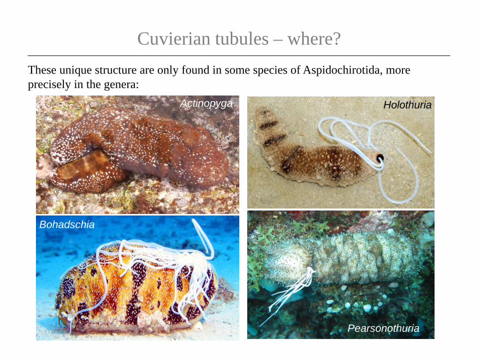
Cuvierian tubules – where?These unique structure are only found in some species of Aspidochirotida, more precisely in the genera:
Actinopyga Holothuria
Pearsonothuria
Bohadschia

What are the Cuvierian tubules ?They are intracoelomic caeca that generally occur in great number (up to 600) and attach to the basal part of the left respiratory tree.
Ct
Left respiratory treeGonad Rete mirabile
Ring
Gut
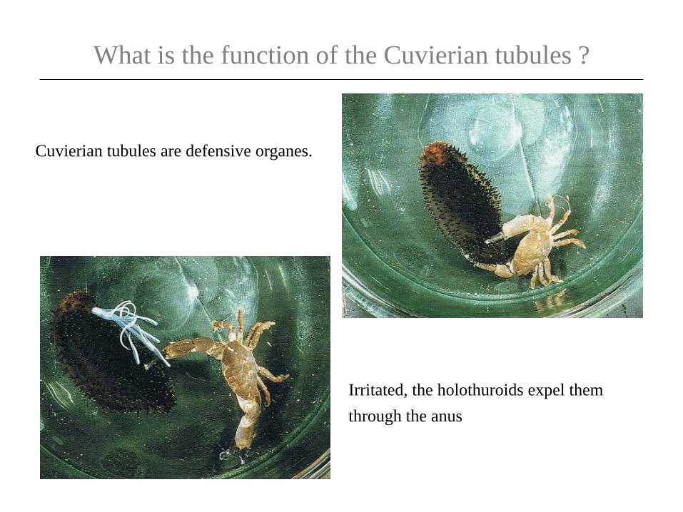
What is the function of the Cuvierian tubules ?
Cuvierian tubules are defensive organes.
Irritated, the holothuroids expel them through the anus

What is the function of the Cuvierian tubules ?
Quiescent tubules are not sticky.
The elongated tubules do not adhere to mucus-cover surfaces, thus preventing them from sticking to the holothuroid body.
The expelled tubules lengthen, become sticky and rapidly immobilize most organisms with which they come in contact!

How do Cuvierian tubules elongate?
Expelled tubules are always in the process of elongation.
Tubules elongate gradually from their basal extremity.
Tubules elongation is irreversible and considerable (up to 20 times their original length!).
Elongating tubules become progressively translucent while their diameter remains unchanged.
Elongation results from powerful entry of water into the tubule
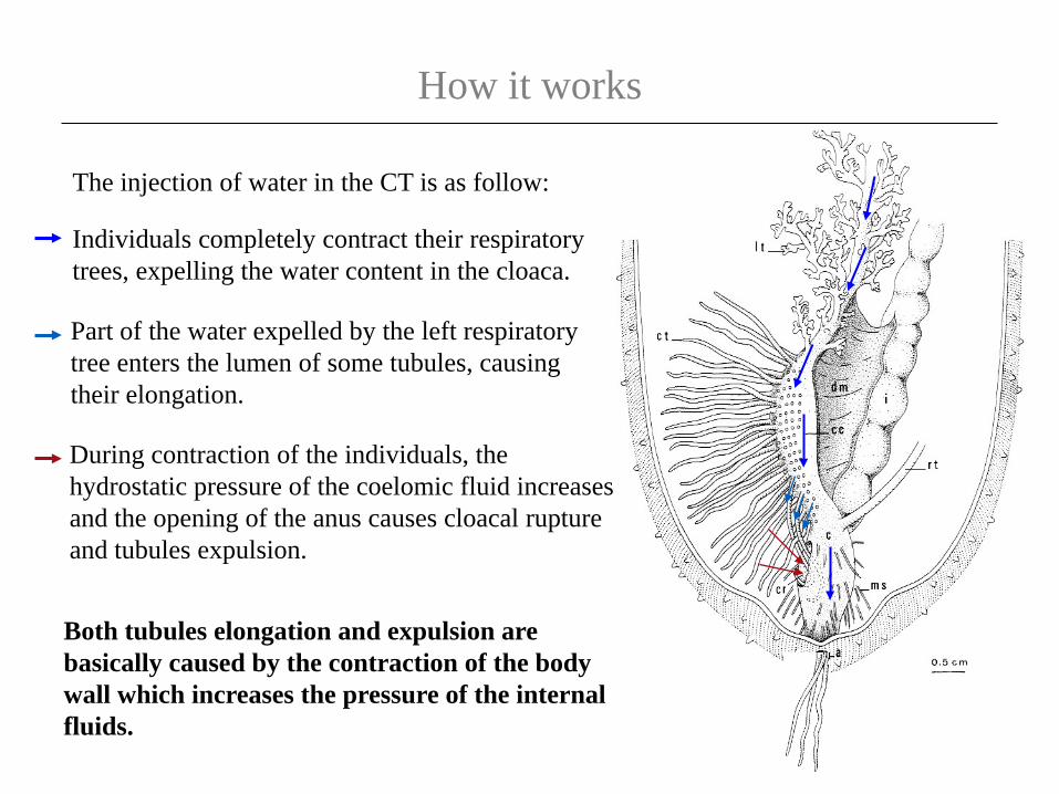
How it works
Both tubules elongation and expulsion are basically caused by the contraction of the body wall which increases the pressure of the internal fluids.
The injection of water in the CT is as follow:
Individuals completely contract their respiratory trees, expelling the water content in the cloaca.
Part of the water expelled by the left respiratory tree enters the lumen of some tubules, causing their elongation.
During contraction of the individuals, the hydrostatic pressure of the coelomic fluid increases and the opening of the anus causes cloacal rupture and tubules expulsion.
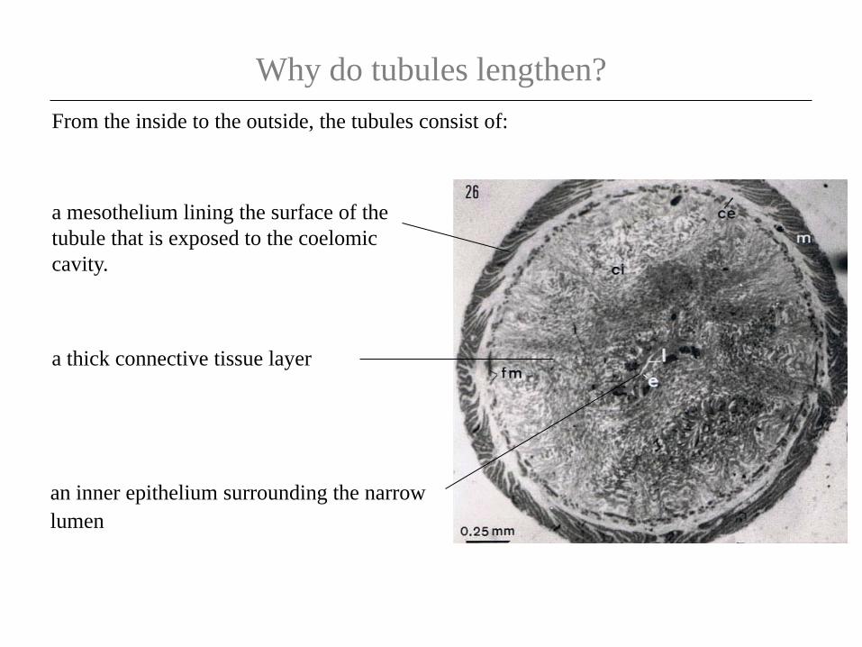
Why do tubules lengthen?From the inside to the outside, the tubules consist of:
an inner epithelium surrounding the narrow lumen
a thick connective tissue layer
a mesothelium lining the surface of the tubule that is exposed to the coelomic cavity.

The inner epithelium
The lumen of the tubule is lined by a highly convoluted epithelium.
The basal process contains piles of 5 to 25 spherules; the content of each spherule is released in the tubule lumen during elongation and could give the rigidity to an elongated tubule.
The basal part of epithelial cells consist of one to three elongated process penetrating deeply in the connective tissue.

The connective tissueThe connective tissue forms 90% of the tubule’s thickness and encloses up to six imprecated collagen helices, each of them being parallel to the long axis of the tubule.
*
*
It also includes longitudinal and circular muscle fibers

The mesothelium
The mesothelium is the tissue layer responsible for adhesion.
It is a pseudostratified epithelium made up of two superposed cell layers:
an outer layer of adluminal cells
Granular cells are filled with densely packed granules enclosing the substance responsible of adhesion.
an inner layer of granular cells which is highly folded along the long axis of the tubule.
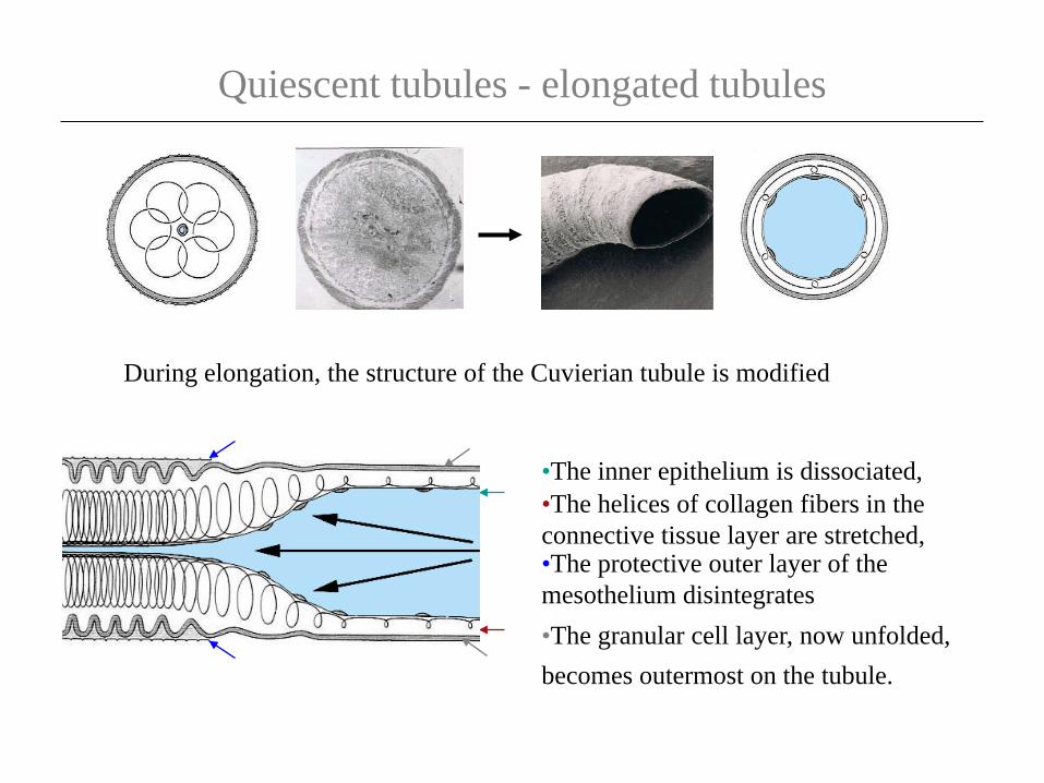
Quiescent tubules - elongated tubules
•The inner epithelium is dissociated,•The helices of collagen fibers in the connective tissue layer are stretched,
•The granular cell layer, now unfolded, becomes outermost on the tubule.
•The protective outer layer of the mesothelium disintegrates
During elongation, the structure of the Cuvierian tubule is modified
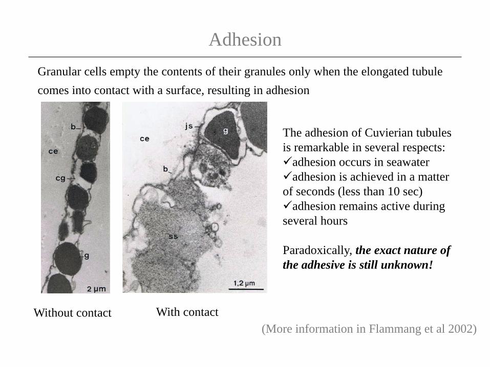
AdhesionGranular cells empty the contents of their granules only when the elongated tubule comes into contact with a surface, resulting in adhesion
The adhesion of Cuvierian tubules is remarkable in several respects: adhesion occurs in seawateradhesion is achieved in a matter of seconds (less than 10 sec)adhesion remains active during several hours
Paradoxically, the exact nature of the adhesive is still unknown!
Without contact With contact(More information in Flammang et al 2002)

Maintaining the line of Defense
Expelled tubules autotomize at their attachment point on the left respiratory tree and are left behind as the holothuroid crawls away.
As only a portion of the tubules are emitted at one time, the total number may suffice for several responses.
After expulsion, the lost tubules are readily regenerated, making them a formidable defense mechanism.
(More information in VandenSpiegel et al 2000)

Variation
All functional Cuvierian tubules (i.e. the expelled ones) investigated so far have almost the same structure. The structure varies only in species where Cuvierian tubules are not expelled.
Microthele Actinopyga

The So-called Cuvierian organs in MicrotheleHolothuria (Microthele)
The fine structure of the tubules closely resembles those of the Holothuria sp.. In contrast, basic behavioral differences occur as the tubules of Microthele sp. cannot elongated nor become sticky, or be expelled by individuals.
It is almost certain that these structure have no defensive role. Yet they are well developed organs that occur in great number in all observed individuals, but from the observation we made it is not possible to ascribe a possible function for these peculiar organs.
Some species of the subgenus Microthele possesses numerous branched tubules whose external surface shows alternative folds which are perpendicular to the long axis of the tubule.
(More information in VandenSpiegel 1995)

The So-called Cuvierian organs in Microthele
Cuvierian Tubules
Cloaca
GonadRing

The So-called Cuvierian organs in ActinopygaGenus Actinopyga
When observed in vivo, the Cuvierian organs in the genus Actinopyga consist of a reddish tuft of branched tubules (no more than 10 tubules per individual).
Tubules attach independently to the basal part of the left respiratory tree

The So-called Cuvierian organs in Actinopyga
Cuvierian tubules Cloaca

The So-called Cuvierian organs in Actinopyga
Two main parts can be distinguished along an actinopygid Cuvierian tubule
The tubule trunk
The tubule branches

The So-called Cuvierian organs in ActinopygaThe tubule trunk
The trunk is a smooth tube with a narrow lumen, it cannot be elongated and has the overall tissue organization of the respiratory
tree.
The spherule itself is made of an enlarged spherical central core containing vesicular cells
The tubule branches have no lumen and consists of a central rachis and many peripheral spherules.
The structure of the rachis resembles that of the tubule trunk.
The tubule branches

The So-called Cuvierian organs in Actinopyga
From the way actinopygid Cuvierian tubules attach and their overall histology they could be homologous to those of the other holothuroids but here again, it appears almost certain that these organs have no defensive role.
Additional information is required concerning the structure of the spherule’s vesicular cells an their changes - for example during growth and/or during the annual cycle of the species - before a possible function can be ascribe to these peculiar organs
(More information in VandenSpiegel et al 1993)

Thank you for your attention