Cutaneous Radiation Injury: Fact Sheet for Physicians · FACT SHEET Cutaneous Radiation Injury:...
13
June 29, 2005 Page 1 of 13 FACT SHEET Cutaneous Radiation Injury: Fact Sheet for Physicians Injury to the skin and underlying tissues from acute exposure to a large external dose of radiation is referred to as cutaneous radiation injury (CRI). Acute radiation syndrome (ARS) 1 will usually be accompanied by some skin damage; however, CRI can occur without symptoms of ARS. This is especially true with acute exposures to beta radiation or low-energy x-rays, because beta radiation and low-energy x-rays are less penetrating and less likely to damage internal organs than gamma radiation is. CRI can occur with radiation doses as low as 2 Gray (Gy) or 200 rads 2 and the severity of CRI symptoms will increase with increasing doses. Most cases of CRI have occurred when people inadvertently came in contact with unsecured radiation sources from food irradiators, radiotherapy equipment, or well depth gauges. In addition, cases of CRI have occurred in people who were overexposed to x-radiation from fluoroscopy units. Early signs and symptoms of CRI are itching, tingling, or a transient erythema or edema without a history of exposure to heat or caustic chemicals. Exposure to radiation can damage the basal cell layer of the skin and result in inflammation, erythema, and dry or moist desquamation. In addition, radiation damage to hair follicles can cause epilation. Transient and inconsistent erythema (associated with itching) can occur within a few hours of exposure and be followed by a latent, symptom-free phase lasting from a few days to several weeks. After the latent phase, intense reddening, blistering, and ulceration of the irradiated site are visible. Depending on the radiation dose, a third and even fourth wave of erythema are possible over the ensuing months or possibly years. In most cases, healing occurs by regenerative means; however, large radiation doses to the skin can cause permanent hair loss, damaged sebaceous and sweat glands, atrophy, fibrosis, decreased or increased skin pigmentation, and ulceration or necrosis of the exposed tissue. With CRI, it is important to keep the following things in mind: • The visible skin effects depend on the magnitude of the dose as well as the depth of penetration of the radiation. • Unlike the skin lesions caused by chemical or thermal damage, the lesions caused by radiation exposures do not appear for hours to days following exposure, and burns and other skin effects tend to appear in cycles. • The key treatment issues with CRI are infection and pain management. 3 1 See “Acute Radiation Syndrome: A Fact Sheet for Physicians” at http://www.bt.cdc.gov/radiation/arsphysicianfactsheet.asp. 2 Both the Gray (Gy) and the rad are units of absorbed dose and reflect the amount of energy deposited in a mass of tissue (1 Gy = 100 rads). In this document, the absorbed dose refers to that dose received by at least 10 cm 2 of the basal cell layer of the skin. The referenced absorbed dose levels in this document are assumed to be from beta, gamma, or x-radiation. Neutron or proton radiation produces many of the health effects described herein at lower absorbed dose levels. 3 On occasion a patient might also be contaminated with radioactive material. To address patient decontamination, please go to the following Web site: http://www.orau.gov/reacts/emergency.htm .
-
Upload
truongminh -
Category
Documents
-
view
234 -
download
0
Transcript of Cutaneous Radiation Injury: Fact Sheet for Physicians · FACT SHEET Cutaneous Radiation Injury:...
June 29, 2005 Page 1 of 13
FACT SHEET
Cutaneous Radiation Injury: Fact Sheet for Physicians Injury to the skin and underlying tissues from acute exposure to a large external dose of radiation is referred to as cutaneous radiation injury (CRI). Acute radiation syndrome (ARS)1 will usually be accompanied by some skin damage; however, CRI can occur without symptoms of ARS. This is especially true with acute exposures to beta radiation or low-energy x-rays, because beta radiation and low-energy x-rays are less penetrating and less likely to damage internal organs than gamma radiation is. CRI can occur with radiation doses as low as 2 Gray (Gy) or 200 rads2 and the severity of CRI symptoms will increase with increasing doses. Most cases of CRI have occurred when people inadvertently came in contact with unsecured radiation sources from food irradiators, radiotherapy equipment, or well depth gauges. In addition, cases of CRI have occurred in people who were overexposed to x-radiation from fluoroscopy units. Early signs and symptoms of CRI are itching, tingling, or a transient erythema or edema without a history of exposure to heat or caustic chemicals. Exposure to radiation can damage the basal cell layer of the skin and result in inflammation, erythema, and dry or moist desquamation. In addition, radiation damage to hair follicles can cause epilation. Transient and inconsistent erythema (associated with itching) can occur within a few hours of exposure and be followed by a latent, symptom-free phase lasting from a few days to several weeks. After the latent phase, intense reddening, blistering, and ulceration of the irradiated site are visible. Depending on the radiation dose, a third and even fourth wave of erythema are possible over the ensuing months or possibly years. In most cases, healing occurs by regenerative means; however, large radiation doses to the skin can cause permanent hair loss, damaged sebaceous and sweat glands, atrophy, fibrosis, decreased or increased skin pigmentation, and ulceration or necrosis of the exposed tissue. With CRI, it is important to keep the following things in mind:
• The visible skin effects depend on the magnitude of the dose as well as the depth of penetration of the radiation.
• Unlike the skin lesions caused by chemical or thermal damage, the lesions caused by radiation exposures do not appear for hours to days following exposure, and burns and other skin effects tend to appear in cycles.
• The key treatment issues with CRI are infection and pain management.3
1 See “Acute Radiation Syndrome: A Fact Sheet for Physicians” at http://www.bt.cdc.gov/radiation/arsphysicianfactsheet.asp. 2 Both the Gray (Gy) and the rad are units of absorbed dose and reflect the amount of energy deposited in a mass of tissue (1 Gy = 100 rads). In this document, the absorbed dose refers to that dose received by at least 10 cm2 of the basal cell layer of the skin. The referenced absorbed dose levels in this document are assumed to be from beta, gamma, or x-radiation. Neutron or proton radiation produces many of the health effects described herein at lower absorbed dose levels. 3 On occasion a patient might also be contaminated with radioactive material. To address patient decontamination, please go to the following Web site: http://www.orau.gov/reacts/emergency.htm.
Cutaneous Radiation Injury: Fact Sheet for Physicians (continued from previous page)
June 29, 2005 Page 2 of 13
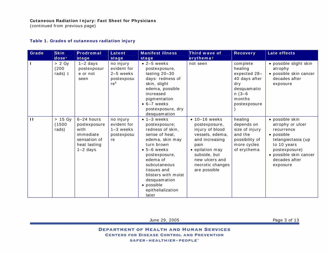
Stages and Grades of CRI CRI will progress over time in stages and can be categorized by grade, with characteristics of the stages varying by grade of injury, as shown in Table 1. Appendix A gives a detailed description of the various skin responses to radiation, and Appendix B provides color photographs of examples of some of these responses.
Prodromal stage (within hours of exposure)—This stage is characterized by early erythema (first wave of erythema), heat sensations, and itching that define the exposure area. The duration of this stage is from 1 to 2 days. Latent stage (1–2 days postexposure)—No injury is evident. Depending on the body part, the larger the dose, the shorter this period will last. The skin of the face, chest, and neck will have a shorter latent stage than will the skin of the palms of the hands or the soles of the feet. Manifest illness stage (days to weeks postexposure)—The basal layer is repopulated through proliferation of surviving clonogenic cells. This stage begins with main erythema (second wave), a sense of heat, and slight edema, which are often accompanied by increased pigmentation. The symptoms that follow vary from dry desquamation or ulceration to necrosis, depending on the severity of the CRI (see Table 1). Third wave of erythema (10–16 weeks postexposure, especially after beta exposure)—The exposed person experiences late erythema, injury to blood vessels, edema, and increasing pain. A distinct bluish color of the skin can be observed. Epilation may subside, but new ulcers, dermal necrosis, and dermal atrophy (and thinning of the dermis layer) are possible. Late effects (months to years postexposure; threshold dose ~10 Gy or 1000 rads)—Symptoms can vary from slight dermal atrophy (or thinning of dermis layer) to constant ulcer recurrence, dermal necrosis, and deformity. Possible effects include occlusion of small blood vessels with subsequent disturbances in the blood supply (telangiectasia); destruction of the lymphatic network; regional lymphostasis; and increasing invasive fibrosis, keratosis, vasculitis, and subcutaneous sclerosis of the connective tissue. Pigmentary changes and pain are often present. Skin cancer is possible in subsequent years. Recovery (months to years)
Cutaneous Radiation Injury: Fact Sheet for Physicians (continued from previous page)
June 29, 2005 Page 3 of 13
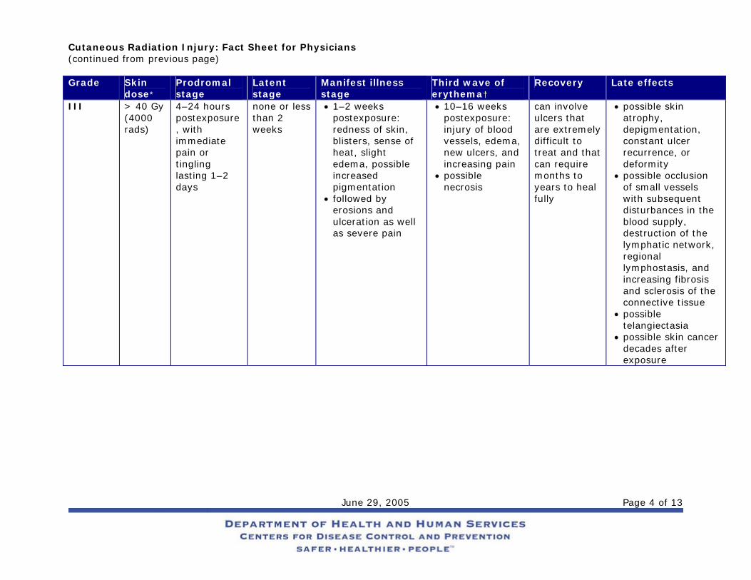
Table 1. Grades of cutaneous radiation injury
Grade Skin
dose* Prodromal stage
Latent stage
Manifest illness stage
Third wave of erythema†
Recovery Late effects
I > 2 Gy (200 rads) ‡
1–2 days postexposure or not seen
no injury evident for 2–5 weeks postexposure§
• 2–5 weeks postexposure, lasting 20–30 days: redness of skin, slight edema, possible increased pigmentation
• 6–7 weeks postexposure, dry desquamation
not seen complete healing expected 28–40 days after dry desquamation (3–6 months postexposure)
• possible slight skin atrophy
• possible skin cancer decades after exposure
II > 15 Gy (1500 rads)
6–24 hours postexposure with immediate sensation of heat lasting 1–2 days
no injury evident for 1–3 weeks postexposure
• 1–3 weeks postexposure; redness of skin, sense of heat, edema, skin may turn brown
• 5–6 weeks postexposure, edema of subcutaneous tissues and blisters with moist desquamation
• possible epithelialization later
• 10–16 weeks postexposure, injury of blood vessels, edema, and increasing pain
• epilation may subside, but new ulcers and necrotic changes are possible
healing depends on size of injury and the possibility of more cycles of erythema
• possible skin atrophy or ulcer recurrence
• possible telangiectasia (up to 10 years postexposure)
• possible skin cancer decades after exposure
Cutaneous Radiation Injury: Fact Sheet for Physicians (continued from previous page)
June 29, 2005 Page 4 of 13
Grade Skin dose*
Prodromal stage
Latent stage
Manifest illness stage
Third wave of erythema†
Recovery Late effects
III > 40 Gy (4000 rads)
4–24 hours postexposure, with immediate pain or tingling lasting 1–2 days
none or less than 2 weeks
• 1–2 weeks postexposure: redness of skin, blisters, sense of heat, slight edema, possible increased pigmentation
• followed by erosions and ulceration as well as severe pain
• 10–16 weeks postexposure: injury of blood vessels, edema, new ulcers, and increasing pain
• possible necrosis
can involve ulcers that are extremely difficult to treat and that can require months to years to heal fully
• possible skin atrophy, depigmentation, constant ulcer recurrence, or deformity
• possible occlusion of small vessels with subsequent disturbances in the blood supply, destruction of the lymphatic network, regional lymphostasis, and increasing fibrosis and sclerosis of the connective tissue
• possible telangiectasia
• possible skin cancer decades after exposure
Cutaneous Radiation Injury: Fact Sheet for Physicians (continued from previous page)
June 29, 2005 Page 5 of 13
Grade Skin dose*
Prodromal stage
Latent stage
Manifest illness stage
Third wave of erythema†
Recovery Late effects
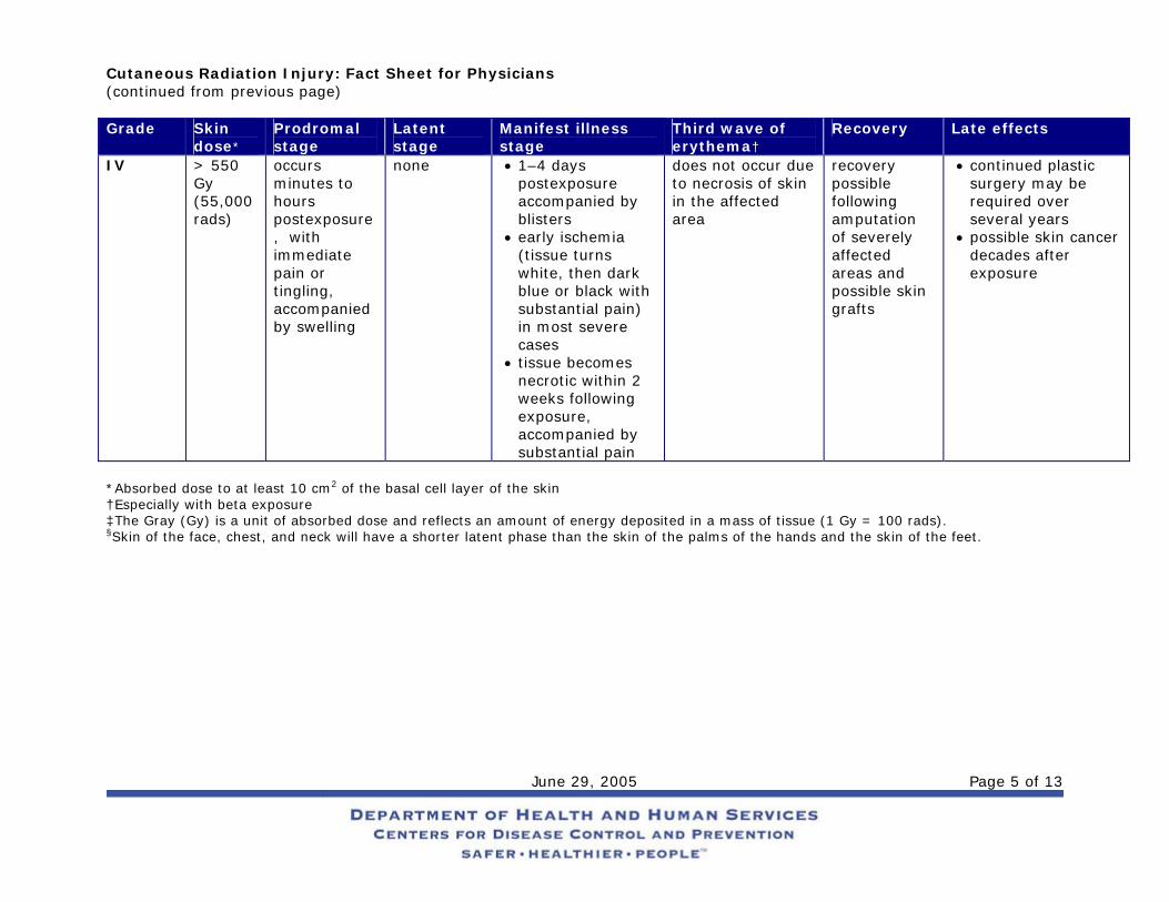
IV > 550 Gy (55,000 rads)
occurs minutes to hours postexposure, with immediate pain or tingling, accompanied by swelling
none • 1–4 days postexposure accompanied by blisters
• early ischemia (tissue turns white, then dark blue or black with substantial pain) in most severe cases
• tissue becomes necrotic within 2 weeks following exposure, accompanied by substantial pain
does not occur due to necrosis of skin in the affected area
recovery possible following amputation of severely affected areas and possible skin grafts
• continued plastic surgery may be required over several years
• possible skin cancer decades after exposure
*Absorbed dose to at least 10 cm2 of the basal cell layer of the skin †Especially with beta exposure ‡The Gray (Gy) is a unit of absorbed dose and reflects an amount of energy deposited in a mass of tissue (1 Gy = 100 rads). §Skin of the face, chest, and neck will have a shorter latent phase than the skin of the palms of the hands and the skin of the feet.
Cutaneous Radiation Injury: Fact Sheet for Physicians (continued from previous page)
June 29, 2005 Page 6 of 13
Patient Management Diagnosis
The signs and symptoms of CRI are as follows: • Intensely painful burn-like skin injuries (including itching, tingling, erythema, or edema) without a
history of exposure to heat or caustic chemicals Note: Erythema will not be seen for hours to days following exposure, and its appearance is cyclic.
• Epilation • A tendency to bleed • Possible signs and symptoms of ARS As mentioned previously, local injuries to the skin from acute radiation exposure evolve slowly over time, and symptoms may not manifest for days to weeks after exposure. Consider CRI in the differential diagnosis if the patient presents with a skin lesion without a history of chemical or thermal burn, insect bite, or skin disease or allergy. If the patient gives a history of possible radiation exposure (such as from a radiography source, x-ray device, or accelerator) or a history of finding and handling an unknown metallic object, note the presence of any of the following: erythema, blistering, dry or wet desquamation, epilation, ulceration. Regarding lesions associated with CRI be aware that, • days to weeks may pass before lesions appear; • unless patients are symptomatic, they will not require emergency care; and • lesions can be debilitating and life threatening after several weeks.
Medical follow-up is essential, and victims should be cautioned to avoid trauma to the involved areas. Initial Treatment
Localized injuries should be treated symptomatically as they occur, and radiation injury experts should be consulted for detailed information. Such information can be obtained from the Radiation Emergency Assistance Center/Training Site (REAC/TS) at www.orau.gov/reacts/ or (865) 576-1005. As with ARS, if the patient also has other trauma, wounds should be closed, burns covered, fractures reduced, surgical stabilization performed, and definitive treatment given within the first 48 hours after injury. After 48 hours, surgical interventions should be delayed until hematopoietic recovery has occurred. A baseline CBC and differential should be taken and repeated in 24 hours. Because cutaneous radiation injury is cyclic, areas of early erythema should be noted and recorded. These areas should also be sketched and photographed, if possible, ensuring that the date and time are recorded. The following should be initiated as indicated:
• Supportive care in a clean environment (a burn unit if one is available) • Prevention and treatment of infections • Use of the following:
o Medications to reduce inflammation, inhibit protealysis, relieve pain, stimulate regeneration, and improve circulation
o Anticoagulant agents for widespread and deep injury • Pain management • Psychological support
Cutaneous Radiation Injury: Fact Sheet for Physicians (continued from previous page)
June 29, 2005 Page 7 of 13
Recommendations for Treatment by Stage The following recommendations for treatment by stage of the illness were obtained by summarizing recommendations from Ricks et al. (226) and Gusev et al. (231), but they do not represent official recommendations of CDC.
Prodromal Stage—Use antihistamines and topical antipruriginous preparations, which act against itch and also might prevent or attenuate initiation of the cycle that leads to the manifestation stage. Anti-inflammatory medications such as corticosteroids and topical creams, as well as slight sedatives, may prove useful. Latent Stage—Continue anti-inflammatory medications and sedatives. At midstage, use proteolysis inhibitors, such as Gordox®. Manifestation Stage—Use repeated swabs, antibiotic prophylaxis, and anti-inflammatory medications, such as Lioxasol®, to reduce bacterial, fungal, and viral infections o Apply topical ointments containing corticosteroids along with locally acting antibiotics and vitamins. o Stimulate regeneration of DNA by using Lioxasol® and later, when regeneration has started,
biogenic drugs, such as Actovegin® and Solcoseril®. o Stimulate blood supply in third or fourth week using Pentoxifylline® (contraindicated for patients
with atherosclerotic heart disease). o Puncture blisters if they are sterile, but do not remove them as long as they are intact. o Stay alert for wound infection. Antibiotic therapy should be considered according to the individual
patient’s condition. o Treat pain according to the individual patient’s condition. Pain relief is very difficult and is the most
demanding part of the therapeutic process. o Debride areas of necrosis thoroughly but cautiously.
Treatment of Late Effects
After immediate treatment of radiation injury, an often long and painful process of healing will ensue. The most important concerns are the following: • Pain management • Fibrosis or late ulcers
Note: Use of medication to stimulate vascularization, inhibit infection, and reduce fibrosis may be effective. Examples include Pentoxifylline®, vitamin E, and interferon gamma. Otherwise, surgery may be required.
• Necrosis • Plastic/reconstructive surgery
Note: Surgical treatment is common. It is most effective if performed early in the treatment process. Full-thickness graft and microsurgery techniques usually provide the best results.
• Psychological effects, such as posttraumatic stress disorder • Possibility of increased risk of skin cancer later in life
For More Assistance Technical assistance can be obtained from the Radiation Emergency Assistance Center/Training Site (REAC/TS) at (865) 576-3131 (M-F, 8 AM to 4:30 PM EST) or (865) 576-1005 (after hours), or at http://www.orau.gov/reacts/, and from the Medical Radiobiology Advisory Team (MRAT) at (301) 295-0316. Also, more information can be obtained from the CDC Health Alert Network at http://www.bt.cdc.gov or 1-800-311-3435.
Cutaneous Radiation Injury: Fact Sheet for Physicians (continued from previous page)
June 29, 2005 Page 8 of 13
References
Gusev IA, Guskova AK, Mettler FA, Jr., editors. Medical Management of Radiation Accidents. 2nd ed. New York: CRC Press, Inc.; 2001.
Hall EJ. Radiobiology for the Radiologist. 5th ed. New York: Lippincott Williams & Wilkins; 2000.
International Commission on Radiological Protection (ICRP). The Biological Basis for Dose Limitation in the Skin. ICRP Publication 59. Annals of the ICRP Volume 22, No. 2. New York: Pergamon Press, 1991.
National Council on Radiation Protection and Measurements (NCRP). Biological Effects and Exposure Limits for “Hot Particles.” NCRP Report No. 130. Bethesda, Maryland: NCRP, 1999.
National Council on Radiation Protection and Measurements (NCRP). Management of Terrorist Events Involving Radioactive Material. NCRP Report No. 138. Bethesda, Maryland: NCRP, 2001.
Ricks RC, Berger ME, O’Hare FM, Jr, editors. The Medical Basis for Radiation Accident Preparedness: The Clinical Care of Victims. REAC/TS Conference on the Medical Basis for Radiation Accident Preparedness. New York: Parthenon Publishing, 2002.
Walker RI, Cerveny TJ, editors. Textbook of Military Medicine: Part 1: Warfare, Weaponry, and the Casualty. Medical Consequences of Nuclear Warfare. Armed Forces Radiobiology Research Institute (AFRRI). Bethesda, Maryland: 1989.
Cutaneous Radiation Injury: Fact Sheet for Physicians (continued from previous page)
June 29, 2005 Page 9 of 13
Appendix A: Responses of the Skin to Radiation Acute epidermal necrosis (time of onset: < 10 days postexposure; threshold dose: ~550 Gy or 55,000 rads)—
Interphase death of postmitotic keratinocytes in the upper visible layers of the epidermis (may occur with high-dose, low-energy beta irradiation)
Acute ulceration (time of onset: < 14 days postexposure; threshold dose: ~20 Gy or 2000 rads)—
Early loss of the epidermis—and to a varying degree, deeper dermal tissue—that results from the death of fibroblasts and endothelial cells in interphase
Dermal atrophy (time of onset: > 26 weeks postexposure; threshold dose: ~10 Gy or 1000 rads)—
Thinning of the dermal tissues associated with the contraction of the previously irradiated area Dermal necrosis (time of onset > 10 weeks postexposure; threshold dose: ~20 Gy or 2000 rads)—
Necrosis of the dermal tissues as a consequence of vascular insufficiency Dry desquamation (time of onset: 3–6 weeks postexposure; threshold dose: ~8 Gy or 800 rads)—
Atypical keratinization of the skin caused by the reduction in the number of clonogenic cells within the basal layer of the epidermis
Early transient erythema (time of onset: within hours of exposure; threshold dose: ~2 Gray [Gy] or 200 rads)—
Inflammation of the skin caused by activation of a proteolytic enzyme that increases the permeability of the capillaries
Epilation (time of onset: 14–21 days; threshold dose: ~3 Gy or 300 rads)—
Hair loss caused by the depletion of matrix cells in the hair follicles Late erythema (time of onset: 8–20 weeks postexposure; threshold dose: ~20 Gy or 2000 rads)—
Inflammation of the skin caused by injury of blood vessels. Edema and impaired lymphatic clearance precede a measured reduction in blood flow.
Invasive fibrosis (time of onset: months to years postexposure; threshold dose: ~20 Gy or 2000 rads)—
Method of healing associated with acute ulceration, secondary ulceration, and dermal necrosis that leads to scar tissue formation
Main erythema (time of onset: days to weeks postexposure; threshold dose: ~3 Gy or 300 rads)—
Inflammation of the skin caused by hyperaemia of the basal cells and subsequent epidermal hypoplasia (see photos 1 and 2)
Moist desquamation (time of onset: 4–6 weeks postexposure; threshold dose: ~15 Gy or 1500 rads)—
Loss of the epidermis caused by sterilization of a high proportion of clonogenic cells within the basal layer of the epidermis
Secondary ulceration (time of onset: > 6 weeks postexposure; threshold dose: ~15 Gy or 1500 rads)—
Secondary damage to the dermis as a consequence of dehydration and infection when moist desquamation is severe and protracted because of reproductive sterilization of the vast majority of the clonogenic cells in the irradiated area
Cutaneous Radiation Injury: Fact Sheet for Physicians (continued from previous page)
June 29, 2005 Page 10 of 13
Telangiectasia (time of onset: > 52 weeks postexposure; threshold dose for moderate severity at 5 years: ~40 Gy or 4000 rads)—
Atypical dilation of the superficial dermal capillaries
Cutaneous Radiation Injury: Fact Sheet for Physicians (continued from previous page)
June 29, 2005 Page 11 of 13
Appendix B: Images
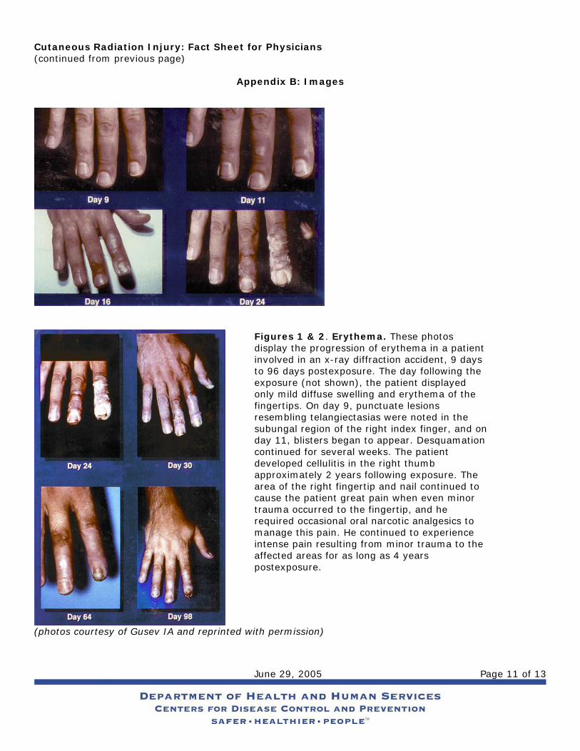
Figures 1 & 2. Erythema. These photos display the progression of erythema in a patient involved in an x-ray diffraction accident, 9 days to 96 days postexposure. The day following the exposure (not shown), the patient displayed only mild diffuse swelling and erythema of the fingertips. On day 9, punctuate lesions resembling telangiectasias were noted in the subungal region of the right index finger, and on day 11, blisters began to appear. Desquamation continued for several weeks. The patient developed cellulitis in the right thumb approximately 2 years following exposure. The area of the right fingertip and nail continued to cause the patient great pain when even minor trauma occurred to the fingertip, and he required occasional oral narcotic analgesics to manage this pain. He continued to experience intense pain resulting from minor trauma to the affected areas for as long as 4 years postexposure.
(photos courtesy of Gusev IA and reprinted with permission)
Cutaneous Radiation Injury: Fact Sheet for Physicians (continued from previous page)
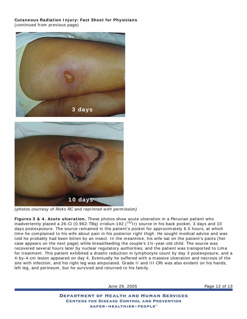
(photos courtesy of Ricks R Figures 3 & 4. Acute ulceinadvertently placed a 26-Cdays postexposure. The soutime he complained to his wtold he probably had been bcase appears on the next precovered several hours latfor treatment. This patient 4-by-4-cm lesion appearedsite with infection, and his rleft leg, and perineum, but
3 days
10 daysJune 29, 2005 Page 12 of 13
C and reprinted with permission)
ration. These photos show acute ulceration in a Peruvian patient who i (0.962-TBq) irridiun-192 (192Ir) source in his back pocket, 3 days and 10 rce remained in the patient’s pocket for approximately 6.5 hours, at which ife about pain in his posterior right thigh. He sought medical advice and was itten by an insect. In the meantime, his wife sat on the patient’s pants (her
age) while breastfeeding the couple’s 1½-year-old child. The source was er by nuclear regulatory authorities, and the patient was transported to Lima exhibited a drastic reduction in lymphocyte count by day 3 postexposure, and a on day 4. Eventually he suffered with a massive ulceration and necrosis of the ight leg was amputated. Grade II and III CRI was also evident on his hands, he survived and returned to his family.
Cutaneous Radiation Injury: Fact Sheet for Physicians (continued from previous page)
June 29, 2005 Page 13 of 13
26 days
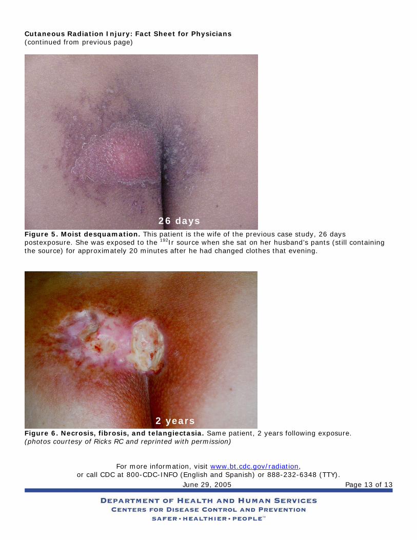
Figure 5. Moist desquamation. This patient is the wife of the previous case study, 26 days postexposure. She was exposed to the 192Ir source when she sat on her husband’s pants (still containing the source) for approximately 20 minutes after he had changed clothes that evening.
2 years
Figure 6. Necrosis, fibrosis, and telangiectasia. Same patient, 2 years following exposure. (photos courtesy of Ricks RC and reprinted with permission)
For more information, visit www.bt.cdc.gov/radiation, or call CDC at 800-CDC-INFO (English and Spanish) or 888-232-6348 (TTY).