CURRENTISSUESINORGANPRESERVATION Monograph - Final.pdf · Histidine 198mmol/L Buffer Mannitol...
20
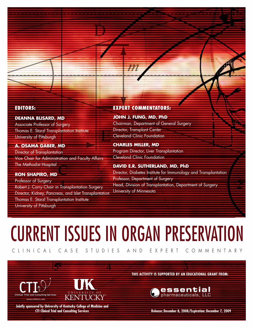
EDITORS: DEANNA BLISARD, MD Associate Professor of Surgery Thomas E. Starzl Transplantation Institute University of Pittsburgh A. OSAMA GABER, MD Director of Transplantation Vice Chair for Administration and Faculty Affairs The Methodist Hospital RON SHAPIRO, MD Professor of Surgery Robert J. Corry Chair in Transplantation Surgery Director, Kidney, Pancreas, and Islet Transplantation Thomas E. Starzl Transplantation Institute University of Pittsburgh EXPERT COMMENTATORS: JOHN J. FUNG, MD, PhD Chairman, Department of General Surgery Director, Transplant Center Cleveland Clinic Foundation CHARLES MILLER, MD Program Director, Liver Transplantation Cleveland Clinic Foundation DAVID E.R. SUTHERLAND, MD, PhD Director, Diabetes Institute for Immunology and Transplantation Professor, Department of Surgery Head, Division of Transplantation, Department of Surgery University of Minnesota Release: December 8, 2008/Expiration: December 7, 2009 CURRENT ISSUES IN ORGAN PRESERVATION C L I N I C A L C A S E S T U D I E S A N D E X P E R T C O M M E N TA R Y THIS ACTIVITY IS SUPPORTED BY AN EDUCATIONAL GRANT FROM: Jointly sponsored by University of Kentucky College of Medicine and CTI Clinical Trial and Consulting Services
Transcript of CURRENTISSUESINORGANPRESERVATION Monograph - Final.pdf · Histidine 198mmol/L Buffer Mannitol...
EDITORS:
DEANNA BLISARD, MDAssociate Professor of SurgeryThomas E. Starzl Transplantation InstituteUniversity of Pittsburgh
A. OSAMA GABER, MDDirector of TransplantationVice Chair for Administration and Faculty AffairsThe Methodist Hospital
RON SHAPIRO, MDProfessor of SurgeryRobert J. Corry Chair in Transplantation SurgeryDirector, Kidney, Pancreas, and Islet TransplantationThomas E. Starzl Transplantation InstituteUniversity of Pittsburgh
EXPERT COMMENTATORS:
JOHN J. FUNG, MD, PhDChairman, Department of General SurgeryDirector, Transplant CenterCleveland Clinic Foundation
CHARLES MILLER, MDProgram Director, Liver TransplantationCleveland Clinic Foundation
DAVID E.R. SUTHERLAND, MD, PhDDirector, Diabetes Institute for Immunology and TransplantationProfessor, Department of SurgeryHead, Division of Transplantation, Department of SurgeryUniversity of Minnesota
Release: December 8, 2008/Expiration: December 7, 2009
CURRENT ISSUES IN ORGAN PRESERVATIONC L I N I C A L C A S E S T U D I E S A N D E X P E R T C O M M E N T A R Y
THIS ACTIVITY IS SUPPORTED BY AN EDUCATIONAL GRANT FROM:
Jointly sponsored by University of Kentucky College of Medicine andCTI Clinical Trial and Consulting Services
CURRENT ISSUES IN ORGAN PRESERVATIONCLINICAL CASE STUDIES AND EXPERT COMMENTARY
CONTENTS
Introduction ..................................................................................................3
Mechanisms of Tissue Injury ............................................................................4
Static Cold Preservation Solutions ....................................................................5
Clinical Trial Review ........................................................................................7
Liver Transplantation ................................................................................7
Pancreas Transplantation ..........................................................................9
Case Studies: Pancreas Transplantation ..........................................................11
Expert Commentary: Pancreas Transplant Case Studies ......................................13
Case Studies: Liver Transplantation ................................................................14
Expert Commentary: Liver Transplant Case Studies............................................16
Conclusion ..................................................................................................17
2
COURSE DESCRIPTIONNeeds AssessmentEvery effort is made to utilize each available organ for transplantation and to optimizesubsequent patient and graft survival. Although there are many factors that impactimmediate graft survival, a prominent topic continues to be the quality of organpreservation. Cold storage and specialized organ preservation solutions are used tominimize the damaging effects of cold ischemia and reperfusion on organ function.1
Optimal organ preservation is becoming increasingly important as the characteristicsof donor organs change. Donor organs are being donated from older and sickerpopulations and being subjected to longer ischemia times.1 Increases in cold ischemiatimes are associated with higher graft loss across all organ types.2
Several different organ preservation solutions are currently used in the United States,and new solutions are under development. University of Wisconsin (UW) solution(Viaspan®) has been the most widely used solution for multi-organ protection sinceits introduction in 1987. However, many institutions have begun to use HistidineTryptophan Ketoglutarate (HTK) solution (Custodiol®) in their transplant practice.Several studies have shown HTK to be as effective as UW for liver, kidney andpancreas protection.3-5
Medicine Accreditation StatementThis activity has been planned and implemented in accordance with the EssentialsAreas and Policies of the Accreditation Council and Continuing Medical Education(ACCME) through the joint sponsorship of University of Kentucky College of Medicineand CTI Clinical Trial and Consulting Services. The University of Kentucky Collegeof Medicine is accredited by the ACCME to provide continuing medical educationfor physicians.
The University of Kentucky College of Medicine designates this educational activityfor a maximum of one (1.0) AMA PRA Category 1 Credit TM towards the AMAPhysician’s Recognition Award. Each physician should claim only those hoursof credit actually spent in the educational activity.
The University of Kentucky College of Medicine presents this activity for educationalpurposes only. Participants are expected to utilize their own expertise and judgmentwhile engaged in the practice of medicine. The content of the presentation isprovided solely by presenters who have been selected for presentations becauseof recognized expertise in their field.
Target AudienceThis educational activity is intended for medical professionals involved in themanagement of transplant patients including: transplant physicians, surgeons,and organ procurement personnel.
EditorsDeanna Blisard, MDAssociate Professor of SurgeryThomas E. Starzl Transplantation InstituteUniversity of PittsburghPittsburgh, PA
A. Osama Gaber, MDDirector of TransplantationVice Chair for Administration and Faculty AffairsThe Methodist HospitalHouston, TX
Ron Shapiro, MDProfessor of SurgeryRobert J. Corry Chair in Transplantation SurgeryDirector, Kidney, Pancreas, and Islet TransplantationThomas E. Starzl Transplantation InstituteUniversity of PittsburghPittsburgh, PA
Expert CommentatorsJohn J. Fung, MD, PhDChairman, Department of General SurgeryDirector, Transplant CenterCleveland Clinic FoundationCleveland, OH
Charles Miller, MDProgram Director, Liver TransplantationCleveland Clinic FoundationCleveland, OH
David E.R. Sutherland, MD, PhDDirector, Diabetes Institute for Immunology and TransplantationProfessor, Department of SurgeryHead, Division of Transplantation, Department of SurgeryUniversity of MinnesotaMinneapolis, MN
3
Faculty DisclosureIt is the policy of the University of Kentucky to ensure balance, independence,objectivity and scientific rigor in all of its educational activities. In accordance withthe policy of the University of Kentucky, faculty are asked to disclose any affiliationor financial interest that may affect the content of this activity.
The faculty reported the following:
John Fung, MD, PhD has served on Essential Pharmaceuticals Speaker bureau
A. Osama Gaber, MD has received consultation fees from Essential Pharmaceuticals
Commercial SupportSupport for this activity is provided by an unrestricted educational grant fromEssential Pharmaceuticals, LLC.
DevelopmentThe educational activity was developed by:
CTI Clinical Trial and Consulting Services
G. Mark Baillie PharmD, MHASenior Research Scientist
Eliezer Katz, MD, FACSVice President of Medical Affairs
Procedures to Obtain Credit1. Complete the monograph in its entirety
2. Upon completion, visit www.CEcentral.com/getcredit
3. Enter activity code MEN09144
4. Login or register
5. Complete posttest and evaluation
6. A printable certificate will be issued
Learning ObjectivesAfter completing this CME activity, participants will be able to:
1. Differentiate among commonly used solid organ preservation solutions.
2. Discuss factors that influence the outcome in organ preservation.
3. Describe the impact of organ preservation solutions on transplant outcomes.
INTRODUCTIONAs the need for organ transplantation increases and the waiting list continues to grow,successful utilization and transplantation of as many available donor organs as possiblehas become paramount. While short-term outcomes have continued to improve andthe incidence of acute rejection episodes has decreased, there has been only modestimprovement in longer term outcomes, due to a variety of factors. Clearly, thegreater use of extended criteria donors (ECD) and donation after cardiac death(DCD) donors should be considered as a contributing factor, which reduce bothshort and long-term graft survival. These donors contribute an increasingly largerpercentage of organs for transplantation — older donors and DCD account forthe major growth in deceased donors over the past 5 years.2, 6, 7
The maintenance of organ viability during recovery and preservation remains a keyfactor in successful outcomes. Knowledge surrounding the impact of brain death,warm and cold ischemia, and reperfusion injury on post-transplant organ functionhas increased dramatically in recent years.8, 9
Increasing awareness of the effects of ischemia-reperfusion (I/R) injury on transplantoutcomes has sparked a revived interest in cold storage preservation solutions.Optimal selection and use of preservation solutions to minimize the tissue damageand injury during the ischemic period to avoid post-transplant complications is anemerging area of research.8
The purpose of this monograph is to critically evaluate selected currently availableand widely used preservation solutions in liver and pancreas transplantation basedupon published literature. Case studies and expert commentary will be usedto illustrate key considerations of the impact of organ preservation solutions onpost-transplant outcomes and how this impacts the dynamically changing natureof organ donor utilization in the United States.
4
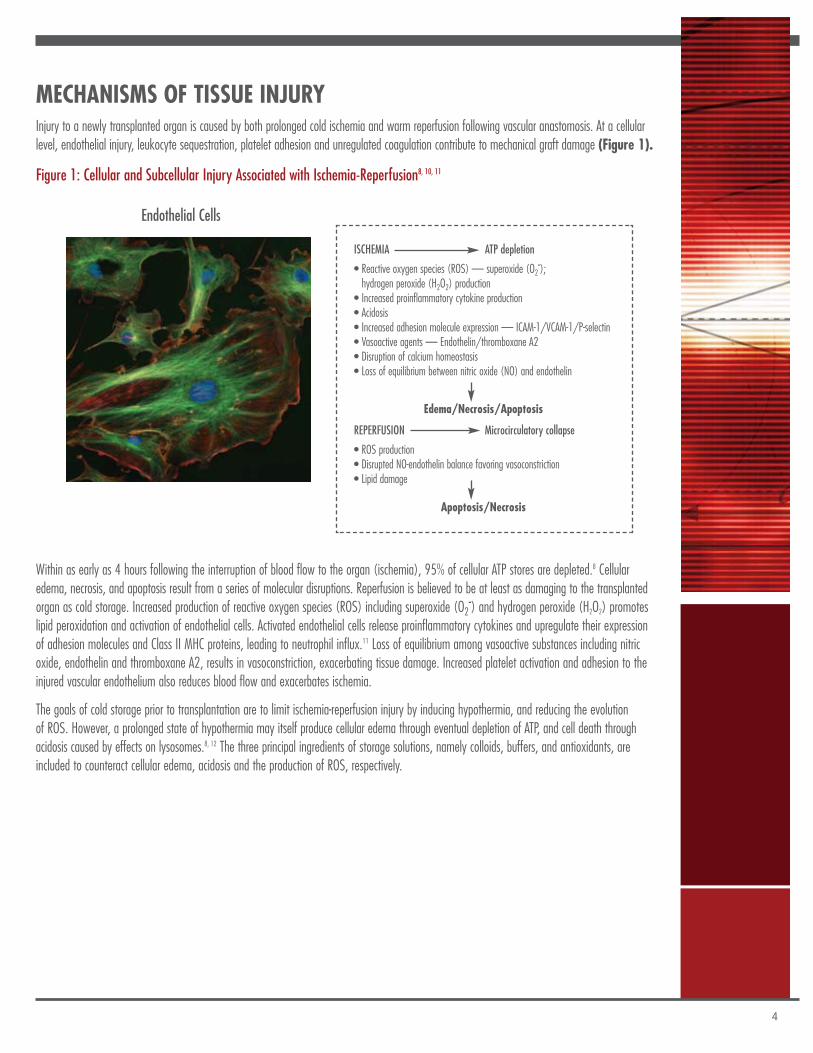
MECHANISMS OF TISSUE INJURYInjury to a newly transplanted organ is caused by both prolonged cold ischemia and warm reperfusion following vascular anastomosis. At a cellularlevel, endothelial injury, leukocyte sequestration, platelet adhesion and unregulated coagulation contribute to mechanical graft damage (Figure 1).
Figure 1: Cellular and Subcellular Injury Associated with Ischemia-Reperfusion8, 10, 11
Endothelial Cells
Within as early as 4 hours following the interruption of blood flow to the organ (ischemia), 95% of cellular ATP stores are depleted.8 Cellularedema, necrosis, and apoptosis result from a series of molecular disruptions. Reperfusion is believed to be at least as damaging to the transplantedorgan as cold storage. Increased production of reactive oxygen species (ROS) including superoxide (O2-) and hydrogen peroxide (H2O2) promoteslipid peroxidation and activation of endothelial cells. Activated endothelial cells release proinflammatory cytokines and upregulate their expressionof adhesion molecules and Class II MHC proteins, leading to neutrophil influx.11 Loss of equilibrium among vasoactive substances including nitricoxide, endothelin and thromboxane A2, results in vasoconstriction, exacerbating tissue damage. Increased platelet activation and adhesion to theinjured vascular endothelium also reduces blood flow and exacerbates ischemia.
The goals of cold storage prior to transplantation are to limit ischemia-reperfusion injury by inducing hypothermia, and reducing the evolutionof ROS. However, a prolonged state of hypothermia may itself produce cellular edema through eventual depletion of ATP, and cell death throughacidosis caused by effects on lysosomes.8, 12 The three principal ingredients of storage solutions, namely colloids, buffers, and antioxidants, areincluded to counteract cellular edema, acidosis and the production of ROS, respectively.
ISCHEMIA ATP depletion
• Reactive oxygen species (ROS) — superoxide (O2-);hydrogen peroxide (H2O2) production
• Increased proinflammatory cytokine production• Acidosis• Increased adhesion molecule expression — ICAM-1/VCAM-1/P-selectin• Vasoactive agents — Endothelin/thromboxane A2• Disruption of calcium homeostasis• Loss of equilibrium between nitric oxide (NO) and endothelin
Edema/Necrosis/Apoptosis
REPERFUSION Microcirculatory collapse
• ROS production• Disrupted NO-endothelin balance favoring vasoconstriction• Lipid damage
Apoptosis/Necrosis
STATIC COLD PRESERVATION SOLUTIONSThe mainstay of all clinically utilized preservation methods is the induction of hypothermia to reduce the metabolic requirements of the organ, and exsanguination of themicrovasculature to facilitate optimal reperfusion. Specialty preservation solutions were developed to further extend acceptable cold ischemia times and to counteract thedeleterious adverse effects associated with hypothermia. The method of flushing the organ with perfusate, immersing it in cold preservation solution, and keeping it at 4°C,has been used for more than 25 years and has shown to be a simple, easy way to transport and store an organ. Both new and current organ preservation solutions areconstantly being evaluated and reformulated to find the optimal bridge to the ischemic gap during organ transport from donor to recipient.
Several solutions are utilized for static cold storage preservation of solid organs. Each solution may vary in composition; however, the goals of therapy with each solutionare similar: to maximize organ function after reperfusion, to prevent cellular edema, and to delay cellular destruction. As such, essential components of current solutionsprevent edema by being hyperosmolar and serve as buffers to maintain a proper pH balance.8, 12, 13 Free radical scavengers, calcium antagonists, complement regulators,and anti-platelet agents may provide additional protection from ischemia-reperfusion injury.12
Preservation solutions have evolved throughout the years, with Euro-Collins, Marshall, and Ross-Marshall citrate solution being among the earliest solutions developed.8 AScientific Registry of Transplant Recipients (SRTR) analysis from 2007 indicated that University of Wisconsin (UW) solution and Histidine Tryptophan Ketoglutarate (HTK)solution were used as the final flush solution in 63% and 28% of kidneys, respectively.2 The use of HTK has increased as the use of UW solution has decreased in the lastthree years.14 Since these two solutions represent approximately 90% of preservation solution use, they will serve as the focus of this comparison.
University of Wisconsin (UW) SolutionUniversity of Wisconsin solution was initially developed as an experimental pancreas transplant preservation solution, but has been widely used for preservation of kidney,pancreas and liver transplanted organs since the late 1980s. It has also been used successfully in heart and intestinal transplantation.
The composition of UW solution was developed through systematic experiments designed to understand the principles of organ damage and protection mechanisms. Thecomponents of UW, as illustrated in Table 1, include buffers, adenosine, oxygen radical scavengers, and the colloid carrier hydroxyethyl starch (HES)8 (Table 1). The twometabolically inert components with relatively large relative molecular size, lactobionate and raffinose, serve as impermeable osmotic agents to delay and minimize cellularedema. HES is a non-toxic colloid used to prevent expansion of the extracellular space, the viscosity of the solution. Glutathione and allopurinol are included as free radicalscavengers and adenosine is used as an adenosine triphosphate (ATP) precursor. Allopurinol is a xanthine oxidase inhibitor that has a protective effect when used before anischemic insult.15 The omission of glucose is intended to minimize intracellular acidosis.16 UW solution was conceived as an intracellular preservation solution, with a highpotassium and low sodium concentration to mimic an intracellular environment. UW solution has an osmolality of 320 mOsm/L and a room temperature pH of 7.4.
Although UW has been the historical standard preservation solution of choice, recent research has suggested potential disadvantages, in addition to a relatively high cost. Highviscosity caused from the 5% HES included in UW has been reported to result in poor initial perfusion of the graft.17, 18 The relationship of viscosity of a solution to temperaturedemonstrates that the viscosity of UW at 4°C is three times greater than a crystalloid solution such as water. This may also be exacerbated by the presence of the inertmacromolecules within UW, that precipitate at colder temperatures, potentially lodging within capillaries and causing poor regional perfusion.19 Additional evidencedemonstrates that the insulin within UW solution may exacerbate hepatic I/R injury by accelerating the depletion of the graft’s ATP supply.20
Histidine Tryptophan Ketoglutarate (HTK) SolutionHTK was originally developed and introduced as a cardioplegia solution for open heart surgery in the 1970s. It was subsequently found to be effective in liver, kidney, and pancreasorgan preservation.21 HTK is composed of the potent buffer histidine and two additional substrates, tryptophan and ketoglutarate (Table 1). The buffering action of histidineretards the decline in tissue pH during ischemia. Tryptophan, an amino acid, acts as membrane stabilizer and free radical scavenger. Another amino acid, ketoglutarate, servesas a substrate for anaerobic metabolism during preservation. In contrast to UW solution, HTK solution is an extracellular preservation solution with high sodium and low potassiumconcentrations. The electrolyte composition prevents triggering of energy consuming activation processes.22 A lowered concentration of potassium improves the washout ofblood during organ recovery of by removing the vasoconstrictive effect associated with high potassium solutions.
Compared to UW solution, HTK has a much lower viscosity, even at temperatures seen during cold preservation. However, a concern surrounding HTK has been the large volumeof solution historically utilized for organ preservation.22 Nevertheless, recent data suggests that lower volumes of HTK solution may be as effective.3, 23 A recent case seriesinvestigation suggested safe organ preservation could be achieved without large volume infusion, based on similar clinical outcomes, with an average of 600mL additionalHTK used, compared with UW solution.23
5
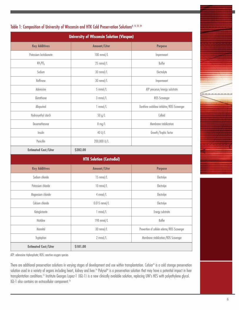
Table 1: Composition of University of Wisconsin and HTK Cold Preservation Solutions8, 13, 22, 24
There are additional preservation solutions in varying stages of development and use within transplantation. Celsior® is a cold storage preservationsolution used in a variety of organs including heart, kidney and liver.24 Polysol® is a preservation solution that may have a potential impact in livertransplantation conditions.25 Institute Georges Lopez-1 (IGL-1) is a new clinically available solution, replacing UW’s HES with polyethylene glycol.IGL-1 also contains an extracellular component.26
6
University of Wisconsin Solution (Viaspan)
Key Additives Amount/Liter Purpose
Potassium lactobionate 100 mmol/L Impermeant
KH2PO4 25 mmol/L Buffer
Sodium 30 mmol/L Electrolyte
Raffinose 30 mmol/L Impermeant
Adenosine 5 mmol/L ATP precursor/energy substrate
Glutathione 3 mmol/L ROS Scavenger
Allopurinol 1 mmol/L Xanthine oxididase inhibitor/ROS Scavenger
Hydroxyethyl starch 50 g/L Colloid
Dexamethasone 8 mg/L Membrane stabilization
Insulin 40 U/L Growth/Trophic factor
Penicillin 200,000 U/L
Estimated Cost/Liter $282.00
HTK Solution (Custodiol)
Key Additives Amount/Liter Purpose
Sodium chloride 15 mmol/L Electrolye
Potassium chloride 10 mmol/L Electrolye
Magnesium chloride 4 mmol/L Electrolye
Calcium chloride 0.015 mmol/L Electrolye
Ketoglutarate 1 mmol/L Energy substrate
Histidine 198 mmol/L Buffer
Mannitol 30 mmol/L Prevention of cellular edema/ROS Scavenger
Tryptophan 2 mmol/L Membrane stabilization/ROS Scavenger
Estimated Cost/Liter $181.00
ATP: adenosine triphosphate; ROS: reactive oxygen species
7
CLINICAL TRIAL REVIEWUniversity of Wisconsin solution has been considered the standard preservation solution in deceased donor abdominal transplantation. As new preservation solutions, such asHTK, have been introduced, comparison studies have been performed to identify similarities and differences in unique organ transplant donor and recipient populations. Thefollowing section will review selected recent clinical trials comparing HTK solution with UW solution, in both pancreas and liver transplantation.
Liver TransplantationDespite experience in Europe dating back to the late 1980s, the first reported experience with HTK in liver transplantation in the United States came from the University ofPittsburgh in 2002.27 Since then, multiple studies comparing HTK to UW solution in liver transplantation have been published (Table 2).
Feng and colleagues conducted a systematic review to compare the safety and efficacy of UW and HTK solutions.28 They reviewed 10 published full text papers, including 11comparisons in 1200 patients, in deceased donor and living related donor liver transplants, to assess patient and graft survival, liver function, and biliary complications. Therewere no statistically significant differences in patient survival, graft survival, primary non-function or acute rejection between UW and HTK. There was a statistically significantincrease in bile production associated with HTK use (95% CI, 18.65-57.47; p=0.0001).
Chan and colleagues conducted a prospective study comparing safety and efficacy of UW and HTK solutions in a consecutive series of 60 right lobe adult-to-adult living donorliver transplants.29 Main outcomes were post-transplant liver biochemistry, prothrombin time, recipient morbidity, and graft and patient survival. There were no significantdifferences in the outcomes measures. The investigators suggested the low potassium content of the HTK offered logistical advantages. There may be less concern for thesmaller amount of potassium entering the systemic circulation upon clamp release. In addition, the lower viscosity of HTK solution allows flushing of the hepatic artery onthe back table by gravity alone, reducing the risk of intimal injury due to additional pressure.
A large case series comparing the perioperative and first year outcomes of liver transplantation using UW solution or HTK also demonstrated no significant differences in 1 monthgraft function and 12 month graft or patient survival.23 There was also no difference in the incidence of primary non-function (UW 1.5%, HTK 0.5%; p=NS). In contrast toprevious reports using large volume flushes (10 to 20 L), in this case series, the HTK-preserved livers received an average of only 600mL more preservation than the liverspreserved with UW (3800mL HTK vs. 3200mL UW). Because of this, the investigators suggested that large volume infusion of HTK solution may be unnecessary for safeorgan preservation and may allow for even greater cost savings to be realized with HTK as compared to UW solution.
In a prospective single center analysis, Mangus and colleagues compared HTK and UW for immediate function and long-term transplant outcomes in whole organ DCD livertransplants (n=698), performing additional survival analysis on ECD subgroups.5 Fewer biliary complications were observed in the HTK group, including any need for biliaryevaluation (HTK 51% vs. UW 60%; p=0.02) or the presence of bile duct stones or sludge (HTK 3.8% vs. UW 11%; p=0.001). The authors suggested that this may be dueto improved flushing of the biliary microcirculation with HTK solution, as it is less viscous. There was no difference between the UW or HTK groups in graft loss within 7 daysof transplant in either standard criteria donor or extended criteria donor donors. In addition, patient and graft survival and graft function were equivalent between ECD liverpreserved with either UW solution or HTK solution.
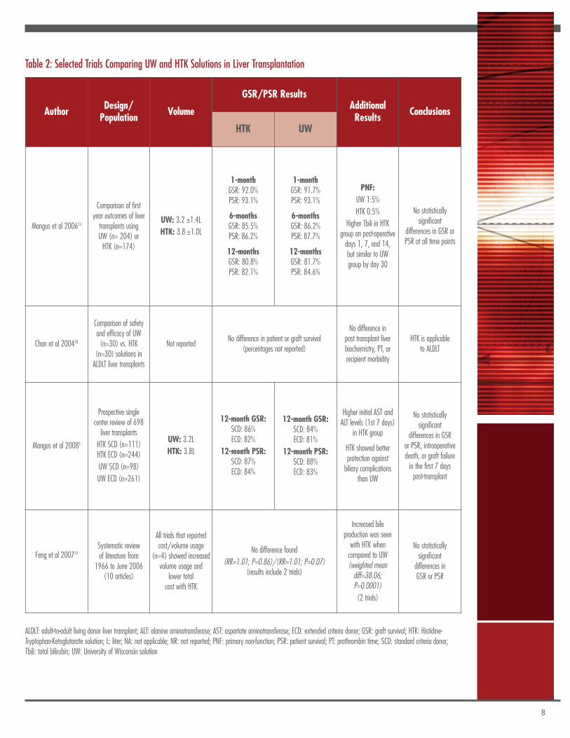
Table 2: Selected Trials Comparing UW and HTK Solutions in Liver Transplantation
8
ALDLT: adult-to-adult living donor liver transplant; ALT: alanine aminotransferase; AST: aspartate aminotransferase; ECD: extended criteria donor; GSR: graft survival; HTK: Histidine-Tryptophan-Ketoglutarate solution; L: liter; NA: not applicable; NR: not reported; PNF: primary non-function; PSR: patient survival; PT: prothrombin time; SCD: standard criteria donor;Tbili: total bilirubin; UW: University of Wisconsin solution
Author Design/Population Volume
GSR/PSR ResultsAdditionalResults Conclusions
HTK UW
Mangus et al 200623
Comparison of firstyear outcomes of livertransplants usingUW (n= 204) orHTK (n=174)
UW: 3.2 ±1.4LHTK: 3.8 ±1.0L
1-monthGSR: 92.0%PSR: 93.1%
6-monthsGSR: 85.5%PSR: 86.2%
12-monthsGSR: 80.8%PSR: 82.1%
1-monthGSR: 91.7%PSR: 93.1%
6-monthsGSR: 86.2%PSR: 87.7%
12-monthsGSR: 81.7%PSR: 84.6%
PNF:UW 1.5%HTK 0.5%
Higher Tbili in HTKgroup on post-operativedays 1, 7, and 14,but similar to UWgroup by day 30
No statisticallysignificant
differences in GSR orPSR at all time points
Chan et al 200428
Comparison of safetyand efficacy of UW(n=30) vs. HTK
(n=30) solutions inALDLT liver transplants
Not reportedNo difference in patient or graft survival
(percentages not reported)
No difference inpost transplant liverbiochemistry, PT, orrecipient morbidity
HTK is applicableto ALDLT
Mangus et al 20085
Prospective singlecenter review of 698liver transplantsHTK SCD (n=111)HTK ECD (n=244)UW SCD (n=98)UW ECD (n=261)
UW: 3.2LHTK: 3.8L
12-month GSR:SCD: 86%ECD: 82%
12-month PSR:SCD: 87%ECD: 84%
12-month GSR:SCD: 84%ECD: 81%
12-month PSR:SCD: 88%ECD: 83%
Higher initial AST andALT levels (1st 7 days)
in HTK group
HTK showed betterprotection againstbiliary complications
than UW
No statisticallysignificant
differences in GSRor PSR, intraoperativedeath, or graft failurein the first 7 dayspost-transplant
Feng et al 200729Systematic reviewof literature from
1966 to June 2006(10 articles)
All trials that reportedcost/volume usage
(n=4) showed increasedvolume usage and
lower totalcost with HTK
No difference found(RR=1.01; P=0.86)/(RR=1.01; P=0.07)
(results include 2 trials)
Increased bileproduction was seenwith HTK whencompared to UW(weighted meandiff=38.06;P=0.0001)(2 trials)
No statisticallysignificantdifferences inGSR or PSR
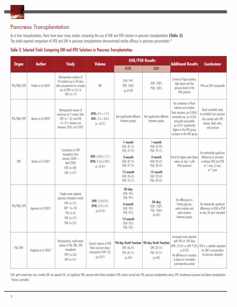
Pancreas TransplantationAs in liver transplantation, there have been many studies comparing the use of UW and HTK solution in pancreas transplantation (Table 3).The initial reported comparison of HTK and UW in pancreas transplantation demonstrated similar efficacy in pancreas preservation.30
Table 3: Selected Trials Comparing UW and HTK Solutions in Pancreas Transplantation
9
Organ Author Study VolumeGSR/PSR Results
Additional Results ConclusionsHTK UW
PTA/PAK/SPK Potdar et al 200432
Retrospective analysis of33 patients up to 30 daysafter procurement to compare
use of HTK (n=16) toUW (n=17)
NRGSR: 94%PSR: 100%(p=0.49)
GSR: 100%PSR: 100%
A trend of high amylase,high lipase and lowglucose levels in the
HTK patients
HTK and UW comparable
PTA/PAK/SPK Alonso et al 200833
Retrospective review ofoutcomes at 2 centers afterHTK (n= 16) and UW(n= 81) solution use
between 2001 and 2007
HTK: 4.9 ± 1.2 LUW: 2.6 ± 0.8 L
(p <0.01)
Not significantly differentbetween groups
Not significantly differentbetween groups
No correlation of flushvolume and amylase
Peak amylase (p=0.004),octreotide use (p=0.03),and graft pancreatitis(p=0.01) significantlyhigher in the HTK groupcompare to the UW group
Need controlled studyto establish best practicesUse caution with HTKvolume, flush rates,
and pressure
SPK Becker et al 20073
Comparison in SPKtransplants fromJanuary 2000 –April 2006HTK (n=48)UW (n=47)
UW: 4.84±1.11 LHTK: 9.65±2.00 L
(p <0.01)
1-monthGSR: 87.5%PSR: 97.9%
3-monthGSR: 87.5%PSR: 97.9%
12-monthGSR: 85.4%PSR: 95.7%
1-monthGSR: 87.0%PSR: 95.7%
3-monthGSR: 82.6%PSR: 89.4%
12-monthGSR: 82.6%PSR: 89.4%
Trend for higher peak lipasevalues on day 1 withHTK treatment
No statistically significantdifferences in pancreasor kidney GSR and PSRat 1 mos, 3 mos,
or 1 year
PTA/PAK/SPKAgarwal et al 200531
Single center updatedpancreas transplant results
HTK (n=75)UW* (n=10)PTA (n=6)SPK (n=57)PAK (n=22)
UW: 3.4±0.8 LHTK: 3.9±1.0 L
(p=0.26)
30-dayGSR: 96%PSR: 99%
6-monthGSR: 94%PSR: 95%
12-monthGSR: 92%PSR: 93%
30-dayGSR: 100%PSR: 100%(p=NS)
No differences infasting glucose,serial amylase andpeak amylasebetween groups
No statistically significantdifferences in GSR or PSRon day 30 post transplant
PTA/SPKEnglesbe et al 200634
Retrospective, multi-centerreview in PTA, PAK, SPK
transplantsHTK (n=36)UW (n=41)
Greater volume of HTKflush used per donorcompared to UW (2L)
(p<0.01)
90-day Graft FunctionSPK: 86.4%PTA: 85.7%(p=NS)
90-day Graft FunctionSPK: 87.5%PTA: 94.1%(p=NS)
Increased acute rejectionwith HTK at 180 days
(HTK: 25.0% vs UW: 9.8%;p<0.05)
No difference in amylaseor lipase in immediatepost-operative period
HTK is a suitable substitutefor UW in preservationof pancreas allografts
GSR: graft survival rate; mos: months; NR: not reported; NS: not significant; PAK: pancreas after kidney transplant; PSR: patient survival rate; PTA: pancreas transplantation alone; SPK: simultaneous pancreas and kidney transplantation*historic controlled
An updated analysis from the same group compared 78 pancreas transplants flushed with HTK compared to historical controls preserved withUW (n=10). Comparable 30-day and 1-year patient and graft survival, along with similar fasting blood glucose and serum amylase levels at allpost-transplantation intervals were demonstrated. Thirty day and 1 year patient survival with HTK was 99% and 93%; graft survival was 96%and 92%. There were no cases of primary non-function.31
Controversy exists regarding the use of HTK in pancreas transplantation due to the larger volume of HTK needed and the susceptibility of thepancreas to edema.3 However, comparable graft function and patient and graft 30-day survival was reported by Potdar et al. in UW preserved(n=17) versus HTK preserved (n=16) pancreas transplants.32 Subjective observations recorded during back table preparation and after reperfusionnoted that pancreas allografts flushed with HTK were more edematous, although pancreatitis was seen with equal frequency (HTK, 23%; UW,18%; p = 0.71). It should be noted that this study utilized larger volumes of HTK than the Indiana experience.
A retrospective review of pancreas transplants included SPK, PTA, and PAK; from 2 transplant centers (n=97) flushed with either HTK (n=16) orUW solution (n=81) revealed a greater rate of graft loss due to thrombosis in the HTK group (3/16; 19%) than in the UW group (3/81; 4%)(p=0.05).33 The authors stated that they did not necessarily believe that HTK solution inadequately replaced UW in pancreas transplantation, butthat caution should be used with respect to the volume of flush, flush rate, and pressure when using HTK.
Interestingly, Englesbe et al observed more acute rejection during the first 6 months post-transplant with HTK compared to UW solution (25%vs. 9.8% in pancreas transplants overall; 27% vs. 0% in SPK transplants; both p < 0.05).34 However, there were similar outcomes reported withrespect to technical graft loss, 90 day graft function, and rate of pancreatic leak/abscess. In addition, there was no significant difference betweenthe 2 groups in postoperative amylase and lipase.
10
11
CASE STUDIES: PANCREAS TRANSPLANTATIONAs evidenced by the clinical trial comparisons of HTK and UW solutions previously described, it is often challenging to translate the results of these studies into clinical practice.
The following case studies describe actual patient experiences. While we agree that it is somewhat difficult to ascribe particular patient and graft outcomes in complex solidorgan transplant recipients to the choice of cold storage preservation solution, these case study descriptions and their associated expert commentaries highlight importantcurrent clinical considerations in differentiating among preservation solutions.
Case Study 1KB is a 52 year old female with end-stage renal disease secondary to Type 1 diabetes mellitus. She underwent her second simultaneouskidney-pancreas transplant on August 1, 2007.
Patient Medical History
The patient received her first simultaneous pancreas-kidney transplant 5 years earlier utilizing a standard UW flush and preservation protocol. The pancreas failed due to pancreasthrombosis in the early postoperative period and was removed. Renal allograft failure due to chronic tubular atrophy/interstitial fibrosis occurred after repeated episodes ofacute cellular rejection and frequent urinary tract infections. She required daily insulin therapy and had increasing renal dysfunction prior to her second transplant, but had notyet returned to hemodialysis.
Donor Information
The donor for the second simultaneous pancreas-kidney transplant was a 26 year old male who had been in a motor vehicle accident and had sustained lethal intracranialhemorrhage and was declared brain dead. The donor was from the local organ procurement area. The cold ischemia time for the pancreas was 15 hours 23 minutes; for thekidney, cold ischemia time was 18 hours 21 minutes. In situ flushing and cold preservation for both organs were with 7-8 liters HTK solution as per local practice. The flushafter the pancreas back table was with approximately 300mL of HTK solution.
Transplant Course
The transplant surgical procedure was uneventful. Both pancreas and kidney had good function in the early post-transplant period. Insulin was never needed post- transplant.The patient was preconditioned with alemtuzumab. Tacrolimus monotherapy was initiated as maintenance immunosuppression.
Post-operatively, the patient developed a urinary tract infection and pneumonia and required inpatient rehabilitation due to severe deconditioning. Both allografts continuedto function well. She experienced one episode of pancreas rejection, which was successfully treated with a corticosteroid bolus and recycle, and the addition of mycophenolatemofetil to the maintenance immunosuppressive regimen.
The patient remains off insulin therapy or oral hypoglycemic agents. Laboratory values at her one year follow-up visit were serum creatinine 1.1 mg/dL , BUN 19 mg/dL,fasting blood glucose 92 mg/dL, amylase 74 U/L, and lipase 62 U/L.
12
Case Study 2JB is a 56 year old male with Type 1 diabetes mellitus since age 2. In August 2006, he underwent pancreastransplantation (pancreas after kidney) after receiving deceased donor kidney transplants in 1981 and in 2002.
Patient Medical History
In addition to Type 1 diabetes mellitus, the patient also had a past medical history of hypertension, rheumatoid arthritis, stroke, and coronaryartery disease, with a previous myocardial infarction.
Donor Information
The donor was a 20 year old female deceased donor from Utah who was receiving 3 vasopressors prior to pancreas recovery. HLA matchingshowed 0 antigen mismatch. The pancreas cold ischemia time was 15 hours 39 minutes. Flushing and cold preservation were with HTK solution.However, the volume of in situ flush with HTK was not known as the pancreas came from an outside organ procurement organization. The backtable flush was with approximately 300mL of HTK solution.
Transplant Course
The surgical procedure was uneventful. The patient was preconditioned with alemtuzumab and was placed on tacrolimus and mycophenolatemofetil maintenance immunosuppression. Postoperatively, the patient developed allograft pancreatitis with a peak lipase of 14,914 U/L. Thelipase decreased to a low of 641 U/L on post-operative day 11, but elevated again with resumption of oral intake. The allograft was studiedwith ultrasound and CT scans and found to have a pseudocyst in the tail. This was managed conservatively with eventual resolution.
The recipient remains well, with labs from his two year follow-up visit showing a serum creatinine of 1.6 mg/dL, BUN 24 mg/dL, fasting bloodglucose 111 mg/dL, amylase 66 U/L and lipase 174 U/L. He remains on no insulin.
EXPERT COMMENTARY: PANCREAS TRANSPLANT CASE STUDIESDavid E.R. Sutherland, MD, PhD
Cold preservation of the pancreas evolved over the years and proved to be a challenge compared to the cold preservation of other solid organs. The vascular and parenchymalstructure of the pancreas makes cold preservation less effective. Several cold preservation solutions have been used historically. At the University of Minnesota,pancreas perfusion preservation was initially performed using silica gel filtered plasma.35 Although this solution proved to be effective with good graft functionpost-transplantation, the inherent risks of disease transmission associated with the use of pooled human plasma was of great concern. UW solution was developed by Dr.Belzer and associates mainly to address the challenges of preserving the pancreas for transplantation. We successfully used UW solution for about 18 years and moved tothe use of HTK solution 2 years ago due to supply and cost considerations.
The two cases presented here from the University of Pittsburgh illustrate the current successful use of HTK solution for the cold preservation of a pancreas allograft. Withthe exception of this being a retransplant, the first case represents a straightforward SPK transplant. The cold ischemia time of 15 hours, which may seem somewhat longto some surgeons, is actually not excessive when compared to registry data. In an analysis of the International Pancreas Transplant Registry (IPTR), the pancreas preservationtime was less than 12 hours in 40% of cases and greater than 24 hours in only 5%.36 A clear correlation between outcome and preservation time could be observed inSPK transplant (p=0.05), with the cutoff at more than 23 hours of preservation. In multivariate models, the difference was confirmed where a longer preservation timewas associated with an increased hazard ratio (HR) of 1.35 (p=0.11) for overall SPK pancreas graft failure and 1.46 (p=0.18) for technical failure. This is in contrast toa solitary pancreas transplant where no impact of preservation time on overall graft failure or technical failure was observed. In addition, according to the IPTR analysis,donor age appears to have a greater impact on graft functional survival and technical failure rates than preservation. While we strive to keep the cold ischemia time of thepancreas as short as possible, our goal at the University of Minnesota is to transplant the pancreas in less than 24 hours.
The second case describes the recipient of another SPK transplant who developed allograft pancreatitis postoperatively. Early graft pancreatitis remains a common cause oftechnical failure after pancreas transplantation, with nearly 20% of patients affected in some series.37 Etiology of graft pancreatitis and thrombosis is considered multi-factorial.Suggested causes include reperfusion injury, mechanical stricture, trauma, donor obesity, and prolonged ischemia. Flush volume and pressure are also important risk factorsfor graft pancreatitis. In general, I believe larger volumes and higher flush pressures are detrimental. Volume and pressure of pancreas flush, however, may not always beeasily controlled in the context of a multi-organ recovery.
There is a perception of a possible association between flush and preservation with HTK solution and an increase in the risk of pancreas graft pancreatitis and thrombosis.However, no clear or definitive determination can be made as there has been only one prospective comparison of HTK and UW solutions in pancreas transplantation; in thestudy no differences in outcomes were found.38 Most published experiences related to this issue are single-center, retrospective reviews with relatively small numbers ofpatients. A recent brief report from Alonso and colleagues attempted to determine whether there was a basis for suspecting a causal relationship.33 Other studies of HTKsolution in pancreas transplantation have not observed an association between HTK and graft pancreatitis.4, 32, 34 At present, no evidence based claims can be made on therisk for post-transplantation pancreatitis when HTK has been used for cold preservation. It should also be noted that there is little evidence that there is less graft pancreatitiswith UW solution. However, in a recent review of the UNOS database outcomes for pancreas transplants preserved in HTK versus UW Solution, HTK preservationwas associated with an increased risk of graft loss (hazard ratio 1.30, p=0.01).39 Unmeasured confounding variables such as surgeon technique, volume of initial flushat the time of organ recovery, organ quality, differences in immunosuppression protocols, and actual causes of graft loss, emphasize the inherent limitations of a retrospectiveregistry review on one hand and underscore the need for prospective randomized studies on the other.
As reviewed earlier in this monograph, most trials have demonstrated that the overall safety and efficacy of HTK solution is comparable to that of UW solution inpancreas transplantation.
13
14
CASE STUDIES: LIVER TRANSPLANTATION
Case Study 3CD is a 47 year old male with end-stage liver disease secondary to Hepatitis C cirrhosis, complicated byhepatocellular carcinoma.
Patient Medical History
The patient was found to have four lesions in both the right and left lobes and was treated with two sessions of transarterial chemo-embolizationin the subsequent 12 months. He was listed for liver transplant by his biological MELD score of 16, since he remained outside of Milan criteria.He underwent a deceased donation after cardiac death (DCD) transplant on August 14, 2007.
Donor Information
The donor was 48 year old DCD donor from outside the local OPO, but within the region, who fell from a loading truck and sustained skull fracturesand blunt head trauma. The donor did not progress to brain death, but in light of the extensive brain injury, the family requested donation aftercardiac death. The donor was not breathing above the ventilator, but was not on any vasopressors. A donor team was dispatched. Prior toextubation 30,000 units of systemic heparin was administered. The donor quickly succumbed and was pronounced dead 12 minutes later. Afterthe required 3 minute standoff, recovery commenced with aortic cannulation and flush with 6 liters of HTK. During this time, venting of the venacava was done trans-diaphramatically and the liver and kidneys were procured. In situ flushing and cold preservation for both organs were withHTK solution. The initial impression by the donor surgeon was that the liver was well perfused, although the right lobe remained somewhat firm.The kidneys were placed on the pump, as per local practice, and were transplanted unremarkably into two kidney recipients with immediatefunction and cold ischemia times of 21 and 26 hours. The cold ischemia time for the liver was 10 hours, 13 minutes.
Transplant Course
Upon exploration, there were five hepatocellular carcinoma lesions with gross macronodular cirrhosis and extensively vascularized adhesions to theanterior abdominal wall. The technical summary of the operation showed a piggyback operation without venovenous bypass, end-to-side cavocavalanastomosis, portal vein-to-portal vein end-to-end anastomosis, hepatic artery-to-hepatic artery anastomosis, and duct-to-duct anastomosis withoutT-tube. After portal vein revascularization, 20 mg of recombinant tissue plasminogen activator (rTPA) was instilled into the hepatic artery andclamped for 20 minutes. At this time, the hepatic artery was backbled and the liver re-arterialized.
Following revascularization, although there was minimal post-reperfusion syndrome, there was moderate fibrinolysis which was treated withcoagulation factor replacement. At the end of the case, the liver was making modest amounts of bile and the serum lactate peaked at 4.4 mg/dL.
The following day, ALT peaked at 2,454 IU/L and the INR at 2.5, while the AST peaked at 9,219 IU/L on post-operative day 2. The totalbilirubin peaked on post-operative day 7 at 21.5 mg/dL. A liver biopsy demonstrated moderate cholestasis with hepatocyte dropout consistentwith moderate ischemia/reperfusion injury, without evidence of rejection. An endoscopic retrograde cholangiopancreatography (ERCP) demonstratednormal bile ducts without obstruction or leak and a biliary stent was placed after instrumentation. The bilirubin began to decrease immediatelythereafter and the patient was discharged on post-operative day 14 with a total bilirubin of 8.8 mg/dL. By post-operative day 21, the bilirubinhad dropped to 2.7 mg/dL and was normal by post-operative day 32. The patient was seen again for stent removal on POD 45 and thecholangiogram was normal. At the one year follow-up, the liver function tests were normal and magnetic resonance cholangiopancreatography(MRCP) revealed no evidence of biliary anomalies. There was no evidence of HCC recurrence on computed tomography (CT) imaging. HCV levelswere 850,000 U/mL at one year and the one year protocol biopsy revealed minimal inflammation without fibrosis.
15
Case Study 4The patient is a 52 year old male patient with end stage liver disease and cirrhosis due to hepatitis C virus infection.
Patient Medical History
A 2.5 cm solitary hepatocellular carcinoma (HCC) lesion had been identified within the left lobe of the liver during the pre-transplant evaluation. The HCC was treated withtranscatheter arterial chemoembolization (TACE) and open radiofrequency ablation. The patient was admitted to the intensive care unit (ICU) and required intubatation due tothe development of intractable ascites and hepatic hydrothorax. Continued decompensation was evidenced by prerenal azotemia, renal failure and overall physical deconditioning.Pre-transplant imaging confirmed cirrhosis of the liver, identified a stable liver mass in the left lobe and raised the possibility of mesenteric vein/portal vein thrombosis. TheMELD score was 27.
Donor Information
The donor was a 47 year old African American male with history of hypertension. The cause of death was intracranial hemorrhage. He had been receiving treatment for thehypertension for several years, with evidence of compliance. There was only mild elevation of liver enzymes following resuscitation. He was 5 foot, 7 inches tall and weighed 84kg.
On visual inspection during organ recovery, the liver appeared to be of normal size and had adequate capillary refill. In situ flush and cold preservation was performed with5 liters of HTK solution. The cold ischemic time was 6 hours.
Transplant Course
During the transplant procedure, massive portal hypertension was noted. Retro-caval dissection was deemed too dangerous due to the extent of collateralization. Pre-cavaldissection was undertaken and the hepatic veins were controlled, allowing caval anastomosis in a piggyback fashion. Although the portal vein appeared to be partially patentby palpation, it was discovered to have cavernous transformation grade IV portal vein thrombosis when transected. This finding precluded direct portal anastomosis. Attempts todeclot the portal vein were unsuccessful. Alternative revascularization on the superior mesenteric vein was also not possible as the clot had extended to the complete length ofthe superior mesenteric vein. Dissection around the renal vein was not possible because of the extensive collateralization and the presence of retroperitoneal adhesions relatedto the prior open radiofrequency ablation procedure. To achieve portal flow, arterialization of the donor portal vein from an accessory hepatic artery was performed. Arterialrevascularization was performed using a supraceliac aortic graft that was anastomosed to the donor common hepatic artery.
This unusual revascularization procedure required an extended period of time during which the liver allograft remained in the surgical field, prior to reperfusion. The liver allograftsustained approximately 45 minutes of warm ischemic time until hepatic artery revascularization, as well as an additional 50 minutes of warm ischemia while the dissectionand arterialization of the portal vein was completed. At the end of the transplant procedure, variceal banding was completed due to persistent portal hypertension. A doublevalve peritoneovenous Denver shunt was also placed.
Immediate graft function was observed post-transplant with peak AST and ALT in the 900-1100 IU/L range that recovered to normal in 72 hours. The patient was extubatedin 24 hours and made an uneventful recovery until released from the hospital 11 days post-transplant. On follow-up six months post-transplant, the patient exhibited onlyminimal ascites, with excellent liver and kidney function.
16
EXPERT COMMENTARY: LIVER TRANSPLANT CASE STUDIESJohn J. Fung, MD, PhD and Charles Miller, MD
UW solution has been the “gold standard” for liver preservation and is still widely used. The liver transplant cases presented illustrate thesuccessful use of HTK solution in complicated cases. The increased use of HTK preservation solution in the last few years reflects a continuoussearch for better preservation techniques. At least two studies have recently documented a higher incidence of late biliary complicationsassociated with the use of UW solution for liver preservation.29, 40 Beginning in 2002, the University of Pittsburgh was the first US liver transplantprogram to use HTK solution. The lower viscosity of HTK compared to UW solution offered a potential advantage in lowering biliary complicationrates by providing for a better flush of the biliary system and its microvasculature. The increased use of ECDs and especially DCDs underscoresthe need for optimal preservation of the liver as a whole and the biliary system in particular.
In DCD, formerly known as non-heart beating donors (NHBD), the adverse outcomes associated with higher graft loss due to primarynon-function and increased risk for biliary strictures, the use of HTK has been suggested to provide improved early graft survival and decreasedbiliary stricture rates, especially when combined with intra-arterial thrombolytic therapy.41, 42
The pathophysiology of graft dysfunction in DCD transplantation represents the most extreme manifestation of the ischemia/reperfusionpathway seen in preserved allografts, but magnified by varying degrees of microcirculatory thrombosis. The impact of this vascular stasis andthrombosis depends on the organ — with kidneys this is manifested by a high rate of acute tubular necrosis and delayed graft function;with livers, there is a higher incidence of macrovascular thrombosis, primary non-function leading to a higher rate of early retransplantation,and more concerningly, an increased rate of late biliary strictures leading to increased morbidity and mortality. Recent experiences withcrystalloid based preservation solutions combined with thrombolytic agents suggest that better outcomes can be obtained by enhancingmicrocirculatory integrity and minimizing post-perfusion microthrombosis.
The impact of DCD in liver transplantation has been assessed from the SRTR database.41 DCD graft survival was significantly lower than donarbrain death grafts with 1- and 3-year graft survival of 70.2% and 63.3% for DCD recipients versus 80.4% and 72.1% for donor brain deathgraft recipients. Recipients of a DCD graft had a greater incidence of primary non-function (11.8 vs. 6.4%) and re-transplantation (13.9% vs.8.3%) compared with DBD recipients. The experience with DCD in the era of a colloid based preservation solution (i.e. UW solution), whichconstitutes the bulk of the SRTR DCD experience, suggests that almost three times as many DCD livers (up to 35-50% of livers) will sufferfrom ischemic-type biliary strictures (ITBS) as compared to donor brain death donor livers.43 This is due to the unique nature of blood supplyfor the bile duct, which depends solely on hepatic artery blood supply via a vascular plexus assuring bile duct viability.26 In DCD, the mandatedperiod of warm ischemia imposed during the declaration of death in the controlled DCD donor and during the logistical delays inherent in theprocess of consent in the uncontrolled DCD donor, leads to stagnation of blood in the microcirculation. Although heparin is permitted in manyDCD procurements, removal of formed blood elements from the microvasculature is dependent on adequate flushing.
Since flow through a hollow vessel is inversely related to the viscosity to the third power, the more viscous UW solution is less likely to penetrateand adequately flush the biliary plexus, potentially leading to ITBS.44
Crystalloid based preservation solutions, such as Histidine-Tryptophan-Ketogluterate (HTK) and Celsior, have the physical advantage of beingsignificantly less viscous than UW solution. In addition, UW solution has been shown to develop adenosine crystals at sub-zero temperatures,which can further cause microcirculatory complications.19 As a result, crystalloid solutions are more likely to flush stagnant blood from themicrovasculature in DCD organs than colloid based preservation solutions. Indeed, HTK has been shown to be associated with lower rates ofbiliary strictures than UW solution and the use of HTK solution for liver preservation was validated in a large series of deceased donor transplantscompleted at Indiana University.23, 45, 46
If DCD livers are to be used in greater frequency, the outcomes will need to be predictably similar to standard donar brain death. How canthis be achieved? Some hints can be derived from other clinical settings, as well as in animal and human DCD studies. The development ofhepatic artery thrombosis (HAT) following liver transplantation is one of the most devastating complications, with a high risk of subsequentbiliary tract ischemia and necrosis, resulting in a high rate of retransplantation. In 1996, in a series of 17 patients that developed HAT earlyafter liver transplant, an approach of immediate thrombectomy with the use of thrombolytic therapy directly instilled into the hepatic arteryand revascularization demonstrated that 88% of liver allografts could be successfully treated with long-term patency of the hepatic arteryand more importantly, no patient developed ITBS.47
The research group from the University of Bonn demonstrated graft alterations including erythrocyte aggregation and thrombus formation, which affected equilibration ofthe preservation solution within the microvasculature preventing effective cold preservation in a rat NHBD model. The compromised microvascular perfusion and release ofliver enzymes associated with the use of UW solution for the initial flushout of livers was markedly attenuated by the additional warm preflush with Ringer’s lactatedsolution containing streptokinase.48 Other animal models have also validated the use of thrombolytic agents in DCD.49
Investigators at the University of Newcastle upon Tyne then conducted a double-blinded, randomized, controlled trial of streptokinase preflush or placebo in human DCDkidneys. Following declaration of cardiac death, a solution containing therapeutic doses of streptokinase was given prior to initiation of infusion of preservation solution, inthis case, Euro-Collins solution. DCD kidneys were machine-preserved and transplanted within 24 hours of procurement. These investigators noted that kidneys from thestreptokinase-treated donors had a significantly better appearance at procurement and there was a higher proportion of kidneys transplanted through the use of streptokinase(63.6% with streptokinase vs. 42.6% with placebo). Given that the majority of DCD were uncontrolled, the high incidence of delayed graft function was not surprising, butall streptokinase treated kidneys recovered function.50
At our own center, we have utilized a protocol of tissue plasminogen activator (TPA) use along with systemic heparin, preferentially given in situ, before declaration of death,followed by preservation flush with HTK. In those instances where TPA or heparin cannot be used before declaration of death, heparin is given along with HTK and the liveris cold-preserved. After portal revascularization in the recipient and the liver is warmed, and 20 mg of TPA in a volume of 7 – 10 cc of normal saline is instilled into thedonor hepatic artery and clamped for 20-30 minutes. The hepatic artery is then back-bled to allow excess TPA to be discarded in order to minimize its introduction into therecipient circulation.44 These practical experiences address some concerns, such as exacerbation of ischemia-reperfusion injury and enhancement of operative bleeding.51
Relevant to the current cases is the finding that outcomes were good, even with cold ischemic time extending beyond 14 hours, and with the long warm ischemic timesdocumented in Case 4. In the latter case, the use of HTK solution was associated with acceptable early graft function and good liver and kidney function at 6 months offollow up.
These two technically challenging cases illustrate the effectiveness of HTK preservation solution in liver transplantation and although studies have shown that outcomes aresimilar when either UW or HTK solution is used, the reduced viscosity and better microcirculatory penetration of HTK solution may represent an advantage especially in avoidinglong-term biliary complications. In the future, however, large-scale clinical trials will be required to definitively evaluate the impact of different preservation solutions on graftfunction and on long-term biliary complications.
CONCLUSIONThe consistent reality of organ shortage for transplantation is the motivator for the increased use of marginal donors (ECD and DCD). The use of marginal donors underscores the importanceof effective and safe organ preservation techniques. Cold preservation of organs for transplantation utilizing various solutions has been the most common preservation technique. UW andHTK are the most common preservation solutions used. An overall assessment of the literature indicates that the use of UW and HTK solutions produce similar outcomes with respect to graftand patient survival.
The challenges in regards to preservation and early graft function are somewhat different between different organs and organ-specific consideration should be given to the useof any preservation technique and solution.
As discussed in this monograph, the use of a crystalloid preservation solution, like HTK, likely provides for improved cold preservation in liver transplantation, particularly in liver allograft fromDCD donors. For pancreas preservation, most publications support the conclusion that both HTK and UW cold preservation solutions provide effective preservation with comparable outcomes.The recent retrospective analysis of the UNOS database, demonstrating a negative effect of HTK pancreas preservation as compared to UW, carries the deficiencies of a retrospective,multicenter analysis and underscores the need for prospective studies.
Technical advantages related to the lack of need for additives, and low viscosity may increase the appeal of HTK solution in certain transplant settings. However, according to package labels,only 6-8L of UW solution is required for perfusion, compared to 10-12L of HTK.22, 52 Nevertheless, studies have documented the successful use of lower volumes of HTK, without compromisingoutcomes. In evaluating the cost of preservation fluids, acquisition, additives required for the use of UW solution, and total volume required must be considered.
In this activity, we have compared the composition, rationale for use, and outcomes of transplantation of the pancreas and the liver preserved with UW or HTK solutions. The lower osmolarityand lower concentration of electrolytes and histidine buffer systems inherent to HTK solution may provide a theoretical rationale for the choice of the less costly preservation solution by somecenters. Clinical data support the conclusion that outcomes are at least comparable following the use of either solution in pancreas or liver transplantation.
Careful analysis of both the advantages and disadvantages of preservation solutions for liver and pancreas transplantation has become important as the use of marginal donors (ECD andDCD) continues to increase.
17
1. Neto JS, Nakao A, Kimizuka K, et al. Protection of transplant-induced renal ischemia-reperfusion injury withcarbon monoxide. Am J Physiol Renal Physiol. Nov 2004;287(5):F979-989.
2. 2007 Annual Report of the U.S. Organ Procurement and Transplantation Network and the Scientific Registryof Transplant Recipients: Transplant Data 1997-2006. Health Resources and Services Administration,Healthcare Systems Bureau, Division of Transplantation, Rockville, MD.
3. Becker T, Ringe B, Nyibata M, et al. Pancreas transplantation with histidine-tryptophan-ketoglutarate (HTK)solution and University of Wisconsin (UW) solution: is there a difference? Jop. 2007;8(3):304-311.
4. Agarwal A, Powelson JA, Goggins WC, Milgrom ML, Fridell JA. Organ preservation with histidine-tryptophanketogluatarate solution in clinical pancreas transplantation: an update of the indiana university experience.Transplant Proc. Mar 2008;40(2):498-501.
5. Mangus RS, Fridell JA, Vianna RM, et al. Comparison of histidine-tryptophan-ketoglutarate solution andUniversity of Wisconsin solution in extended criteria liver donors. Liver Transpl. Mar 2008; 14(3):365-373.
6. Leichtman AB, Cohen D, Keith D, et al. Kidney and pancreas transplantation in the United States, 1997-2006: the HRSA Breakthrough Collaboratives and the 58 DSA Challenge. Am J Transplant. Apr 2008;8(4 Pt 2):946-957.
7. Freeman RB, Jr., Steffick DE, Guidinger MK, Farmer DG, Berg CL, Merion RM. Liver and intestine transplanta-tion in the United States, 1997-2006. Am J Transplant. Apr 2008;8(4 Pt 2):958-976.
8. Maathuis MH, Leuvenink HG, Ploeg RJ. Perspectives in organ preservation. Transplantation. May 272007;83(10):1289-1298.
9. Anaya-Prado R, Delgado-Vazquez JA. Scientific basis of organ preservation. Curr Opin Organ Transplant. Apr2008;13(2):129-134.
10. Montalvo-Jave EE, Escalante-Tattersfield T, Ortega-Salgado JA, Pina E, Geller DA. Factors in the pathophysiologyof the liver ischemia-reperfusion injury. J Surg Res. Jun 1 2008;147(1):153-159.
11. Koo DD, Welsh KI, Roake JA, Morris PJ, Fuggle SV. Ischemia/reperfusion injury in human kidney transplantation:an immunohistochemical analysis of changes after reperfusion. Am J Pathol. Aug 1998;153(2):557-566.
12. Ahmad N, Hostert L, Pratt JR, Billar KJ, Potts DJ, Lodge JP. A pathophysiologic study of the kidney tubule tooptimize organ preservation solutions. Kidney Int. Jul 2004;66(1):77-90.
13. Wilson CH, Brook NR, Talbot D. Preservation solutions for solid organ transplantation. Mini Rev Med Chem.Oct 2006;6(10):1081-1090.
14. Sung RS, Galloway J, Tuttle-Newhall JE, et al. Organ donation and utilization in the United States, 1997-2006. Am J Transplant. Apr 2008;8(4 Pt 2):922-934.
15. Southard JH, van Gulik TM, Ametani MS, et al. Important components of the UW solution. Transplantation.Feb 1990;49(2):251-257.
16. El-Wahsh M. Liver graft preservation: an overview. Hepatobiliary Pancreat Dis Int. Feb 2007; 6(1):12-16.17. Tojimbara T, Wicomb WN, Garcia-Kennedy R, et al. Liver transplantation from non-heart beating donors in
rats: influence of viscosity and temperature of initial flushing solutions on graft function. Liver Transpl Surg.Jan 1997;3(1):39-45.
18. Pirenne J, Van Gelder F, Coosemans W, et al. Type of donor aortic preservation solution and not coldischemia time is a major determinant of biliary strictures after liver transplantation. Liver Transpl. Jun2001;7(6):540-545.
19. Tullius SG, Filatenkow A, Horch D, et al. Accumulation of crystal deposits in abdominal organs followingperfusion with defrosted University of Wisconsin solutions. Am J Transplant. Aug 2002; 2(7):627-630.
20. Li XL, Man K, Ng KT, Lee TK, Lo CM, Fan ST. Insulin in UW solution exacerbates hepatic ischemia/reperfusioninjury by energy depletion through the IRS-2/SREBP-1c pathway. Liver Transpl. Sep 2004;10(9):1173-1182.
21. Bretschneider HJ. Myocardial protection. Thorac Cardiovasc Surg. Oct 1980;28(5):295-302.22. CUSTODIOL [package insert]. Newtown, PA: Essential Pharmaceuticals, LLC; 2006.23. Mangus RS, Tector AJ, Agarwal A, Vianna R, Murdock P, Fridell JA. Comparison of histidine-tryptophan-ketog-
lutarate solution (HTK) and University of Wisconsin solution (UW) in adult liver transplantation. Liver Transpl.Feb 2006;12(2):226-230.
24. Bellamy CA, Nicely B, Mattice BJ, Teaster R. Comparative analysis of clinical efficacy and cost betweenUniversity of Wisconsin solution and histidine-tryptophan-ketoglutarate. Prog Transplant. Sep 2008;18(3):166-171; quiz 172.
25. Hata K, Tolba RH, Wei L, et al. Impact of polysol, a newly developed preservation solution, on cold storageof steatotic rat livers. Liver Transpl. Jan 2007;13(1):114-121.
26. Maathuis MH, Ottens PJ, van Goor H, et al. Static cold storage preservation of ischemically damagedkidneys. a comparison between IGL-1 and UW solution. Transpl Int. May 2008;21(5):473-482.
27. Eghtesad B, Fontes P, Cacciarelli T, Jain A, Sindhi R, Geller D. Comparison of histidine-tryptophan-ketogluarate(HTK) solution vs. University of Wisconsin (UW) solution for organ preservation in liver transplantation.Hepatology. 2002;36(Pt 2):Abstract 2052.
28. Feng L, Zhao N, Yao X, et al. Histidine-tryptophan-ketoglutarate solution vs. University of Wisconsin solutionfor liver transplantation: a systematic review. Liver Transpl. Aug 2007;13(8):1125-1136.
29. Chan SC, Liu CL, Lo CM, Fan ST. Applicability of histidine-tryptophan-ketoglutarate solution in right lobeadult-to-adult live donor liver transplantation. Liver Transpl. Nov 2004;10(11):1415-1421.
30. Fridell JA, Agarwal A, Milgrom ML, Goggins WC, Murdock P, Pescovitz MD. Comparison of histidine-tryptophan-ketoglutarate solution and University of Wisconsin solution for organ preservation in clinical pancreastransplantation. Transplantation. Apr 27 2004;77(8):1304-1306.
31. Agarwal A, Murdock P, Pescovitz MD, Goggins WC, Milgrom ML, Fridell JA. Follow-up experience usinghistidine-tryptophan ketoglutarate solution in clinical pancreas transplantation. Transplant Proc. Oct2005;37(8):3523-3526.
32. Potdar S, Malek S, Eghtesad B, et al. Initial experience using histidine-tryptophan-ketoglutarate solution inclinical pancreas transplantation. Clin Transplant. Dec 2004;18(6):661-665.
33. Alonso D, Dunn TB, Rigley T, et al. Increased pancreatitis in allografts flushed with histidine-tryptophan-ketoglutarate solution: a cautionary tale. Am J Transplant. Sep 2008;8(9):1942-1945.
34. Englesbe MJ, Moyer A, Kim DY, et al. Early pancreas transplant outcomes with histidine-tryptophan-ketoglutarate preservation: a multicenter study. Transplantation. Jul 15 2006;82(1):136-139.
35. Sutherland DE, Gruessner RW, Dunn DL, et al. Lessons learned from more than 1,000 pancreas transplantsat a single institution. Ann Surg. Apr 2001;233(4):463-501.
36. Gruessner AC, Sutherland DE. Pancreas transplant outcomes for United States (US) and non-US cases asreported to the United Network for Organ Sharing (UNOS) and the International Pancreas Transplant Registry(IPTR) as of June 2004. Clin Transplant. Aug 2005;19(4):433-455.
37. Humar A, Ramcharan T, Kandaswamy R, Gruessner RW, Gruessner AC, Sutherland DE. Technical failures afterpancreas transplants: why grafts fail and the risk factors — a multivariate analysis. Transplantation. Oct 272004;78(8):1188-1192.
38. Schneeberger S, Biebl M, Steurer W, et al. A prospective randomized multicenter trial comparing histidine-tryptophane-ketoglutarate versus University of Wisconsin perfusion solution in clinical pancreas transplantation.Transpl Int. Oct 24 2008.
39. Stewart ZA, Cameron, A.M., Singer, A.L., Dagher, N.N., Montgomery, R.A., Segev, D.L. Histidine-Tryptophan-Ketoglutarate (HTK) is associated with reduced graft survival in pancreas transplantation. Am J Transplant.2008;8:1-5.
40. Canelo R, Hakim NS, Ringe B. Experience with hystidine tryptophan ketoglutarate versus UniversityWisconsin preservation solutions in transplantation. Int Surg. Jul-Sep 2003;88(3):145-151.
41. Abt P, Crawford M, Desai N, Markmann J, Olthoff K, Shaked A. Liver transplantation from controllednon-heart-beating donors: an increased incidence of biliary complications. Transplantation. May 272003;75(10):1659-1663.
42. Eghtesad B, Aucejo F, Fung JJ. Preservation solutions in liver transplantation: what are the options?Liver Transpl. Feb 2006;12(2):196-198.
43. Foley DP, Fernandez LA, Leverson G, et al. Donation after cardiac death: the University of Wisconsinexperience with liver transplantation. Ann Surg. Nov 2005;242(5):724-731.
44. Fung JJ, Eghtesad B, Patel-Tom K. Using livers from donation after cardiac death donors — a proposalto protect the true Achilles heel. Liver Transpl. Dec 2007;13(12):1633-1636.
45. Welling TH, Heidt DG, Englesbe MJ, et al. Biliary complications following liver transplantation in the modelfor end-stage liver disease era: effect of donor, recipient, and technical factors. Liver Transpl. Jan2008;14(1):73-80.
46. Testa G, Malago M, Nadalin S, et al. Histidine-tryptophan-ketoglutarate versus University of Wisconsin solutionin living donor liver transplantation: results of a prospective study. Liver Transpl. Aug 2003;9(8):822-826.
47. Pinna AD, Smith CV, Furukawa H, Starzl TE, Fung JJ. Urgent revascularization of liver allografts after earlyhepatic artery thrombosis. Transplantation. Dec 15 1996;62(11):1584-1587.
48. Yamauchi J, Schramm R, Richter S, Vollmar B, Menger MD, Minor T. Improvement of microvascular graftequilibration and preservation in non-heart-beating donors by warm preflush with streptokinase.Transplantation. Feb 27 2003;75(4):449-453.
49. Sugimoto R, Date H, Sugimoto S, et al. Post-mortem administration of urokinase in canine lung transplantationfrom non-heart-beating donors. J Heart Lung Transplant. Sep 2006;25(9):1148-1153.
50. Gok MA, Shenton BK, Buckley PE, et al. How to improve the quality of kidneys from non-heart-beatingdonors: a randomised controlled trial of thrombolysis innon-heart-beating donors. Transplantation. Dec 27 2003;76(12):1714-1719.
51. Porte RJ, Clavien PA. Preflush with plasminogen activator in non-heart-beating donors: is it worth it?Transplantation. May 15 2000;69(9):1769-1771.
52. Viaspan package insert. Wilmington, DE: Dupont Pharma, 1998.
REFERENCES
18
Visit our website at www.CTIFacts.com©2008 CTI Clinical Trial and Consulting ServicesAll Rights Reserved. Printed in the U.S.A.
None of the content of this enduring material may be reproduced in any form without prior permission of CTI Clinical Trial and Consulting Services. The opinionsexpressed in this enduring material are those of the authors and do not necessarily reflect the opinions or recommendations of the affiliated institutions,Essential Pharmaceuticals, the publisher or any other persons. Any procedures, medications or other courses of diagnosis or treatment discussed or suggestedin this enduring material should not be used by clinicians without evaluation of their patients’ conditions, assessments of possible contraindications or dangersin use, review of applicable manufacturer’s product information, and comparison with the recommendations of their authorities.
C L I N I C A L C A S E S T U D I E S A N D E X P E R T C O M M E N T A R Y
Jointly sponsored by University of Kentucky College of Medicine andCTI Clinical Trial and Consulting Services