Current trends in rapid tests for mycotoxins
34
Current trends in rapid tests for mycotoxins Nolan, P., Auer, S., Spehar, A., Elliott, C. T., & Campbell, K. (2019). Current trends in rapid tests for mycotoxins. Food Additives and Contaminants: Part A, 36(5), 800-814. https://doi.org/10.1080/19440049.2019.1595171 Published in: Food Additives and Contaminants: Part A Document Version: Peer reviewed version Queen's University Belfast - Research Portal: Link to publication record in Queen's University Belfast Research Portal Publisher rights © 2019 Taylor & Francis Group, LLC. This work is made available online in accordance with the publisher’s policies. Please refer to any applicable terms of use of the publisher. General rights Copyright for the publications made accessible via the Queen's University Belfast Research Portal is retained by the author(s) and / or other copyright owners and it is a condition of accessing these publications that users recognise and abide by the legal requirements associated with these rights. Take down policy The Research Portal is Queen's institutional repository that provides access to Queen's research output. Every effort has been made to ensure that content in the Research Portal does not infringe any person's rights, or applicable UK laws. If you discover content in the Research Portal that you believe breaches copyright or violates any law, please contact [email protected]. Download date:15. Feb. 2022
Transcript of Current trends in rapid tests for mycotoxins
Current trends in rapid tests for mycotoxins
Nolan, P., Auer, S., Spehar, A., Elliott, C. T., & Campbell, K. (2019). Current trends in rapid tests for mycotoxins.Food Additives and Contaminants: Part A, 36(5), 800-814. https://doi.org/10.1080/19440049.2019.1595171
Published in:Food Additives and Contaminants: Part A
Document Version:Peer reviewed version
Queen's University Belfast - Research Portal:Link to publication record in Queen's University Belfast Research Portal
Publisher rights© 2019 Taylor & Francis Group, LLC. This work is made available online in accordance with the publisher’s policies. Please refer to anyapplicable terms of use of the publisher.
General rightsCopyright for the publications made accessible via the Queen's University Belfast Research Portal is retained by the author(s) and / or othercopyright owners and it is a condition of accessing these publications that users recognise and abide by the legal requirements associatedwith these rights.
Take down policyThe Research Portal is Queen's institutional repository that provides access to Queen's research output. Every effort has been made toensure that content in the Research Portal does not infringe any person's rights, or applicable UK laws. If you discover content in theResearch Portal that you believe breaches copyright or violates any law, please contact [email protected].
Download date:15. Feb. 2022

For Peer Review Only
Current Trends in Rapid tests for Mycotoxins
Journal: Food Additives and Contaminants
Manuscript ID TFAC-2018-534.R1
Manuscript Type: Review
Date Submitted by the Author: 04-Mar-2019
Complete List of Authors: Nolan, Philana; Institute for Global Food Security, Auer, SannaSpehar, AnnaElliott, Chris; Queens University, Belfast, School of Biological SciencesCampbell, Katrina; Queen University, Institute of Agri-Food and Land Use
Methods/Techniques: Screening - biosensor, Screening - immunoassays
Additives/Contaminants: Mycotoxins - fusarium
Food Types: Beverages, Animal feed, Cereals and grain
Abstract: There is an ample number of commercial testing kits available for
http://mc.manuscriptcentral.com/tfac Email: [email protected]
Food Additives and Contaminants

For Peer Review Only
mycotoxin analysis on the market today, including enzyme-linked immunosorbent assays, membrane-based immunoassays, fluorescence polarisation immunoassays and fluorometric assays. It can be observed from the literature, not only are developments and improvements on going for these assays but there are also novel assays being developed using biosensor technology. This review focuses on both the currently available methods and recent innovative methods for mycotoxin testing. Furthermore, it highlights trends that are influencing assay developments such as multiplexing capabilities and rapid on-site analysis, indicating the possible detection methods that will shape the future market.
Page 1 of 31
http://mc.manuscriptcentral.com/tfac Email: [email protected]
Food Additives and Contaminants
123456789101112131415161718192021222324252627282930313233343536373839404142434445464748495051525354555657585960

For Peer Review Only
Current trends in rapid tests for mycotoxins Philana Nolan 1, Sanna Auer 2, Anna Spehar 2, Christopher T. Elliott 1 and Katrina Campbell1*
1. Institute for Global Food Security, School of Biological Sciences, Queen’s University
Belfast, David Keir building, Stranmillis Road, Belfast, UK BT9 5AG. 2. BioMensio Limited, Hermiankatu 6-8H, 33720 Tampere, Finland.
* Corresponding author: Katrina Campbell
Institute for Global Food Security
School of Biological Sciences
E-mail: [email protected]
Abstract
There is an ample number of commercial testing kits available for mycotoxin analysis on the
market today, including enzyme-linked immunosorbent assays, membrane-based
immunoassays, fluorescence polarisation immunoassays and fluorometric assays. It can be
observed from the literature that not only are developments and improvements ongoing for
these assays but there are also novel assays being developed using biosensor technology.
This review focuses on both the currently available methods and recent innovative methods
for mycotoxin testing. Furthermore, it highlights trends that are influencing assay
developments such as multiplexing capabilities and rapid on-site analysis, indicating the
possible detection methods that will shape the future market.
Keywords: mycotoxin; immunoassay; biosensor; on-site detection; rapid detection method;
multiplex detection
Introduction
Naturally occurring food and feed contaminants are unavoidable making them a major issue
in global food safety, particularly those that pose serious health concerns to humans and
animals. Mycotoxins are one such example. These are secondary metabolites of low-
molecular weight that are produced by various fungi, the genera of which include
Page 2 of 31
http://mc.manuscriptcentral.com/tfac Email: [email protected]
Food Additives and Contaminants
123456789101112131415161718192021222324252627282930313233343536373839404142434445464748495051525354555657585960

For Peer Review Only
Aspergillus, Penicillium and Fusarium (Marin et al. 2013; Sweeney et al. 1998). Mycotoxins
mainly enter the food chain because of fungal colonization either of pre-harvest susceptible
crops, or during the time between harvesting and drying, or during storage (Alshannaq et al.
2017). The most agriculturally significant mycotoxins in terms of being a health threat to
humans and animals include aflatoxins (AF), fumonisins (FUM), deoxynivalenol (DON) and
other trichothecenes (T2 and HT2 toxins), ochratoxin A (OTA) and zearalenone (ZEN)
(Raiola et al. 2015) (Table 1). The ergot alkaloids (EA), which are produced by fungi of the
genus Claviceps, are another class of mycotoxins tested for in food and feed. The three
main groups of EA’s are the clavine alkaloids, lysergic acids and ergopeptines. Ergotamine
and ergovaline are some of the EAs most commonly tested for (Crews 2015). Whilst
mycotoxin remediation seems an ideal solution to the problem, many commonly used
physical and chemical methods raise issues regarding safety and nutritional loss of food and
feed (Ji et al. 2016).
The difficulties associated with remediation combined with the health risks of
mycotoxins has provoked other control methods to ensure food safety. This is utilising
accurate and reliable food testing methods to prevent food and feed contaminated with high
levels of mycotoxins reaching the consumer market. The detection of mycotoxins has been
ongoing for approximately 50 years (Wong and Lewis 2017). Maximum levels at which
mycotoxins are permitted in food and animal feed have been set in Regulation (EC)
1881/2006 (EC, 2006a) and subsequent amendments under European legislation and
implemented by Member States. This has subsequently driven the development and
commercialisation of methods for mycotoxin analysis in food and feed.
A myriad of methods for mycotoxin analysis are available and their usage is
dependent on a variety of factors. For example, liquid chromatography tandem mass
spectrometry (LC−MS/MS) is currently extensively used as a confirmatory reference method
as it allows the simultaneous determination of different mycotoxins. Other chromatographic-
based methods exist which can be coupled with ultraviolet (UV), diode array (DAD), and
fluorescence (FLD) detectors including thin-layer chromatography, or high-performance
liquid chromatography (HPLC), the latter of which is more sensitive and reliable, particularly
when coupled FLD’s and subject to adequate clean-up (De Santis et al. 2017; Matabaro et
al. 2017; Man et al. 2017). These methods however are limited to large commercial
companies, reference and academic laboratories with skilled technicians and expensive
laboratory equipment. Furthermore, they are generally time-consuming and labour-intensive
(Ahmed et al. 2017). In this regard rapid commercial test kits for mycotoxin analysis are used
as an alternative for more user-friendly, inexpensive, robust detection.
Page 3 of 31
http://mc.manuscriptcentral.com/tfac Email: [email protected]
Food Additives and Contaminants
123456789101112131415161718192021222324252627282930313233343536373839404142434445464748495051525354555657585960

For Peer Review Only
The majority of test kits on the market today are based on an immunoassay format
which relies on the ability of an antibody to bind to a specific antigen structure (target
molecule). As mycotoxins are relatively small molecules (less than 1 kDa) a ‘sandwich’ type
immunoassay in which multiple antibodies bind to a single toxin is not suitable, rather a
competitive assay is generally employed (Li et al. 2014; Maragos 2009). In a competitive
immunoassay, the analyte of interest from a sample will affect the detection and
measurement of a competitor, meaning the result generated is an indirect measurement of
the analyte of interest. The properties of the competitor will vary depending on the assay but
regardless, every competitor must be capable of triggering a signal that can be detected and
measured by the device/reader in place for that assay. Conventional and currently
manufactured immunoassay based test kits for screening of mycotoxins can be used by
importers, traders, and food and feed manufacturers. These include Enzyme Linked
Immuno-Sorbent Assays (ELISAs), Lateral Flow Immunoassays (LFIA) and fluorometric
assays that use two sample clean-up methods; immunoaffinity column (IAC) and solid phase
extraction (SPE) column clean-up (Alshannaq et al. 2017; Berthiller et al. 2017; Li et al.
2014; Turner et al. 2015) (Table 2).
Assay requirements are evolving which has an impact on the performance of the
current above mentioned immunological test kits. The main current trends to be met include
portability and multi-toxin detection. The reason for portability becoming increasingly
important lies in the growing demand for on-site testing. On-site testing can occur at points
in the food production process. For example, upon reception of raw materials to a feed mill,
analysis of the material can be conducted in a low technology environment using a portable
test kit. Results can be obtained rapidly as samples are not required to be sent off to
reference laboratories for analysis. This also prevents the food production process slowing
down. On-site testing can therefore be considered a time-efficient and cost-effective choice.
Multi-toxin testing eliminates the need to purchase and run multiple single-toxin tests for one
sample batch. Therefore, portable multi-toxin testing devices suitable for onsite testing would
give added cost and time-saving benefits. Of course, another factor in producing a cost-
effective assay lies in the actual fabrication and overall cost of the test itself. Furthermore,
the length of time it takes to generate a result is another important factor in time-efficient
assays.
This review will give a description of commercially available immunological test kits
for mycotoxin analysis as well as highlighting advantages and disadvantages of the
methodology. Following this, any progression in immunological test kits in regard to current
trends in mycotoxin analysis will be discussed as well as any limitations of the assay
hindering progression. Moreover, there are also advancements in the development of
Page 4 of 31
http://mc.manuscriptcentral.com/tfac Email: [email protected]
Food Additives and Contaminants
123456789101112131415161718192021222324252627282930313233343536373839404142434445464748495051525354555657585960

For Peer Review Only
biosensor platforms offering to meet current trends in mycotoxin analysis which will also be
discussed in this review.
Commercially available Test Kits As is evident from Table 2, the market today for mycotoxin test kits is already very
competitive. Some recent developments include the lateral flow devices being sold by R-
Biopharm that utilise a mobile application on a smartphone to analyse colour signal in lieu of
a reader, specifically for AFs, T2/HT2, ZEN and FUM. Another advance in that market from
Randox is the development of Biochip Array Technology (BAT), a chemiluminescent based
assay capable of multiplex analysis. The chips, which are specifically available for mycotoxin
testing, include the Myco10, 7, 5 and flex arrays. The Randox device is laboratory based
and whilst there is a portable device on offer, it is not currently specified to be used with the
mycotoxin specific arrays. Of the companies in Table 2, 67% offer ELISAs, 75% offer lateral
flow devices and 25% offer fluorometric assays. These assays along with some others will
be discussed in more detail below.
Enzyme-linked Immunosorbent Assay ELISAs are traditional for mycotoxin detection. Most commercial ELISAs employ a direct
competitive immunoassay format (Figure 1(a)), using an antibody coated 96 well microtiter
plate (Li et al. 2014). Sample/standard is added into the appropriate wells of the plate
followed by an enzyme-coupled mycotoxin conjugate solution. During an incubation period,
competition occurs between any mycotoxin present in the sample/standard and the enzyme-
conjugated mycotoxin for a limited number of antibody binding sites. After incubation the
plate is washed to remove any unbound mycotoxin and an enzyme substrate is added. This
results in a colour reaction occurring between the enzyme substrate and any enzyme-
coupled mycotoxin bound to the antibodies immobilised onto the microtiter plate. The
intensity of the colour is therefore inversely proportional to the mycotoxin concentration in
the standard/sample extract. After another short incubation period with the enzyme substrate
for colour development, the reaction is halted by adding a stop solution. The plate is then
placed immediately into a colorimetric reader for optical density measurement.
Commercial ELISA kits are high throughput, selective, sensitive assays that require little
sample preparation (Rahmani et al. 2009; Zheng et al. 2006). Assays have also become
more rapid, for example Romer labs offers ELISAs for DON, OTA, AF’s, T2, ZEN and FUM
with incubation periods of 15 min. On the other hand, most are between 1 and 2 hours. In
some cases, overestimation of results, due to cross-reactivity of antibodies (Zachariasova et
al. 2014), and false positive results due to the matrix effect, can interfere with ELISA
Page 5 of 31
http://mc.manuscriptcentral.com/tfac Email: [email protected]
Food Additives and Contaminants
123456789101112131415161718192021222324252627282930313233343536373839404142434445464748495051525354555657585960

For Peer Review Only
readings. To eliminate these effects, most kits specify the matrices to which the ELISA kit
can be applied. However, this can also be considered a disadvantage of the product.
In terms of meeting current trends, multiplexing abilities of ELISAs have been
demonstrated by Urusov et al. (2015). The group immobilised AFB1, OTA and ZEA in
different wells of a single microplate. The study used a competitive format based on Figure
2(b), were analytes in the sample and biotin conjugated antibodies were added to antigen-
coated wells followed by a streptavidin–polyperoxidase conjugate, which through reaction
with bound biotin-antibody produced a high detectable signal. The developed method was
successfully validated using poultry processing products and corn samples spiked with
known quantities of mycotoxins. The LODs for AFB1, OTA and ZEA in these matrices were
0.24, 1.2 and 3 ng/g, respectively. Furthermore, with the advancements in nanotechnology,
McNamee et al. (2017) demonstrated a multiplex nanoarray based on ELISA. ZEA, T2 and
FUMB1 conjugates were nano-spotted into single wells of a microplate. The sensitivity of the
assay was determined by the IC50 values which were 197.4, 0.7 and 216.7 μg/kg in wheat
and 43.6, 0.5 and 25.9 μg/kg in maize for ZEA, T2 and FUMB1 respectively. The group
highlighted the assays comparison to an ELISA protocol, making it easily adaptable by end-
users accustomed to running ELISAs, whilst providing higher sample throughput with high
sensitivity and accuracy.
Developments in the ELISA technique are also being made with the use of
nanomaterials (Liang et al. 2016; Pei et al. 2018; Xiong et al. 2018). For example,
Hendrickson et al. (2018) used magnetic nanoparticles (MNP) suspended in the reaction
medium as a solid support for antibody binding. This pseudo-homogeneous regime as the
group named it, results in the antibody covering a greater surface area and being more
evenly distributed in the medium which increases antibody-antigen diffusion processes,
reducing the time of the assay. With the use of a magnet, the assay also allows for rapid and
simple separation of the MNP-antibody-antigen complexes. The group referred to this as a
pre-concentration step, which when combined with chemiluminescence detection, achieved
ZEN control in wine with detection limit of 0.03–0.05 ng/g.
Membrane-based Immunoassays Lateral Flow Immunoassay
Lateral flow immunoassays (LFA) also known as lateral flow tests, immunochromatographic
tests or strip tests are typically a competitive format where a labelled antibody is used as a
signal reagent (Figure 2(b)). The label can vary from quantum dots (QDs) to luminescent
nanoparticles to amorphous carbon nanoparticles (Zhang et al. 2017). Traditionally it is a
gold nanoparticle (Krska and Molinelli 2009). The strip itself consists of four parts, the
Page 6 of 31
http://mc.manuscriptcentral.com/tfac Email: [email protected]
Food Additives and Contaminants
123456789101112131415161718192021222324252627282930313233343536373839404142434445464748495051525354555657585960

For Peer Review Only
sample pad, conjugate pad, porous membrane and final absorbent pad. If the strip is
freestanding, the labelled antibody is mixed with the sample extract in a microwell before
application to the sample pad. If the strip is enclosed in plastic housing, the sample is
directly added to the sample pad; it then migrates to the conjugate pad, binding to already
immobilized labelled antibody here. Regardless of application, the sample and labelled
antibodies will migrate to the nitrocellulose membrane, which contains a test zone and a
control zone. In a competitive assay, mycotoxin–protein conjugate is coated on the test zone
and the control zone consists of a 2nd antibody. The control zone will always be visible
regardless of the presence or absence of mycotoxin because the 2nd antibody always
captures an anti-2nd labelled antibody. The anti-2nd labelled antibody resides on the
conjugate pad and flows with the sample and labelled antibodies over the test and control
zones. This is an important part of the assay as it ensures that the sample has in fact flowed
through the device. In a negative sample, the free labelled antibody binds to the mycotoxin–
protein conjugate on the test zone, forming a visible line. In a positive sample, the labelled
antibody will not bind to the mycotoxin-protein conjugate on the test zone as the binding
sites of the antibody will be saturated from the toxin in the contaminated sample. As a result,
no visible line will form (Maragos and Busman, 2010). Line visibility at the test zone is
dependent on the degree of sample contamination. LFAs will have a cut-off level which is
the point of discrimination between positive and negative samples. A positive sample with a
mycotoxin concentration equal to or beyond the assay cut-off level will therefore result in no
visible line in the test zone. This level must meet the regulatory requirements for the
maximum permissible level of contamination.
LFAs are strong competitors on the market for mycotoxin detection (Tripathi et al. 2018). The
method can give qualitative and/or semi-quantitative results, is simple, and capable of
generating results within minutes (for example Afla-V by Vicam, 4 min). Furthermore, LFAs
and their readers are easily portable, making them ideal for on-site analysis. In recent years,
LFAs have been demonstrated as capable of multiplex mycotoxin determination. Song et al.
(2014) demonstrated this in a recent paper where an LFA was developed for qualitative
and/or semi-quantitative determination of AFB1, ZEN, DON and their analogues (AFs,
ZENs, DONs) in cereal samples. The LFA device had multiple test lines, each with a
different mycotoxin-conjugate (DON-BSA, ZEN-BSA and AFB1-BSA). The monoclonal
antibodies adopted were class specific, so the LFA strip could simultaneously detect three
groups of mycotoxins in a single assay. The assay was rapid (15 min) and both visual LODs
and calculated LODs (0.05, 1, and 3 μg/kg, respectively) were lower than the EU maximum
levels. Recoveries also ranged from 80% to 122%. Foubert et al. (2017) used green, orange,
and red epoxy-functionalized silica-coated QDs as a signal reagent, rather than gold
Page 7 of 31
http://mc.manuscriptcentral.com/tfac Email: [email protected]
Food Additives and Contaminants
123456789101112131415161718192021222324252627282930313233343536373839404142434445464748495051525354555657585960

For Peer Review Only
nanoparticles, conjugated to anti-ZEN, anti-DON, and anti-T2 mAb, respectively, for ZEN,
DON and T2 toxin detection. The LFA also gave a fast result (15 min) with a low false-
negative rate (<5%). Results were easy to interpret visually by having a different colour
correspond to a different toxin. Validation studies on multiplex lateral flow devices have also
been conducted by Lattanzio et al (2013). The study demonstrated the ruggedness of the
test and showed a false positive rate lower than 6 %. Furthermore, the group considered the
test to have significant economic benefits when using it under real-world conditions.
Flow-Through Immunoassay
Flow-Through tests are in principle based on the competitive format commonly used in
ELISAs. However, rather than anti-mycotoxin antibody being bound to a microwell, it is
bound to a membrane upon filter paper (Figure 1(a)), (Trucksess et al. 1994). The test relies
on the sample extract flowing through filter paper and any mycotoxin present binding to the
antibody on the membrane. Along with the sample extract, enzyme-conjugated mycotoxin is
added, and competition between it and any mycotoxin in the sample occurs for free antibody
binding sites on the membrane. Following this an enzyme substrate solution is added. If the
sample is negative, then the antibody binding sites become saturated with the enzyme-
conjugated mycotoxin and a colour reaction will occur between the enzyme and substrate.
However, if a sample is positive, the mycotoxin will bind to the antibody binding sites and no
colour reaction will take place upon addition of the substrate. Therefore, the colour intensity
is inversely proportional to the amount of toxin present in the sample.
Flow-Through immunoassays can give qualitative and/or semi-quantitative results,
are simple and capable of generating results within minutes (for example OCHRACARD by
R-Biopharm, 5 min). In terms of limitations of the test, membrane saturation and high cut-off
values can lead to inaccurate interpretation of results (Beloglazova et al. 2017). Membrane
saturation can occur when the volume of sample used exceeds that which can be absorbed
by the system. This can affect colour development and subsequently the final readings.
However, the addition of more absorption layers can reduce potential membrane saturation.
Furthermore, recent studies have also aimed to optimise cut-off values. From 2000-2010
there have been developments in Flow-Through tests from single to two-analyte analysis
(Paepens et al. 2004, Saha et al. 2007; Sibanda et al. 2000). More recently, multiplexing has
been shown possible with the flow-through approach. (Burmistrova et al. 2014; Ediage et al.
2013). developed a flow-through test for multiplex screening of ZEN, DON, AFB1 and OTA.
The group tested cereal-based feed ingredients and compound feeds (wheat, barley,
soybean, wheat bran, rice, rice bran, maize, rapeseed meal, sunflower meal) and various
types of complete feed (duckling feed, swine feed, broiler feed, piglet feed).
Page 8 of 31
http://mc.manuscriptcentral.com/tfac Email: [email protected]
Food Additives and Contaminants
123456789101112131415161718192021222324252627282930313233343536373839404142434445464748495051525354555657585960

For Peer Review Only
The developed assay revealed cut-off levels for ZEN, DON, AFB1 and OTA that were
50, 200, 1, and 10 μg/kg, respectively, which comply with European regulations No
401/2006 (EC 2006b) and 519/2014 (EC 2014). Sample pre-treatment involved extraction,
dilution and solid-phase extraction by addition of C18 sorbent followed by final filtration of
the supernatant. The numbers of false-positive and false-negative outcomes were <5%,
which is consistent with the Commission Decision 2002/657/EC (EC 2002). Furthermore,
according to Burmistrova et al (2014) and Beloglazova et al (2017), it is expected that
separate test zones in a multiplex Flow-Through assay minimise non-specific interactions
between immunoreagents compared to the test zones in a multiplex lateral flow assay. This
is due to the sample solution simultaneously contacting the separate immunoreagent test
zones in a Flow-Through assay, compared to a lateral flow, where cross-influence of
immunoreagents can be affected by the liquid running from the front of the system and
passing all test zones (Beloglazova et al. 2017). Furthermore, Flow-Through assays and
their readers are easily portable, making them ideal for on-site analysis.
Membrane based immunoassays (lateral flow and Flow-Through) are progressing to
meet current trends as both have shown the possibility of multiplex testing and both are very
suitable for on-site testing. Furthermore the tests are very quick, generating results within
minutes.
Fluorescence Polarization Immunoassay The fluorescent polarization immunoassay (FPIA) is based on the principle that when a
fluorescent molecule in solution is exposed to polarized light at its excitation wavelength the
resulting emission is depolarized. The polarization is a measure of the orientation of the
fluorescence emission from both horizontal and vertical directions. Small fluorescent
molecules have higher rates of rotation and lower polarization than larger molecules. The
interaction of a fluorophore with a relatively large molecule such as an antibody reduces the
rate of the rotation motion of the fluorophore, resulting in an increase in observed
polarization. This polarisation increase can be detected and measured, making this
phenomenon suitable for the development of a competitive immunoassay for mycotoxin
detection (Smith and Eremin, 2009). In a competitive format the mycotoxin in the sample
competes with a tracer (mycotoxin-fluorophore tracer) for binding sites on a mycotoxin-
specific antibody (Figure 1(b)). In a negative sample, binding of the antibody to the tracer
increases polarization whereas in a positive sample, lesser antibody is bound to the tracer,
reducing polarization. The polarization value is thus inversely proportional to mycotoxin
concentration.
Page 9 of 31
http://mc.manuscriptcentral.com/tfac Email: [email protected]
Food Additives and Contaminants
123456789101112131415161718192021222324252627282930313233343536373839404142434445464748495051525354555657585960
For Peer Review Only
FPIA has a homologous format conducted in solution phase, which unlike a
heterogeneous immunoassay such as ELISA does not require the separation of the free and
bound tracer nor the use of an enzymatic reaction. This is beneficial, as no separation and
washing steps are required which reduces assay time, increases throughput and eliminates
incubation steps for colour development and overall simplifies the method for ease-of-use.
On the other hand, matrix effects can cause interference in reading of results if samples with
coloured compounds are not correctly pre-treated (Valenzano et al. 2014). Rapid FPIA
procedures have been developed since the early 2000’s for the determination of many
mycotoxins including DON, ZEN, FUMs, AFs and OTA, a review of which was done by
Maragos (2009). More recently Bondarenko and Eremin (2012) compared the effect that
various fluorescein-based tracers had on the sensitivity of detection of ZEN and OTA. The
study found that not only were the best tracers ethylenediamine thiocarbamoylfluorescein
(EDF) and aminomethylfluorescein (AMF) but that using OTA-AMF gave a lower detection
limit of 1.5 ng/ml compared to OTA-EDF, which was 6 ng/ml in a model system. When
carried out using spiked grain samples, LODs for ZEN and OTA with their respective best
tracers were 15 and 10 µg/kg, respectively. In 2014, Sheng et al. tested an antiAFB1 mAb
with the aim of developing a total AF assay. The antibody had high cross reactivities of
100%, 65.7%, 143%, 111.4% to AFB1, AFB2, AFG1 and AFM1, respectively, the LOD was
13.12 ng/mL and total analysis time was 5 minutes. Li et al. (2016) developed a multi-
wavelength fluorescence polarization immunoassay (MWFPIA) for multiplexed detection of
DON, T-2 toxin and FUMB1 in maize which were labelled with different dyes for
discrimination during detection. Under optimal conditions, the LODs using MWFPIA were
242.0 μg/kg for DON, 17.8 μg/kg for T-2 toxin and 331.5 μg/kg for FUMB1, providing
sufficient sensitivity to meet the action levels of these three contaminants in maize as set by
the EU. Twenty naturally contaminated maize samples were tested using MWFPIA and
HPLC–MS/MS, with correlation coefficients (R2) of 0.97 for DON and 0.99 for FUMB1. FPIA
requires sample pre-treatment to decrease the matrix effects and obtain accurate
polarisation readings, otherwise there is a risk of overestimation of results. Furthermore,
sample pre-treatment increases total assay time (30 min in total for Li et al 2016).
Nevertheless, the application is still suitable for onsite analysis provided reliable sample pre-
treatment is in place.
Fluorometric Assay In a fluorometric assay, after a preliminary solvent extraction from the solid matrix, typically
with a mixture of acetonitrile or methanol and water, the mycotoxin extract is commonly
cleaned up/enriched by SPE or IAC. The purified sample is then put into a fluorometer for
Page 10 of 31
http://mc.manuscriptcentral.com/tfac Email: [email protected]
Food Additives and Contaminants
123456789101112131415161718192021222324252627282930313233343536373839404142434445464748495051525354555657585960

For Peer Review Only
analysis. Extract purification is required for eliminating matrix effects and potential
fluorescent compounds that could generate false-positive results. Although analysis time
and cost are increased in this case, the analytical method benefits from increased sensitivity
and robustness (Huertas-Pérez et al. 2016; Nilüfer and Boyacıoǧlu, 2002). There are many
commercially available SPE and IAC products available for on-site mycotoxin analysis
(Table 2). Şenyuva et al (2010) have also reviewed these products extensively. IAC and
SPE coupled with fluorometric meters give semi-quantitative and results within minutes
(NeoColumn for Aflatoxin <10 min) and do not require expensive laboratory equipment.
Principle of Immunoaffinity and Solid Phase Extraction clean up columns
IACs work by immobilizing anti-mycotoxin to a solid support contained within a small plastic
column. The solid support is commonly an inexpensive material such as agarose gel or
cellulose, or synthetic organic supports including acrylamide polymers, copolymers or
derivatives, polymethacrylate derivatives and polyethersulfone matrixes. When a sample
extract is passed through the column, any mycotoxin present will selectively bind to the
immobilized antibody and what remains of the extract is flushed out through the column in a
washing step, typically done with water. A solvent is then passed through the column
removing the mycotoxin from the antibody and eluting it from the column. In the now purified
sample extract, the solvent can be evaporated off, leaving a concentrated mycotoxin
sample. An important additional step to this assay is the fluorescent derivatization step to
either enhance the fluorescence of a mycotoxin or render the mycotoxin fluorescent before
measuring in a fluorometer. The natural fluorescence of mycotoxins such as the aflatoxins
may be chemically enhanced via reaction with trifluoroacetic acid, bromine, or iodine (Wacoo
et al. 2014). Other mycotoxins such as DON that are not naturally fluorescent must be
derivatized with zirconyl nitrate and ethylenediamine in methanol for example (Malone et al.
1998).
SPE makes use of a sorbent agent, known as the ‘stationary phase’, loaded inside a
plastic column (like that of IAC) or supported on a disc. The sample passes through the
stationary phase and depending on the type of sorbent material being used, either the
analyte of interest will be captured and retained inside the column or disc while impurities
are filtered out, or it will capture matrix contaminants leaving the analyte of interest left in the
eluted sample (Huertas-Pérez et al. 2016). The latter is known as a one-step SPE column,
packed with a porous frit at the top of the column packing, in a durable plastic tube with
plastic caps at both ends. Sample extract is added to the sample reservoir and a rubber
syringe plunger, or a similar device, is used to push the sample extract through the one-step
SPE column. The purified extract collected at either the lower or upper end of the tube
Page 11 of 31
http://mc.manuscriptcentral.com/tfac Email: [email protected]
Food Additives and Contaminants
123456789101112131415161718192021222324252627282930313233343536373839404142434445464748495051525354555657585960
For Peer Review Only
contains the mycotoxin, which can immediately be derivatized and placed in a fluorometer
for analysis.
Usage and developments in IAC and SPE
MycoSep® columns (Romer Labs) are an example of a commercial one step SPE column
clean-up which has been coupled with fluorometry analysis (Malone et al. 1998; Malone et
al. 2000). Companies typically recommend fluorometric readers too, for example, Romer
Labs offer a FQ-Reader with their FluoroQuant Aflatoxin test kits which are based on either
immunoaffinity column clean-up or solid phase extraction. VICAM also offer IAC kits with
their Series-4EX Fluorometer specifically to be used with the following IACs: AflaTest, AflaB,
AflaM1 FL+, FumoniTest, OchraTest and ZearalaTest.
In a study by Longobardi et al. (2013) the group used a commercial IAC coupled to
SPE for extraction of OTA from red wine samples before direct fluorometric measurement
with a spectrofluorometer. The LOD in spiked red wine samples was 0.2 ng/ml, recoveries
ranged from 94.5-105.4% and the total analysis time was 30 min. A good correlation (r2 =
0.9765) was observed between OTA levels obtained with the fluorometer and HPLC,
showing the results are reliable, with the added advantage of using a simple benchtop
fluorometer which evidently reduces the cost and time of analysis. Commercial IACs have
also been used in evaluation studies such as that by Li et al. (2014) to evaluate the safety of
food in the Yangtze Delta region of China. The group tested for ZEN, OTA, AFs and AFB1
using four different IACs for each toxin (or group of toxins) and four different preliminary
extraction techniques. As no IAC coupled with fluorometry was available for DON, HPLC-UV
was used. Whilst the study allowed for a reliable assessment of mycotoxins in the Yangtze
Delta region, it exemplifies how a high-throughput, affordable, multiplex method of detection
would have enabled a more cost-effective and time-efficient sample analysis for the group.
Developments in SPE have branched to usage of engineered receptors. For
example, in a study by Ali et al. (2010) the group successfully used molecularly imprinted
polymer (MIP) as a selective sorbent for the retention of OTA from cereal extracts. The study
compared capacity of the MIP-SPE method with IAC. The capacity in this case was the
maximum amount of compound that was retained by the sorbent. For MIP-SPE the
recoveries of the extraction were linear up to 5000 ng/g−1, whereas for IAC, when percolating
an extract containing 900-4300 ng/g−1 of OTA, only 650ng were detected in the elute. These
results indicate a decrease of recoveries in IAC caused by overloading of the capacity. In a
study by Sergeyeva et al (2017) SPE with an MIP membrane as a stationary phase was
developed and used with a fluorescent sensor system (spectrofluorometer from Perkin-
Elmer, UK) capable of detecting AFB1 within the range 14–500ng/mL−1. In this paper, the
Page 12 of 31
http://mc.manuscriptcentral.com/tfac Email: [email protected]
Food Additives and Contaminants
123456789101112131415161718192021222324252627282930313233343536373839404142434445464748495051525354555657585960

For Peer Review Only
group aimed to highlight the potential in synthesis of nanostructured polymers (1–10 nm
artificial receptor sites) capable of selective recognition of mycotoxins for usage as novel
selective layers in portable biosensor technology. Furthermore, MIP membranes would also
improve the storage stability of the biosensor as biological reagents such as antibodies and
antigens most commonly used are evidently less stable than MIPs.
Today, most recent developments seen across literature in IAC or SPE are coupled
with LC-MS/MS or HPLC analysis with fluorometry rather than developments in the
traditional fluorometric kits (Wang et al. 2016; Anene et al. 2016). This is likely because LC-
MS/MS and HPLC analysis are the current reference methods of choice capable of multiplex
analysis. In fact, there are even commercial IACs for multiple mycotoxin clean up (Vicam)
that can be used prior to LCMS/MS. In terms of commercial, portable methods, perhaps
SPE integrated with MIP coupled with fluorometric biosensors is the future progression in
SPE technology.
Biosensor Development A biosensor device typically has three components (1) the detection layer (2) the transducer
and (3) the output system. The detection layer can either consist of biological material (e.g.
tissue, microorganisms, organelles, cell receptors, enzymes, antibodies, nucleic acids,
natural products etc.), a biologically derived material (e.g. recombinant antibodies,
engineered proteins, aptamers etc.) or a biomimic (e.g. synthetic receptors, biomimetic
catalysts, combinatorial ligands, imprinted polymers) (Chauhan et al. 2016). The detection
layer is integrated within the transducer so that upon interaction with the sample, a binding
event or reaction occurring at the detection layer will generate an electronic signal via the
transducer that is measured by the output system.
Biosensors today are advancing enormously as analytical devices in food safety with
the integration of many modern developing technologies including but not limited to signal
transduction technology (magneto, piezo, optical and direct electrochemical techniques and
micro-electro-mechanical systems), microfluidics (droplet or digital) and immobilization
technologies (Lin and Guo, 2016). The integration of aptamers in particular with biosensing
technology (aptasensor) is also becoming increasingly prominent. Aptamers are composed
of single stranded oligonucleotides (DNA or RNA), capable of interacting with analytes.
There are many reasons for aptasensor progression in recent years, a comprehensive
review of which was done by Rhouati et al (2017). The review points out how the chemical
production of aptamers is cheaper than that of antibodies. Furthermore, aptamers are easily
labelled (with fluorescent dyes, enzymes) and unlike antibodies, can be regenerated and
reused for other analyses.
Page 13 of 31
http://mc.manuscriptcentral.com/tfac Email: [email protected]
Food Additives and Contaminants
123456789101112131415161718192021222324252627282930313233343536373839404142434445464748495051525354555657585960

For Peer Review Only
Some examples of biosensors that have been applied to mycotoxin analysis are
listed below and in Table 3. The potential barriers to commercialisation are also discussed.
Optical Sensors Surface Plasmon Resonance
Since the early 2000’s, commercially-available laboratory-based systems founded on the
principle of surface plasmon resonance have been effectively used for the detection of
mycotoxins in a variety of matrices including DON in wheat (Meneely et al. 2010), AFM1 in
milk (Wang et al. 2009), OTA in cereals and beverages (Yuan et al. 2009), T2/HT2 in cereals
(Meneely et al. 2012) and ZEN in sorghum (Edupuganti et al. 2013). SPR is an optical
phenomenon that occurs when polarised light hits a metal film between two media of
different refractive index. Biosensors based on SPR typically use a thin gold metal film at the
interface of a glass prism and a sample solution (Meneely and Elliott 2014). Typically, light
coming from the side of higher refractive index (glass prism), is partly reflected and partly
refracted. However, above a critical angle of incidence, total internal reflection will occur
(Ying et al. 2012). Under these conditions, an electromagnetic field component of the light,
the evanescent wave, will penetrate the lower refractive index medium (the sample solution).
When there is a thin metal film at the interface between the high and low refractive mediums,
the evanescent wave interacts with the free oscillating electrons of the thin metal layer
resulting in excitation of surface plasmons. Energy from the incident light dissipates in the
metal film, resulting in a decrease in the intensity of the reflected light. This is the
phenomenon of surface plasmon resonance (Hodnik et al. 2009). In SPR biosensing, the
gold thin film acts as a sensing surface and is modified with biorecognition probes (antigen-
conjugate). Antibody is mixed with the sample solution prior to running it over the sensor
surface so that any free mycotoxin in the sample will bind to the mixed antibody resulting in
no antibody binding to the probes on the sensor surface (Figure 2(a)). However, in a
negative sample, the free antibody will bind to the probes on the sensor surface. A binding
event changes the resonance frequency of the surface plasmons since the refractive index
of the medium at the interface is changing. This change in resonance, changes the intensity
of the reflected light which is detected by the biosensor device as a binding event and is
expressed in arbitrary units known as resonance units. These binding events provide a
relative response (relative to the baseline) and are measured against a calibration curve,
allowing determination of the sample concentration. Each cycle produces a plot known as a
sensorgram, a plot of response against time, detailing binding events in real time which is a
key advantage of the technique (Xu et al. 2016). SPR based biosensors are also reliable,
Page 14 of 31
http://mc.manuscriptcentral.com/tfac Email: [email protected]
Food Additives and Contaminants
123456789101112131415161718192021222324252627282930313233343536373839404142434445464748495051525354555657585960

For Peer Review Only
label-free, sensitive and have the added advantage of reusability with regeneration of the
biosensor chip surface.
There have been variations to the immunoassay format of SPR to include the
combination of SPR with enzyme-derivatised sensors and MIPs. Fluorescence spectroscopy
and the use of gold nanoparticles have also been coupled with SPR for signal enhancement.
Mennely et al. (2014) extensively reviewed SPR methods and highlighted the need for
development and manufacture of portable SPR instruments demonstrating multiplexing
capabilities at lower costs. Since then many studies have been focusing on these aspects of
SPR development. Nevertheless, in a later review by Dahlin, (2015) it was highlighted that
no published studies developing portable or multiplex SPR devices were including a fair
benchmark test against state-of-the-art SPR. A year later, a publication by Joshi et al. (2016)
aimed to address all of the above mentioned issues by developing a 6-plex competitive
inhibition immunoassay for mycotoxins in barley on a prototype portable nanostructured
iSPR instrument which has a nanostructured gold sensor surface, eliminating the need for a
prism. The group first developed a double 3-plex assay, which involved using a well-
established benchtop SPR instrument and two biosensor chips. One chip was used for the
detection of DON, ZEN, T-2 toxin and a second chip for the detection of OTA, FUMB1 and
AFB1. The ovalbumin (OVA) conjugates of mycotoxins were immobilized on the chips via
amine coupling. Upon injection of mixed antibodies at a fixed concentration, with sample or
standard, over a chip with the immobilized mycotoxin–OVA conjugates, the SPR response
was recorded. The chips could be used for up to 60 cycles after regeneration with 10 mM
HCl and 20 mM NaOH after each run. The LODs in barley (in µg kg-1) were 26 for DON, 6 for
ZEN, 0.6 for T-2, 3 for OTA, 2 for FUMB1 and 0.6 for AFB1. In accordance with the EU
regulatory limits, results could be validated for DON, T-2, ZEN and FUMB1 while for OTA
and AFB1 sensitivities should be improved. With these point-of-reference results the group
transferred the assay to a 6-plex format (one chip with all six toxins), using the same bio-
reagents, in the nanoplasmonics instrument and compared the two assays. The 6-plex
portable iSPR assay allowed detection of DON (64 µg/kg), T-2 (26 µg/kg), ZEN (96 µg kg)
and FUMB1 (13 µg/kg) at relevant EU levels, although it was less sensitive. The prototype
iSPR was therefore shown to have potential for future development for application in rapid
in-field and at-line screening of multiple mycotoxins.
From the literature, SPR biosensors from Biacore AB are demonstrated as applicable
for mycotoxin testing. Whilst SPR biosensors are widely studied in academic research, their
commercialisation for mycotoxin analysis is limited by several factors. For example, the
technology and data analysis are not currently usable by non-experts and miniaturisation
needs to be optimised to maintain high sensitivity, both of which limit on-site testing.
Page 15 of 31
http://mc.manuscriptcentral.com/tfac Email: [email protected]
Food Additives and Contaminants
123456789101112131415161718192021222324252627282930313233343536373839404142434445464748495051525354555657585960

For Peer Review Only
Furthermore, labelling of reagents to achieve higher sensitivities adds to the overall cost of
the potential commercial product. These issues would need to be addressed to achieve an
SPR biosensor equivalent test kit.
Mass-Sensitive Sensors Mass-sensitive sensors are based on electroacoustic technology and are often referred to
within the scientific literature as electroacoustic sensors. Electroacoustic technology relies on
the transformation of acoustic energy into electric energy or vice versa. This transformation
can occur in piezoelectric material “sandwiched” between two electrodes, one of which is
functionalised (typically coated with antigen-conjugate) to sense the analyte of interest
(Figure 2(a)). When a piezoelectric material is exposed to a force and is mechanically
deformed it will generate an electric dipole and an electric voltage. This effect can work in
reverse, meaning that if an alternating voltage is applied to a piezoelectric material, it will
oscillate and this is known as the piezoelectric effect (Ferreira et al. 2009). Oscillation of the
material at its resonant frequency generates an acoustic wave which propagates through the
bulk of the material, in a direction perpendicular to the surface. When a sample meets the
coated electrode, any binding event will result in slowing of oscillation on the piezoelectric
materials surface, thus change the property of the acoustic wave. The wave alteration is
detected as a frequency shift from the resonant frequency. This is directly proportional to the
mass bound on the electrode, the relationship between which was described by Sauerbrey
(1959). Better mass sensitivities are achievable by operating at higher frequencies.
Quartz Crystal Microbalance
The development of quartz crystal microbalances (QCM) for bio-sensing relies on the
piezoelectric effect exhibited by quartz crystals (Vashist and Vashist, 2011). A QCM
transducer consists of a thin quartz crystal disk in between two gold electrodes, one of which
is functionalized to sense the analyte of interest. Like SPR, the sensor is typically coated
with antigen-conjugate. QCM has been investigated as a promising transducer for mycotoxin
detection providing real-time analysis and high sensitivity. The technology is also simple to
operate and offers portability due to the small size of the transducer. Similar to SPR, the
functionalised surface of a QCM chip can be regenerated for multiple biosensing runs
(Vashist and Vashist 2011). However, QCM assays have suffered as often insufficient signal
is generated (frequency change) that is needed for detection in the ng-pg range. In a study
by Karczmarczyk et al. (2017) using a QCM based sensing device to detect OTA in red wine,
the group aimed to overcome this issue. To reach a lower limit of detection, the signal was
amplified using secondary antibody conjugated to gold nanoparticles (AuNPs). Initially, due
Page 16 of 31
http://mc.manuscriptcentral.com/tfac Email: [email protected]
Food Additives and Contaminants
123456789101112131415161718192021222324252627282930313233343536373839404142434445464748495051525354555657585960

For Peer Review Only
to the very low concentration of the primary antibody needed to allow inhibition, the recorded
signal for frequency change was weak (Δf =17.97 Hz). Hence, an additional high mass
provided by Ab2-AuNPs injection was applied resulting in a signal enhancement (Δf=52.53
Hz) and consequently an improvement of immunoassay sensitivity. This resulted in a linear
detection range of 0.2–40 ng mL−1 with an LOD of 0.16 ng mL−1, which is one order of
magnitude lower than LOD specified by EU legislation concerning the limit of OTA in food.
However, the downside to this is that the assay is not label-free and perhaps more time
consuming compared to other biosensor techniques on the market for mycotoxin analysis
such as SPR. Other methods of enhancing signal response have been explored including
the coating process of the QCM transducer. In a study by Chauhan et al. (2016), the group
found that in buffer conditions an LOD of 0.008 ng/ml could be achieved with a label-free
non-competitive assay by using an electrochemical quartz crystal microbalance (EQCM)
based immunosensor coated with self-assembled monolayers of hexandithiol (HDT) and
gold nanoparticles (AuNPs) followed by aflatoxin B1 antibody.
QCM biosensors are the most well-established acoustic wave type sensors and
commercially available QSense biosensors by the company Biolin Scientific, Sweden can be
used for mycotoxin detection. However, commercialisation of a QCM biosensor test kit
equivalent is limited by several factors. For example, whilst quartz plates are 330-55 μm
thick with a frequency of 5-20 MHz, it is possible to achieve higher mass sensitivities with
thinner materials. However, attempting to make quartz crystals thinner is an expensive
process. Higher frequency (1-5 GHz) resonators have been developed by thin film
technology, through the emergence of Film Bulk Acoustic Resonators (Webner et al. 2006;
García-Farrera et al. 2017).
Film Bulk Acoustic Resonators
Whilst biosensors based on thin film bulk acoustic resonators (FBAR) have not yet been
used in mycotoxin analysis, literature highlights the potential of this technology as a highly
sensitive technique that could be exploited for the detection of low molecular weight
compounds like mycotoxins. An FBAR transducer is based on a similar principle to QCM,
however, unlike QCM, the piezoelectric material used in FBAR transducers is generally a
thin film (0.5-3μm thick) of aluminium nitride (AlN) or zinc oxide (ZnO) (Chen et al. 2015;
Rughoobur et al. 2018). AIN is a popular choice due to its high values of chemical inertness,
acoustic longitudinal wave velocity, electrical resistivity and its piezoelectric behaviour
(García-Farrera et al. 2017). With these thin piezoelectric films, FBAR biosensors can
ultimately provide better mass sensitivities than QCM as the FBAR transducer operates at
higher frequencies. This nano-manufacturing process is known as the ‘bottom-up’ approach,
Page 17 of 31
http://mc.manuscriptcentral.com/tfac Email: [email protected]
Food Additives and Contaminants
123456789101112131415161718192021222324252627282930313233343536373839404142434445464748495051525354555657585960

For Peer Review Only
building materials up from atomic- and molecular-scale components. QCM however uses the
‘top-down’ fabrication process, reducing the size of large quartz crystals. Due to the small
size of the FBAR transducers, many of them can be integrated within a small area which
gives an added advantage of high throughput and cost-effective analysis, with the possibility
of developing a hand held portable device (Zhang et al 2018).
Conclusion and Future Outlook The market today for mycotoxin test kits is already a competitive one with multiple
companies selling variations of similar products. However, judging from the literature it is
likely that within the next decade, the market will consist of test kits that are hand-held digital
biosensors capable of performing multiplex analysis. One contrasting assay development,
for mainly biosensors, is that which regards either the use of label-free detection or the
addition of labels such as nanoparticles, to increase sensitivity. Here, a trade-off between
sensitivity and cost can be made. There is also a chance that using aptamers or MIPs in
commercially available assays will become more popular, however, immunoassays are still
the current detection method of choice.
References Ahmed, MU, Zourob, M, and Tamiya. E. 2017. Food Biosensors. The Royal Society of
Chemistry: UK, Cambridge.
Ali, WH, Derrien, D, Alix, F, Pérollier, C, Lépine, O, Bayoudh, S, Chapuis-Hugon, F, Pichon,
V. 2010. Solid-phase extraction using molecularly imprinted polymers for selective extraction
of a mycotoxin in cereals. Journal of Chromatography A, 1217: 6668-6673.
Alshannaq, A and Yu, JH. 2017. Occurrence, Toxicity, and Analysis of Major Mycotoxins in
Food. International Journal of Environmental Research and Public Health, 14: 632.
Anene, A, Hosni, K, Chevalier, Y, Kalfat, R, Hbaieb, S. 2016. Molecularly imprinted polymer
for extraction of patulin in apple juice samples. Food Control, 70: 90-95.
Anfossi, L, Giovannoli, C, Baggiani, C. 2016. Mycotoxin detection. Current Opinion in
Biotechnology, 37: 120-126.
Beloglazova, NV, Graniczkowska, K, Ediage, EN, Averkieva, O and De Saeger, S. 2017.
Sensitive Flow-through Immunoassay for Rapid Multiplex Determination of Cereal-borne
Mycotoxins in Feed and Feed Ingredients. Journal of Agricultural and Food Chemistry, 65:
7131-7137.
Page 18 of 31
http://mc.manuscriptcentral.com/tfac Email: [email protected]
Food Additives and Contaminants
123456789101112131415161718192021222324252627282930313233343536373839404142434445464748495051525354555657585960
For Peer Review Only
Berthiller, F, Brera, C, Iha, MH, Krska, R, Lattanzio, VMT, MacDonald, S, Malone, RJ,
Maragos, C, Solfrizzo, M, Stranska-Zachariasova, M, Stroka, J, Tittlemier, SA. 2017.
Developments in mycotoxin analysis: an update for 2015-2016. World Mycotoxin Journal,
10: 5-29.
Bondarenko, AP and Eremin, SAJ. 2012. Determination of zearalenone and ochratoxin a
mycotoxin in grain by fluorescence polarization immunoassay. Journal of Analytical
Chemistry, 67: 790.
Burmistrova, NA, Rusanova, YT, Yurasov, NA, Goryacheva, IY and De Saeger, S. 2014.
Multidetection of mycotoxins by membrane-based flow-through immunoassay. Food Control,
46: 462469.
Chauhan, R, Sing, J, Sachdev, T, Basu, T and Malhotra, BD. 2016. Recent advances in
mycotoxins detection. Biosensors and Bioelectronics, 81: 532–545.
Chauhan, R, Singh, J, Solanki, PR, Manaka, T, Iwamoto, M, Basu, T and Malhotra, BD.
2016. Labelfree piezoelectric immunosensor decorated with gold nanoparticles: Kinetic
analysis and biosensing application. Sensors and Actuators B: Chemical, 222: 804-814.
Chen, G, Zhao, X, Wang, X, Jin, H, Li, S, Dong, S, Flewitt, AJ, Milne, WI and Luo. JK. 2015.
Film bulk acoustic resonators integrated on arbitrary substrates using a polymer support
layer. Scientific Reports, 5: 9510.
Crews, C. 2015. Analysis of Ergot Alkaloids. Toxins, 7: 2024–2050.
Dahlin. AB. 2015. Sensing applications based on plasmonic nanopores: The hole story.
Analyst, 140: 4748-4759.
Daly, SJ, Keating, GJ, Dillon, PP, Manning, BM, O'Kennedy, R and Lee, HA. 2000.
Development of Surface Plasmon Resonance-Based Immunoassay for Aflatoxin B1. Journal
of Agricultural and Food Chemistry, 48: 5097-5104.
De Santis, B, Debegnach, F, Gregori, E, Russo, S, Marchegiani, F, Moracci, G and Brera, C.
2017. Development of a LC-MS/MS Method for the Multi-Mycotoxin Determination in
Composite Cereal Based Samples. Toxins, 9:169.
Edupuganti, SR, Edupuganti, OP and O’Kennedy, R. 2013. Generation of anti-zearalenone
scFv and its incorporation into surface plasmon resonance-based assay for the detection of
zearalenone in sorghum. Food Control, 34: 668-674.
Page 19 of 31
http://mc.manuscriptcentral.com/tfac Email: [email protected]
Food Additives and Contaminants
123456789101112131415161718192021222324252627282930313233343536373839404142434445464748495051525354555657585960

For Peer Review Only
[EC] European Commission. 2002. Commission Decision of 12 August 2002 implementing
Council Directive 96/23/EC concerning the performance of analytical methods and the
interpretation of results. Official J Eur Union. L221:8-36
[EC] European Commission. 2006a. Commission Regulation (EC) No 1881/2006 of 19
December 2006 setting maximum levels for certain contaminants in foodstuffs. Official J Eur
Union. L364:5-24.
[EC] European Commission. 2006b. Commission Regulation (EC) Commission Regulation
(EC) No 401/2006 of 23 February 2006 laying down the methods of sampling and analysis
for the official control of the levels of mycotoxins in foodstuffs Official J Eur Union. L70:12.
[EC] European Commission. 2014. Commission Regulation (EU) No 519/2014 of 16 May
2014 amending Regulation (EC) No 401/2006 as regards methods of sampling of large lots,
spices and food supplements, performance criteria for T-2, HT-2 toxin and citrinin and
screening methods of analysis. Official J Eur Union. L147:29-43.
Ferreira, GN, Da-Silva, A-C and Tomé, B. 2009. Acoustic wave biosensors: physical models
and biological applications of quartz crystal microbalance. Trends in biotechnology, 27: 689-
697.
Foubert, A, Beloglazova, NV, Gordienko, A, Tessier, MD, Drijvers, E, Hens, Z and De
Saeger, S. 2017. Development of a Rainbow Lateral Flow Immunoassay for the
Simultaneous Detection of Four Mycotoxins. Journal of Agricultural and Food Chemistry, 65:
7121-7130.
García-Farrera, B, Murillo AE, Melo-Máximo DV, Salas, O, Melo-Máximo, L and Oseguera,
J. 2017. Effect of the Electrode Layer on the Deposition of AlN for Biosensors. Journal of
Bio- and TriboCorrosion, 3: 15.
Hendrickson, O.D, Chertovich, JO, Zherdev, AV, Sveshnikov, PG and Dzantiev, BB. 2018.
Ultrasensitive magnetic ELISA of zearalenone with pre-concentration and chemiluminescent
detection. Food Control, 84: 330-338.
Hodnik, V and Anderluh, G. 2009. Toxin Detection by Surface Plasmon Resonance.
Sensors, 9: 1339– 1354.
Ji, C, Fan, Y and Zhao, L. 2016. Review on biological degradation of mycotoxins. Animal
Nutrition, 2: 127-133.
Page 20 of 31
http://mc.manuscriptcentral.com/tfac Email: [email protected]
Food Additives and Contaminants
123456789101112131415161718192021222324252627282930313233343536373839404142434445464748495051525354555657585960

For Peer Review Only
Joint FAO/WHO Expert Committee on Food Additives (JECFA). 2017. Evaluation of certain
food additives (Eighty-fourth report of the Joint FAO/WHO Expert Committee on Food
Additives) WHO Technical Report Series, No.1007
Joshi, S, Segarra-Fas, A, Peters, J, Zuilhof, H, A. van Beek, T and Nielen, MWF. 2016.
Multiplex surface plasmon resonance biosensing and its transferability towards imaging
nanoplasmonics for detection of mycotoxins in barley. Royal Society of Chemistry, 141:
1307-1318.
Joshi, S, Annida, R.M, Zuilhof, H, Van Beek, T.A and Nielen, M.W.F. 2016. Analysis of
Mycotoxins in Beer Using a Portable Nanostructured Imaging Surface Plasmon Resonance
Biosensor. Journal of Agricultural and Food Chemistry, 64, 43: 8263-8271.
Karczmarczyk, A, Haupt, K, Feller, KH. 2017. Development of a QCM-D biosensor for
Ochratoxin A detection in red wine. Talanta, 166: 193-197.
Karczmarczyk, A, Dumbiak-Szepietowska, M, Vorobii, M, Rodriguez-Emmenegger, C,
Dostalek, J and Feller, K. 2016. Sensitive and rapid detection of aflatoxin M1 in milk utilizing
enhanced SPR and p(HEMA) brushes. Biosensors and Bioelectronics, 81: 159-165.
Krska, R and Molinelli A. 2009. Rapid test strips for analysis of mycotoxins in food and feed.
Analytical and Bioanalytical Chemistry, 393: 67.
Lattanzio, V.M. T, von Holst, C, Visconti, A. 2013. Experimental design for in-house
validation of a screening immunoassay kit. The case of a multiplex dipstick for Fusarium
mycotoxins in cereals. Analytical and Bioanalytical Chemistry, 24:7773–7782.
Li, W, Powers, S and Dai, SY. 2014. Using commercial immunoassay kits for mycotoxins:
‘joys and sorrows’? Rapid methods for mycotoxin detection. World Mycotoxin Journal, 7: 417
– 430.
Li, R, Wang, X, Zhou, T, Yang, D, Wang, Q and Zhou, Y. 2014. Occurrence of four
mycotoxins in cereal and oil products in Yangtze Delta region of China and their food safety
risks. Food Control, 35: 117-122.
Li, C, Wen, K, Mi, T, Zhang, X, Zhang, H, Zhang, S, Shen, J and Wang, Z. 2016. A universal
multiwavelength fluorescence polarization immunoassay for multiplexed detection of
mycotoxins in maize. Biosensors and Bioelectronics, 79: 258-265.
Page 21 of 31
http://mc.manuscriptcentral.com/tfac Email: [email protected]
Food Additives and Contaminants
123456789101112131415161718192021222324252627282930313233343536373839404142434445464748495051525354555657585960
For Peer Review Only
Liang, Y, Huang, X, Yu, R, Zhou, Y and Xiong, Y. 2016. Fluorescence ELISA for sensitive
detection of ochratoxin A based on glucose oxidase-mediated fluorescence quenching of
CdTe QDs. Analytica Chimica Acta. 936: 195-201.
Lin, X and Guo, X. 2016. Advances in Biosensors, Chemosensors and Assays for the
Determination of Fusarium Mycotoxins. Toxins, 8: 161.
Longobardi, F, Iacovelli, V, Catucci, L, Panzarini, G, Pascale, M, Visconti, A and Agostiano,
A. 2013. Determination of Ochratoxin A in Wine by Means of Immunoaffinity and
Aminopropyl Solid-Phase Column Cleanup and Fluorometric Detection. Journal of
Agricultural and Food Chemistry, 61: 16041608.
Malone, BR, Humphrey, CW, Romer, TR and Richard, JL. 2000. Determination of aflatoxins
in grains and raw peanuts by a rapid procedure with fluorometric analysis. Journal of AOAC
International, 83: 95-98.
Malone, BR, Humphrey, CW, Romer, TR and Richard, JL. 1998. One-step solid-phase
extraction cleanup and fluorometric analysis of deoxynivalenol in grains. Journal of AOAC
International, 81: 448–452.
Marin, S, Ramos, AJ, Cano-Sancho, G and Sanchis, V. 2013. Mycotoxins: Occurrence,
toxicology, and exposure assessment. Food and Chemistry Toxicology, 60: 218–237.
Man, Y, Liang, G, Li, A, and Pan, L. 2017. Recent Advances in Mycotoxin Determination for
Food Monitoring via Microchip. Toxins, 9: 324.
Maragos, CM and Busman, M. 2010. Rapid and advanced tools for mycotoxin analysis: a
review. Food Additives & Contaminants: Part A, 27: 688-700.
Maragos, CM. 2009. Biosensors for mycotoxin analysis: recent developments and future
prospects. World Mycotoxin Journal, 2: 221-238.
Maragos, C. 2009. Fluorescence Polarization Immunoassay of Mycotoxins: A Review.
Toxins, 1: 196207.
Matabaro, E, Ishimwe, N, Uwimbabazi, E, and Lee, BH. 2017. Current Immunoassay
Methods for the Rapid Detection of Aflatoxin in Milk and Dairy Products. Comprehensive
Reviews in Food Science and Food Safety, 16: 808–820.
McNamee, SE, Bravin, F, Rosar, G, Elliott, CT, Campbell, K. 2017. Development of a
nanoarray capable of the rapid and simultaneous detection of zearalenone, T2-toxin and
Fumonisin. Talanta, 164: 368-376.
Page 22 of 31
http://mc.manuscriptcentral.com/tfac Email: [email protected]
Food Additives and Contaminants
123456789101112131415161718192021222324252627282930313233343536373839404142434445464748495051525354555657585960

For Peer Review Only
Meneely, J, Fodey, T, Armstrong, L, Sulyok, M, Krska, R and Elliott, C. 2010. Rapid Surface
Plasmon Resonance Immunoassay for the Determination of Deoxynivalenol in Wheat,
Wheat Products, and Maize-Based Baby Food. Journal of Agricultural and Food Chemistry,
58: 8936-8941.
Meneely, JP, Quinn, JG, Flood, EM, Hajšlová, J and Elliott, CT. 2012. Simultaneous
screening for T2/HT-2 and deoxynivalenol in cereals using a surface plasmon resonance
immunoassay. World Mycotoxin Journal, 5: 117-126.
Meneely, JP and Elliott, CT. 2014. Rapid surface plasmon resonance immunoassays for the
determination of mycotoxins in cereals and cereal-based food products. Special issue: Rapid
methods for mycotoxin detection. World Mycotoxin Journal, 7: 491-505.
Nilüfer, D and Boyacıoǧlu D. 2002. Comparative Study of Three Different Methods for the
Determination of Aflatoxins in Tahini. Journal of Agricultural and Food Chemistry, 50: 3375-
3379.
Njumbe, EE, Di Mavungu, JD, Goryacheva, IY, Peteghem, CV and De Saeger, S. 2012.
Multiplex flowthrough immunoassay formats for screening of mycotoxins in a variety of food
matrices. Analytical and Bioanalytical Chemistry, 403: 265-278.
Paepens, C, De Saeger, S, Sibanda, L, Barna-Vetró, I, Léglise, I, Van Hove, F and Van
Peteghem, C. 2004. A flow-through enzyme immunoassay for the screening of fumonisins in
maize. Analytica Chimica Acta, 523: 229-235.
Pei, K, Xiong, Y, Xu, B, Wu, K, Li, X, Jiang, H and Xiong, H. 2018. Colorimetric ELISA for
ochratoxin A detection based on the urease-induced metallization of gold nanoflowers.
Sensors and Actuators B: Chemical, 262: 102-109.
Pirincci, S, S, Ertekin, Ö, Laguna, D.E, Özen, F.Ş, Özturk, Z.Z and Özturk, S. 2018. Label-
free QCM immunosensor for the detection of ochratoxin A. Sensors, 18, 4.
Rahmani, A, Jinap, S and Soleimany, F. 2009. Qualitative and Quantitative Analysis of
Mycotoxins. Comprehensive Reviews in Food Science and Food Safety, 8: 202–251.
Raiola, A, Tenore, GC, Manyes, L, Meca, G and Ritieni, A. 2015. Risk analysis of main
mycotoxins occurring in food for children: An overview. Food and Chemical Toxicology, 84:
169-180.
Rughoobur, G, Sugime, H, DeMiguel-Ramos, M, Mirea, T, Zheng, S, Robertson, J, Iborra, E,
Flewitt, AJ. 2018. Carbon nanotube isolation layer enhancing in-liquid quality-factors of thin
Page 23 of 31
http://mc.manuscriptcentral.com/tfac Email: [email protected]
Food Additives and Contaminants
123456789101112131415161718192021222324252627282930313233343536373839404142434445464748495051525354555657585960

For Peer Review Only
film bulk acoustic wave resonators for gravimetric sensing. Sensors and Actuators B:
Chemical, 261: 398407.
Saha, D, Acharya, D, Roy, D, Shrestha, D and Dhar, TK. 2007. Simultaneous enzyme
immunoassay for the screening of aflatoxin B1 and ochratoxin A in chili samples. Analytica
Chimica Acta, 584: 343-349.
Sauerbrey, G. 1959. Verwendung Von Schwingquarzen Zur Wagung Dunner Schichten Und
Zur Mikrowagung. Zeitschrift für Physik, 155: 206–222.
Şenyuva, HZ, Gilbert, J. 2010. Immunoaffinity column clean-up techniques in food analysis:
A review. Journal of Chromatography B, 878: 115-132.
Sergeyeva, T, Yarynka, D, Piletska, E, Lynnik, R, Zaporozhets, O, Brovko, O, Piletsky, S,
El'skaya, A. 2017. Fluorescent sensor systems based on nanostructured polymeric
membranes for selective recognition of Aflatoxin B1. Talanta, 175: 101-107.
Sibanda, L., De Saeger, S, Van Peteghem, C, Grabarkiewicz-Szczesna J and Tomczak, M.
2000. Detection of T-2 Toxin in Different Cereals by Flow-Through Enzyme Immunoassay
with a Simultaneous Internal Reference. Journal of Agricultural and Food Chemistry, 48:
5864-5867.
Sheng, YJ, Eremin, S, Mi, TJ, Zhang, SX, Shen, JZ and Wang, ZH. 2014. The Development
of a Fluorescence Polarization Immunoassay for Aflatoxin Detection. Biomedical and
Environmental Sciences, 27:126-9.
Smith, DS and Eremin, SA. 2008. Fluorescence polarization immunoassays and related
methods for simple, high-throughput screening of small molecules. Analytical and
Bioanalytical Chemistry, 391: 1499.
Song, S, Liu, N, Zhao, Z, Ediage, EN, Wu, S, Sun, C, De Saeger, S and Wu, A. 2014.
Multiplex Lateral Flow Immunoassay for Mycotoxin Determination. Analytical Chemistry, 86:
4995-5001.
Sweeney, M.J. and Dobson, D.W., 1998. Mycotoxin production by Aspergillus, Fusarium and
Penicillium species. The International Journal of Food Microbiology, 43: 141-158.
Tripathi, P, Upadhyay, N and Nara, S. 2018. Recent advancements in lateral flow
immunoassays: A journey for toxin detection in food. Critical Reviews in Food Science and
Nutrition, 58: 1715-1734.
Page 24 of 31
http://mc.manuscriptcentral.com/tfac Email: [email protected]
Food Additives and Contaminants
123456789101112131415161718192021222324252627282930313233343536373839404142434445464748495051525354555657585960

For Peer Review Only
Trucksess, MW and Stack, ME. 1994. Enzyme-linked immunosorbent assay of total
aflatoxins B1, B2 and G1 in corn: follow-up collaborative study. Journal of Aoac
International, 77: 655-658.
Turner, NW, Bramhmbhatt, H, Szabo-Vezse, M, Poma, A, Coker, R, and Piletsky, S.A. 2015.
Analytical methods for determination of mycotoxins: An update (2009–2014). Analytica
Chimica Acta, 901: 12-33.
Urusov, AE, Zherdev, AV, Petrakova, AV, Sadykhov, EG, Koroleva, OV and Dzantiev, BB.
2015. Rapid Multiple Immunoenzyme Assay of Mycotoxins. Toxins, 7: 238–254.
Vashist, SK, and Vashist, P. 2011. Recent Advances in Quartz Crystal Microbalance-Based
Sensors. Journal of Sensors, 2011: 13.
Wacoo, AP, Wendiro, D, Vuzi, PC, and Hawumba, JF. 2014. Methods for Detection of
Aflatoxins in Agricultural Food Crops. Journal of Applied Chemistry, 2014: 15.
Wang, M, Jiang, N, Xian, H, Wei, D, Shi, L and Feng, X. 2016. A single-step solid phase
extraction for the simultaneous determination of 8 mycotoxins in fruits by ultra-high-
performance liquid chromatography tandem mass spectrometry. Journal of Chromatography
A, 1429: 22-29.
Weber, J, Albers, WM, Tuppurainen, J, Link, M, Gabl, R, Wersing, W, Schreiter, M. 2006.
Shear mode FBARs as highly sensitive liquid biosensors. Sensors and Actuators A:
Physical, 128: 84-88.
Wong, YC and Lewis, RJ. 2017. Analysis of Food Toxins and Toxicants. John Wiley & Sons
Ltd: UK
Xiong, Y, Pei, K, Wu, Y, Duan, H, Lai, W and Xiong, Y. 2018. Plasmonic ELISA based on
enzymeassisted etching of Au nanorods for the highly sensitive detection of aflatoxin B1 in
corn samples. Sensors and Actuators B: Chemical, 267: 320-327.
Zhang, X, Yu, X, Wen, KT, Li, C, Mari, GM, Jiang, H, Shi, W, Shen, J and Wang, Z. 2017.
Multiplex Lateral Flow Immunoassays Based on Amorphous Carbon Nanoparticles for
Detecting Three Fusarium Mycotoxins in Maize. Journal of agricultural and food chemistry,
65: 8063-8071.
Xu, L, Zhang, Z, Zhang, Q and Li, P. 2016. Mycotoxin Determination in Foods Using
Advanced Sensors Based on Antibodies or Aptamers. Toxins, 8: 239.
Page 25 of 31
http://mc.manuscriptcentral.com/tfac Email: [email protected]
Food Additives and Contaminants
123456789101112131415161718192021222324252627282930313233343536373839404142434445464748495051525354555657585960
For Peer Review Only
Li, Y, Liu, X and Lin, Z. 2012. Recent developments and applications of surface plasmon
resonance biosensors for the detection of mycotoxins in foodstuffs. Food Chemistry, 132:
1549-1554.
Yuan, J, Deng, D, Lauren, DR, Aguilar, M-I and Wu, Y. 2009. Surface plasmon resonance
biosensor for the detection of ochratoxin A in cereals and beverages. Analytica Chimica
Acta, 656: 63-71.
Zachariasova, M, Cuhra, P and Hajslova, J. 2014. Cross-reactivity of rapid immunochemical
methods for mycotoxins detection towards metabolites and masked mycotoxins: the current
state of knowledge Special issue: Rapid methods for mycotoxin detection. World Mycotoxin
Journal, 7: 449 – 464.
Zhang, Y, Luo, J, Flewitt, A.J, Cai, Z, Zhao, X. 2018. Film bulk acoustic resonators (FBARs)
as biosensors: A review. Biosensors and Bioelectronics, 116: 1-15.
Zheng, MZ, Richard, JL, and Binder, J. 2006. A Review of Rapid Methods for the Analysis of
Mycotoxins. Mycopathologia, 161: 261.
Zhu, Z, Feng, M, Zuo, L, Zhu, Z, Wang, F, Chen, L, Li, J, Sang, G and Luo, S. 2015. An
aptamer based surface plasmon resonance biosensor for the detection of ochratoxin A in
wine and peanut oil. Biosensors and Bioelectronics, 65: 320-326.
Page 26 of 31
http://mc.manuscriptcentral.com/tfac Email: [email protected]
Food Additives and Contaminants
123456789101112131415161718192021222324252627282930313233343536373839404142434445464748495051525354555657585960
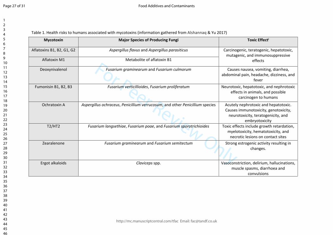
For Peer Review Only
Mycotoxin Major Species of Producing Fungi Toxic Effecti
Aflatoxins B1, B2, G1, G2 Aspergillus flavus and Aspergillus parasiticus
Aflatoxin M1 Metabolite of aflatoxin B1
Carcinogenic, teratogenic, hepatotoxic, mutagenic, and immunosuppressive
effects
Deoxynivalenol Fusarium graminearum and Fusarium culmorum Causes nausea, vomiting, diarrhea, abdominal pain, headache, dizziness, and
feverFumonisin B1, B2, B3 Fusarium verticillioides, Fusarium proliferatum Neurotoxic, hepatotoxic, and nephrotoxic
effects in animals, and possible carcinogen to humans
Ochratoxin A Aspergillus ochraceus, Penicillium verrucosum, and other Penicillium species Acutely nephrotoxic and hepatotoxic. Causes immunotoxicity, genotoxicity,
neurotoxicity, teratogenicity, and embryotoxicity
T2/HT2 Fusarium langsethiae, Fusarium poae, and Fusarium sporotrichioides Toxic effects include growth retardation, myelotoxicity, hematotoxicity, and
necrotic lesions on contact sitesZearalenone Fusarium graminearum and Fusarium semitectum Strong estrogenic activity resulting in
changes.
Ergot alkaloids Claviceps spp. Vasoconstriction, delirium, hallucinations, muscle spasms, diarrhoea and
convulsions
Table 1. Health risks to humans associated with mycotoxins (information gathered from Alshannaq & Yu 2017)
Page 27 of 31
http://mc.manuscriptcentral.com/tfac Email: [email protected]
Food Additives and Contaminants
123456789101112131415161718192021222324252627282930313233343536373839404142434445464748495051525354555657585960
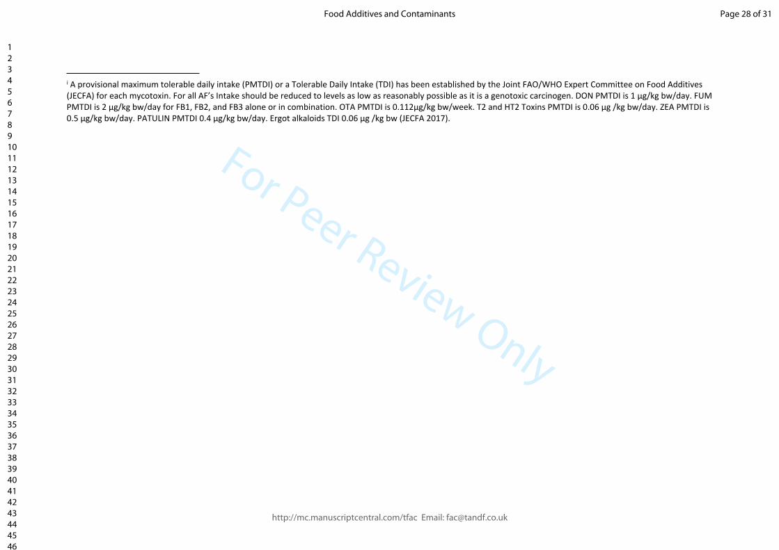
For Peer Review Only
i A provisional maximum tolerable daily intake (PMTDI) or a Tolerable Daily Intake (TDI) has been established by the Joint FAO/WHO Expert Committee on Food Additives (JECFA) for each mycotoxin. For all AF’s Intake should be reduced to levels as low as reasonably possible as it is a genotoxic carcinogen. DON PMTDI is 1 μg/kg bw/day. FUM PMTDI is 2 μg/kg bw/day for FB1, FB2, and FB3 alone or in combination. OTA PMTDI is 0.112μg/kg bw/week. T2 and HT2 Toxins PMTDI is 0.06 μg /kg bw/day. ZEA PMTDI is 0.5 µg/kg bw/day. PATULIN PMTDI 0.4 µg/kg bw/day. Ergot alkaloids TDI 0.06 μg /kg bw (JECFA 2017).
Page 28 of 31
http://mc.manuscriptcentral.com/tfac Email: [email protected]
Food Additives and Contaminants
123456789101112131415161718192021222324252627282930313233343536373839404142434445464748495051525354555657585960

For Peer Review Only
i Neogen offer a fluorometric assay for AF’sii Randox offer an ELISA for testing Ergot Alkaloids. iii Romer Labs offer a fluorometric assay for AF’siv Vicam offer fluorometric assay’s for AF’s, ZEN, OTA and FUM.
Mycotoxin
Company Product Type AF DON T2/HT2 ZEN OTA FUM
ELISA ElbascienceLFA - - -
ELISA - - - - - -Envirologics
LFA
ELISA EuroproximaLFA - - - - - -
ELISA - HelicaLFA - - - - - -
ELISA Neogeni
LFA
ELISA R-Biopharm LFA
ELISA - - - - -Randoxii
LFA - - - - - -
ELISA Romer Labsiii
LFA - -
ELISA Tecna Diagnostics LFA - -
ELISA - - - - - -Vicamiv
LFA -
ELISA - - - - - -Unisensor
LFA
Table 2. Companies that sell commercial test kit products for mycotoxins
Page 29 of 31
http://mc.manuscriptcentral.com/tfac Email: [email protected]
Food Additives and Contaminants
123456789101112131415161718192021222324252627282930313233343536373839404142434445464748495051525354555657585960

For Peer Review Only
Table 3 Different biosensor techniques developed for mycotoxin analysis
i Quartz crystal microbalance with dissipation monitoring.
Biosensing Device Matrix Analyte LOD (µg kg−1) Reference
SPR Milk AFM1 0.018Karczmarczyk
et al 2016
iSPR Beer and barley DON, OTA 17, 7
Joshi et al 2016 (2)
iSPR BarleyDON, T-2, ZEA,
FB1 64, 26, 96, 13Joshi et al 2016 (1)
SPR Wine and peanut oil OTA 0.005
Zhu et al 2015
QCM Buffer OTA 17.2Pirincci et al
2018
QCM-Di Red wine OTA 0.16 Karczmarczyk et al 2017
EQCM Cereal AFB1 0.008Chauhan et
al 2016
Page 30 of 31
http://mc.manuscriptcentral.com/tfac Email: [email protected]
Food Additives and Contaminants
123456789101112131415161718192021222324252627282930313233343536373839404142434445464748495051525354555657585960

For Peer Review Only
Figure 1. A competitive immunoassay format where the antibody is either (a) immobilised on the solid support surface or (b) suspended in solution.
Figure 1 & 2 Key:
= Analyte in sample/standard = Antibody
= Labelled analyte = Labelled Antibody
= Solid support
Figure2. A competitive immunoassay format where the antigen is immobilised on the solid support surface and the antibody is either (a) not labelled or (b) labelled.
Page 31 of 31
http://mc.manuscriptcentral.com/tfac Email: [email protected]
Food Additives and Contaminants
123456789101112131415161718192021222324252627282930313233343536373839404142434445464748495051525354555657585960

For Peer Review Only
Page 32 of 31
http://mc.manuscriptcentral.com/tfac Email: [email protected]
Food Additives and Contaminants
123456789101112131415161718192021222324252627282930313233343536373839404142434445464748495051525354555657585960