Current Treatment Options for Cervical Leiomyomas: A ...
15
medicina Review Current Treatment Options for Cervical Leiomyomas: A Systematic Review of Literature Federico Ferrari 1 , Sara Forte 2 , Gaetano Valenti 3 , Laura Ardighieri 4 , Fabio Barra 5, * , Valentina Esposito 6 , Enrico Sartori 2 and Franco Odicino 2 Citation: Ferrari, F.; Forte, S.; Valenti, G.; Ardighieri, L.; Barra, F.; Esposito, V.; Sartori, E.; Odicino, F. Current Treatment Options for Cervical Leiomyomas: A Systematic Review of Literature. Medicina 2021, 57, 92. https://doi.org/10.3390/medicina 57020092 Academic Editor: Udo Jeschke Received: 12 December 2020 Accepted: 19 January 2021 Published: 21 January 2021 Publisher’s Note: MDPI stays neutral with regard to jurisdictional claims in published maps and institutional affil- iations. Copyright: © 2021 by the authors. Licensee MDPI, Basel, Switzerland. This article is an open access article distributed under the terms and conditions of the Creative Commons Attribution (CC BY) license (https:// creativecommons.org/licenses/by/ 4.0/). 1 Department of Obstetrics and Gynecology, Spedali Civili of Brescia, 25123 Brescia, Italy; [email protected] 2 Department of Clinical and Experimental Sciences, University of Brescia, 25123 Brescia, Italy; [email protected] (S.F.); [email protected] (E.S.); [email protected] (F.O.) 3 Department of General Surgery and Medical-Surgical Specialties, Institute of Obstetrics and Gynecology, University of Catania, 95123 Catania, Italy; [email protected] 4 Department of Pathology, Spedali Civili of Brescia, 25123 Brescia, Italy; [email protected] 5 Academic Unit of Obstetrics and Gynaecology, IRCCS Ospedale Policlinico San Martino, 16100 Genova, Italy 6 Department of Gynecology and Obstetrics, Università degli Studi di Milano, 20122 Milan, Italy; [email protected] * Correspondence: [email protected]; Tel.: +39-334-943-7959 Abstract: Background and objectives: Cervical leiomyomas are a rare benign disease. Although they are mainly treated surgically, currently, there is not a standardized treatment for cervical leiomyomas. This study aims to summarize current literature evidence about treatment options for cervical leiomyomas. Materials and methods: A systematic research of the literature was conducted in Scopus, PubMed/MEDLINE, ScienceDirect, and the Cochrane Library, including observational prospective and retrospective studies, case series and case reports. We collected data regarding studies related to treatment options for cervical leiomyomas, evaluating the following aspects: study design, population, treatment type, rate of surgical complications, and fertility outcome. Results: According to literature research, 38 articles were included. Among 214 patients, the weighted average age was 39.4 years-old; 23 patients were pregnant. Most of the leiomyomas (78%) were extracervical; in 22% of cases (29 patients) were intracervical; 188 patients (88%) received surgical treatment, 6 (3%) received exclusive conservative management and 21 (10%) underwent interventional radiology treatment. One hundred twenty-seven patients (67.5%) underwent myomectomy, while 54 (28.7%) and 7 (3.7%) hysterectomy and trachelectomy, respectively. Cervical myomectomy was performed by open surgery in 21 out of 127 cases (16.5%), while in 92 (72.4%) and 6 (4.7%) patients the surgical approach was performed by traditional and robot-assisted laparoscopy, respectively. The total rate of surgical complications was 5.6%. Conclusion: Surgery is the primary therapeutic option for cervical leiomyomas with a low rate of surgical complications. Interventional radiology techniques have reported promising but still limited results. Keywords: leiomyoma; cervix; hysterectomy; cervical leiomyoma 1. Introduction Uterine leiomyomas (fibroids or myomas) are one of the most common benign smooth muscle tumors in women, having a prevalence of 20–40% after the age of 35 [1,2]. Ninety- five percent of leiomyomas occur in the uterine corpus [3]. Cervical leiomyomas are very uncommon with a frequency of 0.6% [4]; rarely, myomas can be located in other sites of the urogenital tract [5]. Classical symptoms related to myomas are abnormal uterine bleeding (AUB), chronic pelvic pain, dysmenorrhea; bulk-related symptoms can sometimes be observed [6]; similar clinical presentation can also be found in pregnant women affected by uterine leiomyomas [7]. Medicina 2021, 57, 92. https://doi.org/10.3390/medicina57020092 https://www.mdpi.com/journal/medicina
Transcript of Current Treatment Options for Cervical Leiomyomas: A ...
medicina
Review
Current Treatment Options for Cervical Leiomyomas:A Systematic Review of Literature
Federico Ferrari 1 , Sara Forte 2 , Gaetano Valenti 3, Laura Ardighieri 4 , Fabio Barra 5,* , Valentina Esposito 6,Enrico Sartori 2 and Franco Odicino 2
�����������������
Citation: Ferrari, F.; Forte, S.; Valenti,
G.; Ardighieri, L.; Barra, F.; Esposito,
V.; Sartori, E.; Odicino, F. Current
Treatment Options for Cervical
Leiomyomas: A Systematic Review of
Literature. Medicina 2021, 57, 92.
https://doi.org/10.3390/medicina
57020092
Academic Editor: Udo Jeschke
Received: 12 December 2020
Accepted: 19 January 2021
Published: 21 January 2021
Publisher’s Note: MDPI stays neutral
with regard to jurisdictional claims in
published maps and institutional affil-
iations.
Copyright: © 2021 by the authors.
Licensee MDPI, Basel, Switzerland.
This article is an open access article
distributed under the terms and
conditions of the Creative Commons
Attribution (CC BY) license (https://
creativecommons.org/licenses/by/
4.0/).
1 Department of Obstetrics and Gynecology, Spedali Civili of Brescia, 25123 Brescia, Italy;[email protected]
2 Department of Clinical and Experimental Sciences, University of Brescia, 25123 Brescia, Italy;[email protected] (S.F.); [email protected] (E.S.); [email protected] (F.O.)
3 Department of General Surgery and Medical-Surgical Specialties, Institute of Obstetrics and Gynecology,University of Catania, 95123 Catania, Italy; [email protected]
4 Department of Pathology, Spedali Civili of Brescia, 25123 Brescia, Italy; [email protected] Academic Unit of Obstetrics and Gynaecology, IRCCS Ospedale Policlinico San Martino, 16100 Genova, Italy6 Department of Gynecology and Obstetrics, Università degli Studi di Milano, 20122 Milan, Italy;
[email protected]* Correspondence: [email protected]; Tel.: +39-334-943-7959
Abstract: Background and objectives: Cervical leiomyomas are a rare benign disease. Althoughthey are mainly treated surgically, currently, there is not a standardized treatment for cervicalleiomyomas. This study aims to summarize current literature evidence about treatment options forcervical leiomyomas. Materials and methods: A systematic research of the literature was conductedin Scopus, PubMed/MEDLINE, ScienceDirect, and the Cochrane Library, including observationalprospective and retrospective studies, case series and case reports. We collected data regardingstudies related to treatment options for cervical leiomyomas, evaluating the following aspects: studydesign, population, treatment type, rate of surgical complications, and fertility outcome. Results:According to literature research, 38 articles were included. Among 214 patients, the weighted averageage was 39.4 years-old; 23 patients were pregnant. Most of the leiomyomas (78%) were extracervical;in 22% of cases (29 patients) were intracervical; 188 patients (88%) received surgical treatment, 6(3%) received exclusive conservative management and 21 (10%) underwent interventional radiologytreatment. One hundred twenty-seven patients (67.5%) underwent myomectomy, while 54 (28.7%)and 7 (3.7%) hysterectomy and trachelectomy, respectively. Cervical myomectomy was performed byopen surgery in 21 out of 127 cases (16.5%), while in 92 (72.4%) and 6 (4.7%) patients the surgicalapproach was performed by traditional and robot-assisted laparoscopy, respectively. The total rate ofsurgical complications was 5.6%. Conclusion: Surgery is the primary therapeutic option for cervicalleiomyomas with a low rate of surgical complications. Interventional radiology techniques havereported promising but still limited results.
Keywords: leiomyoma; cervix; hysterectomy; cervical leiomyoma
1. Introduction
Uterine leiomyomas (fibroids or myomas) are one of the most common benign smoothmuscle tumors in women, having a prevalence of 20–40% after the age of 35 [1,2]. Ninety-five percent of leiomyomas occur in the uterine corpus [3]. Cervical leiomyomas are veryuncommon with a frequency of 0.6% [4]; rarely, myomas can be located in other sitesof the urogenital tract [5]. Classical symptoms related to myomas are abnormal uterinebleeding (AUB), chronic pelvic pain, dysmenorrhea; bulk-related symptoms can sometimesbe observed [6]; similar clinical presentation can also be found in pregnant women affectedby uterine leiomyomas [7].
Medicina 2021, 57, 92. https://doi.org/10.3390/medicina57020092 https://www.mdpi.com/journal/medicina
Medicina 2021, 57, 92 2 of 15
Various classifications of cervical myomas have been proposed in the past years;most of them are based on their location, distinguishing subserosal myomas (definedextracervical type) from myomas located within the cervix (defined intracervical type) [8,9].
The most common therapy for cervical leiomyomas is represented by surgery; how-ever, compared to myomas of the uterine corpus, which are treated with conservativemyomectomy [10,11] or hysterectomy [12], the surgical treatment of cervical leiomyomascan be more difficult; this is due to the risk of intraoperative hemorrhage and to thepotential injuries to the adjacent organs that are often dislocated and contiguous to thecervical leiomyoma; this may cause a subverted anatomy of the pelvis requiring experi-enced surgeons [13]; furthermore, the position of the leiomyoma in the cervix poses anextra challenge in the surgical approach in case of a fertility-sparing approach. Currently, astandard surgical treatment for the leiomyomas of the cervix still lacks; therefore, the thera-peutic approach depends on the characteristics of the patient, the desire for fertility andthe experience of the individual centers and surgeons [14]. In recent years, few experiencesof interventional radiology procedures have been reported in those patients interested inpreserving their uterus [15].
The aim of this study is to provide an overview of the different treatment options forcervical leiomyomas reported in the literature so far and to give a snapshot of the currentstate of care for these benign tumors.
2. Material and Methods2.1. Study Design
This study is a systematic review of the literature on cervical leiomyomas and theirtreatments. The review follows the Preferred Reporting Items for Systematic Reviews andMeta-Analyses (PRISMA) guidelines [16].
2.2. Inclusion Criteria
The study aimed to ask the following PICOS items.Population: Women with cervical leiomyoma regardless of the co-occurrence of other
types of uterine myomas.Intervention: Surgical, radiological, medical, or conservative management for cervical
myomas.Comparators: No comparators.Outcomes: To identify the most frequently therapeutic approaches used for treating
cervical leiomyomas.Study design: Observational prospective and retrospective studies, case series and case
reports were included.Language: Only manuscripts in English language were considered.
2.3. Search Strategy
A systematic search of the literature was conducted in Scopus, PubMed/MEDLINE,ScienceDirect and the Cochrane Library from their inception to August 2020. A combi-nation of keywords was used as following “Cervical myoma” OR “Cervical leiomyoma”AND “surgical treatment” OR “alternative treatment” OR “non-surgical treatment” OR “in-terventional radiology treatment” OR “uterine artery embolization” OR “medical therapy”OR “GnRH agonists” OR “selective estrogen receptor modulator”.
2.4. Study Selection and Data Extraction
Two authors (SF and VE) independently screened titles and abstracts from the stud-ies in the search results. The eligible studies were then assessed for inclusion based ontheir full text. An additional manual search of reference lists was then performed bya third author (GV) not to miss relevant or recent publications. Disagreements on theeligibility of studies were resolved by a fourth author (FF). Three authors (SF, VE andFB) extracted the following data: study features (authors, year of publication, number of
Medicina 2021, 57, 92 3 of 15
cases), population characteristics (age at the diagnosis, fertility status), characteristics ofthe disease (clinical presentation, radiological features, size and site of myoma, histologyin patients underwent surgery), management in case of surgical or conservative treatment(preoperative medical treatment or other types of preoperative procedures, surgical access,intra-operative findings and additional intra-operative procedures, type of treatment),management in case of interventional radiology treatment (type of treatment, volumereduction, efficacy in symptom resolution) and follow-up. Three additional authors (LA,ES and FO) double-checked data extraction. This study was done following the HelsinkiDeclaration, conforms to the Consensus-based Clinical Case Reporting Guideline Develop-ment (http://www.equator-network.org/) the Committee on Publication Ethics (COPE)guidelines (http://publicationethics.org/).
2.5. Aim of the Systematic Review
This study aimed to summarize the available evidence in the literature concerning thetreatment of cervical myomas.
2.6. Data Synthesis
We followed a standardized, pre-specified format for data extraction. The data ex-tracted were evaluated by analyzing the following topics: surgical and conservative treat-ment for cervical myomas (considering patients’ age at the diagnosis, fertility status, clinicalpresentation, radiological features, site of myoma, preoperative medical treatment or othertypes of preoperative procedures, surgical access, intraoperative findings and additionalintraoperative procedures, type of treatment, histology, follow-up) and interventionalradiology treatment (pregnancy status, age at the diagnosis, symptoms, site, maximumdiameter, mean volume and vascularity on aortography of the myoma, type of treatment,myoma mass infarction, volume reduction rate, technique successful and symptoms resolu-tion, follow-up with MRI) for cervical myomas. Depending on their position relative to theendocervical canal, the lesions were classified as extracervical (including subserosal andintramural cervical myomas) and intracervical. Since there was significant heterogeneitybetween studies, quantitative data synthesis was not possible.
3. Results3.1. Systematic Review of the Literature
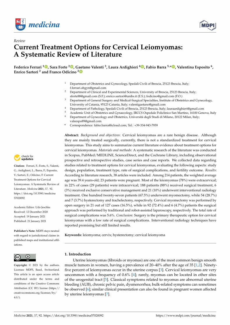
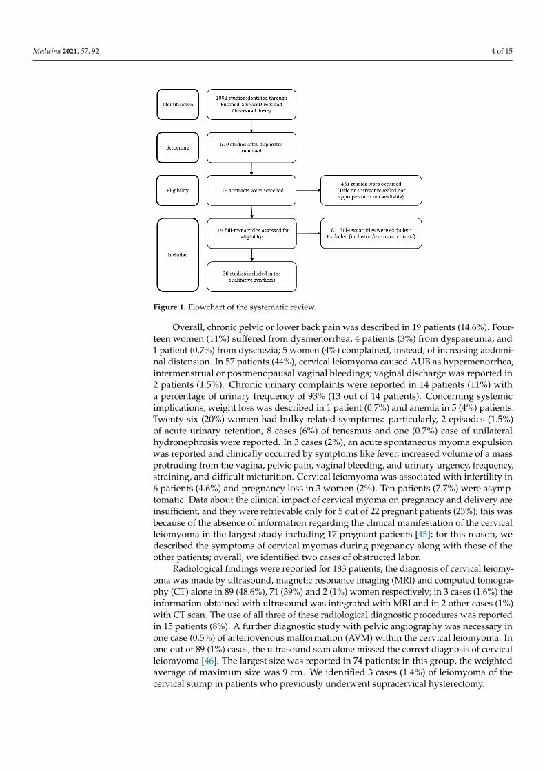
The search strategy provided a total of 570 articles, after removing duplicates. Afterscreening of manuscript titles, 119 full text and abstracts were considered eligible. Ofthese, 81 studies were subsequently excluded after the examination of the abstract andfull text: 29 manuscripts were excluded because only the abstract was retrieved, 24 asthey did not have English language and 28 because they did not have an adequate popu-lation and intervention. Definitively, 38 articles were included in our systematic review(Figure 1) [3,5–40]. Data synthesis was available in Tables 1 and 2.
3.2. Characteristics of Patients, Symptoms and Radiological Findings
A total of 214 women were included. The average age values were not retrievable in threearticles [17–19] and were thus only available for 187 patients; the weighted average age was39.4 years old. Fertility status of the patients was obtained from 24 studies [4,8,19–27,36–40]in 57 patients; among them, 28 (49%) patients were in premenopausal status, 5 (9%) inpostmenopausal status, 23 patients (40%) were pregnant, and one patient (2%) was inpuerperal status.
The description of the symptoms was retrievable in 32 studies [8,17,19–22,24,25,29,33–38,40–44] enrolling 130 patients.
Medicina 2021, 57, 92 4 of 15Medicina 2021, 57, x FOR PEER REVIEW 4 of 18
Medicina 2021, 57, x. https://doi.org/10.3390/xxxxx www.mdpi.com/journal/medicina
Figure 1. Flowchart of the systematic review.
3.2. Characteristics of Patients, Symptoms and Radiological Findings A total of 214 women were included. The average age values were not retrievable in
three articles [17–19] and were thus only available for 187 patients; the weighted average age was 39.4 years old. Fertility status of the patients was obtained from 24 studies [4,8,19–27,36–40] in 57 patients; among them, 28 (49%) patients were in premenopausal status, 5 (9%) in postmenopausal status, 23 patients (40%) were pregnant, and one patient (2%) was in puerperal status.
The description of the symptoms was retrievable in 32 studies [8,17,19–22,24,25,29,33–38,40–44] enrolling 130 patients.
Overall, chronic pelvic or lower back pain was described in 19 patients (14.6%). Four-teen women (11%) suffered from dysmenorrhea, 4 patients (3%) from dyspareunia, and 1 patient (0.7%) from dyschezia; 5 women (4%) complained, instead, of increasing ab-dominal distension. In 57 patients (44%), cervical leiomyoma caused AUB as hypermen-orrhea, intermenstrual or postmenopausal vaginal bleedings; vaginal discharge was re-ported in 2 patients (1.5%). Chronic urinary complaints were reported in 14 patients (11%) with a percentage of urinary frequency of 93% (13 out of 14 patients). Concerning systemic implications, weight loss was described in 1 patient (0.7%) and anemia in 5 (4%) patients. Twenty-six (20%) women had bulky-related symptoms: particularly, 2 episodes (1.5%) of acute urinary retention, 8 cases (6%) of tenesmus and one (0.7%) case of unilateral hydro-nephrosis were reported. In 3 cases (2%), an acute spontaneous myoma expulsion was reported and clinically occurred by symptoms like fever, increased volume of a mass pro-truding from the vagina, pelvic pain, vaginal bleeding, and urinary urgency, frequency, straining, and difficult micturition. Cervical leiomyoma was associated with infertility in 6 patients (4.6%) and pregnancy loss in 3 women (2%). Ten patients (7.7%) were asymp-tomatic. Data about the clinical impact of cervical myoma on pregnancy and delivery are insufficient, and they were retrievable only for 5 out of 22 pregnant patients (23%); this was because of the absence of information regarding the clinical manifestation of the cer-vical leiomyoma in the largest study including 17 pregnant patients [45]; for this reason, we described the symptoms of cervical myomas during pregnancy along with those of the other patients; overall, we identified two cases of obstructed labor.
Radiological findings were reported for 183 patients; the diagnosis of cervical leio-myoma was made by ultrasound, magnetic resonance imaging (MRI) and computed to-mography (CT) alone in 89 (48.6%), 71 (39%) and 2 (1%) women respectively; in 3 cases
Figure 1. Flowchart of the systematic review.
Overall, chronic pelvic or lower back pain was described in 19 patients (14.6%). Four-teen women (11%) suffered from dysmenorrhea, 4 patients (3%) from dyspareunia, and1 patient (0.7%) from dyschezia; 5 women (4%) complained, instead, of increasing abdomi-nal distension. In 57 patients (44%), cervical leiomyoma caused AUB as hypermenorrhea,intermenstrual or postmenopausal vaginal bleedings; vaginal discharge was reported in2 patients (1.5%). Chronic urinary complaints were reported in 14 patients (11%) witha percentage of urinary frequency of 93% (13 out of 14 patients). Concerning systemicimplications, weight loss was described in 1 patient (0.7%) and anemia in 5 (4%) patients.Twenty-six (20%) women had bulky-related symptoms: particularly, 2 episodes (1.5%)of acute urinary retention, 8 cases (6%) of tenesmus and one (0.7%) case of unilateralhydronephrosis were reported. In 3 cases (2%), an acute spontaneous myoma expulsionwas reported and clinically occurred by symptoms like fever, increased volume of a massprotruding from the vagina, pelvic pain, vaginal bleeding, and urinary urgency, frequency,straining, and difficult micturition. Cervical leiomyoma was associated with infertility in6 patients (4.6%) and pregnancy loss in 3 women (2%). Ten patients (7.7%) were asymp-tomatic. Data about the clinical impact of cervical myoma on pregnancy and delivery areinsufficient, and they were retrievable only for 5 out of 22 pregnant patients (23%); this wasbecause of the absence of information regarding the clinical manifestation of the cervicalleiomyoma in the largest study including 17 pregnant patients [45]; for this reason, wedescribed the symptoms of cervical myomas during pregnancy along with those of theother patients; overall, we identified two cases of obstructed labor.
Radiological findings were reported for 183 patients; the diagnosis of cervical leiomy-oma was made by ultrasound, magnetic resonance imaging (MRI) and computed tomogra-phy (CT) alone in 89 (48.6%), 71 (39%) and 2 (1%) women respectively; in 3 cases (1.6%) theinformation obtained with ultrasound was integrated with MRI and in 2 other cases (1%)with CT scan. The use of all three of these radiological diagnostic procedures was reportedin 15 patients (8%). A further diagnostic study with pelvic angiography was necessary inone case (0.5%) of arteriovenous malformation (AVM) within the cervical leiomyoma. Inone out of 89 (1%) cases, the ultrasound scan alone missed the correct diagnosis of cervicalleiomyoma [46]. The largest size was reported in 74 patients; in this group, the weightedaverage of maximum size was 9 cm. We identified 3 cases (1.4%) of leiomyoma of thecervical stump in patients who previously underwent supracervical hysterectomy.
Medicina 2021, 57, 92 5 of 15
3.3. Location of the Cervical Leiomyomas
In 131 patients, lesions were classified as extracervical or intracervical. Overall, 78%(n = 102) of the leiomyomas were extracervical and 22% (n = 29) were intracervical. Inthree cases included in the extracervical group (3%) [26,31,33] the mass hanged from theanterior lip of the portio and protruded out from the vagina; the cervical external ostiumwas completely covered by the myoma, while the cervical canal was not involved and inone of these cases the patient was pregnant at 36 weeks of gestation [26].
In four studies [17,26,44,47] including 63 patients, the location of the cervical leiomy-omas was reported neither was deductible from the text, tables or figures.
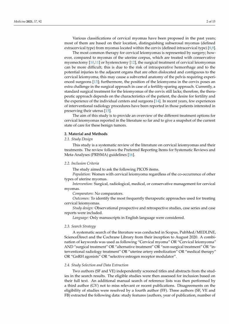
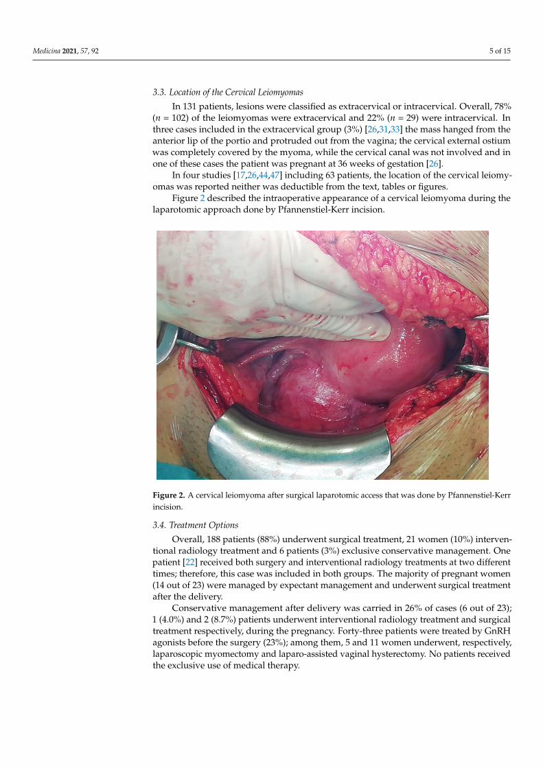
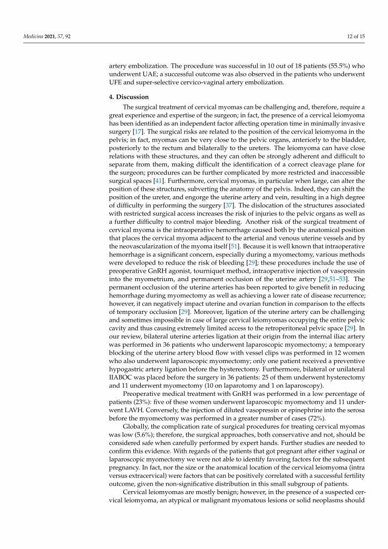
Figure 2 described the intraoperative appearance of a cervical leiomyoma during thelaparotomic approach done by Pfannenstiel-Kerr incision.
Medicina 2021, 57, x FOR PEER REVIEW 5 of 18
Medicina 2021, 57, x. https://doi.org/10.3390/xxxxx www.mdpi.com/journal/medicina
(1.6%) the information obtained with ultrasound was integrated with MRI and in 2 other cases (1%) with CT scan. The use of all three of these radiological diagnostic procedures was reported in 15 patients (8%). A further diagnostic study with pelvic angiography was necessary in one case (0.5%) of arteriovenous malformation (AVM) within the cervical leiomyoma. In one out of 89 (1%) cases, the ultrasound scan alone missed the correct di-agnosis of cervical leiomyoma [46]. The largest size was reported in 74 patients; in this group, the weighted average of maximum size was 9 cm. We identified 3 cases (1.4%) of leiomyoma of the cervical stump in patients who previously underwent supracervical hysterectomy.
3.3. Location of the Cervical Leiomyomas In 131 patients, lesions were classified as extracervical or intracervical. Overall, 78%
(n = 102) of the leiomyomas were extracervical and 22% (n = 29) were intracervical. In three cases included in the extracervical group (3%) [26,31,33] the mass hanged from the ante-rior lip of the portio and protruded out from the vagina; the cervical external ostium was completely covered by the myoma, while the cervical canal was not involved and in one of these cases the patient was pregnant at 36 weeks of gestation [26].
In four studies [17,26,44,47] including 63 patients, the location of the cervical leiomy-omas was reported neither was deductible from the text, tables or figures.
Figure 2 described the intraoperative appearance of a cervical leiomyoma during the laparotomic approach done by Pfannenstiel-Kerr incision.
Figure 2. A cervical leiomyoma after surgical laparotomic access that was done by Pfannenstiel-Kerr incision.
3.4. Treatment Options Overall, 188 patients (88%) underwent surgical treatment, 21 women (10%) interven-
tional radiology treatment and 6 patients (3%) exclusive conservative management. One patient [22] received both surgery and interventional radiology treatments at two differ-ent times; therefore, this case was included in both groups. The majority of pregnant women (14 out of 23) were managed by expectant management and underwent surgical treatment after the delivery.
Figure 2. A cervical leiomyoma after surgical laparotomic access that was done by Pfannenstiel-Kerrincision.
3.4. Treatment Options
Overall, 188 patients (88%) underwent surgical treatment, 21 women (10%) interven-tional radiology treatment and 6 patients (3%) exclusive conservative management. Onepatient [22] received both surgery and interventional radiology treatments at two differenttimes; therefore, this case was included in both groups. The majority of pregnant women(14 out of 23) were managed by expectant management and underwent surgical treatmentafter the delivery.
Conservative management after delivery was carried in 26% of cases (6 out of 23);1 (4.0%) and 2 (8.7%) patients underwent interventional radiology treatment and surgicaltreatment respectively, during the pregnancy. Forty-three patients were treated by GnRHagonists before the surgery (23%); among them, 5 and 11 women underwent, respectively,laparoscopic myomectomy and laparo-assisted vaginal hysterectomy. No patients receivedthe exclusive use of medical therapy.
Medicina 2021, 57, 92 6 of 15
Table 1. Surgical and conservative treatment of cervical myomas.
Author, Year CasesAge at Diagnosis(Years, Mean Age,
Range)Fertility Status Symptoms Imaging
AssessmentSite of
Leiomy-oma
PreoperativeProcedures Surgical Access Type of Treatment Follow-Up
Hashim, 2020 1 27 Premenopausal PP; UrS US; MRI Extracervical None Laparotomy MyomectomyUneventful postoperative
course.No recurrence at six months.
Zhang, 2018 1 22 PuerperalFever; protruding
mass from thevagina
US Intracervical None Vaginal MyomectomyUneventful postoperative
course.Pregnancy two years later.
Kaneda, 2017 22
TAH49.5
(36–62)AM35.5
(28–40)
Unknown Unknown MRI Extracervical Placement ofbilateral IIABOC
Laparotomy withvasopressin injection
Hysterectomy12 (54.6%)
Myomectomy10 (45.4%) AM
Uneventful postoperativerecovery.
Wong, 2017 1 36 Premenopausal Bloating abdomen US; MRI Extracervical None Laparotomy Trachelectomy Uneventful postoperativerecovery.
Peker, 2016 1 40 PremenopausalChronic PP,
dyspareunia,primary infertility
US Extracervical None Laparoscopy Myomectomy Uneventful postoperativerecovery
Giannella, 2016 1 18 Premenopausal Dyspareunia US; CT Extracervical None Laparoscopy Myomectomy
Uneventful postoperativerecovery.
No recurrence at eightmonths.
Goel, 2016 1 40 Premenopausal Bloating abdomen;chronic PP US Extracervical None Laparotomy Hysterectomy Unknown
Peng, 2016 1 42 Premenopausal UrS US; CT Extracervical None Laparotomy Myomectomy Uneventful postoperativerecovery
Garzon-Lopez,2015 1 31 Premenopausal ACUR MRI Extracervical None Laparoscopy with
vasopressin injection Myomectomy Postoperative abscess at thesite of the surgical bed
Bidzinski, 2014 1 48 Unknown ACUR US Extracervical None LaparotomyHysterectomy with
preventive hypogastricartery ligation.
Unknown
Gandhi, 2014 1 30 Pregnancy (IItrimester) None US Extracervical None None
Conservativemanagement
Emergency cesareansection for obstructed
labor
Spontaneous prolapse out ofthe vagina four days after
surgery.Placement of rubber ring
pessary. Halving of volume ofthe myoma and no symptoms
after 6 weeks of follow-up
Keriakos andMaher, 2013 1 29 Pregnancy
Vaginal discharge;AUB; obstructed
laborUS Intracervical
Monolateraluterine arteryembolization
previouspregnancy.
Vaginal
Conservativemanagement during
pregnancy.Emergency cesarean
section for obstructedlabor and
myomectomy six weekslater
Pregnancy at 20 weeks ofgestation at follow-up visit.
Medicina 2021, 57, 92 7 of 15
Table 1. Cont.
Author, Year CasesAge at Diagnosis(Years, Mean Age,
Range)Fertility Status Symptoms Imaging
AssessmentSite of
Leiomy-oma
PreoperativeProcedures Surgical Access Type of Treatment Follow-Up
Kamra, 2013 1 28 Pregnancy (36weeks) AUB US Extracervical None Vaginal Myomectomy Unknown
Hsiao, 2013 14 Unknown Unknown Unknown Unknown Unknown Unknown
RALM withvasopressin injection in
6 (43%) casesTLM with vasopressin
injection in8 (57%) cases
Myomectomy Unknown
Ikechebelu, 2012 1 37 Premenopausal
Protruding massfrom the vagina.acute PP; AUB;
fever; Urs
CT Intracervical Broad-spectrumantibiotics Vaginal Myomectomy Uneventful postoperative
recovery
Higuchi, 2012 8 35.5 ± 5.3 (29–44) Unknown
PP;hypermenorrhea;
one case ofmonolateral
hydronephrosis
MRI
Extracervical(n = 6; 75%)Intracervical
(n = 2;33.3%)
GnRH agonists
Laparoscopy with onecase of conversionAnd vasopressin
injection
Myomectomy
One case of retroperitonealhematoma.
50% of childbearing patientsgot pregnant
Chu, 2012 1 55 Postmenopausal Intermittent PP;AUB US Extracervical
Previoussupracervical
hysterectomy forleiomyomas
Laparotomy Trachelectomy Uneventful postoperativerecovery.
Chu, 2012 1 50 Unknown PP; AUB MRI Extracervical
Previoussupracervical
hysterectomy forleiomyomas
Laparoscopy Trachelectomy Uneventful postoperativerecovery.
Soeda, 2012 1 55 Postmenopausal Bloating abdomen
US; MRI; Pelvicangiographyshowing anAVM in the
uterine tumor
Extracervical MonolateralIIABOC Laparotomy Hysterectomy Unknown
Tian and Hu,2012 17 32
(28–41) Pregnancy Unknown US Unknown Unknown Laparotomy in 16(94%) cases
Vaginal delivery1 (6%)
Cesarean section in16 (94%) cases and 9 of
them (56.3%)myomectomy after
delivery and 3 (18.8%)hysterectomy after
delivery; while 4 (25%)did not receive any
treatment
1 (6%) case of intraoperativehemorrhage during
myomectomy treated withhysterectomy;1 (6%) case of
persistent feverunresponsive to treatment,underwent hysterectomy
eight days later forleiomyoma with signs of
infection and necrosis.1 (6%) case of emergency
hysterectomy for postpartumhemorrhage
Pushpalatha,2011 1 50 Postmenopausal Lower abdominal
heaviness; UrS CT Extracervical None Laparotomy Hysterectomy Uneventful postoperativerecovery

Medicina 2021, 57, 92 8 of 15
Table 1. Cont.
Author, Year CasesAge at Diagnosis(Years, Mean Age,
Range)Fertility Status Symptoms Imaging
AssessmentSite of
Leiomy-oma
PreoperativeProcedures Surgical Access Type of Treatment Follow-Up
Takeda, 2011 13 45.8 (56–36)
Premenopausal(n = 11)
Postmenopausal(n = 2)
Menorrhagia;pressure
symptomsUS; MRI; CT
Extracervical(n = 9; 69%)Intracervical(n = 4; 31%)
GnRH agonist inpremenopausal
women andplacement of
bilateral IIABOC
Laparoscopy withbilateral ureteral
stenting andepinephrine injectionin extracervical cases
LAVH
1 (8%) Intraoperativehemorrhage with blood
transfusion.1 (8%) Post-operative infection
Kilpatrick, 2010 1 32 Unknown AUB US Intracervical None Vaginal Myomectomy at 15weeks of pregnancy
Uneventful postoperativecourse.
Spontaneous vaginal deliveryat 38 weeks gestation and
uneventful postpartumcourse.
Chang, 2010 28 38.0 ± 7.0(24–52) Unknown
dysmenorrhea;hypermenorrhoea;
UrS; tenesmus;pregnancy loss;
infertility
US
Extracervical(n = 26;
93%)Intracervical(n = 2; 7%)
GnRHagonists in 7% of
patients
Laparoscopy withbilateral uterine
arteries ligation andvasopressin injection
Myomectomy
Uneventful postoperativerecovery.
Two infertile patientsconceived spontaneously
at 1 and 7 monthspostoperatively
Matsuoka, 2010 16 37.3 ± 4.2(35–40) Unknown
hypermenorrhea;pressure
symptoms;sterility; lower
back pain
MRI
Intracervical(n = 5;31.2%)
Extracervical(n = 11;68.7%)
GnRH agonists Laparoscopy with theinjection of vasopressin
Myomectomywith uterine artery
clipping in 44% of cases
Uneventful postoperativerecovery.
One infertile patient gotpregnant.
No recurrence at four years offollow-up.
Del Priore, 2010 3 Unknown Unknown PP MRI Extracervical None Laparotomy Radical abdominaltrachelectomy
33% recurrent complaints fora new myoma after 12 months
Baum, 2009 1 32 Premenopausalstatus
PP; protrudingmass from the
vaginaUS Extracervical None Vaginal Myomectomy Uneventful postoperative
recovery
Sinha, 2009 24 37 Unknown Menorrhagia(71%) US Unknown Unknown
Laparoscopywith bilateral
uterine arteries ligationat their origin in 50% of
cases
Myomectomy Unknown
Takeda, 2009 1 33 Premenopausal Bloating abdomen;UrS US; MRI; CT Extracervical GnRH agonists;
bilateral IIABOC
Laparoscopy withbilateral ureteral
stentingMyomectomy Post-operative fever
No recurrence at 6 years
Takeuchi, 2006 5 36.2 ± 5.3 Premenopausal Hypermenorrhea;anemia MRI
Extracervical(n = 3; 60%)Intracervical(n = 2; 40%)
GnRH agonists
Laparoscopywith temporary
clipping of the uterineartery and vasopressin
injection
MyomectomyUneventful postoperative
recoveryNo recurrence at 2 years
Sengupta, 2006 1 29 Pregnant(36 weeks)
Chronic vaginaldischarge;
spontaneousexpulsion:
acute severe PPand AUB
US Intracervical None Vaginal Myomectomy (aftercesarean section)
Uneventful postoperativerecovery
No recurrence at 6 months
Medicina 2021, 57, 92 9 of 15
Table 1. Cont.
Author, Year CasesAge at Diagnosis(Years, Mean Age,
Range)Fertility Status Symptoms Imaging
AssessmentSite of
Leiomy-oma
PreoperativeProcedures Surgical Access Type of Treatment Follow-Up
Gurung, 2003 1 37 PremenopausalMass protrudingfrom the vagina;
AUBUnknown Extracervical None Vaginal Hysterectomy Postoperative UTI
Bajo, 1998 21 50(47–60) Unknown Unknown US Intracervical Unknown Unknown Hysterectomy Unknown
Galt, 1957 1 47 Unknown
Lower back pain,dyschezia,
dyspareunia, lossof weight.
Unknown ExtracervicalPrevious
supracervicalhysterectomy
Laparotomy TrachelectomyUneventful postoperative
recoveryNo recurrence at 6 months
PP: pelvic pain; UrS: urinary symptoms; US: ultrasound scan; ACUR: acute urinary retention; AUB: abnormal uterine bleeding; MRI: magnetic resonance imaging; CT: Computer tomography; AVM: Arteriovenousmalformation; TAH: total abdominal hysterectomies; AM: abdominal myomectomies; IIABOC: Internal Iliac Artery Balloon Occlusion Catheter; GnRH: gonadotropin-releasing hormone; RALM: robot-assistedlaparoscopic myomectomy; TLM: traditional laparoscopic myomectomy; LAVH: Laparoscopic-assisted vaginal hysterectomy; UTI: urinary tract infection.
Table 2. Interventional radiology treatment.
Author, year Cases Median Age FertilityStatus Symptoms Site of
Leiomyoma
ImagingAssessmentand Size of
CL in cm
MeanVolume ofCL in mL
Type ofTreatment
Myoma MassInfarction
VolumeReductionRate (%)
TreatmentSuccess
SymptomsResolution
Follow Upwith MRI
de Bruijn,2019 8 37 Unknown
AUB;pressure
symptoms;PP
Unknown MRI Unknown UAE
90% of patients withsolitary CL
60% of patients withconcurrent
non-cervical myomas
85% 100% 75% 3 months
Kim, 2012 10 Unknown Unknown UnknownIntracervical
8 (80%)Extracervical
2 (20%)
MRI;6 ± 1.6 cm 92.6 ± 54.8 UAE
20% of cases complete;60% of cases partial
necrosis; 20% of casesno necrosis;
42.8% 20% Unknown 3 months
Lohle, 2015 1 40 Pregnancy(20 weeks) AUB Extracervical US; MRI Unknown UFE almost complete Unknown 100% 100% 2 months
DeMeritt,2019 1 37 Premenopausal AUB Extracervical
MRI3.6 × 3.2 ×
4.4 cm26.54
Super-selectivecervico-vaginal
arteryembolization
100% 91% 100% 100% Unknown
CL: cervical leiomyoma; AUB: abnormal uterine bleeding; P: intermittent pain not related to the menstrual cycle; UAE: Uterine artery embolization; UFE: Super-selective uterine fibroid embolization.
Medicina 2021, 57, 92 10 of 15
3.4.1. Surgical Treatment
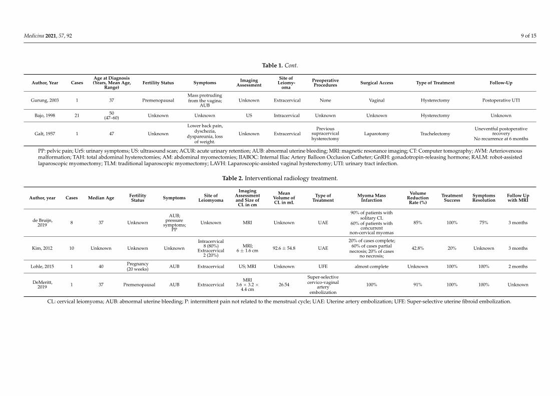
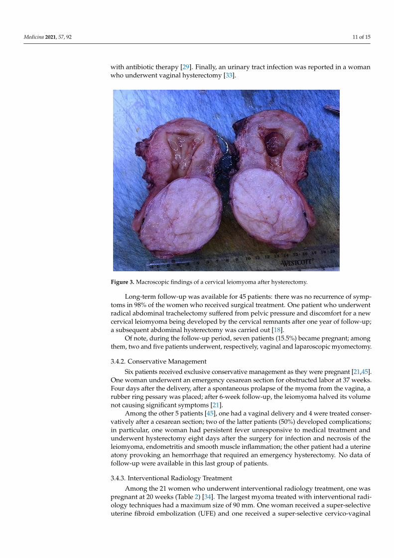
Among patients surgically treated, 127 (67.5%) underwent myomectomy, 54 (28.7%)underwent hysterectomy and 7 (3.7%) underwent trachelectomy. Cervical myomectomywas performed by laparotomy in 21 (16.5%) cases; in this group there were 9 surgicalprocedures (43%) performed at the time of cesarean section after the delivery [48].
Ninety-two (72.4%) and six (4.7%) patients underwent traditional and robot-assistedlaparoscopy both for the cervical myomectomy. In one case (0.8%), the laparoscopy accesswas converted to laparotomy because of the presence of a large submucosal cervical myoma;therefore, there was an overall estimated rate of conversion to laparotomy of 1%. Sevenpatients (5.5%) underwent vaginal myomectomy for treating four intracervical and threeextracervical type leiomyomas; among them, there were two cases of myomectomy inpregnant women at 36 and 15 weeks of gestation [26,30]. The cervical leiomyoma, whichwas removed in the 36 weeks pregnant woman [26], was an extracervical type; despite thesize (55 × 40 millimeters), its removal was possible because there was a broad attachmentto the anterior lip of the cervix, which covered the entire cervical opening and protruded inthe vagina without involving the cervical canal. An intracervical 50 × 30 × 30 millimeterleiomyoma with a pedicle length of 40 mm originating from the left lateral endocervicalcanal close to the internal ostium was removed in the other pregnant woman.
In 91 cases (72%), before the myomectomy, diluted vasopressin or epinephrine wasinjected into the myoma serosa; the surgical access for these patients was laparotomic,laparoscopic and robot-assisted laparoscopic in 10 (11%), 75 (82%) and 6 (7%) cases re-spectively. Fifty-four women (28.7%) underwent total hysterectomy with or withoutsalpingo-oophorectomy; in 19 patients (35%) a laparotomic hysterectomy, in 13 patients(24%) a laparoscopic-assisted vaginal hysterectomy (LAVH) and in one patient (1.8%) avaginal hysterectomy was performed. In 21 cases (39%) the surgical access was not reported.In 3 patients (5.5%) the laparotomic hysterectomy was done after delivery at the time of ce-sarean section. In 85 (45%) cases, the following additional pre or intraoperative proceduresfor preventing the intraoperative bleedings were employed: bilateral uterine arteries liga-tion at their origin from the internal iliac artery (n = 36) and temporarily blocking of uterineartery blood flow with the use of vessel clips (n = 12) during laparoscopic myomectomy;one patient [49] underwent a preventive hypogastric artery ligation before hysterectomy.Bilateral or unilateral internal iliac artery balloon occlusion catheter (IIABOC) was placedbefore the surgery and eventually inflated and deflated during the surgical procedure in36 patients (19%); out of them, 25 women underwent hysterectomy (12 by laparotomyand 13 by laparoscopy) and 11 underwent myomectomy (10 by laparotomy and 1 bylaparoscopy). Seven patients (3.7%) were treated by trachelectomy with laparotomic orlaparoscopic access in 6 (86%) and 1 case (14%), respectively. Three of these patients (43%)had a leiomyoma on the cervical stump; in one of these cases [24], the trachelectomy wasperformed by laparoscopy.
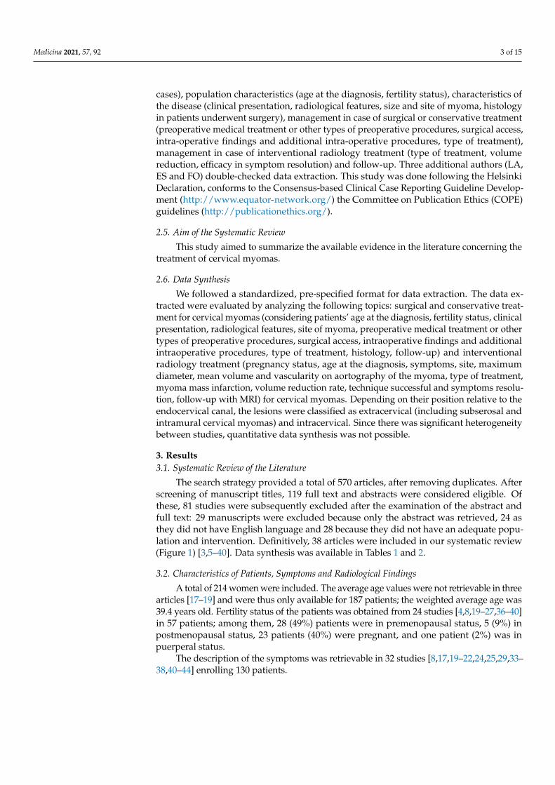
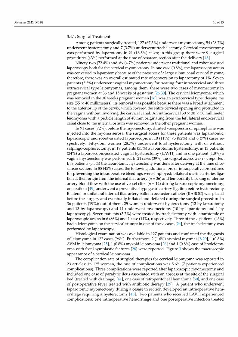
Histological examination was available in 127 patients and confirmed the diagnosisof leiomyoma in 122 cases (96%). Furthermore, 2 (1.6%) atypical myomas [8,20], 1 (0.8%)AVM in leiomyoma [25], 1 (0.8%) myxoid leiomyoma [26] and 1 (0.8%) case of lipoleiomy-oma with focal symplastic features [28] were reported. Figure 3 shows the macroscopicappearance of a cervical leiomyoma.
The complication rate of surgical therapies for cervical leiomyoma was reported in23 articles: in 125 women, the rate of complications was 5.6% (7 patients experiencedcomplications). Three complications were reported after laparoscopic myomectomy andincluded one case of paralytic ileus associated with an abscess at the site of the surgicalbed (treated with drainage) [41], one case of retroperitoneal hematoma [50], and one caseof postoperative fever treated with antibiotic therapy [29]. A patient who underwentlaparotomic myomectomy during a cesarean section developed an intraoperative hem-orrhage requiring a hysterectomy [45]. Two patients who received LAVH experiencedcomplications: one intraoperative hemorrhage and one postoperative infection treated
Medicina 2021, 57, 92 11 of 15
with antibiotic therapy [29]. Finally, an urinary tract infection was reported in a womanwho underwent vaginal hysterectomy [33].
Medicina 2021, 57, x FOR PEER REVIEW 7 of 18
Medicina 2021, 57, x. https://doi.org/10.3390/xxxxx www.mdpi.com/journal/medicina
Figure 3. Macroscopic findings of a cervical leiomyoma after hysterectomy.
The complication rate of surgical therapies for cervical leiomyoma was reported in 23 articles: in 125 women, the rate of complications was 5.6% (7 patients experienced com-plications). Three complications were reported after laparoscopic myomectomy and in-cluded one case of paralytic ileus associated with an abscess at the site of the surgical bed (treated with drainage) [41], one case of retroperitoneal hematoma [50], and one case of postoperative fever treated with antibiotic therapy [29]. A patient who underwent lapa-rotomic myomectomy during a cesarean section developed an intraoperative hemorrhage requiring a hysterectomy [45]. Two patients who received LAVH experienced complica-tions: one intraoperative hemorrhage and one postoperative infection treated with antibi-otic therapy [29]. Finally, an urinary tract infection was reported in a woman who under-went vaginal hysterectomy [33].
Long-term follow-up was available for 45 patients: there was no recurrence of symp-toms in 98% of the women who received surgical treatment. One patient who underwent radical abdominal trachelectomy suffered from pelvic pressure and discomfort for a new cervical leiomyoma being developed by the cervical remnants after one year of follow-up; a subsequent abdominal hysterectomy was carried out [18].
Of note, during the follow-up period, seven patients (15.5%) became pregnant; among them, two and five patients underwent, respectively, vaginal and laparoscopic myomectomy.
Figure 3. Macroscopic findings of a cervical leiomyoma after hysterectomy.
Long-term follow-up was available for 45 patients: there was no recurrence of symp-toms in 98% of the women who received surgical treatment. One patient who underwentradical abdominal trachelectomy suffered from pelvic pressure and discomfort for a newcervical leiomyoma being developed by the cervical remnants after one year of follow-up;a subsequent abdominal hysterectomy was carried out [18].
Of note, during the follow-up period, seven patients (15.5%) became pregnant; amongthem, two and five patients underwent, respectively, vaginal and laparoscopic myomectomy.
3.4.2. Conservative Management
Six patients received exclusive conservative management as they were pregnant [21,45].One woman underwent an emergency cesarean section for obstructed labor at 37 weeks.Four days after the delivery, after a spontaneous prolapse of the myoma from the vagina, arubber ring pessary was placed; after 6-week follow-up, the leiomyoma halved its volumenot causing significant symptoms [21].
Among the other 5 patients [45], one had a vaginal delivery and 4 were treated conser-vatively after a cesarean section; two of the latter patients (50%) developed complications;in particular, one woman had persistent fever unresponsive to medical treatment andunderwent hysterectomy eight days after the surgery for infection and necrosis of theleiomyoma, endometritis and smooth muscle inflammation; the other patient had a uterineatony provoking an hemorrhage that required an emergency hysterectomy. No data offollow-up were available in this last group of patients.
3.4.3. Interventional Radiology Treatment
Among the 21 women who underwent interventional radiology treatment, one waspregnant at 20 weeks (Table 2) [34]. The largest myoma treated with interventional radi-ology techniques had a maximum size of 90 mm. One woman received a super-selectiveuterine fibroid embolization (UFE) and one received a super-selective cervico-vaginal
Medicina 2021, 57, 92 12 of 15
artery embolization. The procedure was successful in 10 out of 18 patients (55.5%) whounderwent UAE; a successful outcome was also observed in the patients who underwentUFE and super-selective cervico-vaginal artery embolization.
4. Discussion
The surgical treatment of cervical myomas can be challenging and, therefore, require agreat experience and expertise of the surgeon; in fact, the presence of a cervical leiomyomahas been identified as an independent factor affecting operation time in minimally invasivesurgery [17]. The surgical risks are related to the position of the cervical leiomyoma in thepelvis; in fact, myomas can be very close to the pelvic organs, anteriorly to the bladder,posteriorly to the rectum and bilaterally to the ureters. The leiomyoma can have closerelations with these structures, and they can often be strongly adherent and difficult toseparate from them, making difficult the identification of a correct cleavage plane forthe surgeon; procedures can be further complicated by more restricted and inaccessiblesurgical spaces [41]. Furthermore, cervical myomas, in particular when large, can alter theposition of these structures, subverting the anatomy of the pelvis. Indeed, they can shift theposition of the ureter, and engorge the uterine artery and vein, resulting in a high degreeof difficulty in performing the surgery [37]. The dislocation of the structures associatedwith restricted surgical access increases the risk of injuries to the pelvic organs as well asa further difficulty to control major bleeding. Another risk of the surgical treatment ofcervical myoma is the intraoperative hemorrhage caused both by the anatomical positionthat places the cervical myoma adjacent to the arterial and venous uterine vessels and bythe neovascularization of the myoma itself [51]. Because it is well known that intraoperativehemorrhage is a significant concern, especially during a myomectomy, various methodswere developed to reduce the risk of bleeding [29]; these procedures include the use ofpreoperative GnRH agonist, tourniquet method, intraoperative injection of vasopressininto the myometrium, and permanent occlusion of the uterine artery [29,51–53]. Thepermanent occlusion of the uterine arteries has been reported to give benefit in reducinghemorrhage during myomectomy as well as achieving a lower rate of disease recurrence;however, it can negatively impact uterine and ovarian function in comparison to the effectsof temporary occlusion [29]. Moreover, ligation of the uterine artery can be challengingand sometimes impossible in case of large cervical leiomyomas occupying the entire pelviccavity and thus causing extremely limited access to the retroperitoneal pelvic space [29]. Inour review, bilateral uterine arteries ligation at their origin from the internal iliac arterywas performed in 36 patients who underwent laparoscopic myomectomy; a temporaryblocking of the uterine artery blood flow with vessel clips was performed in 12 womenwho also underwent laparoscopic myomectomy; only one patient received a preventivehypogastric artery ligation before the hysterectomy. Furthermore, bilateral or unilateralIIABOC was placed before the surgery in 36 patients: 25 of them underwent hysterectomyand 11 underwent myomectomy (10 on laparotomy and 1 on laparoscopy).
Preoperative medical treatment with GnRH was performed in a low percentage ofpatients (23%): five of these women underwent laparoscopic myomectomy and 11 under-went LAVH. Conversely, the injection of diluted vasopressin or epinephrine into the serosabefore the myomectomy was performed in a greater number of cases (72%).
Globally, the complication rate of surgical procedures for treating cervical myomaswas low (5.6%); therefore, the surgical approaches, both conservative and not, should beconsidered safe when carefully performed by expert hands. Further studies are needed toconfirm this evidence. With regards of the patients that got pregnant after either vaginal orlaparoscopic myomectomy we were not able to identify favoring factors for the subsequentpregnancy. In fact, nor the size or the anatomical location of the cervical leiomyoma (intraversus extracervical) were factors that can be positively correlated with a successful fertilityoutcome, given the non-significative distribution in this small subgroup of patients.
Cervical leiomyomas are mostly benign; however, in the presence of a suspected cer-vical leiomyoma, an atypical or malignant myomatous lesions or solid neoplasms should

Medicina 2021, 57, 92 13 of 15
always be excluded, especially when the lesion presents a large size. In the literature,we have identified a percentage of 1.6% of atypical myomas and 1 case of lipoleiomy-oma with focal symplastic features (0.8%). Symplastic leiomyoma is an unusual variantof leiomyoma. It seems that uterine leiomyosarcoma could arise from the preexistingleiomyoma-like areas that often have a symplastic or cellular morphology. As the fre-quency of leiomyosarcomas is only 0.1–0.3% of all leiomyomas, the progression of myomato leiomyosarcoma is rarely observed. Because cellular and symplastic leiomyoma-likeareas are overrepresented in uterine leiomyosarcoma-associated leiomyoma-like areas,leiomyomas with this morphology may be more candidates to malignant transformationthan usual type leiomyomas [28,54]. Thus, it can be said that the symplastic leiomyoma hasa low likelihood of malignant transformation and patient counseling is critical to alleviatethe anxiety associated with such histologic reports [28].
In addition to surgical therapy, interventional radiology techniques for the treatmentof cervical leiomyomas have reported promising but still limited results. In our review,we have found 18 cases of cervical myoma treated with UAE: in particular, one case ofsuper-selective UFE was performed in a pregnant woman and another super-selectivecervico-vaginal artery embolization in a 37 years-old woman; in the last case, the patientbecame pregnant three months after the cervical artery embolization and had a vaginaldelivery at 38 weeks without complications [35]. These techniques can be considered inwomen with a desire to preserve the uterus or who have contraindications to surgery.Although it was showed that pregnancies after uterine embolization have a statisticallysignificantly higher rate for spontaneous abortion (56% vs. 10.5%), risk of malpresentation(20%), and rate of cesarean section (80%) compared to pregnancies after surgical uterineartery occlusion [54], the super-selective embolization of the leiomyoma or cervicovaginalartery is considered a promising option in patients who wish to preserve fertility and/orrefuse surgery.
We have not found data on the use of exclusive medical therapy in cervical myomas.
5. Conclusions
Hysterectomy or myomectomy according to the patient’s age and childbearing re-mains the cornerstone in the treatment of cervical leiomyomas. Since surgery can presentdifficulties, it should be performed by experienced surgeons and can be associated withadditional bleeding prevention procedures. Small but promising evidence regards the useof interventional radiology techniques.
Author Contributions: Conceptualization, F.F. and S.F.; methodology, F.F. and S.F.; software, S.F.;validation, F.F., S.F., G.V., F.B., V.E. and L.A.; formal analysis, F.B., S.F. and L.A.; investigation, F.B.,S.F., G.V. and L.A.; resources, E.S. and F.O.; data curation, F.B., F.F. and S.F.; writing—original draftpreparation, S.F.; writing—review and editing, F.F., S.F. and G.V.; visualization, F.F.; supervision, F.O.;project administration, F.F.; funding acquisition, not applicable. All authors have read and agreed tothe published version of the manuscript.
Funding: This research received no external funding.
Institutional Review Board Statement: Not applicable.
Informed Consent Statement: Not applicable.
Data Availability Statement: Data sharing not applicable.
Acknowledgments: To the medical and nursing staff of the Department of Spedali Civili of Brescia.
Conflicts of Interest: The authors declare no conflict of interest.
Medicina 2021, 57, 92 14 of 15
References1. Solomon, L.A.; Schimp, V.L.; Ali-Fehmi, R.; Diamond, M.P.; Munkarah, A.R. Clinical update of smooth muscle tumors of the
uterus. J. Minim. Invasive Gynecol. 2005, 12, 401–408. [CrossRef]2. Laganà, A.S.; Alonso Pacheco, L.; Tinelli, A.; Haimovich, S.; Carugno, J.; Ghezzi, F.; Mazzon, I.; Bettocchi, S. Management of
Asymptomatic Submucous Myomas in Women of Reproductive Age: A Consensus Statement from the Global Congress onHysteroscopy Scientific Committee. J. Minim. Invasive Gynecol. 2019, 26, 381–383. [CrossRef]
3. Vitale, S.G.; Sapia, F.; Rapisarda, A.M.C.; Valenti, G.; Santangelo, F.; Rossetti, D.; Chiofalo, B.; Sarpietro, G.; La Rosa, V.L.;Triolo, O.; et al. Hysteroscopic morcellation of submucous myomas: A systematic review. Biomed. Res. Int. 2017, 2017, 6848250.[CrossRef]
4. Tiltman, A.J. Leiomyomas of the uterine cervix: A study of frequency. Int. J. Gynecol. Pathol. 1998, 17, 231–234. [CrossRef]5. Ciravolo, G.; Ferrari, F.; Zizioli, V.; Donarini, P.; Forte, S.; Sartori, E.; Odicino, F. Laparoscopic management of a large urethral
leiomyoma. Int. Urogynecol. J. 2019, 30, 1211–1213. [CrossRef]6. Laganà, A.S.; Vergara, D.; Favilli, A.; La Rosa, V.L.; Tinelli, A.; Gerli, S.; Noventa, M.; Vitagliano, A.; Triolo, O.; Rapisarda,
A.M.C.; et al. Epigenetic and genetic landscape of uterine leiomyomas: A current view over a common gynecological disease.Arch. Gynecol. Obstet. 2017, 296, 855–867. [CrossRef]
7. Vitagliano, A.; Noventa, M.; Di Spiezio Sardo, A.; Saccone, G.; Gizzo, S.; Borgato, S.; Vitale, S.G.; Laganà, A.S.; Nardelli, G.B.;Litta, P.S.; et al. Uterine fibroid size modifications during pregnancy and puerperium: Evidence from the first systematic reviewof literature. Arch. Gynecol. Obstet. 2018, 297, 823–835. [CrossRef]
8. Takeuchi, H.; Kitade, M.; Kikuchi, I.; Shimanuki, H.; Kumakiri, J.; Kobayashi, Y.; Kobori, H.; Kinoshita, K. A new enucleationmethod for cervical myoma via laparoscopy. J. Minim. Invasive Gynecol. 2006, 13, 334–336. [CrossRef]
9. Laganà, A.S.; Ciancimino, L.; Mancuso, A.; Chiofalo, B.; Rizzo, P.; Triolo, O. 3D sonohysterography vs hysteroscopy: Across-sectional study for the evaluation of endouterine diseases. Arch. Gynecol. Obstet. 2014, 290, 1173–1178. [CrossRef]
10. Laganà, A.S.; Vitale, S.G.; Muscia, V.; Rossetti, P.; Buscema, M.; Triolo, O.; Rapisarda, A.M.C.; Giunta, L.; Palmara, V.; Granese,R.; et al. Endometrial preparation with Dienogest before hysteroscopic surgery: A systematic review. Arch. Gynecol. Obstet. 2017,295, 661–667. [CrossRef]
11. Laganà, A.S.; Giacobbe, V.; Triolo, O.; Granese, R.; Ban Frangež, H.; Vrtacnik-Bokal, E.; Ietto, C.; Palmara, V.I. Dienogest aspreoperative treatment of submucous myomas for hysteroscopic surgery: A prospective, randomized study. Gynecol. Endocrinol.2016, 32, 408–411. [CrossRef]
12. Ko, J.S.; Suh, C.H.; Huang, H.; Zhuo, H.; Harmanli, O.; Zhang, Y. Association of Race/Ethnicity with Surgical Route andPerioperative Outcomes of Hysterectomy for Leiomyomas. J. Minim. Invasive Gynecol. 2020, S1553-4650, 31126-2. [CrossRef]
13. Soleymani Majd, H.; Ferrari, F.; Gubbala, K.; Campanile, R.G.; Tozzi, R. Latest developments and techniques in gynaecologicaloncology surgery. Curr. Opin. Obstet. Gynecol. 2015, 27, 291–296. [CrossRef]
14. Hu, J.; Tao, X.; Yin, L.; Shi, Y. Successful conservative treatment of cervical pregnancy with uterine artery embolization followedby curettage: A report of 19 cases. BJOG Int. J. Obstet. Gynaecol. 2016, 123, 97–102. [CrossRef]
15. Moher, D.; Liberati, A.; Tetzlaff, J.; Altman, D.G. Preferred reporting items for systematic reviews and meta-analyses: The PRISMAstatement. BMJ 2009, 339, 332–336. [CrossRef]
16. Hsiao, S.-M.; Lin, H.-H.; Peng, F.-S.; Jen, P.-J.; Hsiao, C.-F.; Tu, F.-C. Comparison of robot-assisted laparoscopic myomectomy andtraditional laparoscopic myomectomy. J. Obstet. Gynaecol. Res. 2013, 39, 1024–1029. [CrossRef]
17. Del Priore, G.; Klapper, A.S.; Gurshumov, E.; Vargas, M.M.; Ungar, L.; Smith, J.R. Rescue radical trachelectomy for preservation offertility in benign disease. Fertil. Steril. 2010, 94, 1910.e5–1910.e7. [CrossRef]
18. Kim, M.D.; Lee, M.; Jung, D.C.; Park, S.I.; Lee, M.S.; Won, J.Y.; Lee, D.Y.; Lee, K.H. Limited Efficacy of Uterine Artery Embolizationfor Cervical Leiomyomas. J. Vasc. Interv. Radiol. 2012, 23, 236–240. [CrossRef]
19. Abu Hashim, H.; Al Khiary, M.; El Rakhawy, M. Laparotomic myomectomy for a huge cervical myoma in a young nulligravidawoman: A case report and review of the literature. Int. J. Reprod. Biomed. 2020, 18, 135–144. [CrossRef]
20. Gandhi, A.; Dugad, H.; Shah, Y. A rare presentation of cervical fibroid in pregnancy. Ann. Afr. Med. 2014, 13, 88. [CrossRef]21. Keriakos, R.; Maher, M. Management of Cervical Fibroid during the Reproductive Period. Case Rep. Obstet. Gynecol. 2013, 2013,
984030. [CrossRef] [PubMed]22. Ikechebelu, J.I.; Eleje, G.U.; Okpala, B.C.; Onyiaorah, I.V.; Umeobika, J.C.; Onyegbule, O.A.; Ejikeme, B.T. Vaginal myomectomy
of a prolapsed gangrenous cervical leiomyoma. Niger. J. Clin. Pract. 2012, 15, 358. [CrossRef] [PubMed]23. Chu, C.M.; Acholonu, U.C.; Chang-Jackson, S.-C.R.; Nezhat, F.R. Leiomyoma Recurrent at the Cervical Stump: Report of Two
Cases. J. Minim. Invasive Gynecol. 2012, 19, 131–133. [CrossRef] [PubMed]24. Soeda, S.; Ushijima, J.; Furukawa, S.; Miyajima, M.; Sakuma, K.; Watanabe, T.; Miyazaki, M.; Hashimoto, Y.; Nishiyama, H.;
Fujimori, K. Uterine Arteriovenous Malformation Formed in a Large Uterine Cervical Myoma. Tohoku J. Exp. Med. 2012, 228,181–187. [CrossRef]
25. Kamra, H.T.; Dantkale, S.S.; Birla, K.; Sakinlawar, P.W.; Narkhede, R.R. Myxoid leiomyoma of cervix. J. Clin. Diagn. Res. 2013, 7,2956–2957. [CrossRef]
26. Zhang, J.; Zou, B.; Wang, K. Spontaneous expulsion of a huge cervical leiomyoma from the vagina after cesarean: A case reportwith literature review. Medicine 2018, 97, e11766. [CrossRef]
Medicina 2021, 57, 92 15 of 15
27. Pushpalatha, K.; Kumar, S.; Dinda, A.K.; Sharma, J.B. Symplastic leiomyoma of uterus—A clinico-pathological dilemma. BMJCase Rep. 2011, 2011, bcr0920114835. [CrossRef]
28. Takeda, A.; Koyama, K.; Imoto, S.; Mori, M.; Nakano, T.; Nakamura, H. Temporary endovascular balloon occlusion of the bilateralinternal iliac arteries to control hemorrhage during laparoscopic-assisted vaginal hysterectomy for cervical myoma. Eur. J. Obstet.Gynecol. Reprod. Biol. 2011, 158, 319–324. [CrossRef]
29. Kilpatrick, C.C.; Adler, M.T.; Chohan, L. Vaginal Myomectomy in Pregnancy: A Report of Two Cases. South. Med. J. 2010, 103,1058–1060. [CrossRef]
30. Baum, J.D.; Narinedhat, R. Cervical Myoma Experienced as Prolapse. J. Minim. Invasive Gynecol. 2009, 16, 248–249. [CrossRef]31. Sengupta, S.; Reddy, K.; Pillai, M. Prolapsed cervical fibroid in pregnancy: A challenging obstetric dilemma. J. Obstet. Gynaecol.
2006, 26, 823–824. [CrossRef] [PubMed]32. Gurung, G.; Rana, A.; Bahadur Rana Magar, D. Utero-vaginal prolapse due to portio vaginal fibroma. J. Obstet. Gynaecol. Res.
2003, 29, 157–159. [CrossRef]33. Lohle, P.N.M.; Boekkooi, P.F.; Fiedeldeij, C.A.; Berden, H.J.J.M.; de Jong, W.; Reekers, J.A.; Franx, A.; van Rooij, W.J.J. Selective
Embolisation of a Heavily Bleeding Cervical Fibroid in a Pregnant Woman. Cardiovasc. Intervent. Radiol. 2015, 38, 1649–1653.[CrossRef] [PubMed]
34. DeMeritt, J.S.; Wajswol, E.; Wattamwar, A. Pregnancy after Superselective Embolization of the Cervicovaginal Arteries for aBleeding Cervical Fibroid. J. Vasc. Interv. Radiol. 2019, 30, 1639–1642.e2. [CrossRef] [PubMed]
35. Wong, J.; Tan, G.H.C.; Nadarajah, R.; Teo, M. Novel management of a giant cervical myoma in a premenopausal patient. BMJCase Rep. 2017, 2017, bcr2017221408. [CrossRef] [PubMed]
36. Peker, N.; Gündogan, S.; Sendag, F. Laparoscopic Management of Huge Cervical Myoma. J. Minim. Invasive Gynecol. 2017, 24,345–346. [CrossRef]
37. Giannella, L.; Mfuta, K.; Tuzio, A.; Cerami, L.B. Dyspareunia in a Teenager Reveals a Rare Occurrence: Retroperitoneal CervicalLeiomyoma of the Left Pararectal Space. J. Pediatr. Adolesc. Gynecol. 2016, 29, e9–e11. [CrossRef]
38. Goel, N.; Seth, S. An unusual case of cervical fibroid masquerading as ovarian tumor. J. Mid-Life Health 2016, 7, 144–146. [CrossRef]39. Peng, K.; Jiang, L.-Y.; Teng, S.-W.; Wang, P.-H. Degenerative leiomyoma of the cervix: Atypical clinical presentation and an
unusual finding. Taiwan. J. Obstet. Gynecol. 2016, 55, 293–295. [CrossRef]40. Garzon-Lopez, O.; Garzón-Lopez, F.; Gomez-Ponce, H.; Morgan-Ortiz, F. Laparoscopic Management of a Huge Retro-Cervical
Myoma. J. Minim. Invasive Gynecol. 2015, 22, S218. [CrossRef]41. Chang, W.-C.; Chen, S.; Huang, S.-C.; Chang, D.-Y.; Chou, L.-Y.; Sheu, B.-C. Strategy of cervical myomectomy under laparoscopy.
Fertil. Steril. 2010, 94, 2710–2715. [CrossRef] [PubMed]42. Galt, C.E. Fibroid of the cervical stump. Am. J. Obstet. Gynecol. 1957, 74, 1005–1006. [CrossRef]43. de Bruijn, A.M.; Adriaansens, S.-O.J.H.; Smink, M.; Venmans, A.; Hehenkamp, W.J.K.; Smeets, A.J.; Lopez, A.; Huirne, J.A.F.;
Lohle, P.N.M. Uterine Artery Embolization in Women with Symptomatic Cervical Leiomyomata: Efficacy and Safety. Cardiovasc.Intervent. Radiol. 2019, 42, 371–380. [CrossRef] [PubMed]
44. Tian, J.; Hu, W. Cervical leiomyomas in pregnancy: Report of 17 cases. Aust. N. Z. J. Obstet. Gynaecol. 2012, 52, 258–261. [CrossRef][PubMed]
45. Bajo, J.; Moreno-Calvo, F.J.; Uguet-de-Resayre, C.; Huertas, M.A.; Mateos, F.; Haya, J. Contribution of transvaginal sonography tothe evaluation of benign cervical conditions. J. Clin. Ultrasound 1999, 27, 61–64. [CrossRef]
46. Sinha, R.; Sundaram, M. Cervical Myomectomy with Uterine Artery Ligation at Its Origin. J. Minim. Invasive Gynecol. 2010, 17,S58. [CrossRef]
47. Bidzinski, M.; Siergiej, M.; Radkiewicz, J.; Sikora, S. Acute urinary retention due to cervical myoma—A case report and a reviewof the literature. Polish Gynaecol. 2015, 86, 77–79. [CrossRef]
48. Higuchi, Y.; Okuda, K.; Nakamura, Y.; Hayashi, A.; Hayashi, M.; Fujiyama, F.; Yoshida, Y.; Yamashita, Y.; Terai, Y.; Kamegai,H.; et al. Efficacy and safety of bipolar electrode grasping forceps for laparoscopic myomectomy in uterine cervical myoma. AsianJ. Endosc. Surg. 2012, 5, 126–130. [CrossRef] [PubMed]
49. Ferrari, F.; Forte, S.; Sbalzer, N.; Zizioli, V.; Mauri, M.; Maggi, C.; Sartori, E.; Odicino, F. Validation of an enhanced recovery aftersurgery protocol in gynecologic surgery: An Italian randomized study. Am. J. Obstet. Gynecol. 2020, 223. [CrossRef]
50. Fletcher, H.; Frederick, J.; Hardie, M.; Simeon, D. A Randomized Comparison of Vasopressin and Tourniquet as HemostaticAgents during Myomectomy. Obstet. Gynecol. 1996, 87, 1014–1018. [CrossRef]
51. Liu, W.-M.; Wang, P.-H.; Chou, C.-S.; Tang, W.-L.; Wang, I.-T.; Tzeng, C.-R. Efficacy of combined laparoscopic uterine arteryocclusion and myomectomy via minilaparotomy in the treatment of recurrent uterine myomas. Fertil. Steril. 2007, 87, 356–361.[CrossRef]
52. Cheng, Z.; Yang, W.; Dai, H.; Hu, L.; Qu, X.; Kang, L. Laparoscopic Uterine Artery Occlusion Combined with Myomectomy forUterine Myomas. J. Minim. Invasive Gynecol. 2008, 15, 346–349. [CrossRef]
53. Mittal, K.R.; Chen, F.; Wei, J.J.; Rijhvani, K.; Kurvathi, R.; Streck, D.; Dermody, J.; Toruner, G.A. Molecular and immunohistochem-ical evidence for the origin of uterine leiomyosarcomas from associated leiomyoma and symplastic leiomyoma-like areas. Mod.Pathol. 2009, 22, 1303–1311. [CrossRef]
54. Holub, Z.; Mara, M.; Kuzel, D.; Jabor, A.; Maskova, J.; Eim, J. Pregnancy outcomes after uterine artery occlusion: Prospectivemulticentric study. Fertil. Steril. 2008, 90, 1886–1891. [CrossRef]