Current Surgical Cardiac Procedures
33
Current Surgical Cardiac Procedures By Pam Bayles, RN, BSN
Transcript of Current Surgical Cardiac Procedures

Current Surgical Cardiac Procedures
By Pam Bayles, RN, BSN
2
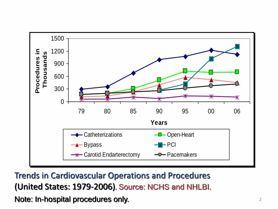
Trends in Cardiovascular Operations and Procedures (United States: 1979-2006). Source: NCHS and NHLBI.
Note: In-hospital procedures only.
0
300
600
900
1200
1500
79 80 85 90 95 00 06
Years
Pro
ce
du
res
in
Th
ou
sa
nd
s
Catheterizations Open-Heart
Bypass PCI
Carotid Endarterectomy Pacemakers

Less invasive techniques
• Endovascular aneurysm repair
• Minimally invasive open heart surgery or
• OPCAB (off-pump coronary artery bypass)
• Robotics
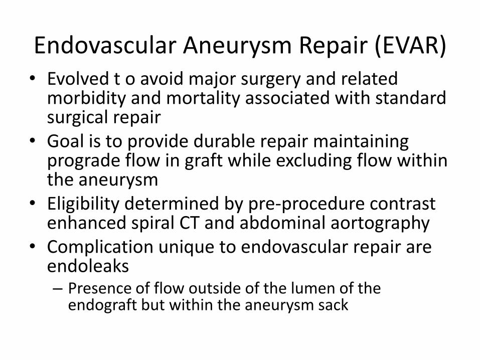
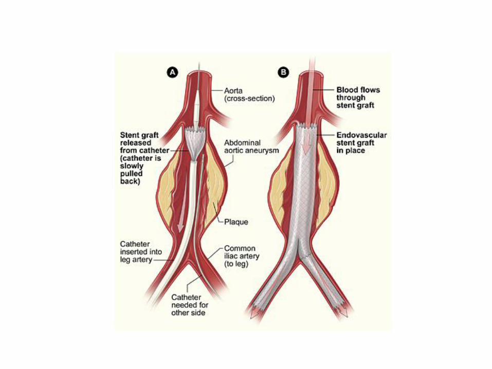
Endovascular Aneurysm Repair (EVAR)• Evolved t o avoid major surgery and related
morbidity and mortality associated with standard surgical repair
• Goal is to provide durable repair maintaining prograde flow in graft while excluding flow within the aneurysm
• Eligibility determined by pre-procedure contrast enhanced spiral CT and abdominal aortography
• Complication unique to endovascular repair are endoleaks– Presence of flow outside of the lumen of the
endograft but within the aneurysm sack

Thoracic Abdominal Aortic Aneurysm

• Potential complication from interrupted blood supply to the spinal cord are life-threatening
– Result in lower extremity weakness
– Paralysis 6%-40%
• Lumbar Drain placed to monitor CSF pressure
– Pressures > 15 considered elevated
– Increase may be corrected by drainage of CSF
• Keep SBP > 120 and < 160. Keep MAP > 90
– Use Neosynephrine to prevent hypotension
– Avoid HTN, which may increase bleeding
• No NTG or Nipride
• Monitor neurological signs Q1hour
– Report any neuro change, weakness, tingling, pain, loss of sensation in lower extremities or buttocks, or is bowel incontinence develops
• Minimally invasive heart surgery
– Performed on beating heart
– Referred to as MIDCAB (minimallly invasive direct coronary artery bypass) or OPCAB (off-pump CABG)
– Smaller incision
– Faster recovery time
– Use of heart-lung machine avoided
– Decreased procedure costs
– Reduced morbidity and mortality
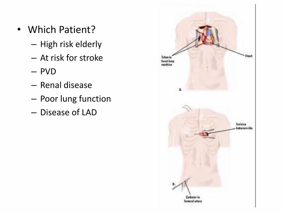
• Which Patient?
– High risk elderly
– At risk for stroke
– PVD
– Renal disease
– Poor lung function
– Disease of LAD
• Complications:
– MIDCAB
• Restenosis – incidence decreasing with experience
• Rib fracture
• Pericarditis
• Conversion to standard sternotomy
• SVT arrhythmias and ST segment elevation may develop
– OPCAB
• Conversion to cardiopulmonary bypass for completion of anastomosis
• Additional surgery to control bleeding r/t use of both internal mammary arteries
• Cerebral complications and atrial fibrillation
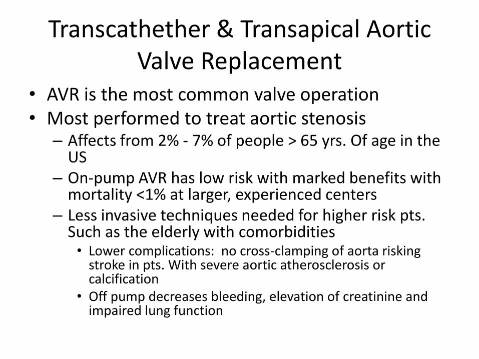
Transcathether & Transapical Aortic Valve Replacement
• AVR is the most common valve operation• Most performed to treat aortic stenosis
– Affects from 2% - 7% of people > 65 yrs. Of age in the US
– On-pump AVR has low risk with marked benefits with mortality <1% at larger, experienced centers
– Less invasive techniques needed for higher risk pts. Such as the elderly with comorbidities• Lower complications: no cross-clamping of aorta risking
stroke in pts. With severe aortic atherosclerosis or calcification
• Off pump decreases bleeding, elevation of creatinine and impaired lung function

Transcatheter & transapical AVR
• In clinical trials
• Indications:
– High risk pts. ( > 20% mortality)
– Severe, symptomatic AS
– Severe, ascending aortic calcification that prevents aortic cannulation or cross-clamping
– Severe radiation damage to chest or other severe chest deformities that would preclude sternotomy
• Does not require removal of native valve
• Held in place by stent or frame
• Local or spinal anesthesia with sedation or general anesthesia in cath lab or OR equipped with fluoroscopy and TEE
• Two approaches for transcatheter AVR– Antegrade, transeptal
• Access via femoral vein and pass catheter to right ventricle
• Puncture septum
• Technically difficult and can damage mitral valve
– Retrograde approach• Femoral artery accessed to reach aortic valve
• Limitations:– Size of artery
– Atheroschlerotic material can be embolized from aorta into distal circulation
• Contrast medium used to verify position of catheter
• Rapid pacing (HR > 150-220/min) needed for Edwards valve to decrease C.O.
• Must be careful not to cover opening of coronary ostia
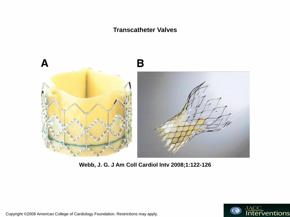
Copyright ©2008 American College of Cardiology Foundation. Restrictions may apply.
Webb, J. G. J Am Coll Cardiol Intv 2008;1:122-126
Transcatheter Valves
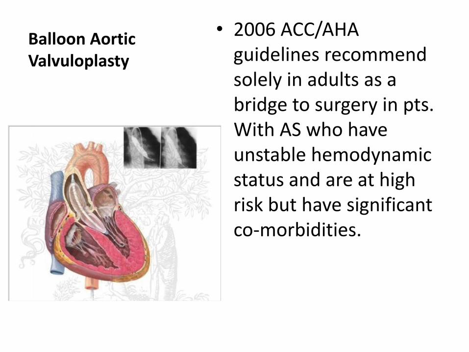
Balloon Aortic Valvuloplasty
• 2006 ACC/AHA guidelines recommend solely in adults as a bridge to surgery in pts. With AS who have unstable hemodynamic status and are at high risk but have significant co-morbidities.
• Transapical approach best
– For pts. With tortuous or small femoral or iliac vessels
– For pts. With severe PVD
– For pts. With heavily calcified aorta
– Quicker
• Access via a 5 to 8cm anterolateral left thoracotomy usually in the 6th ICS
• Pericardium opened and transapical stab incision made in left ventricle
• Balloon valvuloplasty performed to dilate native valve first
• High rate pacing done to decrease C.O.
• Complications
– Vessel rupture
– Dissection
– Pseudoaneurysm
– Bleeding and thrombus formation
– Myocardial perforation
– Cardiac tamponade
– Embolization of calcified material
– Perivalvular regurgitation
Surgical Procedures
• Maze Procedure
– Indicated for pts. Who are intolerant of the arrhythmia, had failure of drug therapy, or had multiple embolic events
– Incisions/lesions created in both atria and both atrial appendages removed
• Theory is that A. fib results from multiple macro-reentry circuits
– 98% successful
Candidates for procedure are:
Have symptoms but medicines fail to control
High risk for embolic events
Atrial fibrillation longer than 6mos. & have enlarged left atrium
Already undergoing mitral valve or other cardiac surgery

Atrial appendages
• Blind pouches attached to each atria
• Contribute nothing to overall function
• In A. Fibrillation, because of no synchrony or uniform contraction of atrial muscle blood sits dormant in appendages
• Therefore, clots tend to occur and often progress and become larger
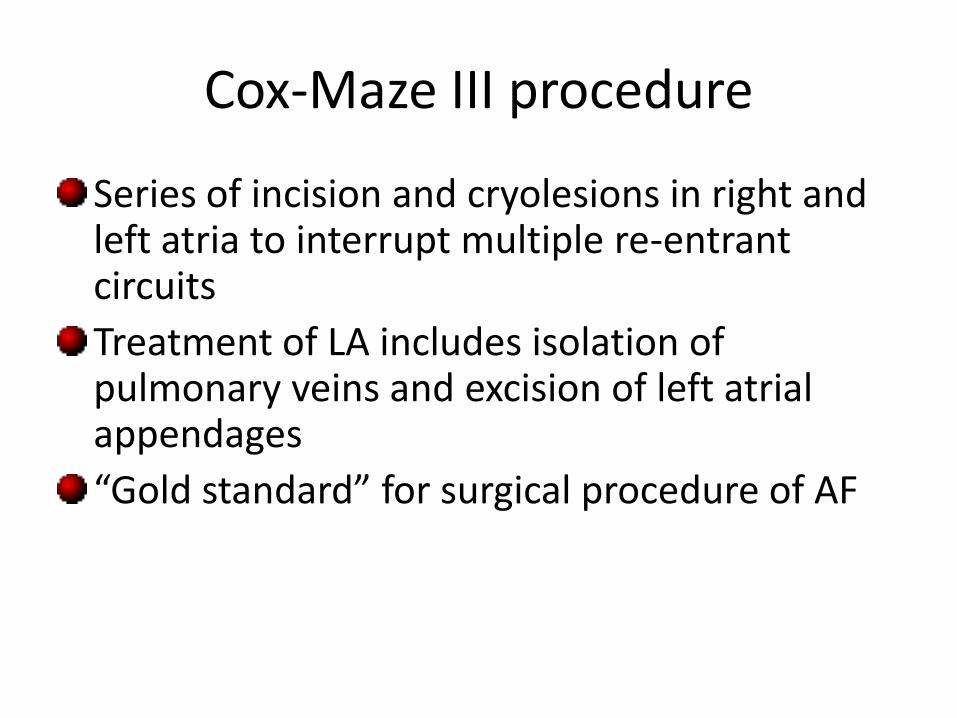
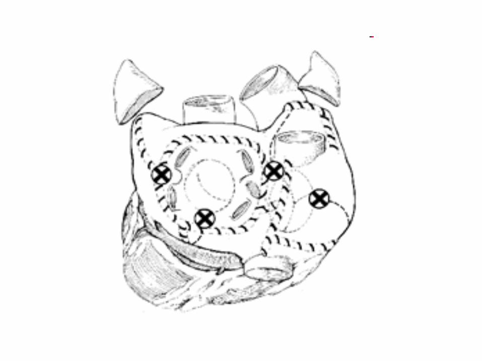
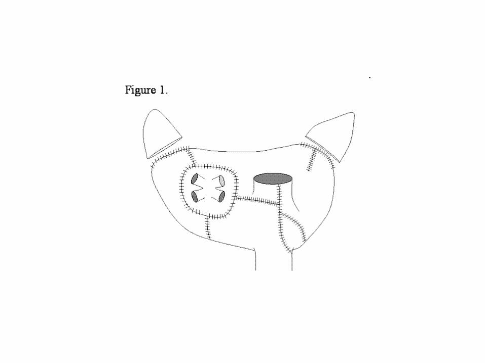
Cox-Maze III procedure
Series of incision and cryolesions in right and left atria to interrupt multiple re-entrant circuits
Treatment of LA includes isolation of pulmonary veins and excision of left atrial appendages
“Gold standard” for surgical procedure of AF
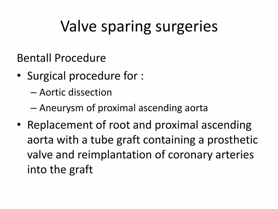
Valve sparing surgeries
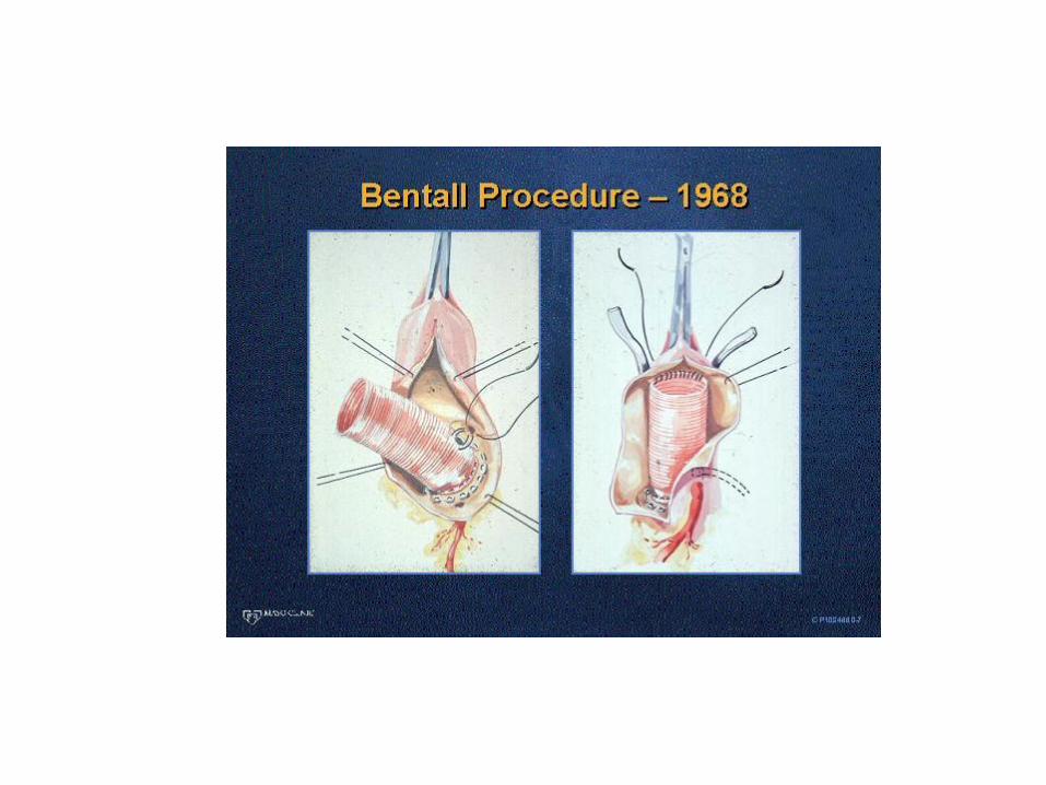
Bentall Procedure
• Surgical procedure for :
– Aortic dissection
– Aneurysm of proximal ascending aorta
• Replacement of root and proximal ascending aorta with a tube graft containing a prosthetic valve and reimplantation of coronary arteries into the graft

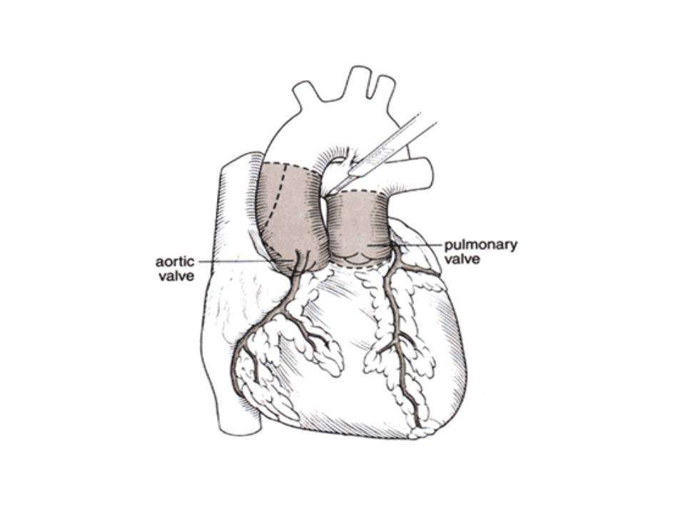
Ross Procedure
Patient’s diseased aortic valve is replaced with their own pulmonary valve
Pulmonary valve replaced with cadaver pulmonary valve
Anticoagulation not required
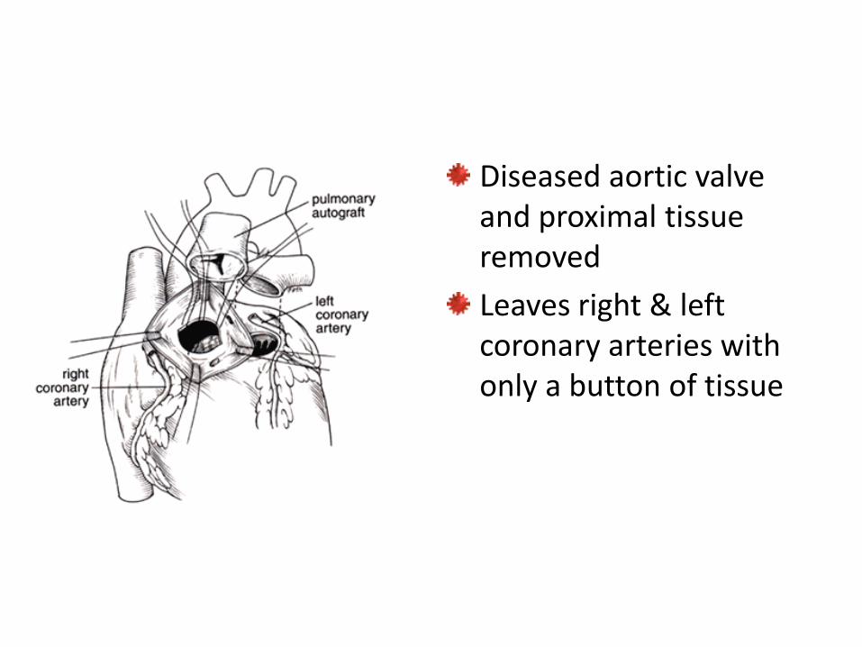
Diseased aortic valve and proximal tissue removed
Leaves right & left coronary arteries with only a button of tissue
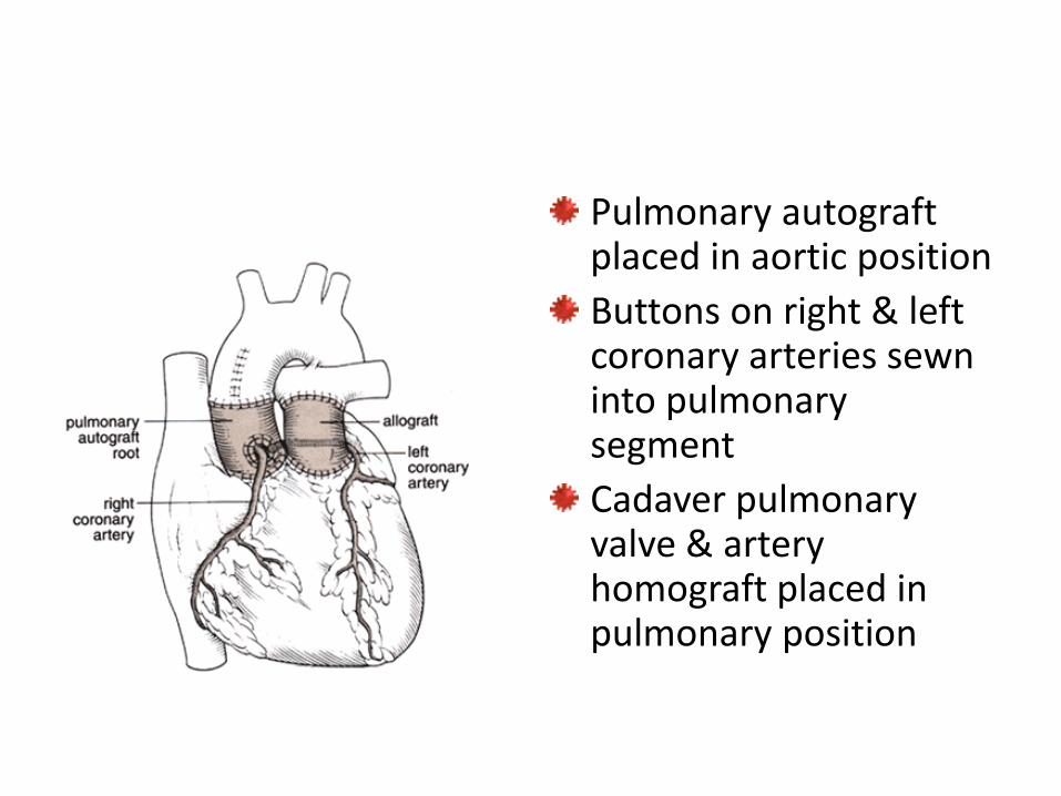
Pulmonary autograft placed in aortic position
Buttons on right & left coronary arteries sewn into pulmonary segment
Cadaver pulmonary valve & artery homograft placed in pulmonary position
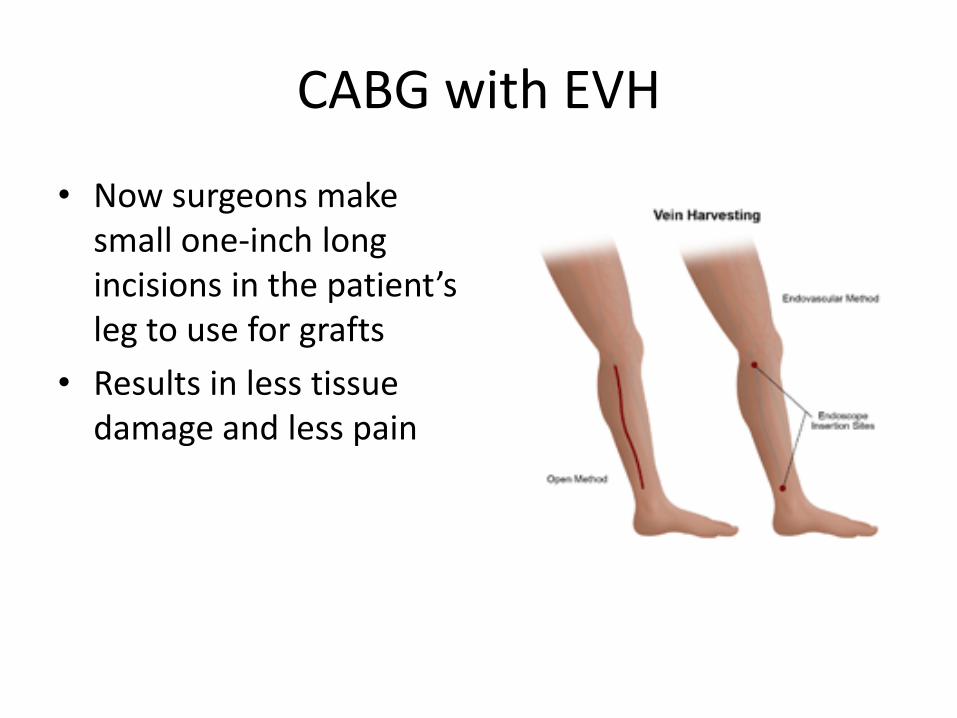
CABG with EVH
• Now surgeons make small one-inch long incisions in the patient’s leg to use for grafts
• Results in less tissue damage and less pain
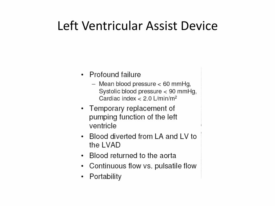
Left Ventricular Assist Device