Current strategies for the restoration of adequate ... DOI: . ... pelvic tilt 12°, and lumbar...
11
Current strategies for the restoration of adequate lordosis during lumbar fusion Cédric Barrey, Alice Darnis Cédric Barrey, Alice Darnis, Department of Spine Surgery, P Wertheimer Hospital, Hospices Civils de Lyon, University Claude Bernard Lyon 1, 69003 Lyon, France Author contributions: Barrey C and Darnis A contributed equally to this work. Open-Access: This article is an open-access article which selected by an in-house editor and fully peer-reviewed by external reviewers. It distributed in accordance with the Creative Commons Attribution Non Commercial (CC BY-NC 4.0) license, which permits others to distribute, remix, adapt, build upon this work non-commercially, and license their derivative works on different terms, provided the original work is properly cited and the use is non-commercial. See: http://creativecommons.org/ licenses/by-nc/4.0/ Correspondence to: Cédric Barrey, MD, PhD, Department of Spine Surgery, P Wertheimer Hospital, Hospices Civils de Lyon, University Claude Bernard Lyon 1, 59 Boulevard Pinel, 69003 Lyon, France. [email protected] Telephone: +33-6-86940037 Fax: +33-4-72357262 Received: December 30, 2013 Peer-review started: December 31, 2013 First decision: March 12, 2014 Revised: July 14, 2014 Accepted: July 27, 2014 Article in press: July 29, 2014 Published online: January 18, 2015 Abstract Not restoring the adequate lumbar lordosis during lumbar fusion surgery may result in mechanical low back pain, sagittal unbalance and adjacent segment degeneration. The objective of this work is to describe the current strategies and concepts for restoration of adequate lordosis during fusion surgery. Theoretical lordosis can be evaluated from the measurement of the pelvic incidence and from the analysis of spatial organization of the lumbar spine with 2/3 of the lordosis given by the L4-S1 segment and 85% by the L3-S1 segment. Technical aspects involve patient positioning on the operating table, release maneuvers, type of instrumentation used (rod, screw-rod connection, interbody cages), surgical sequence and the overall surgical strategy. Spinal osteotomies may be required in case of fixed kyphotic spine. AP combined surgery is particularly efficient in restoring lordosis at L5-S1 level and should be recommended. Finally, not one but several strategies may be used to achieve the need for restoration of adequate lordosis during fusion surgery. Key words: Lumbar lordosis; Pelvis shape; Pelvis incidence; Spinal fusion; Spine surgery; Sagittal balance © The Author(s) 2015. Published by Baishideng Publishing Group Inc. All rights reserved. Core tip: Not restoring the adequate lumbar lordosis during fusion surgery may result in mechanical pain, sagittal unbalance and adjacent segment degeneration. The objective of this paper is to describe the current strategies and concepts for restoration of adequate lordosis during fusion surgery. The amount of lordosis to restore can be precisely evaluated from the analysis of spino-pelvic parameters. Technical tools during surgery involve patient positioning, release maneuvers, type of instrumentation used and surgical sequence. Finally, not one but several strategies may be used to restore the adequate lordosis during fusion surgery. Barrey C, Darnis A. Current strategies for the restoration of adequate lordosis during lumbar fusion. World J Orthop 2015; 6(1): 117-126 Available from: URL: http://www.wjg- net.com/2218-5836/full/v6/i1/117.htm DOI: http://dx.doi. org/10.5312/wjo.v6.i1.117 INTRODUCTION Lumbar fusion is a common surgical procedure for the REVIEW Online Submissions: http://www.wjgnet.com/esps/ Help Desk: http://www.wjgnet.com/esps/helpdesk.aspx DOI: 10.5312/wjo.v6.i1.117 117 January 18, 2015|Volume 6|Issue 1| WJO|www.wjgnet.com World J Orthop 2015 January 18; 6(1): 117-126 ISSN 2218-5836 (online) © 2015 Baishideng Publishing Group Inc. All rights reserved.
Transcript of Current strategies for the restoration of adequate ... DOI: . ... pelvic tilt 12°, and lumbar...
Current strategies for the restoration of adequate lordosis during lumbar fusion
Cédric Barrey, Alice Darnis
Cédric Barrey, Alice Darnis, Department of Spine Surgery, P Wertheimer Hospital, Hospices Civils de Lyon, University Claude Bernard Lyon 1, 69003 Lyon, FranceAuthor contributions: Barrey C and Darnis A contributed equally to this work.Open-Access: This article is an open-access article which selected by an in-house editor and fully peer-reviewed by external reviewers. It distributed in accordance with the Creative Commons Attribution Non Commercial (CC BY-NC 4.0) license, which permits others to distribute, remix, adapt, build upon this work non-commercially, and license their derivative works on different terms, provided the original work is properly cited and the use is non-commercial. See: http://creativecommons.org/licenses/by-nc/4.0/Correspondence to: Cédric Barrey, MD, PhD, Department of Spine Surgery, P Wertheimer Hospital, Hospices Civils de Lyon, University Claude Bernard Lyon 1, 59 Boulevard Pinel, 69003 Lyon, France. [email protected] Telephone: +33-6-86940037 Fax: +33-4-72357262Received: December 30, 2013Peer-review started: December 31, 2013First decision: March 12, 2014Revised: July 14, 2014Accepted: July 27, 2014Article in press: July 29, 2014Published online: January 18, 2015
AbstractNot restoring the adequate lumbar lordosis during lumbar fusion surgery may result in mechanical low back pain, sagittal unbalance and adjacent segment degeneration. The objective of this work is to describe the current strategies and concepts for restoration of adequate lordosis during fusion surgery. Theoretical lordosis can be evaluated from the measurement of the pelvic incidence and from the analysis of spatial organization of the lumbar spine with 2/3 of the lordosis given by the L4-S1 segment and 85% by the L3-S1 segment. Technical aspects involve patient positioning
on the operating table, release maneuvers, type of instrumentation used (rod, screw-rod connection, interbody cages), surgical sequence and the overall surgical strategy. Spinal osteotomies may be required in case of fixed kyphotic spine. AP combined surgery is particularly efficient in restoring lordosis at L5-S1 level and should be recommended. Finally, not one but several strategies may be used to achieve the need for restoration of adequate lordosis during fusion surgery.
Key words: Lumbar lordosis; Pelvis shape; Pelvis incidence; Spinal fusion; Spine surgery; Sagittal balance
© The Author(s) 2015. Published by Baishideng Publishing Group Inc. All rights reserved.
Core tip: Not restoring the adequate lumbar lordosis during fusion surgery may result in mechanical pain, sagittal unbalance and adjacent segment degeneration. The objective of this paper is to describe the current strategies and concepts for restoration of adequate lordosis during fusion surgery. The amount of lordosis to restore can be precisely evaluated from the analysis of spino-pelvic parameters. Technical tools during surgery involve patient positioning, release maneuvers, type of instrumentation used and surgical sequence. Finally, not one but several strategies may be used to restore the adequate lordosis during fusion surgery.
Barrey C, Darnis A. Current strategies for the restoration of adequate lordosis during lumbar fusion. World J Orthop 2015; 6(1): 117-126 Available from: URL: http://www.wjg-net.com/2218-5836/full/v6/i1/117.htm DOI: http://dx.doi.org/10.5312/wjo.v6.i1.117
INTRODUCTIONLumbar fusion is a common surgical procedure for the
REVIEW
Online Submissions: http://www.wjgnet.com/esps/Help Desk: http://www.wjgnet.com/esps/helpdesk.aspxDOI: 10.5312/wjo.v6.i1.117
117 January 18, 2015|Volume 6|Issue 1|WJO|www.wjgnet.com
World J Orthop 2015 January 18; 6(1): 117-126ISSN 2218-5836 (online)
© 2015 Baishideng Publishing Group Inc. All rights reserved.

management of degenerative and spinal deformities. Loss of lordosis after lumbar spine fusion can lead to chronic low back pain, positive sagittal balance with forward inclination of the trunk and adjacent segment degeneration. Identification and restoration of adequate lumbar lordosis for sagittal balance should be an everyday concern for the spine surgeon. However the challenge is to determine the correct amount of lumbar lordosis that each patient requires to maintain optimal sagittal balance. The development of posterior and anterior instrumentation has offered the advantage of a more accurate and more efficient restoration of the alignment of the lumbar spine and having good knowledge of the pelvic and spinal parameters is required to use it as effectively as possible.
WHY IS IT NECESSARY TO RESTORE LORDOSIS?In 1973 Doherty[1] described a symptomatic fixed forward inclination of the trunk due to loss of normal lumbar lordosis following posterior spinal fusion for thoracolumbar scoliosis with Harrington instrumentation. Flatback syndrome, also known as fixed sagittal imbalance, was then described in 1977 by Moe et al[2] with a series of 16 patients with a loss of lumbar lordosis after thoracolumbar fusion. The most common cause of flatback syndrome is iatrogenic secondary to Harrington rod instrumentation[3-10] but there are many other iatrogenic causes such as hypolordotic lumbar fusion for degenerative spondylolysis, scoliosis or stenosis with instability. Failure to maintain lumbar lordosis during a fusion of a degenerative spine can result in accelerated adjacent degeneration, mechanical low back pain and loss of sagittal balance with forward inclination of the trunk, anterior displacement of the center of gravity and compensatory mechanisms such as cervical and thoracic segment hyperextension, knee flexion and hip extension[11-15]. These compensatory mechanisms have adverse effects
such as chronic pain, disability and/or muscle fatigue[15]. Breakdown of the adjacent level has been identified as one cause of postoperative pain and disability[16-18].
The biomechanical effect of postoperative hypolordosis in lumbar fusion on instrumented and adjacent spinal segments has been described by Umehara et al[19] in 2000. Postoperative lumbar hypolordosis accelerate adjacent segment deterioration by loading the motion segment in a nonphysiologic way. The loss of lordosis in the instrumented segments not only affects the adjacent segments, but also increases the load on the posterior spinal implant. The tension in the anterior soft tissue structures decreases, increasing the implant load needed to balance the extension moment. To maintain good balance in the presence of a loss of lordosis, the posterior shear force on the proximal segments increases. This increases the extension moment on the lumbar spine and leads to an increased loading of the posterior implant, with a higher risk of loosening due to repetitive extension loading during activities of daily living. The loading of the posterior column in the segment above the instrumentation increases and may contribute to the degenerative changes (DDD, facet arthritis, listhésis) at the junctional level reported as long-term consequences of lumbar fusion (Figure 1).
Other factors are implicated in adjacent degeneration including rigid fixation, number of levels fused, and health of the adjacent level[19]. Guigui et al[20] showed that adjacent segment degeneration was significantly more common in patients treated earlier for degenerate disc disease than in younger patients with spondylolisthesis. Adjacent segment degeneration has been reported to be more frequent in females[21].
In 2001, Izumi et al[22] analyzed the sagittal lumbar alignment before and after posterior instrumentation and showed that in case of degenerative changes in the adjacent unfused segment the mean lumbar lordotic angles were decreased postoperatively by about 10°. Lazennec et al[23] in 2000 described the difficulty of achieving optimal lumbo-sacral alignment during fusion and showed statistically significant correlation between reduction of sacral inclination and back pain following lumbosacral fusion in the standing position because of undue stress on the sacroiliac joints and on the hips. In 2001, Kumar et al[24] reported 31 patients with radiographic evidence of adjacent level degenerative changes above the level of fusion in a series of 83 patients. The lowest incidence of adjacent segment degeneration was seen in patients with normal C7 sagittal plumb line and normal sacral inclination (8%). The difference between this group and the other groups with abnormality in either the plumb line or the sacral inclination or both was statistically significant.
It is difficult to determine the « good position » for lumbar fusion and the optimal degree of lordosis has not yet be defined[25]. Achieving a strong fusion in the optimal position requires understanding the relationships between the pelvic and spinal parameters in order to determine the theoretical lordosis for each individual (Table 1).
118 January 18, 2015|Volume 6|Issue 1|WJO|www.wjgnet.com
Barrey C et al . Surgical strategies to restore lumbar lordosis
Hypolordotic construct: Stresses at adjacent levels!
a = 24°
Figure 1 Hypolordotic lumbo-sacral fusion with hyperextension of the segment above the instrumentation. Failure to restore a good sagittal balance leads to chronic back pain and early degenerative changes at adjacent level(s).
HOW MUCH LORDOSIS IS IT NECESSARY TO RESTORE ? THE CONCEPT OF THE THEORETICAL LORDOSISThe concept of the theoretical lordosis. Relationships between the components of the lumbo-pelvic complex. To determine the amount of lordosis to restore, we have to introduce the concept of theoretical lordosis deriving from the need for congruence between spinal and pelvic parameters.
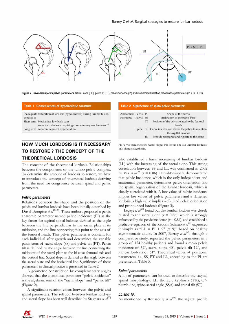
Pelvic parametersRelations between the shape and the position of the pelvis and lumbar lordosis have been initially described by Duval-Beaupère et al[26-29]. These authors proposed a pelvic anatomic parameter named pelvic incidence (PI) as the key factor for sagittal spinal balance, defined as the angle between the line perpendicular to the sacral plate at its midpoint, and the line connecting this point to the axis of the femoral heads. This pelvic parameter is constant for each individual after growth and determines the variable parameters of sacral slope (SS) and pelvic tilt (PT). Pelvic tilt is defined by the angle between the line connecting the midpoint of the sacral plate to the bi-coxo-femoral axis and the vertical line. Sacral slope is defined as the angle between the sacral plate and the horizontal line. Significance of these parameters in clinical practice is presented in Table 2.
A geometric construction by complementary angles showed that the anatomical parameter “pelvic incidence” is the algebraic sum of the “sacral slope” and “pelvic tilt” (Figure 2).
A significant relation exists between the pelvic and spinal parameters. The relation between lumbar lordosis and sacral slope has been well described by Stagnara et al[25]
who established a linear increasing of lumbar lordosis (LL) with the increasing of the sacral slope. This strong correlation between SS and LL was confirmed in 2002 by Vaz et al[30] (r = 0.86). Duval-Beaupère demonstrated that pelvic incidence, which is the only independent and anatomical parameter, determines pelvic orientation and the spatial organization of the lumbar lordosis, which is closely correlated with it. A low value of pelvic incidence implies low values of pelvic parameters and a flattened lordosis; a high value implies well-tilted pelvic orientation and pronounced lordosis (Figure 3).
Legaye et al[28] found out that lumbar lordosis was closely related to the sacral slope (r = 0.86), which is strongly influenced by the pelvic incidence (r = 0.84), and established a predictive equation of the lordosis. Schwab et al[31] expressed it simply as “LL = PI + 9° (± 9)” based on healthy asymptomatic adults. In 2007, Barrey et al[32], through a comparative study, reported the pelvic parameters in a group of 154 healthy patients and found a mean pelvic incidence of 52°, sacral slope 40°, pelvic tilt 12°, and lumbar lordosis of 61°. Theoretical values of positional parameters, i.e., SS, PT and LL, according to the PI are presented in Table 3.
Spinal parametersA lot of parameters can be used to describe the sagittal spinal morphology: LL, thoracic kyphosis (TK), C7-plumb-line, spino-sacral angle (SSA) and spinal tilt (ST).
LL and TKAs mentioned by Roussouly et al[33], the sagittal profile
119 January 18, 2015|Volume 6|Issue 1|WJO|www.wjgnet.com
Figure 2 Duval-Beaupère’s pelvic parameters. Sacral slope (SS), pelvic tilt (PT), pelvic incidence (PI) and mathematical relation between the parameters (PI = SS + PT).
Inadequate restoration of lordosis (hypolordosis) during lumbar fusion exposes to Short term Mechanical low back pain
Anterior unbalance requiring compensatory mechanisms[15]
Long term Adjacent segment degeneration
Table 1 Consequences of hypolordotic construct
Anatomical Pelvis PI Shape of the pelvis Positional Pelvis SS Inclination of the pelvis base
PT Position of the pelvis related to the femoral heads
Spine LL Curve in extension above the pelvis to maintain the sagittal balance
TK Provide resistance and rigidity to the spine
Table 2 Significance of spino-pelvic parameters
PI: Pelvis incidence; SS: Sacral slope; PT: Pelvis tilt; LL: Lumbar lordosis; TK: Thoracic kyphosis.
SS PT PI
SS
PI = SS + PT
PTPI
Barrey C et al . Surgical strategies to restore lumbar lordosis

120 January 18, 2015|Volume 6|Issue 1|WJO|www.wjgnet.com
slope approaches the horizontal. The inflection point is low and posterior, creating a short lordosis with a negative lordosis tilt angle. The upper spine has a significant kyphosis of the thoracolumbar junction and thorax. In his series, the mean global lumbar lordosis of this group was 52°; Type 2 Lordosis. The sacral slope is less than 35°. The apex of the lumbar lordosis is located at base of the L4 vertebral body. The lower arc of lordosis is relatively flat. The inflection point is higher and more anterior, decreasing the lordosis tilt angle but increasing the number of vertebral bodies included in the lordosis. The entire spine is relatively hypolordotic and hypokyphotic. In his series, the mean global lumbar lordosis of this group was 52°; Type 3 Lordosis. The sacral slope is between 35° and 45°. The apex of lumbar lordosis is in the center of the L4 vertebral body. The lower arc of lordosis becomes more prominent. The inflection point is at the thoracolumbar junction, and the lordosis tilt angle is nearly zero. An average of four vertebral bodies constitutes the arc of lordosis. The spine is well balanced. In his series, the mean global lumbar lordosis of this group was 61°; Type 4 Lordosis: The sacral slope is greater than 45°, which is associated with a high pelvic incidence. The apex of the lumbar lordosis is located at the base of the L3 vertebral body or higher. The lower arc of lordosis is prominent, and the lordosis tilt angle is zero or positive. The number of vertebrae in a lordotic orientation is greater than 5, and a state of segmental hyperextension exists. In his series, the mean global lumbar lordosis of this group was 71°.
C7 plumb line, SSA and STAs described by Roussouly et al[34], the C7-plumb-line is the vertical axis begining at the centroid of C7 and the SSA is defined as the angle between a line from the center of C7 to the center of the sacral endplate and the sacral endplate itself. The spinal tilt (ST) is defined as the angle between the line connecting the centers of C7 and S1
of the spine is usually characterized as being kyphotic between T1 and T12, and lordotic between L1 and L5, but this is not necessarily the case. The “thoracic” segment of the spine is located between T1 and the inflection point where the spine transitions from kyphosis to lordosis. The “lumbar” lordosis exists between the inflection point and S1. This determination of kyphotic and lordotic segments is independent of the anatomic location of the thoracolumbar junction at T12-L1. To characterize the lumbar lordosis in normal population, several parameters have to be taken into consideration: the position of the apex of the thoracic and lumbar curves, the position of the inflection point (transition between LL and TK), the number of vertebral bodies in each curvature, total kyphosis and lordosis in degrees, lordosis tilt angle, and the sacral slope (Figure 4).
Based on these considerations, Roussouly established a system to classify each patient as one of four types (Figure 5): Type 1 Lordosis. The sacral slope is less than 35°, which is usually associated with a low pelvic incidence. The apex of the lumbar lordosis is located in the center of L5 vertebral body. The lower arc of lordosis is minimal, decreasing toward zero as the sacral
Figure 3 Low pelvic incidence is usually associated with slight sacral slope and flat lumbar spine, and high pelvic incidence with great sacral slope and more curved lumbar spine[32]. PI: Pelvis incidence; PT: Pelvis tilt.
Figure 4 View of several spinal parameters. Lumbar lordosis (LL), Thoracic kyphosis (TK), Apex of the lordosis, Inflection point. PI: Pelvis incidence; PT: Pelvis tilt; SS: Sacral slope.
PI class PI (°) PTth (°) LLth (°)
Ⅰ < 38 4 PI + 18 Ⅱ 38-47 8 PI + 13 Ⅲ 48-57 12 PI + 9 Ⅳ 58-67 16 PI + 6 Ⅴ 68-77 20 PI + 2 Ⅵ > 78 24 PI - 5
Table 3 Theoretical values for positional pelvis and spinal parameters related to pelvis incidence
PT: Pelvis tilt; LL: Lumbar lordosis; PTth: Theoretical PT; LLth: Theoretical LL. As examples, for PI measured to 40°, expected PT should be 8° and LL should be 53°; for PI measured to 52°, expected PT should be 12° and LL should be 61°; and for PI measured to 64°, expected PT should be 16° and LL should be 70°.
Low PI
PT
PI
PT
PI
High PI
PIPT
SS
Thoracic kyphosis
Lumbar lordosis
Inflection point
Apex of lordosis
Barrey C et al . Surgical strategies to restore lumbar lordosis

121 January 18, 2015|Volume 6|Issue 1|WJO|www.wjgnet.com
and the horizontal axis. There is a geometric association between ST, SSA, and SS: ST = SSA - SS (Figure 6). In a cohort of 153 patients without symptoms of spinal disease the mean SSA was 134.7° and the mean ST was 95.1°.
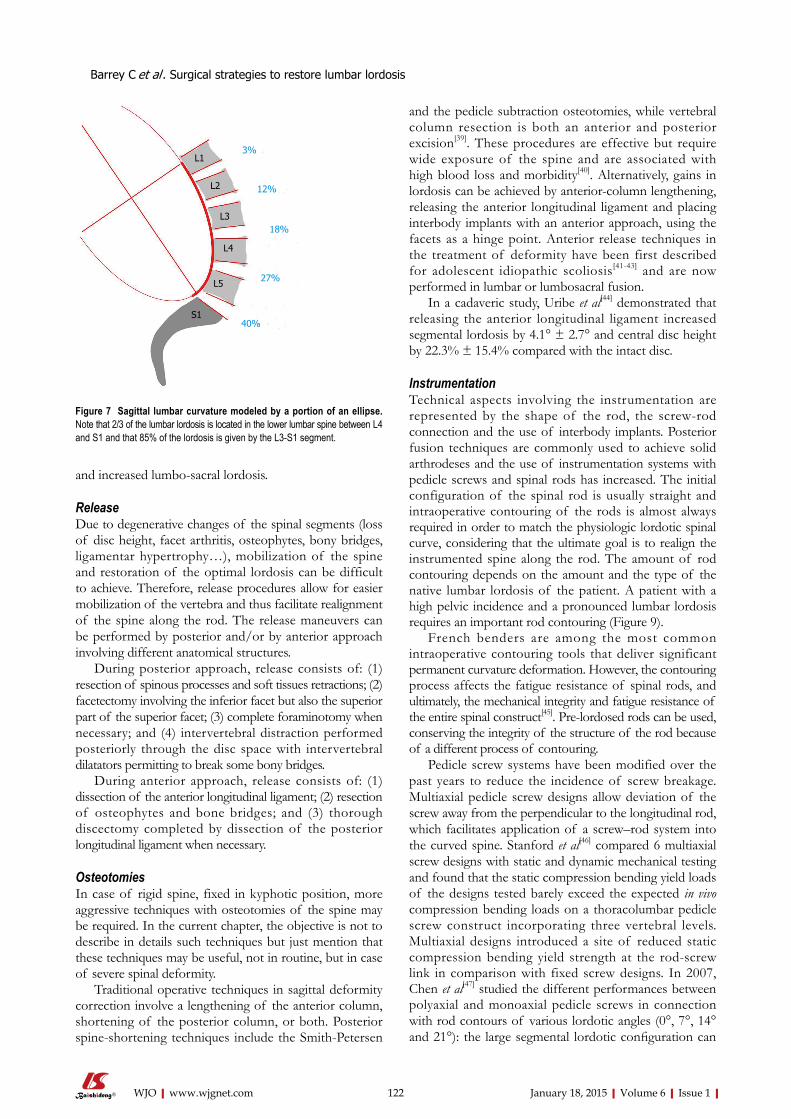
In 1998, Janik et al[35] hypothesized that a simple geometric model in the shape of an ellipse, from T12 to S1, would fit the lumbar lordosis. The elliptical model was approximately an 85° portion of a quadrant and suggested that about 70% of the lumbar lordosis was located between L4 and S1 (Figure 7).
Taking in account the theoretical lordosis for each individual related to the PI and also the normal distribution of the lordosis along the lumbar spine, we can calculate the amount of lordosis to restore according to the length of the construct (Table 4).
HOW TO RESTORE LORDOSIS DURING LUMBOSACRAL FUSION? TECHNICAL KEY POINTSTools and technical key-points to restore lordosis during lumbar fusion surgery are synthesized in Table 5.
Operative positionDifferent operative positions can be used in lumbar spinal surgery, depending on the type of the procedure.
Decompressive procedures are optimally performed in positions incorporating less lordosis, improving access to the spinal canal and intervertebral discs and decreasing blood loss[36], as in the knee-chest position.
At the opposite, lumbar or lumbosacral fusions with internal fixation should be performed in an operative position which recreates physiologic lordosis. In 1996, Stephens et al[37] compared operative tables used commonly for spinal procedures in order to determine which positions reproduce “normal” lumbar lordosis. Ten volunteers without any history of lumbar surgery or symptomatology underwent lateral radiograph in the standing position and in three different kinds of operative position: prone position on the Jackson table, knee flexed at 15°, knee-chest position with hips flexed at 90° on the Andrews table, and intermediate position with hips flexed at 60° (Figure 8). The mean lumbar lordosis angle from L1 to sacrum was 51.7° in the standing position, 52° in the prone position on the Jackson table, 17° in the knee-chest position and 27.3° with the hips flexed at 60°. The decrease in lordosis was statistically significant in the knee-chest and the intermediate position compared with the standing position and the Jackson table.
Another study in 1995 by Peterson et al[38] showed that the “90-90” position on the Hastings frame was associated with significant reduction of total and segmental lordosis in the middle and lower lumbar spine. We therefore recommend positioning prone, as example on a Jackson table, maintained standing lumbar lordosis
Figure 5 Roussouly’s classification of sagittal profiles of the spine in four types[33].
Figure 6 C7 plumb line, spino sacral angle and spinal tilt. C7PL: C7 Plumb Line; SSA: Spino sacral angle; ST: Spinal tilt.
PI LLth Length of fusionL5-S1 L4-S1 L3-S1 L2-S1 L1-S1
40% of LLth 67% of LLth 85% of LLth 97% of LLth 100% of LLth
40° 55° 22° 37° 47° 53° 55° 50° 60° 24° 40° 51° 58° 60° 60° 65° 26° 44° 55° 63° 65° 70° 70° 28° 47° 59° 68° 70°
Table 4 Amount of lordosis to restore related to the pelvis incidence and the length of spinal construct
PT: Pelvis tilt; LLth: Theoretical Lumbat lordosis.
Operative positioning Avoid knee-chest position Release Allow for mobilization of the spinal segments Instrumentation Contouring of the rod
Screw-rod connectionInterbody implants
Spinal osteotomies Indicated only when the spine is fixed in kyphotic position
Surgical sequence AP sequence should be promoted
Table 5 Technical key points permitting to restore lordosis
Type 1SS < 35°
Type 2SS < 35°
Type 335° < SS < 45°
Type 4SS > 45°
SSA
ST
C7PL
Barrey C et al . Surgical strategies to restore lumbar lordosis
122 January 18, 2015|Volume 6|Issue 1|WJO|www.wjgnet.com
and increased lumbo-sacral lordosis.
ReleaseDue to degenerative changes of the spinal segments (loss of disc height, facet arthritis, osteophytes, bony bridges, ligamentar hypertrophy…), mobilization of the spine and restoration of the optimal lordosis can be difficult to achieve. Therefore, release procedures allow for easier mobilization of the vertebra and thus facilitate realignment of the spine along the rod. The release maneuvers can be performed by posterior and/or by anterior approach involving different anatomical structures.
During posterior approach, release consists of: (1) resection of spinous processes and soft tissues retractions; (2) facetectomy involving the inferior facet but also the superior part of the superior facet; (3) complete foraminotomy when necessary; and (4) intervertebral distraction performed posteriorly through the disc space with intervertebral dilatators permitting to break some bony bridges.
During anterior approach, release consists of: (1) dissection of the anterior longitudinal ligament; (2) resection of osteophytes and bone bridges; and (3) thorough discectomy completed by dissection of the posterior longitudinal ligament when necessary.
Osteotomies In case of rigid spine, fixed in kyphotic position, more aggressive techniques with osteotomies of the spine may be required. In the current chapter, the objective is not to describe in details such techniques but just mention that these techniques may be useful, not in routine, but in case of severe spinal deformity.
Traditional operative techniques in sagittal deformity correction involve a lengthening of the anterior column, shortening of the posterior column, or both. Posterior spine-shortening techniques include the Smith-Petersen
and the pedicle subtraction osteotomies, while vertebral column resection is both an anterior and posterior excision[39]. These procedures are effective but require wide exposure of the spine and are associated with high blood loss and morbidity[40]. Alternatively, gains in lordosis can be achieved by anterior-column lengthening, releasing the anterior longitudinal ligament and placing interbody implants with an anterior approach, using the facets as a hinge point. Anterior release techniques in the treatment of deformity have been first described for adolescent idiopathic scoliosis[41-43] and are now performed in lumbar or lumbosacral fusion.
In a cadaveric study, Uribe et al[44] demonstrated that releasing the anterior longitudinal ligament increased segmental lordosis by 4.1° ± 2.7° and central disc height by 22.3% ± 15.4% compared with the intact disc.
InstrumentationTechnical aspects involving the instrumentation are represented by the shape of the rod, the screw-rod connection and the use of interbody implants. Posterior fusion techniques are commonly used to achieve solid arthrodeses and the use of instrumentation systems with pedicle screws and spinal rods has increased. The initial configuration of the spinal rod is usually straight and intraoperative contouring of the rods is almost always required in order to match the physiologic lordotic spinal curve, considering that the ultimate goal is to realign the instrumented spine along the rod. The amount of rod contouring depends on the amount and the type of the native lumbar lordosis of the patient. A patient with a high pelvic incidence and a pronounced lumbar lordosis requires an important rod contouring (Figure 9).
French benders are among the most common intraoperative contouring tools that deliver significant permanent curvature deformation. However, the contouring process affects the fatigue resistance of spinal rods, and ultimately, the mechanical integrity and fatigue resistance of the entire spinal construct[45]. Pre-lordosed rods can be used, conserving the integrity of the structure of the rod because of a different process of contouring.
Pedicle screw systems have been modified over the past years to reduce the incidence of screw breakage. Multiaxial pedicle screw designs allow deviation of the screw away from the perpendicular to the longitudinal rod, which facilitates application of a screw–rod system into the curved spine. Stanford et al[46] compared 6 multiaxial screw designs with static and dynamic mechanical testing and found that the static compression bending yield loads of the designs tested barely exceed the expected in vivo compression bending loads on a thoracolumbar pedicle screw construct incorporating three vertebral levels. Multiaxial designs introduced a site of reduced static compression bending yield strength at the rod-screw link in comparison with fixed screw designs. In 2007, Chen et al[47] studied the different performances between polyaxial and monoaxial pedicle screws in connection with rod contours of various lordotic angles (0°, 7°, 14° and 21°): the large segmental lordotic configuration can
Figure 7 Sagittal lumbar curvature modeled by a portion of an ellipse. Note that 2/3 of the lumbar lordosis is located in the lower lumbar spine between L4 and S1 and that 85% of the lordosis is given by the L3-S1 segment.
L1
L2
L3
L4
L5
S1
3%
12%
18%
27%
40%
Barrey C et al . Surgical strategies to restore lumbar lordosis

123 January 18, 2015|Volume 6|Issue 1|WJO|www.wjgnet.com
decrease the stiffness in the monoaxial screws. Polyaxial screws combined with an interbody cage fixation provide higher compression and flexion stiffness in 21° segmental lordosis and enhance the contact ratio of the interbody cage. However, to our knowledge there is no published data comparing the amount of lordosis restored between monoaxial and polyaxial screws. The angle between the screw and the rod is constant with the monoaxial screws (90°) whereas it is variable with the polyaxial screws. Because of this difference in the rod-screw connection, the amount of lordosis in the fusion may not be as important as the amount of rod contouring when using polyaxial screws (Figure 10).
Interbody fusion techniques have been developed to
provide solid fixation of spinal segments while restoring a proper disc height and sagittal balance[48,49]. The interbody lumbar fusions may be achieved by anterior lumbar interbody fusion (ALIF), transforaminal lumbar interbody fusion (TLIF), posterior lumbar interbody fusion (PLIF), extreme lateral approach (XLIF) or a combined approach.
Segmental lordosis is a fundamental concern: at first, threaded interbody devices for lumbar fusion were placed under interbody distraction between parallel endplates and, as such, had no intrinsic ability to induce a lordotic contour, whereas for patients undergoing fusion with vertically oriented mesh cages combined with posterior compression instrumentation, there was a mean lordotic gain of 5°/segment[50]. Today, ALIF combined with posterior fixation
Figure 8 Different positions on operative table (Figures on the left coming from the work by Stephens et al[37]. Clinical case on the right: Loss of lumbar lordosis in knee-chest position with L2-S1 angle passing from 50° preoperatvely to 28° peroperatively).
Figure 9 The importance of rod contouring depends on the spino-pelvic morphotype. PI: Pelvic incidence.
Prone
Intermediate
Knee-chest
Low PI Intermediate PI High PILow PI
Barrey C et al . Surgical strategies to restore lumbar lordosis
Standing preop Knee-chese preop

124 January 18, 2015|Volume 6|Issue 1|WJO|www.wjgnet.com
has become one of the standard operative procedures for degenerative disorders of the lumbosacral spine[51,52]. The ALIF procedure allows for thorough discectomy, appropriate cleaning of endplates and large bone grafts. Different studies have reported segmental lordosis gain measured over the fused segments from 2° to 11° with ALIF[53-55], 8° with PLIF[56], and 7° with TLIF[57]. The ALIF procedure uses high lordotic cages, allowing more correction of narrowed L5-S1 and L4-L5 discs than those observed in PLIF or TLIF procedure[55]. Restoring 5° to 6° is probable sufficient at L4-L5 level and above, but not enough at L5-S1.
Approach and surgical sequenceIn the clinical setting of fusion at the lumbosacral junction, Soegaard et al[58] demonstrated that the circumferential fusion using the wedge-shaped cage and pedicle screws fixation restored lordosis, attained higher union rate, and had a better functional outcome than the instrumented posterolateral fusion. Combined anterior/posterior arthrodesis procedures are documented in the literature data, most authors focused on fusion success, without relating it to the sequence and details of anterior and posterior procedures[59-61]. In a recent study, Barrey et al[55] demonstrated that combined lumbo-sacral fusion was a safe and efficient surgical technique to obtain a high-quality fusion, restore a proper disc height and appropriate segmental lordosis and provide good clinical and functional outcomes. Lumbo-sacral fusion was achieved by combined approach, anterior then posterior, using anterior PEEK cage filled with BMP and posterior pedicle-screw stabilization. This surgical sequence combines an anterior distraction with an anterior release and the use of a high lordotic cage followed by a posterior pedicle-screw fixation with compression (Figure 11).
Surgery sequencing of this combined approach has also an impact on sagittal alignment and balance. In this
study, the author begins with the anterior step, realigning the spine by the patient position: supine and slightly extension of lumbar spine, and then performs stabilization during the posterior step. Disc height and segmental lordosis L5-S1, L4-L5 and L4-S1 significantly increased postoperatively. Mean correction was approximately 11° for L5-S1 and 6° for L4-L5 and all levels instrumented with cages were fused at the last-time follow-up CT scan. This study suggests that combined AP surgery should be promoted particularly at L5-S1 level.
CONCLUSIONTo conclude, need for restoration of lordosis during lumbar and lumbo-sacral fusion is now well-documented in the literature and well-admitted by spine surgeons. Instrument the spine in a lordotic position signifies to leave the spine in an economic, painless and balanced position.
Optimal lordosis is different for each individual and depends on the spino-pelvic organization of the subject. Analysis of the spino-pelvic parameters and, especially measurement of pelvis incidence, is a crucial step to determine the theoretical lordosis and therefore the amount of lordosis to restore. For harmonious types, we can now evaluate the amount of lordosis to restore according to the levels involved and the length of the construct.
Tools for the restoration of lordosis are not only represented by the instrumentation but also by patient positioning, release procedures and overall surgical strategy. Consequently, there are certainly not one but several surgical strategies permitting to achieve the same objective during lumbar fusion: restore the adequate lumbar lordosis.
REFERENCES1 Doherty J. Complications of fusion in lumbar scoliosis. Proc
Figure 10 Impact of rod-screw connection on lordosis restoration: monoaxial vs polyaxial screws. With mono-axial screws, the connection between the screw and the rod is perpendicular, permitting therefore to position the spine approximately in a similar position compared to the rod. On the contrary, using poly-axial screws, the connection between the screw and the rod is angulated with the spine less lordotic compared to the rod. In the figure, although the contouring of the rod is similar for the two spines, the spine on the right, instrumented with poly-axial screws, is significantly less lordotic.
Figure 11 Posterior and lateral view of combined anterior/posterior arthrodesis using anterior lumbar interbody fusion cage inserted anteriorly and posterior pedicle-screw stabilization[55].
MONO-axial screws POLY-axial screws
A
B
A
C
Barrey C et al . Surgical strategies to restore lumbar lordosis

125 January 18, 2015|Volume 6|Issue 1|WJO|www.wjgnet.com
Scoliosis Res Soc 1973; 55: 438-452 Moe JH, Denis F. The iatrogenic loss of lumbar lordosis.
Orthop Trans 1977; 1: 1313 Swank S, Lonstein JE, Moe JH, Winter RB, Bradford DS.
Surgical treatment of adult scoliosis. A review of two hundred and twenty-two cases. J Bone Joint Surg Am 1981; 63: 268-287 [PMID: 6450768]
4 Swank SM, Mauri TM, Brown JC. The lumbar lordosis below Harrington instrumentation for scoliosis. Spine (Phila Pa 1976) 1990; 15: 181-186 [PMID: 2353253]
5 Lagrone MO, Bradford DS, Moe JH, Lonstein JE, Winter RB, Ogilvie JW. Treatment of symptomatic flatback after spinal fusion. J Bone Joint Surg Am 1988; 70: 569-580 [PMID: 3356724]
6 La Grone MO. Loss of lumbar lordosis. A complication of spinal fusion for scoliosis. Orthop Clin North Am 1988; 19: 383-393 [PMID: 3282206]
7 Casey MP, Asher MA, Jacobs RR, Orrick JM. The effect of Harrington rod contouring on lumbar lordosis. Spine (Phila Pa 1976) 1987; 12: 750-753 [PMID: 3686231]
8 van Dam BE, Bradford DS, Lonstein JE, Moe JH, Ogilvie JW, Winter RB. Adult idiopathic scoliosis treated by posterior spinal fusion and Harrington instrumentation. Spine (Phila Pa 1976) 1987; 12: 32-36 [PMID: 3554557]
9 Aaro S, Ohlén G. The effect of Harrington instrumentation on the sagittal configuration and mobility of the spine in scoliosis. Spine (Phila Pa 1976) 1983; 8: 570-575 [PMID: 6648706 DOI: 10.1097/00007632-198309000-00002]
10 Lu DC, Chou D. Flatback syndrome. Neurosurg Clin N Am 2007; 18: 289-294 [PMID: 17556130 DOI: 10.1016/j.nec.2007.01.007]
11 Potter BK, Lenke LG, Kuklo TR. Prevention and management of iatrogenic flatback deformity. J Bone Joint Surg Am 2004; 86-A: 1793-1808 [PMID: 15292431]
12 Sarwahi V , Boachie-Adjei O, Backus SI , Taira G. Characterization of gait function in patients with postsurgical sagittal (flatback) deformity: a prospective study of 21 patients. Spine (Phila Pa 1976) 2002; 27: 2328-2337 [PMID: 12438980 DOI: 10.1097/01.BRS.0000030304.83145.01]
13 Jang JS, Lee SH, Min JH, Maeng DH. Changes in sagittal alignment after restoration of lower lumbar lordosis in patients with degenerative flat back syndrome. J Neurosurg Spine 2007; 7: 387-392 [PMID: 17933311 DOI: 10.3171/SPI-07/10/387]
14 Gottfried ON, Daubs MD, Patel AA, Dailey AT, Brodke DS. Spinopelvic parameters in postfusion flatback deformity patients. Spine J 2009; 9: 639-647 [PMID: 19482517 DOI: 10.1016/j.spinee.2009.04.008]
15 Barrey C, Roussouly P, Le Huec JC, D’Acunzi G, Perrin G. Compensatory mechanisms contributing to keep the sagittal balance of the spine. Eur Spine J 2013; 22 Suppl 6: S834-S841 [PMID: 24052406]
16 Hayes MA, Tompkins SF, Herndon WA, Gruel CR, Kopta JA, Howard TC. Clinical and radiological evaluation of lumbosacral motion below fusion levels in idiopathic scoliosis. Spine (Phila Pa 1976) 1988; 13: 1161-1167 [PMID: 2974626]
17 Lee CK. Accelerated degeneration of the segment adjacent to a lumbar fusion. Spine (Phila Pa 1976) 1988; 13: 375-377 [PMID: 3388124]
18 Schlegel JD, Smith JA, Schleusener RL. Lumbar motion segment pathology adjacent to thoracolumbar, lumbar, and lumbosacral fusions. Spine (Phila Pa 1976) 1996; 21: 970-981 [PMID: 8726202]
19 Umehara S, Zindrick MR, Patwardhan AG, Havey RM, Vrbos LA, Knight GW, Miyano S, Kirincic M, Kaneda K, Lorenz MA. The biomechanical effect of postoperative hypolordosis in instrumented lumbar fusion on instrumented and adjacent spinal segments. Spine (Phila Pa 1976) 2000; 25: 1617-1624 [PMID: 10870136]
20 Guigui P, Lambert P, Lassale B, Deburge A. Long-term outcome at adjacent levels of lumbar arthrodesis. Rev Chir
Orthop Reparatrice Appar Mot 1997; 83: 685-696 [PMID: 9615139]21 Etebar S, Cahill DW. Risk factors for adjacent-segment failure
following lumbar fixation with rigid instrumentation for degenerative instability. J Neurosurg 1999; 90: 163-169 [PMID: 10199244]
22 Izumi Y, Kumano K. Analysis of sagittal lumbar alignment before and after posterior instrumentation: risk factor for adjacent unfused segment. Eur J Orthop Surg Traumatol 2001; 11: 9-13
23 Lazennec JY, Ramaré S, Arafati N, Laudet CG, Gorin M, Roger B, Hansen S, Saillant G, Maurs L, Trabelsi R. Sagittal alignment in lumbosacral fusion: relations between radiological parameters and pain. Eur Spine J 2000; 9: 47-55 [PMID: 10766077]
24 Kumar MN, Baklanov A, Chopin D. Correlation between sagittal plane changes and adjacent segment degeneration following lumbar spine fusion. Eur Spine J 2001; 10: 314-319 [PMID: 11563617 DOI: 10.1007/s005860000239]
25 Stagnara P, De Mauroy JC, Dran G, Gonon GP, Costanzo G, Dimnet J, Pasquet A. Reciprocal angulation of vertebral bodies in a sagittal plane: approach to references for the evaluation of kyphosis and lordosis. Spine (Phila Pa 1976) 1982; 7: 335-342 [PMID: 7135066]
26 Duval-Beaupère G, Robain G. Visualization on full spine radiographs of the anatomical connections of the centres of the segmental body mass supported by each vertebra and measured in vivo. Int Orthop 1987; 11: 261-269 [PMID: 3623765]
27 Duval-Beaupère G, Schmidt C, Cosson P. A Barycentremetric study of the sagittal shape of spine and pelvis: the conditions required for an economic standing position. Ann Biomed Eng 1992; 20: 451-462 [PMID: 1510296]
28 Legaye J, Duval-Beaupère G, Hecquet J, Marty C. Pelvic incidence: a fundamental pelvic parameter for three-dimensional regulation of spinal sagittal curves. Eur Spine J 1998; 7: 99-103 [PMID: 9629932]
29 Duval-Beaupère, Marty C, Barthel F, Boiseaubert B, Boulay Ch, Commard MC, Coudert V, Cosson P, Descamps H, Hecquet J, Khoury N, Legaye J, Marpeau M, Montigny JP, Mouilleseaux B, Robin G, Schmitt C, Tardieu C, Tassin JL, Touzeau C. Sagittal profile of the spine prominent part of the pelvis. Stud Health Technol Inform 2002; 88: 47-64 [PMID: 15459980]
30 Vaz G, Roussouly P, Berthonnaud E, Dimnet J. Sagittal morphology and equilibrium of pelvis and spine. Eur Spine J 2002; 11: 80-87 [PMID: 11931071]
31 Schwab F, Lafage V, Patel A, Farcy JP. Sagittal plane considerations and the pelvis in the adult patient. Spine (Phila Pa 1976) 2009; 34: 1828-1833 [PMID: 19644334 DOI: 10.1097/BRS.0b013e3181a13c08]
32 Barrey C, Jund J, Noseda O, Roussouly P. Sagittal balance of the pelvis-spine complex and lumbar degenerative diseases. A comparative study about 85 cases. Eur Spine J 2007; 16: 1459-1467 [PMID: 17211522 DOI: 10.1007/s00586-006-0294-6]
33 Roussouly P, Gollogly S, Berthonnaud E, Dimnet J. Classification of the normal variation in the sagittal alignment of the human lumbar spine and pelvis in the standing position. Spine (Phila Pa 1976) 2005; 30: 346-353 [PMID: 15682018]
34 Roussouly P, Gollogly S, Noseda O, Berthonnaud E, Dimnet J. The vertical projection of the sum of the ground reactive forces of a standing patient is not the same as the C7 plumb line: a radiographic study of the sagittal alignment of 153 asymptomatic volunteers. Spine (Phila Pa 1976) 2006; 31: E320-E325 [PMID: 16688022 DOI: 10.1097/01.brs.0000218263.58642.ff]
35 Janik TJ , Harrison DD, Cailliet R, Troyanovich SJ, Harrison DE. Can the sagittal lumbar curvature be closely approximated by an ellipse? J Orthop Res 1998; 16: 766-770 [PMID: 9877403 DOI: 10.1002/jor.1100160620]
36 Tarlov IM. The knee-chest position for lower spinal
Barrey C et al . Surgical strategies to restore lumbar lordosis
126 January 18, 2015|Volume 6|Issue 1|WJO|www.wjgnet.com
operations. J Bone Joint Surg Am 1967; 49: 1193-1194 [PMID: 6038865]
37 Stephens GC, Yoo JU, Wilbur G. Comparison of lumbar sagittal alignment produced by different operative positions. Spine (Phila Pa 1976) 1996; 21: 1802-1806; discussion 1807 [PMID: 8855466]
38 Peterson MD, Nelson LM, McManus AC, Jackson RP. The effect of operative position on lumbar lordosis. A radiographic study of patients under anesthesia in the prone and 90-90 positions. Spine (Phila Pa 1976) 1995; 20: 1419-1424 [PMID: 7676342]
39 Bridwell KH. Decision making regarding Smith-Petersen vs. pedicle subtraction osteotomy vs. vertebral column resection for spinal deformity. Spine (Phila Pa 1976) 2006; 31: S171-S178 [PMID: 16946635 DOI: 10.1097/01.brs.0000231963.72810.38]
40 Bridwell KH, Lewis SJ, Edwards C, Lenke LG, Iffrig TM, Berra A, Baldus C, Blanke K. Complications and outcomes of pedicle subtraction osteotomies for fixed sagittal imbalance. Spine (Phila Pa 1976) 2003; 28: 2093-2101 [PMID: 14501920 DOI: 10.1097/01.BRS.0000090891.60232.70]
41 Dobbs MB, Lenke LG, Kim YJ, Luhmann SJ, Bridwell KH. Anterior/posterior spinal instrumentation versus posterior instrumentation alone for the treatment of adolescent idiopathic scoliotic curves more than 90 degrees. Spine (Phila Pa 1976) 2006; 31: 2386-2391 [PMID: 16985469 DOI: 10.1097/01.brs.0000238965.81013.c5]
42 Lenke LG. Anterior endoscopic discectomy and fusion for adolescent idiopathic scoliosis. Spine (Phila Pa 1976) 2003; 28: S36-S43 [PMID: 12897472 DOI: 10.1097/01.BRS.0000076896.14492.DC]
43 Luhmann SJ, Lenke LG, Kim YJ, Bridwell KH, Schootman M. Thoracic adolescent idiopathic scoliosis curves between 70 degrees and 100 degrees: is anterior release necessary? Spine (Phila Pa 1976) 2005; 30: 2061-2067 [PMID: 16166896]
44 Uribe JS, Smith DA, Dakwar E, Baaj AA, Mundis GM, Turner AW, Cornwall GB, Akbarnia BA. Lordosis restoration after anterior longitudinal ligament release and placement of lateral hyperlordotic interbody cages during the minimally invasive lateral transpsoas approach: a radiographic study in cadavers. J Neurosurg Spine 2012; 17: 476-485 [PMID: 22938554 DOI: 10.3171/2012.8.SPINE111121]
45 Lindsey C, Deviren V, Xu Z, Yeh RF, Puttlitz CM. The effects of rod contouring on spinal construct fatigue strength. Spine (Phila Pa 1976) 2006; 31: 1680-1687 [PMID: 16816763 DOI: 10.1097/01.brs.0000224177.97846.00]
46 Stanford RE, Loefler AH, Stanford PM, Walsh WR. Multiaxial pedicle screw designs: static and dynamic mechanical testing. Spine (Phila Pa 1976) 2004; 29: 367-375 [PMID: 15094532]
47 Chen SH, Mo Lin R, Chen HH, Tsai KJ. Biomechanical effects of polyaxial pedicle screw fixation on the lumbosacral segments with an anterior interbody cage support. BMC Musculoskelet Disord 2007; 8: 28 [PMID: 17349057 DOI: 10.1186/1471-2474-8-28]
48 Stonecipher T, Wright S. Posterior lumbar interbody fusion with facet-screw fixation. Spine (Phila Pa 1976) 1989; 14: 468-471 [PMID: 2718053]
49 Lee CS, Hwang CJ, Lee DH, Kim YT, Lee HS. Fusion rates of instrumented lumbar spinal arthrodesis according to surgical
approach: a systematic review of randomized trials. Clin Orthop Surg 2011; 3: 39-47 [PMID: 21369477 DOI: 10.4055/cios.2011.3.1.39]
50 Klemme WR, Owens BD, Dhawan A, Zeidman S, Polly DW. Lumbar sagittal contour after posterior interbody fusion: threaded devices alone versus vertical cages plus posterior instrumentation. Spine (Phila Pa 1976) 2001; 26: 534-537 [PMID: 11317974]
51 Fraser RD. Interbody, posterior, and combined lumbar fusions. Spine (Phila Pa 1976) 1995; 20: 167S-177S [PMID: 8747273]
52 Fujimaki A, Crock HV, Bedbrook GM. The results of 150 anterior lumbar interbody fusion operations performed by two surgeons in Australia. Clin Orthop Relat Res 1982; (165): 164-167 [PMID: 7075054]
53 Schiffman M, Brau SA, Henderson R, Gimmestad G. Bilateral implantation of low-profile interbody fusion cages: subsidence, lordosis, and fusion analysis. Spine J 2003; 3: 377-387 [PMID: 14588950 DOI: 10.1016/S1529-9430(03)00145-1]
54 Pavlov PW, Meijers H, van Limbeek J, Jacobs WC, Lemmens JA, Obradov-Rajic M, de Kleuver M. Good outcome and restoration of lordosis after anterior lumbar interbody fusion with additional posterior fixation. Spine (Phila Pa 1976) 2004; 29: 1893-1899; discussion 1900 [PMID: 15534411]
55 Barrey CY, Boissiere L, D’Acunzi G, Perrin G. One-stage combined lumbo-sacral fusion, by anterior then posterior approach: clinical and radiological results. Eur Spine J 2013; 22 Suppl 6: S957-S964 [PMID: 24048651 DOI: 10.1007/s00586-013-3017-9]
56 Sears W. Posterior lumbar interbody fusion for degenerative spondylolisthesis: restoration of sagittal balance using insert-and-rotate interbody spacers. Spine J 2005; 5: 170-179 [PMID: 15749617 DOI: 10.1016/j.spinee.2004.05.257]
57 Anand N, Hamilton JF, Perri B, Miraliakbar H, Goldstein T. Cantilever TLIF with structural allograft and RhBMP2 for correction and maintenance of segmental sagittal lordosis: long-term clinical, radiographic, and functional outcome. Spine (Phila Pa 1976) 2006; 31: E748-E753 [PMID: 16985443 DOI: 10.1097/01.brs.0000240211.23617.ae]
58 Soegaard R, Bünger CE, Christiansen T, Høy K, Eiskjaer SP, Christensen FB. Circumferential fusion is dominant over posterolateral fusion in a long-term perspective: cost-utility evaluation of a randomized controlled trial in severe, chronic low back pain. Spine (Phila Pa 1976) 2007; 32: 2405-2414 [PMID: 18090078 DOI: 10.1097/BRS.0b013e3181573b2d]
59 El Masry MA, Badawy WS, Rajendran P, Chan D. Combined anterior interbody fusion and posterior pedicle screw fixation in patients with degenerative lumbar disc disease. Int Orthop 2004; 28: 294-297 [PMID: 15309326 DOI: 10.1007/s00264-004-0587-5]
60 Moore KR, Pinto MR, Butler LM. Degenerative disc disease treated with combined anterior and posterior arthrodesis and posterior instrumentation. Spine (Phila Pa 1976) 2002; 27: 1680-1686 [PMID: 12163733]
61 Sarwat AM, O’Brien JP, Renton P, Sutcliffe JC. The use of allograft (and avoidance of autograft) in anterior lumbar interbody fusion: a critical analysis. Eur Spine J 2001; 10: 237-241 [PMID: 11469736]
P- Reviewer: Roye JR DP S- Editor: Wen LL L- Editor: A E- Editor: Wu HL
Barrey C et al . Surgical strategies to restore lumbar lordosis

© 2015 Baishideng Publishing Group Inc. All rights reserved.
Published by Baishideng Publishing Group Inc8226 Regency Drive, Pleasanton, CA 94588, USA
Telephone: +1-925-223-8242Fax: +1-925-223-8243
E-mail: [email protected] Desk: http://www.wjgnet.com/esps/helpdesk.aspx
http://www.wjgnet.com