Current concepts of autoimmune bullous diseases Advances in … · Pemphigoid group Mucous membrane...
27
Current concepts of autoimmune bullous diseases Advances in pathogenesis Luca Borradori Dept. of Dermatology Inselspital, University Hospital of Berne Switzerland [email protected]
Transcript of Current concepts of autoimmune bullous diseases Advances in … · Pemphigoid group Mucous membrane...

Current concepts of autoimmune bullous diseases
Advances in pathogenesis
Luca Borradori
Dept. of Dermatology
Inselspital, University Hospital of Berne
Switzerland [email protected]

Autoimmune bullous
diseases Bullous pemphigoid

Autoimmune bullous diseases
Bullous pemphigoid

Autoimmunbullöse
Erkrankungen
Pemphigus vulgaris

Autoimmune blistering diseases of the skin
1. Heterogeneous group of diseases with substantial clinical overlap
associated with blistering of skin and/or mucosae
2. Humoral and cellular autoimmune response to disease-specific
target antigens
3. Targeted auto-antigens: molecular components of adhesion
complexes critical for skin integrity (and beyond…)
4. The same targeted antigens are defective in distinct inherited skin
diseases associated with skin fragility, blistering (e.g. epidermolysis
bullosa group), palmoplantar keratoderma, or ectodermal dysplasia

Major adhesion complexes in epidermis and other stratified
epithelia as disease targets of autoimmune bullous disease
Desmosomes
Cell-cell
adhesion complexes
Hemidesmosomes
Junctional adhesion
complexes
Kottke MD et al. J Cell Science 2006

Autoimmune bullous diseases: major groups
1. Diseases with intraepidermal blistering
Loss of cell-cell adhesion
Pemphigus group
2. Diseases with subepidermal blistering
Dermo-epithelial dysadhesion
Pemphigoid group
Mucous membrane pemphigoid
Epidermolysis bullosa acquisita
(Dermatitis herpetiformis)
3. Diseases with “mixed“ type of blistering
Paraneoplastic pemphigus (PNP)

Bullous pemphigoid - Key points
2. Subepidermal blistering with a dermal
neutrophilic and eosinophilic infiltrate
DIF
3. Linear deposits of IgG and/or C3 along the
epidermal basement membrane zone
4. Autoantibodies and autoreactive T cell
response to BP180 and BP230, two
components of hemidesmosomes
Di Zenzo G et al. Clin Dermatol. 2012 30 :3-16.
1. Most frequent autoimmune subepidermal bullous
disease in Europe
• Elderly affected
• Rarely in childhood

ANCHORING
FIBRILS
Collagen VII
Laminin 332
6
BP
180
BP230
4 a6
PLECTIN
BP
Auto-Abs
TARGETS
ANCHORING
FILAMENTS
BASAL
KERATINOCYTE
BP180 et BP230, the target antigens of pemphigoids,
are components of hemidesmosomes

BP autoantibodies recognize multiple antigenic sites
on both BP180 and BP230
COOH
ECD
NH2
Extracellular
Domain (ECD)
Intracellular
Domain (ICD)
NC16A
Collagen 15
C-terminus
LAD-1
BP180
B C
Globular head Rod Globular
repeats
COOH NH2
FP-3A FP-7
rBP55
BP230-C
F G
N1 N2 N3 C1 C2
BP230-N BP230-C1
BP230
Di Zenzo & Borradori , Clin Dermatol 2011
Are BP autoantibodies directed
against BP180 (and BP230) pathogenic?

Pathogenic role of anti-BP180 autoantibodies
Transplacentar passage of anti-BP180 autoantibodies
in gestational pemphigoid causes transient blistering
in the newborn

Demonstration of pathogenic activity of
anti-BP180 auto-antibodies in murine models:
previous limitations
Human anti-BP180 auto-abs fail to
react with mouse BP180 and do
not reproduce bullous pemphigoid
in a passive transfer mouse model
Liu Z et al. J Clin Invest 1993; 92: 2480-8
Need for better models to assess the role of anti-BP180 auto-abs

Generation of BP180-humanized mice
(mBP180 -/- hBP180 +/+) (1)
Nishie W et al. Nature Med. 2007;11: 378-383
‘Humanized’ mice to test the pathogenicity of human anti-BP180 auto-antibodies
mBP180 -/-
mBP180 -/- hBP180 +/+
human BP180 murine BP180
mBP180-/- ,huBP180+/+
mBP180+/- TG-hBP180
Inactivation of
BP180 gene
Transgenic expression
of human BP180
Crossing

BP180-humanized mice injected with either purified anti-BP180 auto-abs or
with a mAb to human BP180 (NC16A) develop a BP-like phenotype
• Erythema and subepidermal blistering
Nishie W et al. Nature Med. 2007;11: 378-383; Li Q et al. J Immunol 2010; 185: 7746-7755
Anti-BP180 IgG auto-antibodies
are pathogenic
• Separation in the lamina lucida
• IgG auto-abs fixation at the DEJ

Blistering by human anti-BP180 abs depends on complement
and neutrophils in a humanized BP180-NC16A mouse model Liu Z et al. J Autoimm 2008; 31: 331-338
Mice depleted of neutrophils develop
no blistering with pathogenic abs
F(ab’)2 fragments of anti-BP180 abs fail
to trigger blistering in injected mice
Murine C3 Murine C3

Rag2-/-
Hum BP180+/+
humBP180-/-
Spleen (T cells & B cells etc.)
Immunization with
recombinant human BP180
No tolerance against human BP180
Transfer
Role of B and T cells in disease development:
active mouse model for bullous pemphigoid
Ujiie H et al. J Immunol 2010;184;2166-2174
Both B cells and CD4-positive T cells are required for disease initiation
in an active mouse model for bullous pemphigoid
Ujiie H et al. J Immunol 2010;184;2166-2174

Pathogenicity of anti-BP180 IgG autoantibodies : current model
DERMIS
DERMAL
VESSEL
IgG
EPIDERMIS
MC
MF
PMN
C3b C5a
BLISTER
FORMATION
Neutrophil
elastase a1-
proteinase
inhibitor
IL-8
MMP-9
IL-8
3
3
1
3
3
3
4 4
3
3
MMP-9
PMN
2
5
6
6
Di Zenzo et al. 2008
Production of autoantibodies, regulation by autoreactive T cells
Complement activation - Neutrophil migration and activation
Eosinophils and mast cells implicated (IgE autoantibodies)
Secretion and/or activation of plasmin, MMPs and neutrophil elastase
Damage of the dermo-epidermal junction

Pemphigus vulgaris / p. foliaceus - Key points
1. Autoimmune blistering diseases of the skin
and/or mucosae
– Start usually in adulthood
– Association with certain HLA class II alleles
– P. foliaceus: endemic areas in Brazil and North Africa
2. Intraepidermal blister formation (acantholysis)
3. Immune response to desmoglein 3 and/or desmoglein 1
4. Desmogleins: components of cell-cell adhesion complexes,
desmosomes Stanley & Amagai. NEJM 2006; 355:1800-1807

Mice without dsg 3 develop a phenotype of PV and hair loss
Dsg 3 -/-
Dsg 3 -/-
Role of dsg 3
promoting cell-cell adhesion
in normal hair structure (anchorage of telogen hair)
Koch PJ et al. JCB 1997; JCS 1998

Pemphigus auto-abs cause acantholysis and blistering in
passive transfer studies in mice
Auto-abs target epitopes on the N-terminus of dsg 3/1
within the cell adhesion surface region
Weakly pathogenic abs (only if combined)
Strongly pathogenic ab (alone)
Futei Y et al. J. Immunol. 2000
Sekiguchi M et al. J. Immunol. 2001
Tsunoda K et al. J. Immunol. 2003
Kawasaki H et al. JID 2006
Intracellular domain
cell

Steric hindrance hypothesis
Strongly pathogenic antibodies in pemphigus react with the
cell adhesion surface of desmogleins
Model of interaction of cadherins at the cell
surface Electronic tomographic mapping
of a desmosome
100 nm
Cellular membrane
He W. et al. Science 2003: 302: 109
Getsios S. et al. Nat Rev. 2004; 5: 271
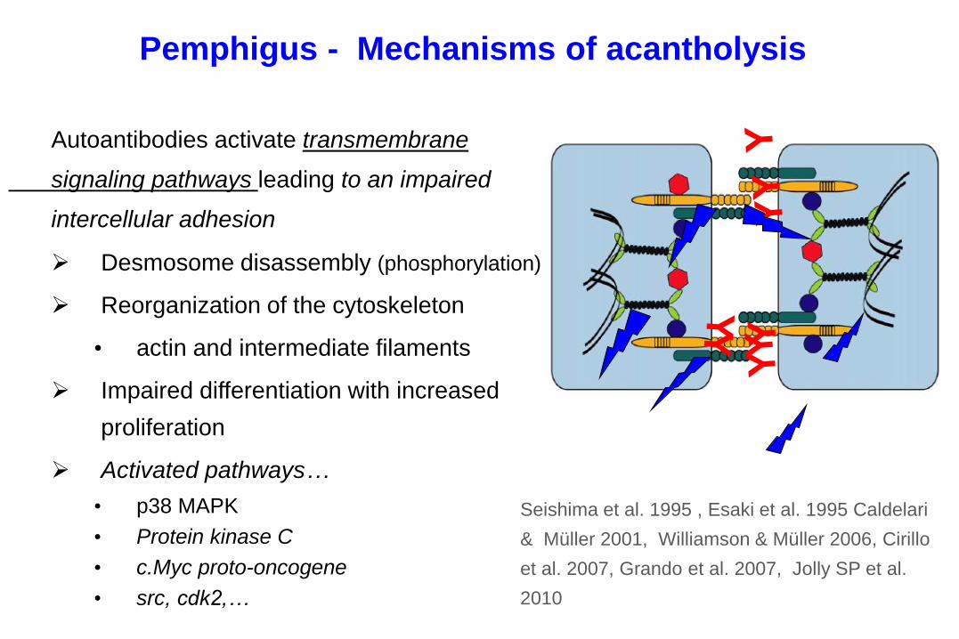
Pemphigus - Mechanisms of acantholysis
Autoantibodies activate transmembrane
signaling pathways leading to an impaired
intercellular adhesion
Desmosome disassembly (phosphorylation)
Reorganization of the cytoskeleton
• actin and intermediate filaments
Impaired differentiation with increased
proliferation
Activated pathways…
• p38 MAPK
• Protein kinase C
• c.Myc proto-oncogene
• src, cdk2,…
Seishima et al. 1995 , Esaki et al. 1995 Caldelari
& Müller 2001, Williamson & Müller 2006, Cirillo
et al. 2007, Grando et al. 2007, Jolly SP et al.
2010

PV IgG-mediated signaling activates p38 MAPK with
reorganization of the cytoskeleton
Berkowitz P et al. JBC 2005
Berkowitz P et al. PNAS 2006
Waschke J et al., JCB 2006
Jolly PS et al JBC 2010
GTPase RhoA Phosph.
Inhibition of p38MAPK prevents clinical blistering
Perspectives: pharmacologic therapy of pemphigus ?
p38 MAPK inhibitors, including KC-706, currently evaluated
PV IgG

Rag2-/-
Dsg3+/+
Dsg3-/-
Spleen (T cells & B cells etc.)
PV mouse model : interaction between antigen-specific
B-T cells is required for disease development
Immunization with
rDsg3
No tolerance against Dsg3
Transfer
Erosions & hair loss Acantholytic blister IgG deposition
kindly provided by M. Amagai
Takashi et al. J Immunol 2009

Hertl M et coll. Marburg
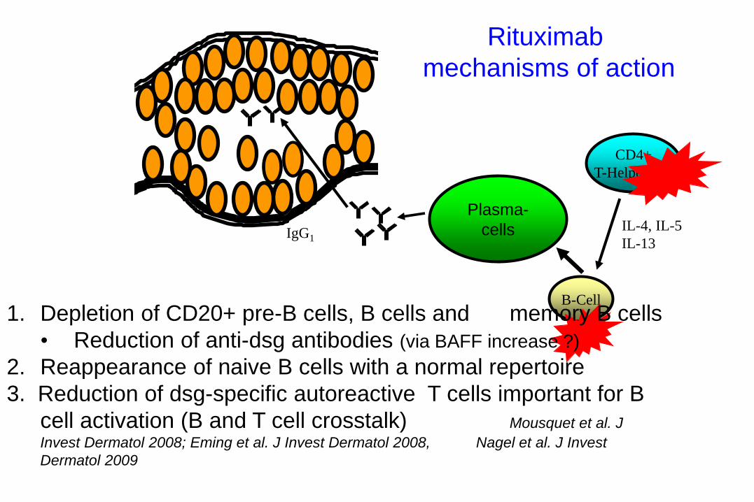
IgG1
B-Cell
CD4+
T-Helper-cell
IL-4, IL-5
IL-13
Plasma-
cells
Rituximab
mechanisms of action
1. Depletion of CD20+ pre-B cells, B cells and memory B cells
• Reduction of anti-dsg antibodies (via BAFF increase ?)
2. Reappearance of naive B cells with a normal repertoire
3. Reduction of dsg-specific autoreactive T cells important for B
cell activation (B and T cell crosstalk) Mousquet et al. J
Invest Dermatol 2008; Eming et al. J Invest Dermatol 2008, Nagel et al. J Invest
Dermatol 2009

Advances in pathogenesis in AIBD
Take home messages and learning objectives
1. Group of rare diseases associated with autoantibodies directed
against structural components of adhesion complexes,
desmosomes and hemidesmosomes
• Targets of both acquired and inherited dermatoses
2. Advances in the understanding of mechanisms leading to blistering
• Bullous pemphigoid: inflammatorry cascade with activation of
complement, inflammatory cells, and proteases
• Pemphigus: steric hindrance, dsg-depleted desmosomes,
activation of signaling pathways


![Pemphigus Herpetiformis [Print] - eMedicine Dermatology Herpetiformis .pdf · • Etiology in the neutrophil-dominant subset of pemphigus herpetiformis includes the following: o In](https://static.fdocuments.net/doc/165x107/603eff65c1246c599955258c/pemphigus-herpetiformis-print-emedicine-herpetiformis-pdf-a-etiology-in.jpg)