Current and emerging partnerships in imaging and radiotherapy by musila mutala
30
-
Upload
kesho-conference -
Category
Health & Medicine
-
view
74 -
download
1
Transcript of Current and emerging partnerships in imaging and radiotherapy by musila mutala


The Incremental Journey So Far
1. Visual determination and/or physical palpation of abnormal masses to guide radiotherapy target.
2. Plain x-rays and anatomical landmarks
3. Crossectional imaging
4. Medical technology advances in delivery of radiotherapy.

Steps of Radiotherapy Process
1. Cancer staging
2. Target volume determination
3. Treatment planning
4. Simulation
5. Radiotherapy delivery
6. Radiotherapy verification
7. Patient monitoring
8. Patient follow up

Imaging needs in each step

Cancer Staging
0To determine the extent of disease
0To determine the intent of radiotherapy- curative, high dose local control, palliative.
Target volume determination
0To define the treatment volumes of the tumour for treatment planning
0To identify the relevant organs-at-risk for treatment planning

Treatment Planning
0To evaluate the defined 3-D target volumes and organs-at-risk for selection of the number of beams, the orientation of the beams, design of the treatment plan, selection of the plan technique, use of intensity modulation or image guided strategy and computing plan distributions.
0To review plan dosimetry in 3D and permit creation of dose-volume histograms.
0To provide opportunity for plan optimization

Simulation
0To determine patient positioning and set up in 3D
0To assess position and use of radiation immobilization aids or anatomical markers.

Radiotherapy Delivery
0To provide accuracy and reliability of treatment delivery
0To offer opportunity for image guided radiotherapy such as adaptive or predictive treatments
0To permit options for target tracking and target gating

Radiotherapy Verification
0To confirm placement of treatment fields or delivery of radiotherapy
0To evaluate in vivo dosimetry
(EPIDs)

Patient Monitoring
0To assess uncertainties in patient tolerance to treatment
Patient Follow up
0To determine tumour response
0To assess treatment related side effects
0To evaluate disease recurrence

0 From as early as the 1970s research on how crossesctionalimaging can contribute to RTP was ongoing especially after the introduction of CT in medical science. For example Gotein et al concluded that improved tumor delineation and target coverage using CT could result in an increase in local tumor control probability by an average of 6% with the chance of five-year survival increasing by an average of 3.5% ⋆. With advances in CT technology now it is possible to provide 3D spatial volumes for better tumour definition and reduce geographical miss during RT.
⋆Goitein M. The utility of computed tomography in radiation therapy; an estimate of outcome. Int J Radiat Oncol Biol Phys 1979; 5: 1799–807.

Note that shaping of the treatment field for A is crude and does not provide estimation of the tumor shape. By using CT scanning, the 3D shape of the tumor can be determined. The treatment fields can then be shaped to the profile of the tumor using multileaf collimation and taking into account the surrounding organs-at-risk such as the heart, esophagus, and lung tissue as demonstrated in B. (Source: Husband and Reznek's Imaging in Oncology ).
An example of a lung radiotherapy case where the anterior treatment field was planned using conventional radiotherapy planning methods with a simple chest radiograph image and (B–D) the same lung case planned using CT simulation planning where the anterior treatment field in shown in B and the corresponding coronal and sagittal views are shown in C and D, respectively.

Improving Therapeutic Ratios
0 Radiotherapy planning studies have outlined that the volume of irradiated tissue can be reduced by 30% to 50% using CFRT compared to conventional planning techniques ⋆.
0 IMRT is an improvement of CRFT in reducing dose to OAR in concave structures e.g the rectum during RT of prostatic cancer.
0 IGRT addresses the temporal and spatial variations that may lead to geographical misses of the target volume and OAR to increase therapeutic ratios.
⋆ Robinson MH, Bidmead AM, Harmer CL. Value of conformal planning in the radiotherapy of soft tissue sarcoma. Clin Oncol (R Coll Radiol) 1992; 4: 290–3

Necessary imaging information for target volume delineation
1. Patient
2. The tumour
3. Nodal regions
4. Organs at risk
(Structured reporting is gaining popularity all over the world as an effective tool of communication between the radiologist and clinician).
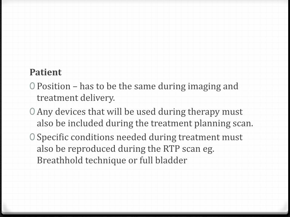
Patient
0 Position – has to be the same during imaging and treatment delivery.
0Any devices that will be used during therapy must also be included during the treatment planning scan.
0 Specific conditions needed during treatment must also be reproduced during the RTP scan eg. Breathhold technique or full bladder

The tumour0 Spatial location0 3D shape and size0 Details of the edge where it forms a border with adjacent
normal tissue0 Details of the adjacent anatomy where no clear boundaries
are defined0 Details of any potential microscopic extension0 Functional tumour activity in relation to anatomical and
morphological status0 Details of spatial variation that may be associated with the
tumour.

Nodal regions
0Mention of the known locoregional nodal sites
0Abnormal size and suspicious features
Organs at risk (OAR)
0Anatomical details including normal variants.
0 Potential temporal spatial variation

Radiologist’s Forte
0 Staging
0 Follow up
0Monitoring

Radio-oncologist’s Field
0Target delineation
0Treatment planning
0 Simulation
0Delivery
0Verification
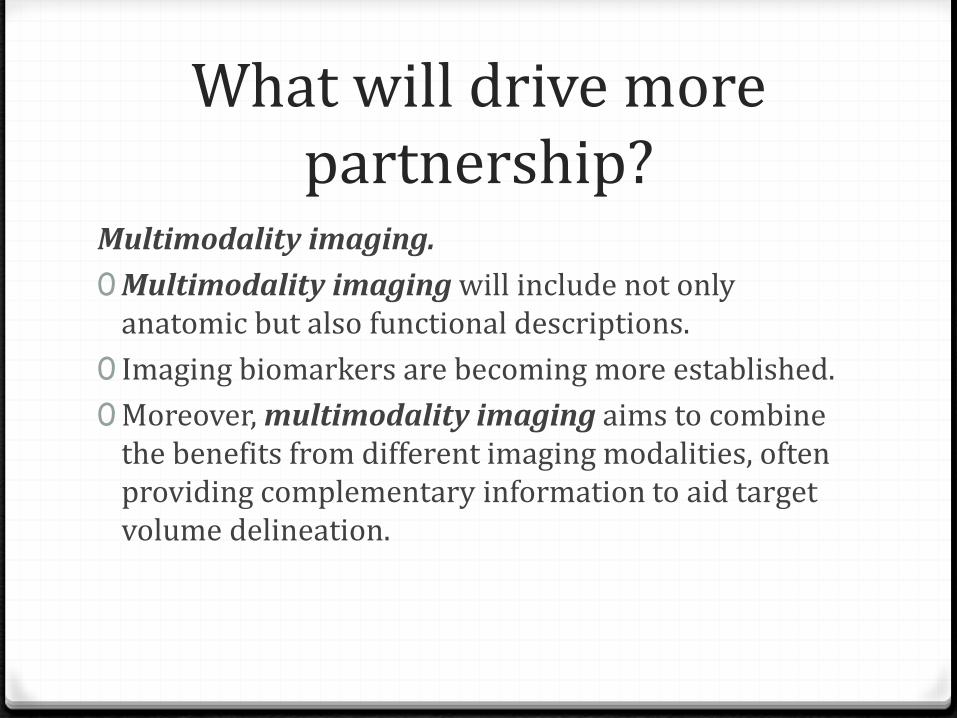
What will drive more partnership?
Multimodality imaging.
0Multimodality imaging will include not only anatomic but also functional descriptions.
0 Imaging biomarkers are becoming more established.
0Moreover, multimodality imaging aims to combine the benefits from different imaging modalities, often providing complementary information to aid target volume delineation.

Multimodality Imaging
CT
0 CT is the current mainstay imaging modality used in RTP.
0 It provides geometrically accurate and reliable cross-sectional images with 3D external contours of the patient.
0 Tissue or electron density information available from the Hounsfield units on the CT slices can be directly used, after applying correction factors, to calculate radiation absorbed doses for planning dosimetry.
0 Limitations in certain anatomical areas such as brain and pelvis
Multimodality Imaging
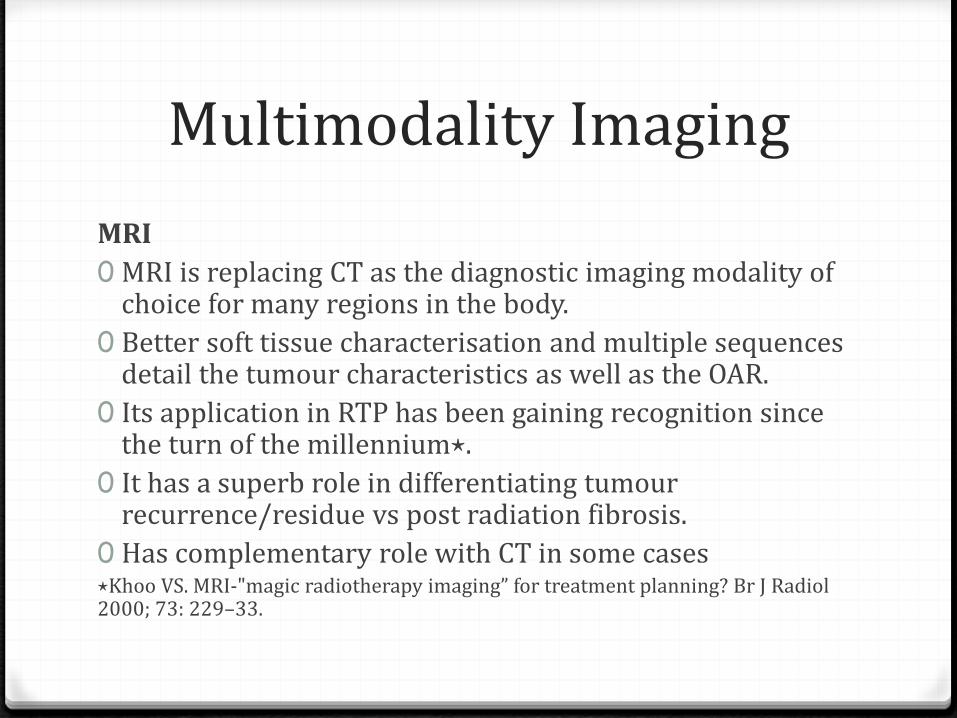
MRI
0 MRI is replacing CT as the diagnostic imaging modality of choice for many regions in the body.
0 Better soft tissue characterisation and multiple sequences detail the tumour characteristics as well as the OAR.
0 Its application in RTP has been gaining recognition since the turn of the millennium⋆.
0 It has a superb role in differentiating tumour recurrence/residue vs post radiation fibrosis.
0 Has complementary role with CT in some cases⋆Khoo VS. MRI-"magic radiotherapy imaging” for treatment planning? Br J Radiol2000; 73: 229–33.
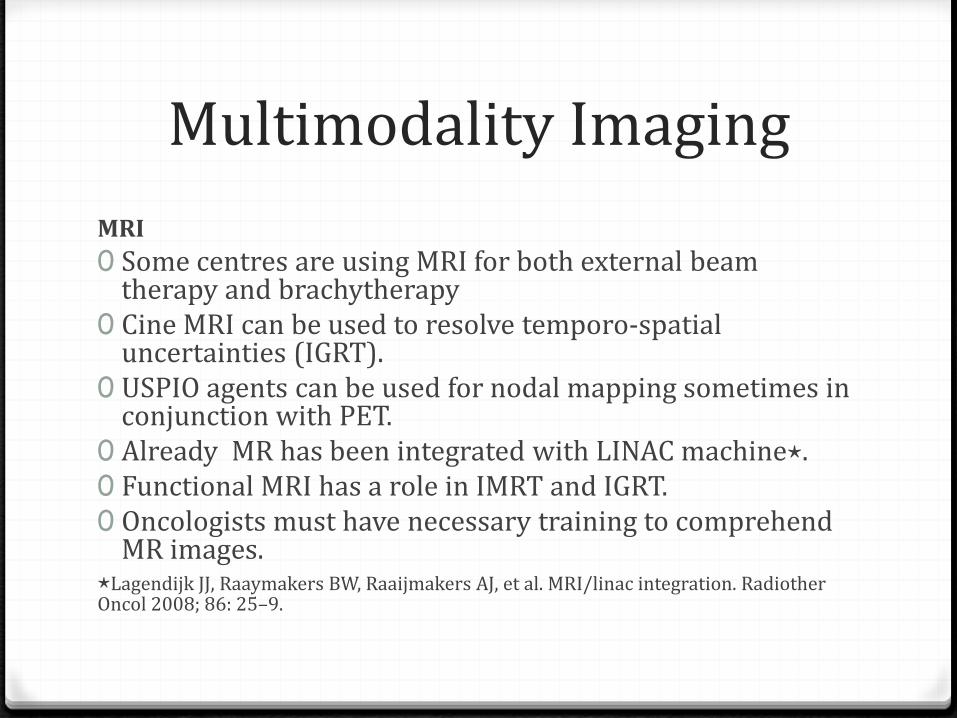
Multimodality Imaging
MRI
0 Some centres are using MRI for both external beam therapy and brachytherapy
0 Cine MRI can be used to resolve temporo-spatial uncertainties (IGRT).
0 USPIO agents can be used for nodal mapping sometimes in conjunction with PET.
0 Already MR has been integrated with LINAC machine⋆.0 Functional MRI has a role in IMRT and IGRT.0 Oncologists must have necessary training to comprehend
MR images.⋆Lagendijk JJ, Raaymakers BW, Raaijmakers AJ, et al. MRI/linac integration. RadiotherOncol 2008; 86: 25–9.
Multimodality Imaging
PET
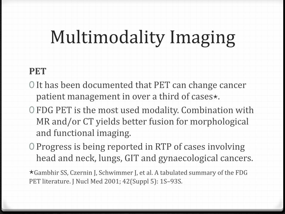
0 It has been documented that PET can change cancer patient management in over a third of cases⋆.
0 FDG PET is the most used modality. Combination with MR and/or CT yields better fusion for morphological and functional imaging.
0 Progress is being reported in RTP of cases involving head and neck, lungs, GIT and gynaecological cancers.
⋆Gambhir SS, Czernin J, Schwimmer J, et al. A tabulated summary of the FDG
PET literature. J Nucl Med 2001; 42(Suppl 5): 1S–93S.
Conclusion
0 Staging of cancer and follow up have a well established partnership between imaging and radiotherapy departments in Kenya currently .
0Target volume delineation , treatment planning, simulation, radiation delivery, verification and monitoring are areas showing potential for more partnership between the two disciplines.

Way Forward
0Dedicated imaging units within radiotherapy departments.
0Dedicated radiological team within the radiotherapy department.
0Personnel training.
0Multidisciplinary teams.

Asanteni


![Islam Aur Esa'eiyat Ka Taqabili Mutala [Urdu]](https://static.fdocuments.net/doc/165x107/577c7df71a28abe054a03d81/islam-aur-esaeiyat-ka-taqabili-mutala-urdu.jpg)