CT and MRI of Acquired Portal Venous System Anomalies · CT and MRI of portal venous system ... the...
6
J Gastrointestin Liver Dis December 2006 Vol.15 No 4, 393-398 Address for correspondence: Ioana Lupescu Department of Radiology Fundeni Hospital Sos.Fundeni, no.258 Bucharest, Romania E-mail: [email protected] CLINICAL IMAGING CT and MRI of Acquired Portal Venous System Anomalies Ioana Lupescu, Narcis Masala, Rãzvan Capsa, Nicoarã Câmpeanu, ªerban Alexandru Georgescu Department of Radiology, Fundeni Hospital, University of Medicine and Pharmacy “Carol Davila”, Bucharest Abstract In this educational presentation, we offer an overview of acquired anomalies of the portal venous system explored by biphasic helical CT and MRI. Portosystemic collateral vessels, cavernous transformation of the portal vein, intrahepatic vascular shunts, aneurysms of the portal venous system, thrombosis of the portal venous system, and gas in the portal venous system will be discussed. For liver surgery and interventional procedures it is necessary to have a correct mapping of normal anatomy, variants, and different pathologies involving the portal venous system. Key words Acquired anomalies - portal venous system - helical computed tomography - magnetic resonance imaging MRI - MR angiography Introduction The portal venous system comprises all the veins draining the abdominal part of the digestive tract, including the lower esophagus but excluding the lower anal canal. Tributaries of the portal vein are the splenic, superior mesenteric, left gastric, right gastric, paraumbilical, and cystic veins. Imaging evaluation of the portal venous system is usually performed with color Doppler ultrasonography, spiral computed tomography (CT), and magnetic resonance imaging (MRI) (1). Invasive techniques such as arterial portography, direct portography, and splenoportography may also be used, but in our days these methods are replaced by MR portography (1). Imaging details Biphasic dynamic contrast material–enhanced helical CT is a useful tool for assessing abnormalities of the portal venous system. Most perfusion alterations are seen during the hepatic arterial phase (HAP), with normal attenuation in the portal venous phase (PVP). Our protocol for biphasic helical CT of the abdomen is: we inject 100 mL of iodinated contrast material at a rate of 3–4mL/sec. HAP images are acquired 20–25 seconds after the start of the injection, and PVP images are acquired 25– 35 seconds later. MR evaluation was performed with a 1.5-T high- performance–gradient MR imaging system and a phased array torso coil. The following pulse sequences were used: T2-weighted fast spin-echo fat-suppressed sequence with a respiratory trigger, T1-weighted gradient echo sequence and 3D MR multiphase angiography fast spoiled gradient echo dynamic fat-suppressed sequence after the admi- nistration of contrast material. Portal hypertension results from a relative or absolute obstruction of the splanchnic blood flow or, less commonly, from increased portal blood flow. Normal portal pressure is 5-10 mm Hg. Portal hypertension is present if portal venous pressure exceeds 10mmHg (1). Portal hypertension can be classified into presinusoidal, sinusoidal and postsinusoidal causes. The obstruction of hepatoportal flow leads to the development of numerous collateral pathways from the high-pressure portal system to the low-pressure systemic circulation. Portosystemic collateral vessels The most common cause of portosystemic collateral vessels is portal hypertension. Other causes of their presence are splenic or splenomesenteric venous compres- sion and obstruction due to neoplasms, pancreatitis, or surgery. Coronary, esophageal, paraesophageal, and gastric collateral vessels Esophageal varices are of major clinical importance
-
Upload
vuongkhuong -
Category
Documents
-
view
232 -
download
0
Transcript of CT and MRI of Acquired Portal Venous System Anomalies · CT and MRI of portal venous system ... the...

CT and MRI of portal venous system
J Gastrointestin Liver DisDecember 2006 Vol.15 No 4, 393-398Address for correspondence: Ioana Lupescu
Department of RadiologyFundeni HospitalSos.Fundeni, no.258Bucharest, RomaniaE-mail: [email protected]
CLINICAL IMAGING
CT and MRI of Acquired Portal Venous System Anomalies
Ioana Lupescu, Narcis Masala, Rãzvan Capsa, Nicoarã Câmpeanu, ªerban Alexandru Georgescu
Department of Radiology, Fundeni Hospital, University of Medicine and Pharmacy “Carol Davila”, Bucharest
AbstractIn this educational presentation, we offer an overview
of acquired anomalies of the portal venous system exploredby biphasic helical CT and MRI. Portosystemic collateralvessels, cavernous transformation of the portal vein,intrahepatic vascular shunts, aneurysms of the portalvenous system, thrombosis of the portal venous system,and gas in the portal venous system will be discussed. Forliver surgery and interventional procedures it is necessaryto have a correct mapping of normal anatomy, variants, anddifferent pathologies involving the portal venous system.
Key wordsAcquired anomalies - portal venous system - helical
computed tomography - magnetic resonance imaging MRI -MR angiography
IntroductionThe portal venous system comprises all the veins
draining the abdominal part of the digestive tract, includingthe lower esophagus but excluding the lower anal canal.Tributaries of the portal vein are the splenic, superiormesenteric, left gastric, right gastric, paraumbilical, and cysticveins.
Imaging evaluation of the portal venous system isusually performed with color Doppler ultrasonography, spiralcomputed tomography (CT), and magnetic resonanceimaging (MRI) (1). Invasive techniques such as arterialportography, direct portography, and splenoportographymay also be used, but in our days these methods are replacedby MR portography (1).
Imaging details Biphasic dynamic contrast material–enhanced helical
CT is a useful tool for assessing abnormalities of the portalvenous system. Most perfusion alterations are seen duringthe hepatic arterial phase (HAP), with normal attenuation inthe portal venous phase (PVP).
Our protocol for biphasic helical CT of the abdomen is:we inject 100 mL of iodinated contrast material at a rate of3–4mL/sec. HAP images are acquired 20–25 seconds afterthe start of the injection, and PVP images are acquired 25–35 seconds later.
MR evaluation was performed with a 1.5-T high-performance–gradient MR imaging system and a phasedarray torso coil. The following pulse sequences were used:T2-weighted fast spin-echo fat-suppressed sequence witha respiratory trigger, T1-weighted gradient echo sequenceand 3D MR multiphase angiography fast spoiled gradientecho dynamic fat-suppressed sequence after the admi-nistration of contrast material.
Portal hypertension results from a relative or absoluteobstruction of the splanchnic blood flow or, less commonly,from increased portal blood flow. Normal portal pressure is5-10 mm Hg. Portal hypertension is present if portal venouspressure exceeds 10mmHg (1). Portal hypertension can beclassified into presinusoidal, sinusoidal and postsinusoidalcauses.
The obstruction of hepatoportal flow leads to thedevelopment of numerous collateral pathways from thehigh-pressure portal system to the low-pressure systemiccirculation.
Portosystemic collateral vesselsThe most common cause of portosystemic collateral
vessels is portal hypertension. Other causes of theirpresence are splenic or splenomesenteric venous compres-sion and obstruction due to neoplasms, pancreatitis, orsurgery.
Coronary, esophageal, paraesophageal,and gastric collateral vesselsEsophageal varices are of major clinical importance
Lupescu et al394
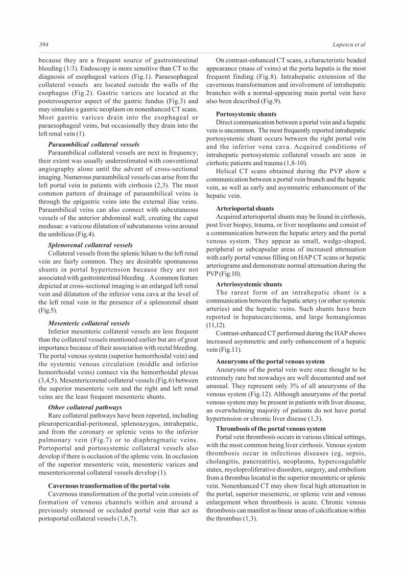
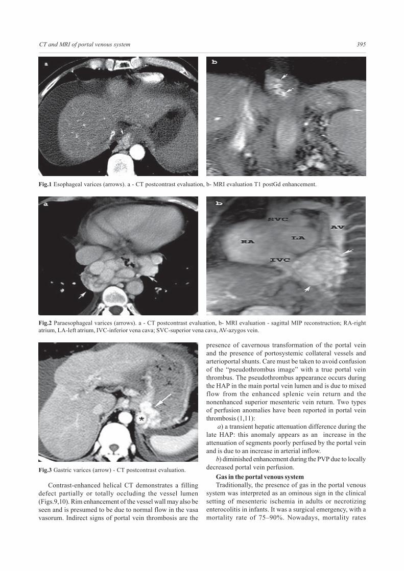
because they are a frequent source of gastrointestinalbleeding (1/3). Endoscopy is more sensitive than CT to thediagnosis of esophageal varices (Fig.1). Paraesophagealcollateral vessels are located outside the walls of theesophagus (Fig.2). Gastric varices are located at theposterosuperior aspect of the gastric fundus (Fig.3) andmay simulate a gastric neoplasm on nonenhanced CT scans.Most gastric varices drain into the esophageal orparaesophageal veins, but occasionally they drain into theleft renal vein (1).
Paraumbilical collateral vesselsParaumbilical collateral vessels are next in frequency;
their extent was usually underestimated with conventionalangiography alone until the advent of cross-sectionalimaging. Numerous paraumbilical vessels can arise from theleft portal vein in patients with cirrhosis (2,3). The mostcommon pattern of drainage of paraumbilical veins isthrough the epigastric veins into the external iliac veins.Paraumbilical veins can also connect with subcutaneousvessels of the anterior abdominal wall, creating the caputmedusae: a varicose dilatation of subcutaneous veins aroundthe umbilicus (Fig.4).
Splenorenal collateral vesselsCollateral vessels from the splenic hilum to the left renal
vein are fairly common. They are desirable spontaneousshunts in portal hypertension because they are notassociated with gastrointestinal bleeding . A common featuredepicted at cross-sectional imaging is an enlarged left renalvein and dilatation of the inferior vena cava at the level ofthe left renal vein in the presence of a splenorenal shunt(Fig.5).
Mesenteric collateral vesselsInferior mesenteric collateral vessels are less frequent
than the collateral vessels mentioned earlier but are of greatimportance because of their association with rectal bleeding.The portal venous system (superior hemorrhoidal vein) andthe systemic venous circulation (middle and inferiorhemorrhoidal veins) connect via the hemorrhoidal plexus(3,4,5). Mesentericorenal collateral vessels (Fig.6) betweenthe superior mesenteric vein and the right and left renalveins are the least frequent mesenteric shunts.
Other collateral pathwaysRare collateral pathways have been reported, including
pleuropericardial-peritoneal, splenoazygos, intrahepatic,and from the coronary or splenic veins to the inferiorpulmonary vein (Fig.7) or to diaphragmatic veins.Portoportal and portosystemic collateral vessels alsodevelop if there is occlusion of the splenic vein. In occlusionof the superior mesenteric vein, mesenteric varices andmesentericorenal collateral vessels develop (1).
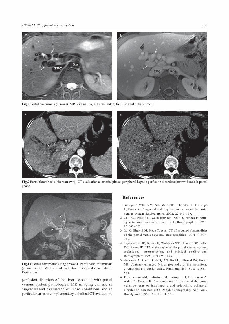
Cavernous transformation of the portal veinCavernous transformation of the portal vein consists of
formation of venous channels within and around apreviously stenosed or occluded portal vein that act asportoportal collateral vessels (1,6,7).
On contrast-enhanced CT scans, a characteristic beadedappearance (mass of veins) at the porta hepatis is the mostfrequent finding (Fig.8). Intrahepatic extension of thecavernous transformation and involvement of intrahepaticbranches with a normal-appearing main portal vein havealso been described (Fig.9).
Portosystemic shuntsDirect communication between a portal vein and a hepatic
vein is uncommon. The most frequently reported intrahepaticportosystemic shunt occurs between the right portal veinand the inferior vena cava. Acquired conditions ofintrahepatic portosystemic collateral vessels are seen incirrhotic patients and trauma (1,8-10).
Helical CT scans obtained during the PVP show acommunication between a portal vein branch and the hepaticvein, as well as early and asymmetric enhancement of thehepatic vein.
Arterioportal shuntsAcquired arterioportal shunts may be found in cirrhosis,
post liver biopsy, trauma, or liver neoplasms and consist ofa communication between the hepatic artery and the portalvenous system. They appear as small, wedge-shaped,peripheral or subcapsular areas of increased attenuationwith early portal venous filling on HAP CT scans or hepaticarteriograms and demonstrate normal attenuation during thePVP (Fig.10).
Arteriosystemic shuntsThe rarest form of an intrahepatic shunt is a
communication between the hepatic artery (or other systemicarteries) and the hepatic veins. Such shunts have beenreported in hepatocarcinoma, and large hemangiomas(11,12).
Contrast-enhanced CT performed during the HAP showsincreased asymmetric and early enhancement of a hepaticvein (Fig.11).
Aneurysms of the portal venous systemAneurysms of the portal vein were once thought to be
extremely rare but nowadays are well documented and notunusual. They represent only 3% of all aneurysms of thevenous system (Fig.12). Although aneurysms of the portalvenous system may be present in patients with liver disease,an overwhelming majority of patients do not have portalhypertension or chronic liver disease (1,3).
Thrombosis of the portal venous systemPortal vein thrombosis occurs in various clinical settings,
with the most common being liver cirrhosis. Venous systemthrombosis occur in infectious diseases (eg, sepsis,cholangitis, pancreatitis), neoplasms, hypercoagulablestates, myeloproliferative disorders, surgery, and embolismfrom a thrombus located in the superior mesenteric or splenicvein. Nonenhanced CT may show focal high attenuation inthe portal, superior mesenteric, or splenic vein and venousenlargement when thrombosis is acute. Chronic venousthrombosis can manifest as linear areas of calcification withinthe thrombus (1,3).
CT and MRI of portal venous system 395
Fig.1 Esophageal varices (arrows). a - CT postcontrast evaluation, b- MRI evaluation T1 postGd enhancement.
Fig.2 Paraesophageal varices (arrows). a - CT postcontrast evaluation, b- MRI evaluation - sagittal MIP reconstruction; RA-rightatrium, LA-left atrium, IVC-inferior vena cava; SVC-superior vena cava, AV-azygos vein.
Fig.3 Gastric varices (arrow) - CT postcontrast evaluation.
Contrast-enhanced helical CT demonstrates a fillingdefect partially or totally occluding the vessel lumen(Figs.9,10). Rim enhancement of the vessel wall may also beseen and is presumed to be due to normal flow in the vasavasorum. Indirect signs of portal vein thrombosis are the
presence of cavernous transformation of the portal veinand the presence of portosystemic collateral vessels andarterioportal shunts. Care must be taken to avoid confusionof the “pseudothrombus image” with a true portal veinthrombus. The pseudothrombus appearance occurs duringthe HAP in the main portal vein lumen and is due to mixedflow from the enhanced splenic vein return and thenonenhanced superior mesenteric vein return. Two typesof perfusion anomalies have been reported in portal veinthrombosis (1,11):
a) a transient hepatic attenuation difference during thelate HAP: this anomaly appears as an increase in theattenuation of segments poorly perfused by the portal veinand is due to an increase in arterial inflow.
b) diminished enhancement during the PVP due to locallydecreased portal vein perfusion.
Gas in the portal venous systemTraditionally, the presence of gas in the portal venous
system was interpreted as an ominous sign in the clinicalsetting of mesenteric ischemia in adults or necrotizingenterocolitis in infants. It was a surgical emergency, with amortality rate of 75–90%. Nowadays, mortality rates

Lupescu et al396
Fig.4 Umbilical vein recanalisation - MRI evaluation. a- coronal MIP reconstruction (black arrows), b- axial MIP reconstruction(white arrows). L-liver, Spl-spleen, GF-gastric fornix.
Fig.5 Portal hypertension. Spleno-renal shunt (arrows) - CTevaluation.
Fig.6 Meseterico-renal collateral vessels (arrows) - MRI- postGdMIP reconstruction. SMV-superior mesenteric vein, PV-portalvein, LRV-left renal vein, Spl V- splenic vein.
Fig.7 Portal hypertension. Communication with LIPV-left inferiorpulmonary vein (arrow). LPA- ;eft pulmonary artery, Ao-aorta.
rather to new and better imaging techniques, which haveallowed recognition of an increasing number of causes ofgas in the portal venous system (1). The accessibility to CTunits has increased the sensitivity for detection of portalvenous system gas. Reported causes of portal venoussystem gas are necrotizing pancreatitis, abdominal abscess,intestinal obstruction, perforated gastric ulcer or carcinoma,diverticulitis, inflammatory bowel disease, abdominal trauma,ingestion of a caustic agent, enema administration,colonoscopy, gastrostomy tubes, and liver transplantation.
Intrahepatic portal vein gas should be differentiated fromaerobilia. The distribution of hepatic gas in patients withaerobilia is central, around the portal hilum, and does notextend to within 2 cm of the liver capsule (Fig.12).
Gas in the mesenteric vein branches should bedifferentiated from pneumoperitoneum.
associated with portal venous system gas have declined to29%–43%. This decline is due not to improved therapy but
Conclusion Biphasic helical CT is a useful tool for evaluation of
CT and MRI of portal venous system 397
Fig.8 Portal cavernoma (arrows). MRI evaluation, a-T2 weighted, b-T1 postGd enhancement.
Fig.9 Portal thrombosis (short arrows) - CT evaluation a- arterial phase: peripheral hepatic perfusion disorders (arrows head), b-portalphase.
perfusion disorders of the liver associated with portalvenous system pathologies. MR imaging can aid indiagnosis and evaluation of these conditions and inparticular cases is complementary to helical CT evaluation.
References1. Gallego C, Velasco M, Pilar Marcuello P, Tejedor D, De Campo
L, Friera A. Congenital and acquired anomalies of the portalvenous system. Radiographics 2002; 22:141–159.
2. Cho KC, Patel YD, Wachsberg RH, Seeff J. Varices in portalhypertension: evaluation with CT. Radiographics 1995;15:609–622.
3. Ito K, Higuchi M, Kada T, et al. CT of acquired abnormalitiesof the portal venous system. Radiographics 1997; 17:897–917.
4. Leyendecker JR, Rivera E, Washburn WK, Johnson SP, DiffinDC, Eason JD. MR angiography of the portal venous system:techniques, interpretation, and clinical applications.Radiographics 1997;17:1425–1443.
5. Shirkhoda A, Konez O, Shetty AN, Bis KG, Ellwood RA, KirschMJ. Contrast-enhanced MR angiography of the mesentericcirculation: a pictorial essay. Radiographics 1998; 18:851–861.
6. De Gaetano AM, Lafortune M, Patriquin H, De Franco A,Aubin B, Paradis K. Cavernous transformation of the portalvein: patterns of intrahepatic and splanchnic collateralcirculation detected with Doppler sonography. AJR Am JRoentgenol 1995; 165:1151–1155.
Fig.10 Portal cavernoma (long arrows). Portal vein thrombosis(arrows head)= MRI postGd evaluation. PV-portal vein. L-liver,P-pancreas.

Lupescu et al398
Fig.12 Pseudoaneurysm of portal vein - MRI- coronal MIPreconstruction poststenotic (arrows head) saccular dilatation ofportal vein (arrows).
Fig.11 Arterio-hepatic shunts - CT evaluation: a-arterial phase: early enhancement of middle hepatic vein and inferior vena cava(arrows), b-portal phase: huge necrotic hepatic tumor (long white arrow).
Fig 13 Portal air (black arrow)- CT evaluation.
7. Song B, Min P, Oudkerk M, et al. Cavernous transformation ofthe portal vein secondary to tumor thrombosis of hepatocellularcarcinoma: spiral CT visualization of the collateral vessels.Abdom Imaging 2000; 25:385–393.
8. Park JH, Cha SH, Han JK, Han MC. Intrahepatic portosystemicvenous shunt. AJR 1990;155:527-528.
9. Mori H, Hayashi K, Fukuda T, et al. Intrahepatic portosystemicvenous shunt: occurrence in patients with and without livercirrhosis. AJR 1987;149:711-714.
10. Quiroga S, Sebastia MC, Moreiras M, Pallisa E, Rius JM, Alvarez-Castells A. Intrahepatic arterioportal shunt: helical CT findings.Eur Radiol 1999; 9:1126–1130.
11. Vilgrain V, Vullierme M, Djabbari M, Sibert A, Menu Y.Liver and vascular abnormalities. J Radiol 2002; 83: 255-268.
12. Chen WP, Chen JH, Hwang JI, et al. Spectrum of transienthepatic attenuation differences in biphasic helical CT. AJR1999:172:419-424.