-Cryptoxanthin and Bone Metabolism: The Preventive Role in ...
14
356 Journal of Health Science, 54(4) 356–369 (2008) — Review — β-Cryptoxanthin and Bone Metabolism: The Preventive Role in Osteoporosis Masayoshi Yamaguchi ∗ Division of Endocrinology and Metabolism and Lipids, Department of Medicine, Emory University School of Medicine, 1639 Pierce Drive, 1305 WMRB, Atlanta, Georgia 30322–0001, U.S.A. (Received May 7, 2008) Bone loss with aging induces osteoporosis. The most dramatic expression of the disease is represented by fractures of the proximal femur. Pharmacologic and nutritional factors may play a role in the prevention of bone loss with aging. β-Cryptoxanthin, a kind of carotenoid, is abundant in Satsuma mandarin orange (Citrus unshiu MARC.). Amoung various carotenoids including β-cryptoxanthin, lutein, lycopene, β-carotene, astaxanthin, and rutin, β-cryptoxanthin has been found to have a unique anabolic effect on bone calcification in vitro. Hesperidin, which is contained in Satsuma mandarin orange, did not have an anabolic effect on bone calcification in vitro. β-Cryptoxanthin has stimulatory effects on osteoblastic bone formation and inhibitory effects on osteoclastic bone resorption in vitro, thereby increasing bone mass. β-Cryptoxanthin has an effect on the gene expression of various proteins which are related to osteoblastic bone formation and mineralization in vitro. β-Cryptoxanthin has inhibitory effects on enzyme activity which is related to osteoclastic bone resororption, and the carotenoid induces apoptosis of mature osteoclastic cells in vitro. Oral administration of β-cryptoxanthin has been shown to have the anabolic effects on bone components in young and aged rats, and the administration has the preventive effects on bone loss in streptozotocin-diabetic rats and ovariectomized rats in vivo. Moreover, the intake of β-cryptoxanthin-reinforced juice for longer periods has been shown to have both stimulatory effects on bone formation and inhibitory effects on bone resorption in healthy human or postmenopausal women in evaluating with serum biochemical markers of bone metabolism in vivo. Thus the intake of dietary β-cryptoxanthin may have a preventive effect on osteoporosis due to stimulating bone formation and due to inhibiting bone resorption. Moreover, epidemiological studies suggest the potential role of β-cryptoxanthin as a sustainable nutritional approach to improving bone health of human subjects. β-Cryptoxanthin is an important food factor in maintaining bone healthy and in preventing osteoporosis. Key words —— β-cryptoxanthin, osteoblastic bone formation, osteoclastic bone resorption, osteoporosis INTRODUCTION Bone mass decreases with increasing age. The decrease in bone mass is due to increased bone resorption and decreased bone formation. Osteo- clasts induce bone resorption, and osteoblasts stim- ulate bone formation. Ovarian hormone deficiency at menopause in women stimulates bone loss. A fur- ther possible cause is the deterioration of osteoblas- tic cell function with increasing age. Osteoporosis is induced with decrease in bone mass as shown in ∗ To whom correspondence should be addressed: Division of Endocrinology and Metabolism and Lipids, Department of Medicine, Emory University School of Medicine, 1639 Pierce Drive, 1305 WMRB, Atlanta, Georgia 30322–0001, U.S.A. Tel.: +1-404-727-2598; Fax: +1-404-727-1300; E- mail: [email protected] Fig. 1, and it is widely recognized as a major public health problem. The most dramatic expression of the disease is represented by fractures of the prox- imal femur for which the number increases as the population ages. 1, 2) Pharmacological and nutritional factors may have the potential effect to prevent bone loss with increasing age. Nutritional factors may be especially important in the prevention of osteo- porosis. 3–5) Chemical factors in food and plants can help to prevent bone loss with increasing age but these factors are poorly understood. Our studies have shown that isoflavones, which are contained in soybeans, or menaquinone-7 (vita- min K 2 ), which is abundant in fermented soybeans, have been demonstrated to increasing bone mass due to stimulating osteoblastic bone formation and
Transcript of -Cryptoxanthin and Bone Metabolism: The Preventive Role in ...

356 Journal of Health Science, 54(4) 356–369 (2008)
— Review —
β-Cryptoxanthin and Bone Metabolism: The PreventiveRole in Osteoporosis
Masayoshi Yamaguchi∗
Division of Endocrinology and Metabolism and Lipids, Department of Medicine, Emory University School of Medicine, 1639 PierceDrive, 1305 WMRB, Atlanta, Georgia 30322–0001, U.S.A.
(Received May 7, 2008)
Bone loss with aging induces osteoporosis. The most dramatic expression of the disease is represented byfractures of the proximal femur. Pharmacologic and nutritional factors may play a role in the prevention of boneloss with aging. β-Cryptoxanthin, a kind of carotenoid, is abundant in Satsuma mandarin orange (Citrus unshiuMARC.). Amoung various carotenoids including β-cryptoxanthin, lutein, lycopene, β-carotene, astaxanthin, andrutin, β-cryptoxanthin has been found to have a unique anabolic effect on bone calcification in vitro. Hesperidin,which is contained in Satsuma mandarin orange, did not have an anabolic effect on bone calcification in vitro.β-Cryptoxanthin has stimulatory effects on osteoblastic bone formation and inhibitory effects on osteoclastic boneresorption in vitro, thereby increasing bone mass. β-Cryptoxanthin has an effect on the gene expression of variousproteins which are related to osteoblastic bone formation and mineralization in vitro. β-Cryptoxanthin has inhibitoryeffects on enzyme activity which is related to osteoclastic bone resororption, and the carotenoid induces apoptosisof mature osteoclastic cells in vitro. Oral administration of β-cryptoxanthin has been shown to have the anaboliceffects on bone components in young and aged rats, and the administration has the preventive effects on bone lossin streptozotocin-diabetic rats and ovariectomized rats in vivo. Moreover, the intake of β-cryptoxanthin-reinforcedjuice for longer periods has been shown to have both stimulatory effects on bone formation and inhibitory effects onbone resorption in healthy human or postmenopausal women in evaluating with serum biochemical markers of bonemetabolism in vivo. Thus the intake of dietary β-cryptoxanthin may have a preventive effect on osteoporosis due tostimulating bone formation and due to inhibiting bone resorption. Moreover, epidemiological studies suggest thepotential role of β-cryptoxanthin as a sustainable nutritional approach to improving bone health of human subjects.β-Cryptoxanthin is an important food factor in maintaining bone healthy and in preventing osteoporosis.
Key words —— β-cryptoxanthin, osteoblastic bone formation, osteoclastic bone resorption, osteoporosis
INTRODUCTION
Bone mass decreases with increasing age. Thedecrease in bone mass is due to increased boneresorption and decreased bone formation. Osteo-clasts induce bone resorption, and osteoblasts stim-ulate bone formation. Ovarian hormone deficiencyat menopause in women stimulates bone loss. A fur-ther possible cause is the deterioration of osteoblas-tic cell function with increasing age. Osteoporosisis induced with decrease in bone mass as shown in
∗To whom correspondence should be addressed: Divisionof Endocrinology and Metabolism and Lipids, Departmentof Medicine, Emory University School of Medicine, 1639Pierce Drive, 1305 WMRB, Atlanta, Georgia 30322–0001,U.S.A. Tel.: +1-404-727-2598; Fax: +1-404-727-1300; E-mail: [email protected]
Fig. 1, and it is widely recognized as a major publichealth problem. The most dramatic expression ofthe disease is represented by fractures of the prox-imal femur for which the number increases as thepopulation ages.1, 2)
Pharmacological and nutritional factors mayhave the potential effect to prevent bone losswith increasing age. Nutritional factors may beespecially important in the prevention of osteo-porosis.3–5) Chemical factors in food and plants canhelp to prevent bone loss with increasing age butthese factors are poorly understood.
Our studies have shown that isoflavones, whichare contained in soybeans, or menaquinone-7 (vita-min K2), which is abundant in fermented soybeans,have been demonstrated to increasing bone massdue to stimulating osteoblastic bone formation and

No. 4 357
to inhibiting osteoclastic bone resorption in vitro[Refs. 3), 4) in Review]. The supplementation ofthese food factors may have preventive effects onbone loss induced in animal model of osteoporosisand in human subjects.
Other food and plant factors (includingflavonoid,6) p-hydroxycinnamic acid,7, 8) the ex-tracts of Sargassum horneri,9, 10) bee pollen ofCistus ladaniferus,11, 12) or wasabi leafstalk13, 14))have also shown to increase bone mass in thefemoral tissues of rats in vitro and in vivo. Foodchemical factors thus play an important role in bonehealth and prevention of bone loss.
Retinol (vitamin A) is known to have a detri-mental effect on bone at high doses. In laboratoryanimals, high levels of vitamin A lead to acceleratedbone resorption, bone fractures, and osteoporoticbone lesions.15) The effects of carotenoids on bonemetabolism, however, have not been fully clarified.Carotenoids are present in fruit and vegetables.β-Cryptoxanthin is a kind of carotenoid which
is abundant in Satsuma mandarin orange (Citrus
Fig. 1. Change in Bone Mass with Increasing AgeBone mass increases with growing and reaches to peak in the
range of age between twenty and twenty-five years. Then bone massgradually decreases with aging. In postomenopausal women, bonemass decreases dramatically, and it induces osteoporosis. It is impor-tant to prevent the decrease in bone mass with increasing age.
unshiu MARC.). The chemical structure of β-cryptoxanthin is shown in Fig. 2. The biologicalfunction of β-cryptoxanthin in animal and human,however, was not clarified thus far. We foundthat β-cryptoxanthin has a unique anabolic effecton bone metabolism;16, 17) such an effect was notseen by lutein, lycopene, or astaxanthin, which isother carotenoids, and flavonoid rutin (quercetin-3-rutinoside). This was the first time finding.
This review introduces the cellular mechanismby which β-cryptoxanthin increases bone mass andits role in the prevention of osteoporosis.
BASIC ASPECTS ON REGULATION OFBONE METABOLISM
Bone contains over 98% of total body calcium.Bone metabolism is regulated by the functions ofosteoblasts and osteoclasts, which are major cells inbone tissues,18–20) as shown in Fig. 3.
Osteoclasts, which develop from hematopoieticprogenitors, are recruited to the site and excavatethe calcified matrix. Then, the cavity is refilled byosteoblasts via a process that occurs in three distinctphases: initiation, progression, and termination.
In the physiologic process of bone turnover, aresorptive stimulus firstly triggers recruitment of os-teoclasts to a site on the bone surface. This isfollowed by active resorption by osteoclasts, afterwhich cells withdraw from the bone surface andmononuclear phagocytic cells appear on the newlyresorbed surface. These cells are then followed byyoung osteoblasts, which begin the bone formationphase.
During the initiation phase, a team of os-teoblasts arising from local mesenchymal stem cellsassembles at the bottom of the cavity and bone for-mation begins. After the resorbed lacunar pit isfilled with new osteoid, osteoblasts become flatterand less active, with the final newly remodeled bonesurface lined by flat lining cells. Remodeling of
Fig. 2. Chemical Structure of β-CryptoxanthinThe molecular weight of this compound is 552. β-Cryptoxanthin is a kind of carotenoid which is abundant in Satsuma mandarin (Citrus unshiu
MARC.).

358 Vol. 54 (2008)
Fig. 3. Regulatory Mechanism in Bone RemodelingBone metabolism is regulated by osteoclasts and osteoblasts. Osteoclasts, which stimulate bone resorption, are generated from stem cells by
interleukin-1 (IL-1), IL-6, M-CSF, and other factors. Osteoclastogenesis is mediated through RANKL produced from osteoblasts by various boneresorption stimulatory factors. Osteoblasts, which stimulate bone formation and mineralization, are differentiated from mesenchymal cells by variousbone formation factors. After bone formation and calcification, osteoblasts are changed to osteocytes in bone matrix.
cancellous bone begins with the retraction of liningcells that cover the bone surface.18)
As bone formation progresses, some osteoblastsare entombed within the matrix as osteocytes but themajority dies by apoptosis. Bone formation termi-nates when the cavity has been refilled, at whichtime the few osteoblasts that remain become theflat lining cells that cover the quiescent surfaces ofbone. Once formed, few osteocytes die. Their via-bility is likely maintained by physiological levels ofmechanical stimulation. When mechanical forcesare reduced, for example in weightlessness, osteo-cytes die by apoptosis. This event appears to act asa beacon for osteoclast recruitment and generationof a new basic multicellular unit, which in turn re-places the old bone containing dead osteocytes withnew bone containing viable osteocytes.
Bone acts as major storage site for gowth fac-tors.20) Growth factors, which are produced by os-teoblasts, diffuse into newly deposited osteoid andare stored in the bone matrx including isulin-likegrowth factors (IGF-I and II), transforming growthfactor-β1 (TGF-β1), platelet-derived growth fac-tor (PDGF), or bone morphologic protein (BMP).These bone-derived factors, which can be liberatedduring subsequent periods of bone resorption, act inan autocrine, paracrine, or delayed paracrine fash-ion in the local microenvironment of the bone sur-face.
It is this process of bone remodeling that makesbone unique among organs and tissues and that
also adds so many levels of complexity, with re-spect to interactions along the remodeling sequenceby systemic influences (hormones), stress actionon trabecular and cortical systems (physical activ-ity/weight bearing), growth factors and cytokinesproduced by the bone cells which act locally on theirown cell types and on the other bone cell types, orfactors that come from nearby cells present in themarrow tissues.
It is interested whether food factors have a rolein the regulation of bone remodeling.
β-CRYPTOXANTHIN AND BONEMETABOLISM
β-Cryptoxanthin Stimulates Bone Formationand Inhibits Bone Resorption in Tissue CultureIn Vitro
Amoung of various carotenoid and flavonoids,β-cryptoxanthin has been shown to have aunique anabolic effect on bone calcification.11, 16, 17)
Culture with β-cryptoxanthin (10−7 or 10−6 M)caused a significant increase in calcium contentand alkaline phosphatase activity in the femoral-diaphyseal (cortical bone) and -metaphyseal (tra-becular bone) tissues in vitro. Lutein, lycopene,and rutin (10−8 to 10−6 M) did not have anabolic ef-fects on calcium contents and alkaline phosphataseactivity in rat femoral-diaphyseal and -meta-physeal tissues. Astaxanthin and β-carotene (10−6
No. 4 359
or 10−5 M) did not have an effect on the femoralcalcium contents. Myricetin, kaempferol, isorham-netin, curcumin, or hesperidin (10−7 to 10−5 M) hadno effect on bone calcium content in tissue cul-tures in vitro.11) Quercetin significantly increasedcalcium content in femoral diaphyseal tissues butnot metaphyseal tissues. Alkaline phosphatase par-ticipates in mineralization in bone tissues.21) β-Cryptoxanthin had a unique anabolic effect on bonecalcification in vitro. The effect of β-cryptoxanthinincreasing bone components was completely pre-vented with cycloheximide, an inhibitor of proteinsynthesis, suggesting that the effect is needed newlyprotein synthesis.17)
β-Cryptoxanthin has been shown to inhibit boneresorption in bone tissue cultures.17) Parathyroidhormone (PTH) or prostaglandin E2 (PGE2), whichis bone-resorbing factor, can stimulate osteoclasticbone resorption in vitro.21–23) Culture with PTH orPGE2 caused a significant decrease in calcium con-tent in the diaphyseal and metaphyseal tissues.17)
This decrease was completely inhibited in the pres-ence of β-cryptoxanthin (10−8 to 10−6 M).17) Also,culture with β-cryptoxanthin completely inhibitedthe PTH- or PGE2-induced increase in medium glu-cose consumption and lactic acid production bybone tissues.17) β-Cryptoxanthin had inhibitory ef-fects on bone resorption in tissue culture in vitro.
Thus β-cryptoxanthin was found to have stim-ulatory effect on bone formation and inhibitory ef-fects on bone resorption in bone tissue culture invitro.
It has been reported that the serum concentra-tion of β-cryptoxanthin due to consumption of veg-etable juice in women is in the range of 1.3× 10⁻⁷
to 5.3× 10⁻⁷ M.24) β-Cryptoxanthin in the range of10−8 to 10−6 M caused a significant anabolic effecton biochemical components in rat femoral tissues invitro, suggesting a physiologic role in the regulationof bone metabolism.
Cellular and Molecular Mechanisms of β-Cryptoxanthin Action in Osteoblastic Cells
The cellular and molecular mechanisms bywhich β-cryptoxanthin stimulates bone formationin bone tissues were examined using cloned os-teoblastic cells in vitro. Osteoblastic MC3T3E1cells were used. β-Cryptoxanthin was found tostimulate the proliferation of osteoblastic cells insubconfluent monolayers in a medium contain-ing 10% fetal bovine serum.25) Culture with β-cryptoxanthin also caused a significant increase in
biochemical components (protein content, alkalinephosphatase activity, and DNA content) of os-teoblastic cells.25) This effect was significantlyabolished in the presence of staurosporine, an in-hibitor of protein kinase C, or PD98059, an in-hibitor of mitosis activated protein kinase (MAP)kinase, although the effect of β-cryptoxanthin in in-creasing cellular biochemical components was notsignificantly prevented by dibucain, an inhibitor ofCa2+/calmodulin-dependent protein kinase.25) Thestimulatory effect of β-cryptoxanthin on osteoblas-tic cell components seems to be partly mediatedthrough signaling factors of protein kinase C orMAP kinase in the cells.
Also, the effects of β-cryptoxanthin in in-creasing the biochemical components in osteoblas-tic cells are completely inhibited in the presenceof 5,6-dichloro-1-β-D-ribofuranosylbenzimidazole(DRB), an inhibitor of RNA polymerase II, suggest-ing that the carotenoid effect results from a stimula-tory effect on transcriptional activity in osteoblasticcells.
The mineralization in osteoblastic cells hasbeen shown to stimulate by prolonged culture withβ-cryptoxanthin.26) The stimulatory effect of β-cryptoxanthin on mineralization may result from thecarotenoid-induced proliferation and differentiationof osteoblastic cells. Whether β-cryptoxanthin stim-ulates gene expression for proteins that involve inbone formation and mineralization in osteoblasticcells, is important.
Then the effect of β-cryptoxanthin on geneexpression in osteoblastic cells using reversetranscription-polymerase chain reaction (RT-PCR)was examined. Culture with β-cryptoxanthin wasfound to stimulate the mRNA expression of IGF-Ior TGF-β1 in osteoblastic cells.25) This finding maysupport the view that β-cryptoxanthin has a stimula-tory effect on transcriptional activity in osteoblasticcells. IGF-I or TGF-β1 is a bone growth factors pro-duced from osteoblasts.27, 28) The stimulatory effectof β-cryptoxanthin on the proliferation of osteoblas-tic cells may be partly mediated through the actionof IGF-I or TGF-β1 produced from the cells.β-Cryptoxanthin (10−7 or 10−6 M) has also been
found to increase the mRNA expression of Runx2,α1(I) collagen, and alkaline phosphatase in os-teoblastic MC3T3-E1 cells.26) Runx2 (Cbfa1) is amember of the runt domain family of transcrip-tion factors and a master regulator of osteoblast dif-ferentiation.29) α1(I) Collagen is a matrix proteinthat is related to bone formation and mineralization

360 Vol. 54 (2008)
Fig. 4. β-Cryptoxanthin Stimulates Osteoblastic Bone Formation and Mineralizationβ-Cryptoxanthin stimulates proliferation and differentiation of osteoblastic cells. The carotenoid stimulates gene expression of Runx2, type I
collagen, alkaline phosphatase, IGF-I, or TGF-β1 that involves in differentiation and mineralization in osteoblastic cells. The increase in these proteinmolecules induces bone formation and mineralization.
in osteoblast lineage cells.30) Alkaline phosphataseparticipates in the mineralization process in os-teoblastic cells.31) β-Cryptoxanthin has a stimula-tory effect on the gene expression of for various pro-teins involved in osteoblasic bone formation. Theeffect of β-cryptoxanthin in stimulating mineraliza-tion in osteoblastic cells is summarized in Fig. 4.
The effects of β-cryptoxanthin in stimulatingRunx2, α1(I) collagen, and alkaline phosphatasemRNA expression in osteoblastic MC3T3-E1 cellswas found to prevent completely in the presenceof DRB,26) supporting the view that the carotenoidstimulates transcriptional activity in osteoblasticMC3T3-E1 cells.
Vitamin A (retinol) may be able to bind to nu-clear receptors in cells. Retinol and β-carotene isshown to inhibit the proliferation of osteoblasticMC3T3-E1 cells as well as DNA synthesis of thecells, due to increasing alkaline phosphatase activ-ity dose dependently (10−9 to 10−7 M).32) We con-firmed that vitamin A (10−7 or 10−6 M) increasesalkaline phosphatase activity in osteoblastic cells.β-Cryptoxanthin (10−7 or 10−6 M) caused a signif-icant increase in alkaline phosphatase activity andprotein content in osteoblastic cells. This effect wasalso seen in the presence of vitamin A (10−6 M).26)
Moreover, the stimulatory effect of β-cryptoxanthinon the expression of Runx2 type 1 and α1(I) col-lagen mRNA was also observed in the presenceof vitamin A.26) Vitamin A did not have a signifi-cant effect on Runx2 type 1 mRNA expression in
osteoblastic MC3T3-E1 cells.Thus the mode of action of β-cryptoxanthin
on gene expression in osteoblastic cells may differfrom that of vitamin A, which is mediated throughthe retinoid X receptor in the nucleus of the cells.26)
It is speculated that β-cryptoxanthin may be ableto bind other receptors (including orphan recep-tors), and that the carotenoid may stimulate tran-scriptional activity in osteoblastic cells. The mech-anism of β-cryptoxanthin action in stimulating pro-liferation, differentiation, and mineralization in os-teoblastic cells is summarized in Fig. 5.
Cellular and Molecular Mechanisms of β-Cryptoxanthin Action in Osteoclasts
Effect on Osteoclastogenesis: The receptor ac-tivator of nuclear factor-kappa B (NF-κB) ligand(RANKL) plays a pivotal role in osteoclastogene-sis from bone marrow cells. RANKL expression isinduced in osteoblastic cells and bone marrow stro-mal cells in response to osteoporotic factors, such asPTH, PGE2, and 1,25-dihydroxyvitamin D3 (VD3),and combined treatment of hematopoietic cells withmacrophage colony-stimulating factor (M-CSF),and the soluble form of RANKL (sRANKL) in-duces osteoclast differentiation in vitro.33, 34) Thereceptor protein RANK (receptor activator of NF-κB) is expressed on the surface of osteoclast pro-genitors.β-Cryptoxanthin (10−8 to 10−6 M) was shown
to have a potent inhibitory effect on osteoclast-like

No. 4 361
Fig. 5. The Cellular Mechanism by Which β-Cryptoxanthin Stimulates Bone Formation and Mineralization in Osteoblastic Cellsβ-Cryptoxanthin (CRP) may bind to orphan receptors in the nucleus of osteoblastic cells, and it stimulates gene expression of bone formation-
related proteins. CRP also stimulates nuclear transcriptional activity mediated through activation of PKC or MAPK in osteoblastic cells.
cell formation in mouse marrow culture in vitro.35)
The inhibitory effect of β-cryptoxanthin onosteoclast-like cell formation was seen at the laterstage of osteoclast differentiation in bone marrowcultures. Culture with β-cryptoxanthin caused amarked inhibition of osteoblast-like cell formationinduced in the presence of PTH, PGE2, VD3,lipopolysaccharide (LPS), or tumor necrosis factor-α (TNF-α). β-Cryptoxanthin also had a significantinhibitory effect on osteoclast-like cell formationinduced by RANKL.35) The inhibitory effect ofβ-cryptoxanthin was equal to that of 17 β-estradiol(E2), calcitonin, genistein, and zinc sulfate, whichcan inhibit osteoclast-like cell formation inducedby bone-resorbing factors.35)
The interaction of RANKL with its receptorRANK leads to the recruitment of the signalingadaptor molecules TNF receptor-associated factors(TRAFs) to the receptor complex and the activationof NF-κB and c-Jun N-terminal kinase (JNK).36, 37)
Protein kinase C family enzyme has a role inregulation of osteoclast formation and functionpotentially by participating in the extracellu-lar signaling-regulated kinase (ERK) signalingpathway of M-CSF and RANKL.38) Phorbol12-myristate 13-acetate (PMA), an activatorof protein kinase C, significantly stimulatedosteoclast-like cell formation in mouse marrowcultures, and the PMA-induced osteoclastogenesisis inhibited in the presence of β-cryptoxanthin.35)
Moreover, β-cryptoxanthin is found to have asignificant inhibitory effect on dibutyryl cyclicadenosine monophosphate (DcAMP)-inducedosteoclast-like cell formation in mouse marrowcultures.35) It is assumed that activation of pro-tein kinase C and protein kinase A pathwaysleads to increased RANKL expression, and thatβ-cryptoxanthin can inhibit protein kinase C- orprotein kinase A-related RANKL expression inosteoclastogenesis.
The effect of β-cryptoxanthin in inhibiting os-teoclastogenesis is summarized in Fig. 6.
Effect on Osteoclastic Function: The effectsof β-cryptoxanthin on mature osteoclasts werealso investigated.38) M-CSF-dependent bone mar-row macrophages were cultured in the presence ofM-CSF and RANKL for 4 days.38) The osteoclas-tic cells formed were further cultured in mediumcontaining β-cryptoxanthin with or without M-CSFand RANKL for 24–72 hr. The number of osteo-clastic cells were significantly decreased in culturewith β-cryptoxanthin (10−7 or 10−6 M) in the pres-ence or absence of M-CSF and RANKL for 72 hr.The β-cryptoxanthin-induced decrease in osteoclas-tic cells was significantly inhibited in the presenceof caspase-3 inhibitor. Agarose gel electrophore-sis showed the presence of low-molecular-weightDNA fragments of adherent cells cultured with β-cryptoxanthin. These findings indicate that thecarotenoid induces apoptotic cell death.
362 Vol. 54 (2008)
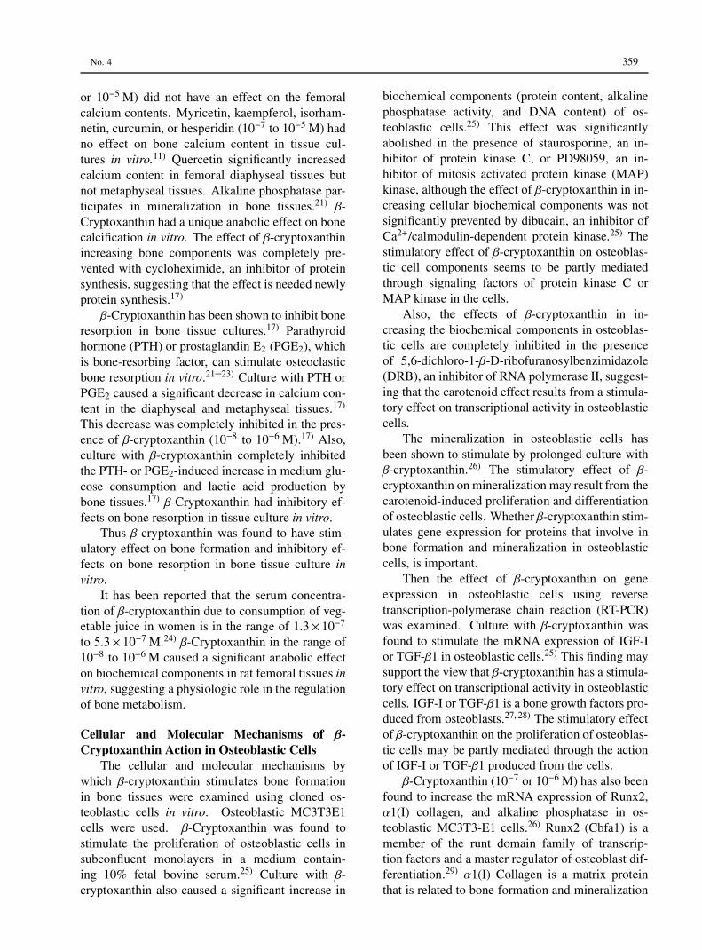
Fig. 6. β-Cryptoxanthin Inhibits Osteoclastogenesis in Bone Marrow Cultureβ-Cryptoxanthin (CRP) inhibits osteoclast formation from mononuclear osteoclasts which are mediated through RANKL and RANK signaling in
bone marrow culture systems. CRP also induces apoptotic cell death of mature osteoclasts. By such a cellular mechanism, CRP inhibits osteoclasticbone resorption. Whether CRP stimulates production of osteoprotegerin (OPG), which inhibits binding of RANKL to RANK receptor, in osteoblasticcells is unknown.
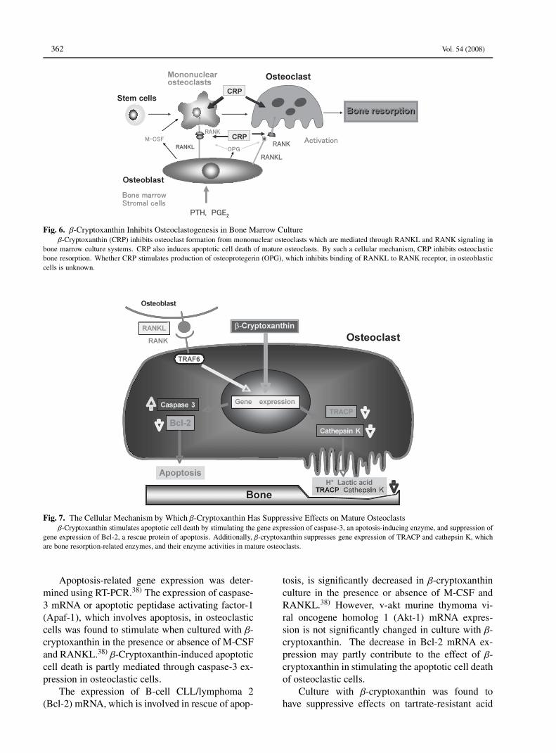
Fig. 7. The Cellular Mechanism by Which β-Cryptoxanthin Has Suppressive Effects on Mature Osteoclastsβ-Cryptoxanthin stimulates apoptotic cell death by stimulating the gene expression of caspase-3, an apotosis-inducing enzyme, and suppression of
gene expression of Bcl-2, a rescue protein of apoptosis. Additionally, β-cryptoxanthin suppresses gene expression of TRACP and cathepsin K, whichare bone resorption-related enzymes, and their enzyme activities in mature osteoclasts.
Apoptosis-related gene expression was deter-mined using RT-PCR.38) The expression of caspase-3 mRNA or apoptotic peptidase activating factor-1(Apaf-1), which involves apoptosis, in osteoclasticcells was found to stimulate when cultured with β-cryptoxanthin in the presence or absence of M-CSFand RANKL.38) β-Cryptoxanthin-induced apoptoticcell death is partly mediated through caspase-3 ex-pression in osteoclastic cells.
The expression of B-cell CLL/lymphoma 2(Bcl-2) mRNA, which is involved in rescue of apop-
tosis, is significantly decreased in β-cryptoxanthinculture in the presence or absence of M-CSF andRANKL.38) However, v-akt murine thymoma vi-ral oncogene homolog 1 (Akt-1) mRNA expres-sion is not significantly changed in culture with β-cryptoxanthin. The decrease in Bcl-2 mRNA ex-pression may partly contribute to the effect of β-cryptoxanthin in stimulating the apoptotic cell deathof osteoclastic cells.
Culture with β-cryptoxanthin was found tohave suppressive effects on tartrate-resistant acid

No. 4 363
phosphatase (TRACP) activity, and it decreasesTRACP and cathepsin K mRNA expressions inosteoclastic cells in the presence or absence ofM-CSF and RANKL.38) These findings suggestthat β-cryptoxanthin can inhibit the enhancementof bone-resorbing activity in osteoclasts. β-Cryptoxanthin could inhibit various bone-resorbingfactors-induced decrease in bone calcium contentand increase in lactic acid production in rat femoraltisuue culture system in vitro.13) Presumably, β-cryptoxanthin has inhibitory effects on the activa-tion of mature osteoclasts.β-Cryptoxanthin has been demonstrated to have
stimulatory effects on apoptotic cell death due toactivating gene expression of its related proteins.The carotenoid also has suppressive effects onTRACP activity and gene expression of enzymesthat involve in bone-resorbing activity in osteoclas-tic cells. The action of β-cryptoxanthin in osteo-clasts is summarized in Fig. 7.
PREVENTIVE ROLE OFβ-CRYPTOXANTHIN IN
OSTEOPOROSIS
As mentioned above, β-cryptoxanthin has beenshown to have a stimulatory effect on osteoblasticbone formation and an inhibitory effect on osteo-clastic bone resorption in vitro. The preventive ef-fect of β-cryptoxanthin on osteoporosis was investi-gated using animal models.
Effect of β-Cryptoxanthin in Animal Models forOsteoporosis
Effects on Young and Aged Rats: The an-abolic effect of β-cryptoxanthin on bone com-ponents in young and aged rats was exa-mined.39) β-Cryptoxanthin (10, 25 or 50 µg/100 gbody weight) was orally administered once dailyfor 7 days to young male rats.39) The administrationof β-cryptoxanthin (25 or 50 µg/100 g body weight)caused a significant increase in calcium content, al-kaline phosphatase activity, and DNA contents inthe femoral-diaphyseal and -metaphyseal tissues.Such an effect is also observed in the femoral tis-sues of aged (50-week-old) female rats.40) Alkalinephosphatase is an enzyme marker of osteoblasts,and the enzyme participates in bone minerali-zation.41) DNA content in bone tissues is an indexof the number of bone cells.42) β-Cryptoxanthin hasbeen shown to have an anabolic effect on bone com-
ponents in rats in vivo.Effect on Bone Loss Induced in Diabetic Rats:
Whether β-cryptoxanthin has a preventive effect onbone loss in the pathophysiologic state was ex-amined. Bone loss has been shown to induce instreoptozotocin (STZ)-diabetic rats.43) Young ratsreceived a single subcutaneous administration ofSTZ (6.0 mg/100 g body weight), and then the an-imals were orally administered β-cryptoxanthin (5or 10 µg/100 g body weight) once daily for 7 or 14days. The administration of STZ caused a signifi-cant decrease in body weight and a significant in-crease in serum glucose, triglyceride, and calciumlevels, indicating a diabetic state. These alterationswere significantly prevented after the administrationof β-cryptoxanthin (5 or 10 µg/100 g) for 14 days.Calcium content, alkaline phosphatase activity, andDNA content in the femoral-diaphyseal and -meta-physeal tissues were significantly decreased inSTZ-diabetic rats. These decreases were signif-icantly prevented after the administration of β-cryptoxanthin (5 or 10 µg/100 g) for 14 days. Thusthe intake of β-cryptoxanthin was found to have pre-ventive effects on STZ-diabetic state and bone lossin STZ-diabetic rats.43)
Effect on Bone Loss in Ovariectomized Rats:Bone loss is induced after ovriectomy (OVX),which is a model of postmenopausal osteoporo-sis. The effect of β-cryptoxanthin on OVX-inducedbone loss was examined.44) β-Cryptoxanthin (5 or10 µg/100 g body weight) was orally administeredonce daily for 3 months to OVX rats. The analysisusing peripheral quantitative computed tomographyshowed that OVX induced a significant decrease inmineral content and mineral density in the femoral-diaphyseal and -metaphyseal tissues. These de-creases were significantly prevented after the ad-ministration of β-cryptoxanthin (5 or 10 µg/100 g).Moreover, OVX induced a significant decrease inbone biochemical components. These decreaseswere completely prevented after the administra-tion of β-cryptoxanthin (5 or 10 µg/100 g). β-Cryptoxanthin was found to have preventive effectson OVX-induced bone loss in vivo.44)
Effects of β-Cryptoxanthin in Healthy Individu-als and Menopausal Women
The effects of β-cryptoxanthin on bonemetabolism in human were investigated usingserum bone metabolic markers. Serum bone-specific alkaline phosphatase and γ-carboxylatedosteocalcin are bone metabolic markers of os-

364 Vol. 54 (2008)
teoblastic bone formation.45, 46) Serum boneTRACP and N-telopeptides of type I colla-gen are metabolic markers of osteoclastic boneresorption.47, 48)
The effects of prolonged intake of juice pre-pared from Satsuma mandarin (Citrus unshiuMARC.) containing β-cryptoxanthin on circulatingbiochemical markers of bone metabolism in sub-jects, including menopausal woman, were exam-ined.49–51)
Effect on Healthy Individuals: Twenty-one vol-unteers (10 males and 11 females) were dividedinto two groups of ten volunteers (5 males and5 females) and eleven volunteers (5 males and 6females), and each group was given sequentiallyjuice (192 ml) containing two different contents ofβ-cryptoxanthin once a day for 28 or 56 days as fol-lows: either regular juice with naturally occurring802 µg β-cryptoxanthin/100 ml or a reinforced juicecontaining 1500 µg β-cryptoxanthin/100 ml.49) Theintake of regular juice for 28 or 56 days in healthysubjects caused a significant increase in serum γ-carboxylated osteocalcin concentration, and the in-take for 56 days produced a significant decrease inserum bone TRACP activity. Moreover, the intakeof the β-cryptoxanthin reinforced juice for 28 or56 days caused a significant increase in serum γ-carboxylated osteocalcin concentration and a corre-sponding decrease in serum bone TRACP activityand N-telopeptide of type I collagen. These find-ings suggest that the intake of β-cryptoxanthin rein-forced juice has a stimulatory effect on osteoblasticbone formation and inhibitory effect on osteoclasticbone resorption in normal individuals.49)
The serum β-cryptoxanthin concentration wassignificantly increased with the intake of regu-lar juice for 56 days.50) This increase was signifi-cantly enhanced after the intake of β-cryptoxanthin-reinforced juice. The intake of regular juice or ofβ-cryptoxanthin-reinforced juice for 56 days causeda significant increase in serum γ-carboxylated os-teocalcin and a significant decrease in serum boneTRACP activity. A possible relationship be-tween serum β-cryptoxanthin and circulating γ-carboxylated osteocalcin concentrations was foundusing the value obtained from all groups for be-fore intake and with the intake of regular juiceand β-cryptoxanthin-reinforced juice. A negativerelationship between serum β-cryptoxanthin con-centation and circulating TRACP activity was ob-served.50) This study shows that a relationship be-tween serum β-cryptoxanthin and circulating bone
metabolic markers is found in healthy individualswith the intake of juice containing β-cryptoxanthin.
Effect on Menopausal Women: Ninety volun-teers, aged 27–65 years (19 men and 71 women),were enrolled in this study.51) The seventy onefemales included 35 premenopausal women (ages,27–50 years) and 36 menopausal women (ages, 46–65 years). Volunteers were divided into four groups;placebo juice without β-cryptoxanthin (5 men and19 women), juice containing β-cryptoxanthin at1.5 mg/200 ml of juice/day (4 men and 17 women),3.0 mg/day (5 men and 17 women), and 6.0 mg/day(5 men and 18 women). Placebo or juice (200 ml)was ingested once a day for 28 or 56 days.
Serum β-cryptoxanthin concentrations were sig-nificantly increased after the intake of juice con-taining β-cryptoxanthin (1.5, 3.0, or 6.0 mg/day)for 28 or 56 days, and the increases were dose-dependent.51) A significant increase in serum β-cryptoxanthin concentration was also observed at28 days at the end of intake, indicating thatthe carotenoid is stable in the serum. Serumβ-cryptoxanthin concentration was in the rangeof 4.20× 10⁻⁷ M to 4.89× 10⁻⁷ M in the placebogroups. The intake of juice reinforced withβ-cryptoxanthin concentration at doses of 1.5,3.0, or 6.0 mg/day significantly increased theserum concentration to 2.43× 10⁻⁶, 4.06× 10⁻⁶,or 5.38× 10⁻⁶ M, respectively.51) These increaseswere about 5- or 10-fold as compared with the valueobtained before intake or after placebo intake. Ithas been reported that the serum concentration ofβ-cryptoxanthin increased due to the consumptionof vegetable juice in women from 1.3× 10⁻⁷ to5.3× 10−7 M.24)
In ninety volunteers (aged 27–65 years), serumbone-specific alkaline phosphatase activity was sig-nificantly increased after the intake of juice con-taining β-cryptoxanthin (3.0 or 6.0 mg/day) for 56days as compared with the value obtained beforeintake.51) γ-Carboxylated osteocalcin concentrationwas significantly increased after the intake of juicecontaining β-cryptoxanthin (3.0 or 6.0 mg/day) for28 or 56 days as compared with the value ob-tained before intake or after the intake of placebojuice.51) Serum TRACP activity and type I colla-gen N-telopeptide concentration were significantlydecreased after the intake of juice containing β-cryptoxanthin (3.0 or 6.0 mg/day) for 28 or 56 daysas compared with the value obtained before intakeor after intake of placebo juice, and significant de-creases were also seen after the intake of 1.5 mg/day

No. 4 365
β-cryptoxanthin as compared with the valueobtained before intake.51)
In menopausal women (36 volunteers), bone-specific alkaline phosphatase activity and γ-carboxylated osteocalcin concentration were signif-icantly increased after the intake of juice contain-ing β-cryptoxanthin (3.0 or 6.0 mg/day) for 56 daysas compared with the value obtained after placebointake.51) Also, this intake caused a significant de-crease in bone TRACP activity and type I colla-gen N-telopeptide concentration. The prolonged in-take of β-cryptoxanthin-reinforced juice has beendemonstrated to have stimulatory effects on os-teoblastic bone formation and inhibitory effects onosteoclastic bone resorption in menopausal women.
Meanwhile, serum calcium, inorganic phos-phorous, and parathyroid hormone (intact) werenot changed after the intake of β-cryptoxanthin-containing juice for 28 or 56 days. Other serumbiochemical findings were not changed after theintake of juice containing β-cryptoxanthin (3.0 or6.0 mg/day) for 56 days. We confirmed the safetyof β-cryptoxanthin in human.51)
As the mentioned above, the intake of juice rein-forced with β-cryptoxanthin (3.0 or 6.0 mg/day) hada significant effect on circulating bone metabolicmarkers in men, premenopausal women, andmenopausal women.51) This indicates that the ef-fects of β-cryptoxanthin in stimulating bone forma-tion and inhibiting bone resorption are present inboth sexes. Interestingly, the intake of juice re-inforced with β-cryptoxanthin (3.0 or 6.0 mg/day)was found to have effects on circulating bonemetabolic markers in menopausal women, indicat-ing that the supplementation of β-cryptoxanthin haspreventive effects on bone loss due to osteoporosisin menopausal women. This preventive effect wasobvious at a dose of β-cryptoxanthin of 3.0 mg/dayin menopausal women. This dose may be suitablein the prevention of osteoporosis in human subjects.
Thus the intake of reinforced juice, which con-tains more β-cryptoxanthin than regular juice, hasbeen demonstrated to have a preventive effect onbone loss that accompanies an increase in age.
EPIDEMIOLOGICAL EVIDENCE FORROLE OF β-CRYPTOXANTHIN IN BONE
HEALTHY
On the based on our findings, it has been re-cently reported that epidemiological studies show
that the intakes of fruit and vegetables containingβ-cryptoxanthin may reduce the risk of osteo-porosis.52–54)
The effect of dietary antioxidants on knee struc-ture in a cohort of healthy, middle-aged subjectswith no clinical knee osteoarthritis is examined.52)
Two hundred and ninety-three healthy adults (meanage = 58.0 years) without knee pain or knee injuryare selected from an existing community-based co-hort. The intake of antioxidant vitamins and foodsources by these individuals was estimated froma food frequency questionnaire at baseline. Thecartilage volume, bone area, cartilage defects andbone marrow lesions were assessed approximately10 years later using magnetic resonance imaging.Higher vitamin C intake was associated with a re-duced risk of bone marrow lesions and with a re-duction in the tibial plateau bone area. There wasan inverse association between fruit intake and thetibial plateau bone area and between fruit intake andthe risk of bone marrow lesions. Neither fruit in-take nor vitamin C intake was significantly associ-ated with the cartilage volume or cartilage defects.Lutein and zeaxanthin intake was associated with adecreased risk of cartilage defects, and vitamin Eintake tended top be positively associated with thetibial plateau bone are only after adjusting for vita-min C intake. The β-cryptoxanthin intake was in-versely associated with the tibial plateau bone areaafter adjusting for vitamin E intake. These observa-tions suggest a beneficial effect of fruit consumptionand vitamin C intake as they are associated with areduction in bone size and the number of bone mar-row lesions, both of which are important in the pato-genesis of knee osteoarthritis.52)
Bone mineral density (BMD) in post-menopausal female subjects has been shownto associate with serum antioxidant carotenoids.A total of six hundred ninety-nine subjects (222males and 477 females) who had received healthexaminations in the town of Mikkabi, ShizuokaPrefecture, Japan, participated in the study.53)
Radial BMD was measured using dual-energyX-ray absorptiometry. The associations of serumcarotenoid concentrations with the dadial BMDwere evaluated cross-sectionally. In male andpre-menopausal female subjects, the six serumcarotenoids were not associated with the radialBMD. On the other hand, in post-menopausalfemale subjects, serum β-cryptoxanthin and β-carotene were weakly but positively correlated withthe radial BMD. After adjustment for confounders,

366 Vol. 54 (2008)
the odds ratio (OR) for the lowest quartile of BMDin the high groups of serum β-cryptoxanthin againstthe lowest quartile was 0.45 in post-menopausalfemale subjects. However, this association wasnot significant after further adjusting for intakes ofminerals and vitamins. Antioxidant carotenoids,especially β-cryptoxanthin, significantly but partlyassociate with the radial BMD in post-menopausalfemale subjects.53)
Seasonal variation of serum α- and β-cryptoxanthin and 25-OH-vitamin D3 in womenwith osteoporosis is examined.54) In six hundredfourty-four women with osteoporosis, serumβ-cryptoxanthin and 25-OH-vitamin D3 showeda weak but significant correlation and exhibited acomplementary seasonal distribution.54) Dietaryintake and serum levels of β-cryptoxanthin havebeen inversely related to different bone and jointdisorders and in vitro and animal studies haveshown that β-cryptoxanthin displays a uniqueanabolic effect on bone calcification. Due to theemerging role of β-cryptoxanthin in bone biology,this study aimed to assess the serum distributionand variability of β-cryptoxanthin and their poten-tial relation to 25-OH-vitamin D3 in women withosteoporosis.
Overall, significant seasonal variations werefound for the three analyses and inter-individualvariation was also high (60–73%). β-Cryptoxanthinand 25-OH-vitamin D3 exhibited a marked comple-mentary seasonal distribution in serum, with vita-min D displaying the highest values in summer andβ-cryptoxanthin in winter.
Given the anabolic effect of β-cryptoxanthin onbone calcification and its complementary seasonaldistribution with respect to 25-OH-vitamin D3, thepotential role of β-cryptoxanthin as a sustainablenutritional approach to improving bone health de-serves to be further evaluated.54)
CONCLUSION
Amoung various carotinoids, β-cryptoxanthinhas a unique anabolic effect on bone mass due tostimulating osteoblastic bone formation and inhibit-ing osteoclastic bone resorption, thereby increasingbone mass. β-Cryptoxanthin modulates gene ex-pression of various proteins that involve in bone for-mation in osteoblasts and bone resorption in osteo-clasts. The intake of dietary β-cryptoxanthin hasbeen shown to have preventive effect on bone loss
in animal models for osteoporosis and restorativeeffect on bone metabolism in menopausal women.The role of β-cryptoxanthin in bone healthy hasbeen also shown in human subjects with epidemi-ological studies. β-Cryptoxanthin may have an im-portant role in prevention of osteoporosis.
Epidemiological studies, moreover, demon-strates the anabolic effect of β-cryptoxanthin onbone calcification and its complementary seasonaldistribution with respect to 25-OH-vitamin D3, aclassic hormonal factor of bone healthy in hu-man subjects,54) suggesting the potential role of β-cryptoxanthin as a sustainable nutritional approachto improving bone health. The physiologic signifi-cance of β-cryptoxanthin in bone healthy, however,remains to be elucidated.
Whether the combination of nutritional factorsexhibits an additive or synergistic effect on bonecomponents has not been fully clarified. Thisknowledge may be important in preventing boneloss with increasing age. The anabolic effect of β-cryptoxanthin on osteoblastic bone formation hasnot been shown to enhance synergistically in thepresence of genistein, menaquinone-7, VD3, orE2, which has a stimulatory effect on osteoblas-tic bone formation.55, 56) The combination of β-cryptoxanthin and zinc at a lower concentration,however, is found to have a synergistic effect onosteoblastic bone formation55–57) and an additivesuppressive effect on osteoclastic cell functions.58)
Zinc has been shown to stimulate osteoblastic boneformation and inhibit osteoclastic bone resorption.59–63) The finding is interested in respect of the de-velopment of new supplement with the compositionof food factors that reveal a potent-anabolic effectin prevention of osteoporosis. It also would be use-ful to identify some of the foods that contain higherlevels of β-cryptoxanthin and zinc.
Additionally, the intake of β-cryptoxanthin withhigher dose may have a pharmacologic role in ther-apy of osteoporosis. Clinical studies are needed inthe development of new drug for osteoporosis ther-apy.
In conclusion, β-cryptoxanthin is a kind ofcarotenoid that has a potential effect in maintainingbone healthy and in preventing osteoporosis.
Acknowledgements The author appreciates Dr.Satoshi Uchiyama who was collaborated in thisstudy, and also thanks Research & Development,Ehime Beverage, Inc., in Matsuyama, Ehime Pre-fecture, Japan that was collaborated in the examina-

No. 4 367
tion of reinforced juice intake in human subjects.
REFERENCES
1) Cooper, C. and Melton, J. III (1995) Epidemiologyof osteoporosis. Trends Endocrinol. Metab., 3, 224–229.
2) Riggs, B. L., Jowsey, J., Kelly, P. J., Jones, J. D. andMaher, F. T. (1969) Effect of sex hormones on bonein primary osteoporosis. J. Clin. Invest., 48, 1065–1072.
3) Bonjour, J. -P., Schurch, M. -A. and Rizzori, R.(1996) Nutritional aspects of hip fracture. Bone, 18,1395–1445.
4) Yamaguchi, M. (2002) Isoflavone and bonemetabolism: Its cellular mechanism and preventiverole in bone loss. J. Health Sci., 48, 209–222.
5) Yamaguchi, M. (2006) Regulatory mechanism offood factors in bone metabolism and prevention ofosteoporosis. Yakugaku Zasshi, 126, 1117–1137.
6) Yamaguchi, M., Hamamoto, R., Uchiyama, S. andIshiyama, K. (2008) Effects of flavonoid on cal-cium content in femoral tissue culture and parathy-roid hormone-stimulated osteoclastogenesis in bonemarrow culture in vitro. Mol. Cell. Biochem., 303,83–88.
7) Lai, Y. L. and Yamaguchi, M. (2006) Phytocompo-nent p-hydroxycinnamic acid stimulates bone for-mation and inhibits bone resorption in rat femoraltissues in vitro. Mol. Cell. Biochem., 292, 45–52.
8) Yamaguchi, M., Lai, Y. L., Uchiyama, S. andNakagawa, T. (2008) Oral administration of phyto-component p-hydroxycinnamic acid prevents boneloss in ovariectomized rats. Mol. Cell. Biochem.,311, 31–36.
9) Yamaguchi, M., Hachiya, S., Hiratsuka, S. andSuzuki, T. (2001) Effect of marine algae extract onbone calcification in the femoral-metaphyseal tis-sues of rats: Anabolic effect of Sargassum horneri.J. Health Sci., 47, 533–538.
10) Uchiyama, S. and Yamaguchi, M. (2002) An-abolic effect of marine alga Sargassum horneri ex-tract on bone components in the femoral-diaphysealand -metaphyseal tissues of young and aged rats invivo. J. Health Sci., 48, 325–330.
11) Yamaguchi, M., Hamamoto, R., Uchiyama, S.,Ishiyama, K. and Hashimoto, K. (2006) Anaboliceffects of bee pollen Cistus ladaniferus extracton bone components in the femoral-diaphysealand -metaphyseal tissues of rats in vitro and in vivo.J. Health Sci., 52, 43–49.
12) Yamaguchi, M., Uchiyama, S. and Nakagawa, T.
(2007) Preventive effects of bee pollen Cistus ladan-iferus extract on bone loss in ovariectomiaed rats invivo. J. Health Sci., 53, 571–575.
13) Yamaguchi, M., Ma, Z. J. and Suzuki, T. (2003)Anabolic effect of wasabi leafstalk (WasabiaJaponica MATSUM.) extract on bone componentsin the femoral-diaphyseal and -metaphyseal tissuesof aged female rats in vitro and in vivo. J. HealthSci., 49, 123–128.
14) Suzuki, T. and Yamaguchi, M. (2004) Purificationof active component in wasabi leafstalk (Wasabiajaponica MATSUM.) extract in stimulating bonecalcification in vitro. J. Health Sci., 50, 483–490.
15) Promislow, J. H. E., Goodman-Gruen, D., Slymen,D. J. and Barret-Connor, E. (2002) Retinol intakeand bone mineral density in the elderly: the ranchobrenardo study. J. Bone Miner. Res., 17, 1349–1358.
16) Yamaguchi, M. and Uchiyama, S. (2003) Effect ofcarotenoid on calcium content and alkaline phos-phatase activity in rat femoral tissues in vitro: Theunique anabolic effect of β-cryptoxanthin. Biol.Pharm. Bull., 26, 1188–1191.
17) Yamaguchi, M. and Uchiyama, S. (2004) β-Cryptoxanthin stimulates bone formation and in-hibits bone resorption in tissue culture in vitro. Mol.Cell. Biochem., 258, 137–144.
18) Parfitt, A. M. (1990) Bone-forming cells in clinicalconditions. In The Osteoblast and Osteocyte, Bone(Hall, B. K., Ed.), vol. 1, Telford Press and CRCPress, Boca Raton, FL, pp. 351–429.
19) Baron, R., Neff, L., Tran Van, P., Nefussi, J. R., andVignery, A. (1986) Kinetic and cytochemical identi-fication of osteoclast precursors and their differenti-ation into multinucleated osteoclasts. Am. J. Pathol.,122, 363–378.
20) Canalis, E., McCarthy, T. and Centrella, M. (1988)Growth factors and the regulation of bone remodel-ing. J. Clin. Invest., 81, 277–281.
21) Klein-Nulend, J., Fall, P. M. and Raisz, L. G.(1990) Comparison of the effects of synthetic humanparathyroid hormone (PTH)-(1-34)-related peptideof malignancy and bovine PTH-(1-34) on bone for-mation and resorption in organ culture. Endocrinol-ogy, 126, 223–227.
22) Graves, L. III and Jilka, R. L. (1990) Compari-son of bone and parathyroid hormone as stimulatorsof osteoclast development and activity in calvarialcell cultures from normal and osteopetrotic (mi/mi)mice. J. Cell. Physiol., 145, 102–109.
23) Klein, D. C. and Raisz, L. G. (1970) Stimulation ofbone resorption in tissue culture. Endocrinology, 86,1436–1440.
24) McEligot, A. J., Rock, C. L., Shanks, T. G., Flatt, S.

368 Vol. 54 (2008)
W., Newman, V., Farber, S. and Pierce, J. P. (1999)Comparison of serum carotenoid responses betweenwomen consuming vegetable juice and women con-suming raw or cooked vegetable. Cancer Epidemi-nol. Biomarkers Prev., 8, 227–231.
25) Uchiyama, S. and Yamaguchi, M. (2005) β-Cryptoxanthin stimulates cell proliferation and tran-scriptional activity in osteoblastic MC3T3-E1cells.Int. J. Mol. Med., 15, 675–681.
26) Uchiyama, S. and Yamaguchi, M. (2005) β-Cryptoxanthin stimulates cell differentiation andmineralization in osteoblastic MC3T3-E1 cells. J.Cell. Biochem., 95, 1224–1234.
27) Centrella, M., McCarthy, T. L. and Canalis, E.(1990) Receptors for insulin-like growth factor-Iand -II in osteoblast-enriched cultures from fetal ratbone. Endocrinology, 126, 39–44.
28) Palcy, S., Bolivar, I. and Goltzman, D. (2000)Role of activator protein 1 transcriptional activity inthe regulation of gene expression by transforminggrowth factor β1 and bone morphogenic protein 2in ROS 17/2.8 osteoblast-like cells. J. Bone Miner,Res., 15, 2352–2361.
29) Komori, T., Yagi, H., Nomura, S., Yamaguchi, A.,Sasaki, K., Deguchi, K., Shimizu, Y., Bronson, R.T., Gao, Y. H., Inada, M., Sato, M., Okamoto, R.,Kitamura, Y., Yoshiki, S. and Kishimoto, T. (1997)Targeted disruption of Cbfa1 results in a comple lackof bone formation owing to maturational arrest ofosteoblasts. Cell, 89, 755–764.
30) Lian, J. B., Stein, G. S., Canalis, E., Roby, P. G.and Boskey, A. L. (1999) Bone formation: Os-teoblast lineage cells, growth factors, matrix pro-teins, and the mineralization process. In Primer onthe Metabolic Bone Diseases and Disorders of Min-eral Metabolism (Favus, M. J., Ed.), 4th edition,Lippincott Williams & Wilkins Press, New York,pp. 14–29.
31) Yohay, D. A., Zhang, J., Thrailkill, K. M., Arthur, J.M. and Quarles, L. D. (1994) Role of serum in thedevelopmental expression of alkaline phosphatase inMC3T3-E1 osteoblasts. J. Cell. Physiol., 158, 467–475.
32) Park, C. K., Ishimi, Y., Ohmura, M., Yamaguchi, M.and Ikegami, S. (1997) Vitamin A and carotenoidstimulate differentiation of mouse osteoblstic cells.J. Nutr. Sci. Vitaminol. (Tokyo), 43, 281–296.
33) Zaidi, M., Blair, H. C., Moonga, B. S., Abe, E.and Huang, C. L. -H. (2003) Osteoclastogenesis,bone resorption, and osteoblast-based therapeutics.J. Bone Miner. Res., 18, 599–609.
34) Tanaka, S., Nakamura, I., Inoue, J. -I., Oda, H. andNakamura, K. (2003) Signal transduction pathways
regulating osteoclast differentiation and function. J.Bone Miner. Metab., 21, 123–133.
35) Uchiyama, S. and Yamaguchi, M. (2004) Inhibitoryeffect of β-cryptoxanthin on osteoclast-like cell for-mation in mouse marrow cultures. Biochem. Phar-macol., 67, 1297–1305.
36) Anderson, D. M., Maraskovsky, E., Billingsley, W.L., Dougall, W. C., Tometsko, M. E., Roux, E.R., Teepe, M. C., DuBose, R. F., Cosman, D. andGailibert, L. (1997) A homologue of the TNF re-ceptor and its ligand enhance T-cell growth anddendritic-cell function. Nature, 390, 175–195.
37) Lee, Z. H., Kwack, K., Kim, K. K., Lee, S. H. andKim, H. -H. (2000) Activation of c-Jun N-terminalkinase and activator protein 1 by receptor activatorof NF-κB. Mol. Pharmacol., 58, 1536–1545.
38) Uchiyama, S. and Yamaguchi, M. (2006) β-Cryptoxanthin stimulates apoptotic cell death andsuppresses cell function in osteoclastic cells:Change in their related gene expression. J. Cell.Biochem., 98, 1185–1195.
39) Uchiyama, S., Sumida, T. and Yamaguchi, M.(2004) Oral administration of β-cryptoxanthin in-duces anabolic effects on bone components in thefemoral tissues of rats in vivo. Biol. Pharm. Bull.,27, 232–235.
40) Uchiyama, S., Sumida, T. and Yamaguchi, M.(2004) Anabolic effect of β-cryptoxanthin on bonecomponents in the femoral tissues of aged rats invivo and in vitro. J. Health Sci., 50, 491–496.
41) Majeska, R. J. and Wuthier, R. E. (1975) Studies onmatrix vesicles isolated from chick epiphyseal car-tilage. Association of pyrophosphatase and ATPaseactivities with alkaline phosphatase. Biochim. Bio-phys. Acta, 391, 51–60.
42) Canalis, E., Centrella, M., Burch, W. and McCarthy,T. L. (1989) Insulin-like growth factor-I mediates se-lective anabolic effects of parathyroid hormone inbone cultures. J. Clin. Invest., 83, 60–65.
43) Uchiyama, S. and Yamaguchi, M. (2005) Oral ad-ministration of β-cryptoxanthin prevents bone lossin streptozotocin-diabetic rats in vivo. Biol. Pharm.Bull., 28, 1766–1769.
44) Uchiyama, S. and Yamaguchi, M. (2006) Oral ad-ministration of β-cryptoxanthin prevents bone lossin ovariectomized rats. Int. J. Mol. Med., 17, 15–20.
45) Price, P. A. (1985) Vitamin K-dependent formationof bone gla protein (osteocalcin) and its function.Vitam. Horm., 42, 65–108.
46) Levy, J. R., Murray, E., Manolagass, S. andOlefsky, J. M. (1986) Demonstration of insulin re-ceptors and modulation of alkaline phosphatase ac-tivity by insulin in rat osteoblastic cells. Endocrinol-

No. 4 369
ogy, 119, 1786–1792.47) Hallen, J. M., Alatalo, S. L., Suminen, H., Cheng,
S., Janekila, A. J. and Vaananen, H. K. (2000)Tartrate-resistant acid phosphatase 5b: A novelserum marker of bone resorption. J. Bone Miner.Res., 15, 1337–1345.
48) Clements, J. D., Herrick, M. V., Singer, F. R.and Eyre, D. R. (1997) Evidence that serum NTx(collagen-type I N-telopeptides) can act as an im-munochemical marker of bone resorption. Clin.Chem., 43, 2058–2063.
49) Yamaguchi, M., Igarashi, A., Uchiyama, S., Morita,S., Sugawara, K. and Sumida, K. (2004) Prolongedintake of jucice (Citrus unshiu) reinforced with β-cryptoxanthin has an effect on circulating bone bio-chemical markers in normal individuals. J. HealthSci., 50, 619–624.
50) Yamaguchi, M., Igarashi, A., Morita, S.,Sumida, T. and Sugawara, K. (2005) Relation-ship between serum β-cryptoxanthin and circulatingbone metabolic markers in healthy individualswith the intake of juice (Citrus unshiu) containingβ-cryptoxanthin. J. Health Sci., 51, 738–743.
51) Yamaguchi, M., Igarashi, A., Uchiyama, S.,Sugawara, K., Sumida, T., Morita, S., Ogawa, H.,Nishitani, M. and Kajimoto, Y. (2006) Effect of β-cryptoxanthin on circulating bone metabolic mark-ers: Intake of juice (Citrus unshiu) supplementedwith β-cryptoxanthin has an effect in menopausalwomen. J. Health Sci., 52, 758–768.
52) Wang, Y., Hodge, A. M., Wluka, A. E., English,D. R., Giles, G. G., O’Sullivan, R., Forbes, A. andCicuttini, F. M. (2007) Effect of antioxidants onknee cartilage and bone in healthy, middle-aged sub-jects: a cross-sectional study. Arthritis Res. Ther., 9,R66.
53) Sugiura, M., Nakamura, M., Ogawa, K., Ikoma,Y., Ando, F. and Yano, M. (2008) Bone mineraldensity in post-menopausal female subjects is as-sociated with serum antioxidant carotenoids. Osteo-poros. Int., 19, 211–219.
54) Granado-Lorencio, F., Olmedilla-Alonso, B.,Herrero-Barbudo, C., Blanco-Navarro, I. and
Perez-Sacristan, B. (2008) Seasonal variationof serum alpha- and beta-cryptoxanthin and 25-OH-vitamin D(3) in women with osteoporosis.Osteoporos. Int., 19, 717–720.
55) Uchiyama, S., Ishiyama, K., Hashimoto, K. andYamaguchi, M. (2005) Synergistic effect of β-cryptoxanthin and zinc sulfate on the bone com-ponent in rat femoral tissues in vitro: The uniqueanabolic effect with zinc. Biol. Pharma. Bull., 28,2142–2145.
56) Yamaguchi, M., Uchiyama, S., Ishiyama, K. andHashimoto, K. (2006) Oral administration in combi-nation with zinc enhances β-cryptoxanthin-inducedanabolic effects on bone components in the femoraltissues of rats in vivo. Biol. Pharm. Bull., 29, 371–374.
57) Uchiyama, S. and Yamaguchi, M. (2008) Anaboliceffect of β-cryptoxanthin in osteoblastic MC3T3-E1cells is enhanced with 17 β-estradiol, genistein, orzinc sulfate in vitro: the unique effect with zinc onRunx2 and α1 (I) collagen mRNA espression. Mol.Cell. Biochem., 307, 209–219.
58) Yamaguchi, M. and Uchiyama, S. (2008) Combi-nation of β-cryptoxanthin and zinc has potent ef-fects on apoptotic cell death and suppression of boneresorption-related gene expression in osteoclasticcells. Int. J. Mol. Med., 22, 221–228.
59) Yamaguchi, M., Oishi, H. and Suketa, Y. (1987)Stimulatory effect of zinc on bone formation in tis-sue culture. Biochem. Pharmacol., 36, 4007–4012.
60) Kishi, S. and Yamaguchi, M. (1994) Inhibitory ef-fect of zinc compounds on osteoclast-like cell for-mation in mouse marrow culture. Biochem. Pharma-col., 48, 1225–1230.
61) Yamaguchi, M. (1998) Role of zinc in bone forma-tion and bone resorption. J. Trace Elem. Ex. Med.,11, 119–135.
62) Yamaguchi, M. (1995) β-Alanyl-L-histidinato zinc:A potent activator in bone formation. Curr. Med.Chem., 1, 356–365.
63) Yamaguchi, M. (2007) Role of zinc in bonemetabolism and preventive effect on bone disorder.Biomed. Res. Trace Elem., 18, 346–366.