Crypt organoids culture as an in vitromodel in drug...
29
DMD Manuscript #75945 1 Title Page Crypt organoids culture as an in vitro model in drug metabolism and cytotoxicity studies. Wenqi Lu, Eva Rettenmeier, Miles Paszek, Mei-Fei Yueh, Robert H. Tukey, Jocelyn Trottier, Olivier Barbier, Shujuan Chen Laboratory of Environmental Toxicology, Department of Pharmacology, University of California, San Diego, La Jolla, CA 92093, U.S.A (W. L., E. R., M. P., M-F. Y., R. H. T., S. C.); Laboratory of Molecular Pharmacology, CHU de Québec Research Centre, Québec (Québec), Canada G1V 4G2 (J. T., O. B.) This article has not been copyedited and formatted. The final version may differ from this version. DMD Fast Forward. Published on May 3, 2017 as DOI: 10.1124/dmd.117.075945 at ASPET Journals on June 15, 2018 dmd.aspetjournals.org Downloaded from
Transcript of Crypt organoids culture as an in vitromodel in drug...
DMD Manuscript #75945
1
Title Page
Crypt organoids culture as an in vitro model in drug metabolism and cytotoxicity studies.
Wenqi Lu, Eva Rettenmeier, Miles Paszek, Mei-Fei Yueh, Robert H. Tukey, Jocelyn Trottier,
Olivier Barbier, Shujuan Chen
Laboratory of Environmental Toxicology, Department of Pharmacology, University of California,
San Diego, La Jolla, CA 92093, U.S.A (W. L., E. R., M. P., M-F. Y., R. H. T., S. C.); Laboratory of
Molecular Pharmacology, CHU de Québec Research Centre, Québec (Québec), Canada G1V
4G2 (J. T., O. B.)
This article has not been copyedited and formatted. The final version may differ from this version.DMD Fast Forward. Published on May 3, 2017 as DOI: 10.1124/dmd.117.075945
at ASPE
T Journals on June 15, 2018
dmd.aspetjournals.org
Dow
nloaded from

DMD Manuscript #75945
2
Running Title Page
Running Title: Intestinal crypt culture in drug metabolism studies
Correspondence should be addressed to Dr. Shujuan Chen [email protected]
9500 Gilman Drive,
La Jolla, CA 92093-0722
Phone: 858-822-1351
Fax: 858-822-0363
Manuscript metrics:
No. of Text pages: 29
No. of Tables: 0
No. of Figures: 4
No. of References: 53
No. of words in the Abstract: 240
No. of words in the Introduction: 504
No. of words in the Discussion: 1274
Abbreviations used in this paper:
CAR, constitutive androstane receptor; IECs, intestinal epithelial cells; ISCs, intestinal stem cells;
PPAR, peroxisome proliferator-activated receptor; PXR, pregnane X receptor; CYP, Cytochrome
P450; UGT, UDP-glucuronosyltransferase; XPGs, xenobiotic processing genes; XNR, xenobiotic
nuclear receptors, DMEs, drug metabolism enzymes; GI, gastrointestinal;
This article has not been copyedited and formatted. The final version may differ from this version.DMD Fast Forward. Published on May 3, 2017 as DOI: 10.1124/dmd.117.075945
at ASPE
T Journals on June 15, 2018
dmd.aspetjournals.org
Dow
nloaded from

DMD Manuscript #75945
3
Abstract
The gastrointestinal tract is enriched with xenobiotic processing proteins that play important roles
in xenobiotic bioactivation, metabolism, and detoxification. The application of genetically modified
mouse models has been instrumental in characterizing the function of xenobiotic processing
genes (XPG) and their proteins in drug metabolism. Here, we report the utilization of 3D crypt
organoid cultures from these animal models to study intestinal drug metabolism and toxicity. With
the successful culturing of crypt organoids, we profiled the abundance of Phase I and Phase II
XPG expression, drug transporter gene expression and xenobiotic nuclear receptor (XNR) gene
expression. Functions of XNRs were examined by treating crypt cells with XNR prototypical
agonists. Real-time quantitative PCR demonstrated that the representative downstream target
genes were induced. These findings were validated from cultures developed from XNR-null mice.
In crypt cultures isolated from Pxr-/- mice, PCN failed to induce Cyp3a11 gene expression; similarly,
WY14643 failed to induce Cyp4a10 in the Pparα-/- crypts. Crypt cultures from control (Ugt1F/F) and
intestinal epithelial cell (IEC) specific Ugt1 null mice (Ugt1∆IEC) were treated with camptothecin
(CPT)-11, an anticancer pro-drug with severe intestinal toxicity that originates from insufficient
UGT1A1 dependent glucuronidation of its active metabolite SN-38. In the absence of Ugt1 gene
expression, Ugt1∆IEC crypt cultures exhibit very limited production of SN-38 glucuronide (SN-38G),
concordant with increased apoptosis in comparison to Ugt1F/F crypt cultures. This study suggests
crypt organoid cultures as an effective in vitro model for studying intestinal drug metabolism and
toxicity.
This article has not been copyedited and formatted. The final version may differ from this version.DMD Fast Forward. Published on May 3, 2017 as DOI: 10.1124/dmd.117.075945
at ASPE
T Journals on June 15, 2018
dmd.aspetjournals.org
Dow
nloaded from
DMD Manuscript #75945
4
Introduction
The gastrointestinal (GI) tract is lined with epithelial cells that express an abundance of
xenobiotic or drug metabolizing enzymes (DMEs) in addition to efflux and influx transporters. Thus,
the absorption of xenobiotics and drugs are subjected to metabolism by DMEs localized in the
mature enterocyte. This process contributes significantly to bioactivation, metabolism, and
detoxification of drugs, all of which are associated with the outcome of drug efficacy and safety.
A greater appreciation attributed to the GI tract and drug metabolism has been achieved by
examining drug metabolism and toxicity in knockout animal models or fully humanized animal
models. For example, human CYP3A4 is considered by many to be one of the most versatile of
the CYPs involved in drug metabolism because it accounts for the metabolism of almost half of
all prescribed medications (Zanger et al., 2008). Van Herwaarden et al (van Herwaarden et al.,
2007; van Waterschoot et al., 2008; van Waterschoot et al., 2009). generated intestine- and liver-
specific expression of human CYP3A4 in a Cyp3a-null background demonstrating intestinal
CYP3A4 activity had a profound impact on systemic exposure of the anticancer drug docetaxel
following its oral administration. Camptothecin (CPT)-11 (Irinotecan), is a prodrug that is
hydrolyzed by carboxylesterase (CES) in different tissues to form the active topoisomerase
inhibitory metabolite SN-38 (Kaneda et al., 1990; Sugimoto et al., 1990), which is further
metabolized by UGT1A1 dependent glucuronidation to form an SN-38 glucuronide (SN-38G) (Iyer
et al., 1998). Classically, the bioactivation and detoxification of CPT-11 have been credited
primarily to hepatic metabolism (Alimonti et al., 2004; Michael et al., 2006). However, recent
studies have confirmed that intestinal CES2 has a greater affinity toward CPT-11 in comparison
to liver CES1 (Hatfield et al., 2011). With the development of Ugt1F/F mice, deletion of the Ugt1
locus in intestinal epithelial cells (Ugt1∆IEC) produced severe CPT-11-induced intestinal toxicity in
comparison to Ugt1F/F mice or mice with a liver specific Ugt1 deletion (Ugt1∆HEP) (Chen et al.,
This article has not been copyedited and formatted. The final version may differ from this version.DMD Fast Forward. Published on May 3, 2017 as DOI: 10.1124/dmd.117.075945
at ASPE
T Journals on June 15, 2018
dmd.aspetjournals.org
Dow
nloaded from
DMD Manuscript #75945
5
2013). This result highlighted that UGT1A1 expression in intestinal tissue is critical in preventing
SN-38 induced toxicity.
Given the importance of the GI tract and the beneficial application of genetically modified
mouse models to examine drug metabolism and toxicity, the development of primary cells cultured
from these mice to examine gene expression and toxicity will provide an important tool to
complement in vivo animal experiments. Sato et al (Sato et al., 2009). established long-term
culture conditions in which one single Lgr5+ stem cell embedded in matrix gel can independently
generate villus-like epithelial domains with the presence of all differentiated IECs. The 3-D culture
is a continuously expanding, self-organizing structure which is reminiscent of normal gut. This
result has been popularly applied in the study of crypt-villus biology, regenerative medicine, and
gene therapy (Sato and Clevers, 2013; Grabinger et al., 2014; Ranga et al., 2014), but its
applications to investigate intestinal epithelial cell specific xenobiotic metabolism has been lacking.
In this article, we report the application of crypt organoid cultures from genetically modified mice
as a model to examine gene expression and drug metabolism.
This article has not been copyedited and formatted. The final version may differ from this version.DMD Fast Forward. Published on May 3, 2017 as DOI: 10.1124/dmd.117.075945
at ASPE
T Journals on June 15, 2018
dmd.aspetjournals.org
Dow
nloaded from

DMD Manuscript #75945
6
Material and methods
Reagents: WY14643, pregnenolone 16α-carbonitrile (PCN), 1,4-Bis-[2-(3,5-
dichloropyridyloxy)]benzene (TCPOBOP), phenobarbital, docosahexaenoic acid (DHA), and β-
naphthoflavone (BNF), N-acetylcysteine, and CPT-11 standard were from Sigma-Aldrich. SN-38
and T0901317 were purchased from Cayman Chemical Company. SN-38 glucuronide was from
Santa Cruz Biotechnology. Internal standards deuterated d10-CPT-11 and d3-SN-38 were
purchased from Toronto Research Chemicals. 2,3,7,8-tetrachlorodibenzo-p-dioxin (TCDD) was
obtained from the National Cancer Institute, National Institutes of Health, Chemical Carcinogen
Reference Standard Repository. Matrigel basement membrane matrix was purchased from BD
biosciences. Advanced DMEM/F12 medium and the supplements including GlutaMax, hepes,
penicillin streptomycin, mouse EGF, N2, and B27 supplements were purchased from Thermo
Fisher Scientific; mouse Noggin was from Peprotech. HEK-293 cells producing R-Spondin 1
protein (293T-HA-Raspol_Fc cell line) was a generous gift from Dr. Calvin Kuo laboratory at
Stanford University (Ootani et al., 2009). The cells were cultured and passaged per standard
protocol. For collecting R-Spondin 1 conditioned media, 293T-HA-Raspol_Fc cells at
approximately 75% confluency were changed to advanced DMEM/F12 medium and cultured for
another week. Medium was then collected by passing through 0.22 µm sterile filter.
Animals: C57BL/6J mice were purchased from Charles River Laboratories. Intestinal specific
Ugt1 deletion mice (Ugt1∆IEC) were generated in our laboratory (Chen et al., 2013). Pparα-/- mice
were purchased from Jackson Laboratories (Lee et al., 1995). Pxr-/- and Car-/- mouse models were
a kind gift from Drs. Ron Evans (Laboratory at Salk Institute, La Jolla) (Xie et al., 2000) and
Masahiko Negishi (Ueda et al., 2002; Yamazaki et al., 2005) (NIEHS), respectively. The proposed
mouse experiments have been approved per the procedures as set by the University of California
San Diego (UCSD) Institutional Animal Care and Use Committee (IACUC). All the proposed
This article has not been copyedited and formatted. The final version may differ from this version.DMD Fast Forward. Published on May 3, 2017 as DOI: 10.1124/dmd.117.075945
at ASPE
T Journals on June 15, 2018
dmd.aspetjournals.org
Dow
nloaded from
DMD Manuscript #75945
7
mouse strains were housed in a pathogen-free facility with automated temperature control and a
12 h x 12 h light/dark cycle.
Isolation of crypt cells and organoid culture. Intestinal crypt cell isolation and organoid
culturing were carried out according to previous publications (Sato et al., 2009; Sato and Clevers,
2013) with several modifications. Briefly, mouse small intestine was dissected, cut longitudinally
and washed briefly by ice-cold DPBS. The tissue was further dissected into small pieces and
incubated in DPBS containing 2 mM EDTA at 4ºC with gentle shaking. One hour later, the EDTA
solution was removed and 10% FBS in DPBS buffer added followed by vigorously shaking to
release villi and crypt cells. The cell solution was passed through tea filter to remove tissue debris,
and then further filtered through a 70 µm cell strainer. The filtrate was centrifuged at 1,000 g for
10 min at 4ºC and the cell pellet washed twice by DPBS. Crypt cells were counted. Approximately
1,000 crypts were suspended into 50 µl ice-cold Matrigel and plated into pre-warmed 24-well
tissue culture plates. Ten minutes after the incubation, 0.5 ml of complete growth medium
(advanced DMEM with GlutaMax, Hepes, Penicillin/streptomycin, N2 and B27 supplements, with
2.5 mM N-acetylcysteine, 0.1 µg/ml mNoggin, 0.05 µg/ml mEGF, and 10% of R-Spondin 1
conditioned medium) was added. The growth of crypt organoids was monitored. Mouse EGF was
added every other day. Fresh medium was added every four days. Cells were normally ready for
passage every 7 days. Crypt cells were exposed to various chemicals on day four and after 24 h
the cells were collected in triplicate for further analysis. The results were described as the average
± SEM, which represent multiple independent experiments with compatible data. Student’s t-test
was used for statistical analysis.
RNA preparation and Reverse Transcription Quantitative PCR (RT-qPCR) analysis. Culture
medium was removed and the plates were kept on ice. Ice-cold DPBS was added to break up the
Matrigel and the organoids released by pipetting several times through P1000 tips. Crypts cells
were collected by centrifugation. Cell pellets were then incubated with 1 mL of TRIzol reagent and
This article has not been copyedited and formatted. The final version may differ from this version.DMD Fast Forward. Published on May 3, 2017 as DOI: 10.1124/dmd.117.075945
at ASPE
T Journals on June 15, 2018
dmd.aspetjournals.org
Dow
nloaded from
DMD Manuscript #75945
8
RNA isolated as outlined by TRIzol kit. RNA concentrations were determined with a Nanodrop
Lite spectrophotometer (Thermo Scientific). Reverse transcription was performed by using 1 µg
of RNA and following instructions from the iScript cDNA Synthesis Kit (Bio-Rad). Real time PCR
(RT-qPCR) experiments were carried out on a CFX96 QPCR system (BioRad) by using
Ssoadvanced SYBR Green reagent (BioRad). Primers were designed through mouse primer
depot (https://mouseprimerdepot.nci.nih.gov/). Transcription levels were quantified with the Ct
value normalized to mouse cyclophilin (Cph).
Protein Preparation and Western Blots. Cells were released from matrigel. Cell lysates were
prepared in a RIPA buffer. After protein concentration determination, protein samples were
subjected to gel electrophoresis by utilizing Nupage 4–12% (wt/vol) Bis-Tris gradient gel
(ThermoFisher). Western blots were developed and imaged using a ChemiDoc Touch Imaging
System (BioRad). The following antibodies were used: goat anti UGT1A (Santa Cruz, sc-27418),
rabbit anti-cleaved caspase 3 (Cell Signaling, #9661), and anti–α-Tubulin (Sigma, T9026).
LC/MS studies of CPT-11, SN-38, and SN-38G. Sample preparation was performed as
previously described (Marangon et al., 2015). Media or cell lysates were centrifuged, the
supernatants were added with 0.1% acetic acid (v/v 1;3) containing the internal standards
deuterated d10-CPT-11 and d3-SN-38 at 15 ng/ml. Standards were prepared by spiking 10 µL of
working solutions in 90 µL of blank medium which sustains subsequent treatments as samples.
Samples were centrifuged and one microliter of the supernatants was injected for analysis by a
Prominence HPLC (Shimazu, Tokyo, Japan) coupled with and API4000 mass spectrometer
(Sciex, Massachusetts, USA). Separation was achieved using a Gemini C18 column (100X4.6mm,
3uM; Phenomenex, Torrance, CA, USA), and MS analysis was achieved using positive mode with
ion spray voltage set at 5000V and a source temperature of 600°C. The following transitions were
used for quantification in MRM mode: CPT-11: m/z 587.4124.2; SN-38: m/z 393.3349.3; SN-
This article has not been copyedited and formatted. The final version may differ from this version.DMD Fast Forward. Published on May 3, 2017 as DOI: 10.1124/dmd.117.075945
at ASPE
T Journals on June 15, 2018
dmd.aspetjournals.org
Dow
nloaded from
DMD Manuscript #75945
9
38G: m/z 569.3392.3, and the internal standards d10-CPT-11: m/z 597.4133.3, and d3-SN-
38: m/z 396.2352.2.
This article has not been copyedited and formatted. The final version may differ from this version.DMD Fast Forward. Published on May 3, 2017 as DOI: 10.1124/dmd.117.075945
at ASPE
T Journals on June 15, 2018
dmd.aspetjournals.org
Dow
nloaded from
DMD Manuscript #75945
10
Results
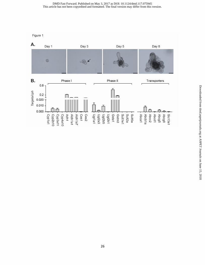
Development of crypt organoids and the expression of drug metabolism enzymes. Each
crypt culture contained multiple cells and normally formed spherical structures on the first day
after the incubation (Fig 1A), followed by budding. Small buds usually could be visualized in two
to four days after the incubation. The crypt region underwent continuous budding events; by day
5 after incubation, the lumen of organoids became distorted and dark, which were filled with
matured enterocytes or apoptotic cells (Fig 1A). Well-developed organoids were collected
between day 7-10 after cells were passaged, and RT-qPCR was conducted to investigate gene
expression patterns. As demonstrated in Figure 1B, Phase I and Phase II XPGs along with
transporter genes were identified in mature crypt organoids. Cytochrome P450 (Cyp) gene
expression for Cyp2b10 and Cyp3a11 along with other XPGs including alcohol dehydrogenase
Adh1, aldehyde dehydrogenase Aldh1a1 and Aldh1a7 were actively expressed, all of which have
been known to exhibit high expression levels in intestine (Vaglenova et al., 2003; Huang et al.,
2009; Renaud et al., 2011). In comparison to carboxylesterase (Ces) 1, Ces2 is known as the
intestinal specific isoform (Hatfield et al., 2011), which is consistent with our results, although RT-
qPCR results cannot provide absolute gene expression levels. Phase II XPGs were highlighted
by gene expression of Gst in addition to the Ugts encoded by the Ugt1 and Ugt2 gene families,
both of which are intestinal enriched genes (Gibbs et al., 1998; Strassburg et al., 1999; Shin et
al., 2009). Efflux drug transporters were also identified in crypt organoids, such as P-glycoprotein
(Pgp, Abcb1a) and Mrp2 (Abcc2), both of which are important transporters of many drugs and
xenobiotics in enterocytes (Sparreboom et al., 1997; Mottino et al., 2000), along with intestinal
specific transporters Abcg5/8 (Berge et al., 2000), which are important sterol efflux pumps.
Functional nuclear receptors observed in organoids that respond to xenobiotic treatment.
Fundamental to understanding the regulatory events associated with DMEs has been a clear
vision characterizing the role of the xenobiotic nuclear receptors (XNRs) in control and expression
This article has not been copyedited and formatted. The final version may differ from this version.DMD Fast Forward. Published on May 3, 2017 as DOI: 10.1124/dmd.117.075945
at ASPE
T Journals on June 15, 2018
dmd.aspetjournals.org
Dow
nloaded from
DMD Manuscript #75945
11
of the xenobiotic processing genes (XPGs) (Rushmore and Kong, 2002; Staudinger et al., 2010;
Chai et al., 2013; Staudinger et al., 2013; Hoffmann and Partridge, 2015). Xenobiotic and
environmental toxicant exposure has the potential to modify XPG expression through interactions
with XNRs. Altered gene expression through activation of XNRs affects drug metabolism as well
as drug-drug interactions, leading to changes in drug efficacy and safety. The gene expression
levels of XNRs in crypt organoids were determined, as shown in Figure 2A. We examined
expression of the genes encoding the aryl hydrocarbon receptor (Ahr), pregnane X receptor (Pxr),
constitutive androstane receptor (Car), peroxisome proliferator-activated receptors (Ppar),
retinoid X receptor (Rxr), and the liver X receptor (Lxr), and RNA encoded by each gene was
detectable. These gene expression patterns are relative to Cph gene expression and do not
reflect comparative expression values of RNA between the genes, since the primers for each
gene may have differences in their annealing properties. To determine their functionality, crypt
organoids were then treated with specific XNR ligands and downstream target gene expression
evaluated. TCDD treatment, the prototypical agonist of the AhR (Quattrochi and Tukey, 1993),
induced Cyp1a1 gene expression (Fig 2B); BNF, an AhR agonist (Sinal et al., 1999), also induced
Cyp1a1 (Fig 2B). Similarly, when crypt organoids were treated with TCPOBOP, a mouse specific
CAR agonist (Tzameli et al., 2000), Cyp2b10 was induced (Fig 2C). Cyp3a11 gene expression
was induced by PCN (Fig 2D), a well-studied PXR ligand (Guo et al., 2002). We observed that
Cyp3a11 was also induced by TCPOBOP and the LXR agonist T0901317, implying that Cyp3a11
gene expression can also be regulated by activated CAR and LXR. Cyp4a10 was largely induced
by the PPARα agonist WY14643 (Fig 2E). Drug transporter genes were also inducible, as shown
in Fig 2F with induction of Abca1 gene expression by the LXR agonist T0901317.
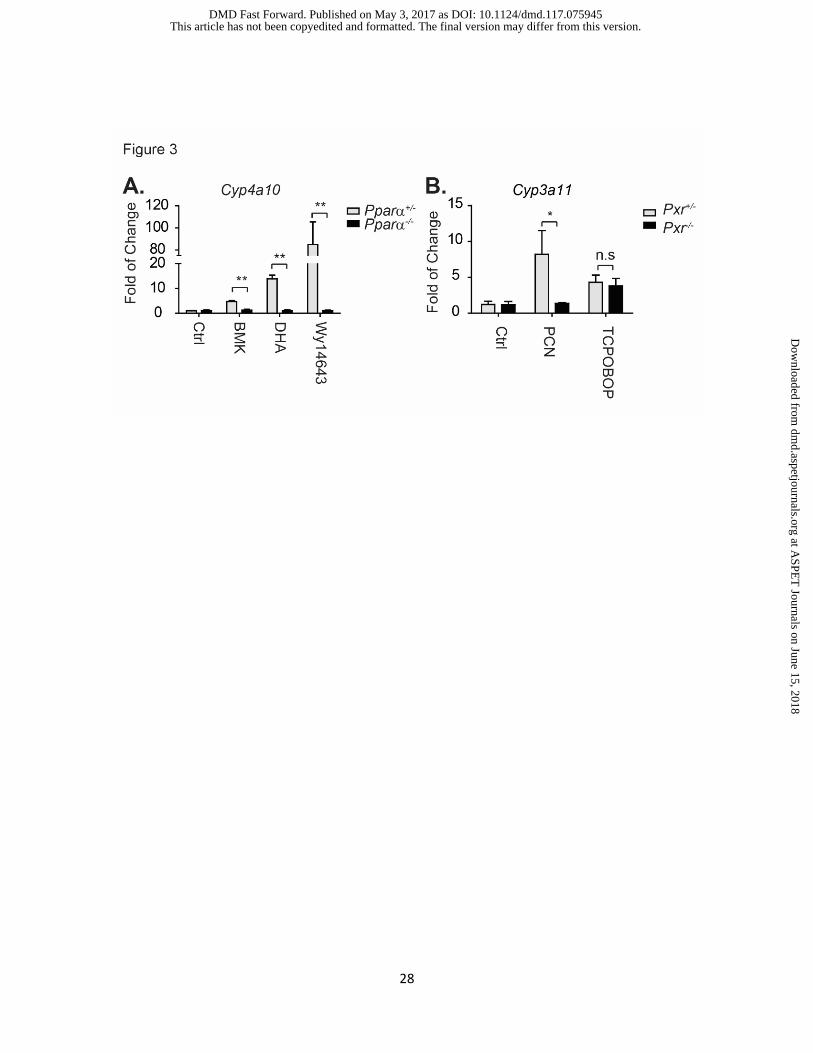
Crypt organoids isolated from XNR-knockout mice for XPG expression studies. Knockout
(KO) mouse models have been used to understand the physiological and pharmacological role of
a specific gene. Knockout mouse models have also been exploited in drug metabolism studies,
This article has not been copyedited and formatted. The final version may differ from this version.DMD Fast Forward. Published on May 3, 2017 as DOI: 10.1124/dmd.117.075945
at ASPE
T Journals on June 15, 2018
dmd.aspetjournals.org
Dow
nloaded from

DMD Manuscript #75945
12
particularly in elucidating XNR-mediated gene regulation of XPGs. Here we have utilized both
global and intestinal specific KO mouse models to investigate the utilization of organoid cultures
in drug metabolism studies. Crypt cells were isolated from both wildtype and Pparα-/- mice, and
after 5 days the organoids were treated with breast milk (1:100 dilution), the fatty acid DHA at 5
µM, and the PPARα agonist WY14643 at 100 µM. Twenty-four hours after treatment, cells were
collected for RT-qPCR analysis. In wildtype cells, breast milk, DHA and WY14643 activated gene
expression of the PPARα targeted gene Cyp4a10; however, these inductions were completely
diminished in crypt organoids isolated from Pparα-/- mice (Fig 3A). Similarly, PCN also lost its
capability in mediating the Cyp3a11 induction in Pxr-/- organoids (Fig 3B). These results confirm
that crypt organoids maintain the genetic characteristics that are exhibited in KO mice following
activation of XNRs.
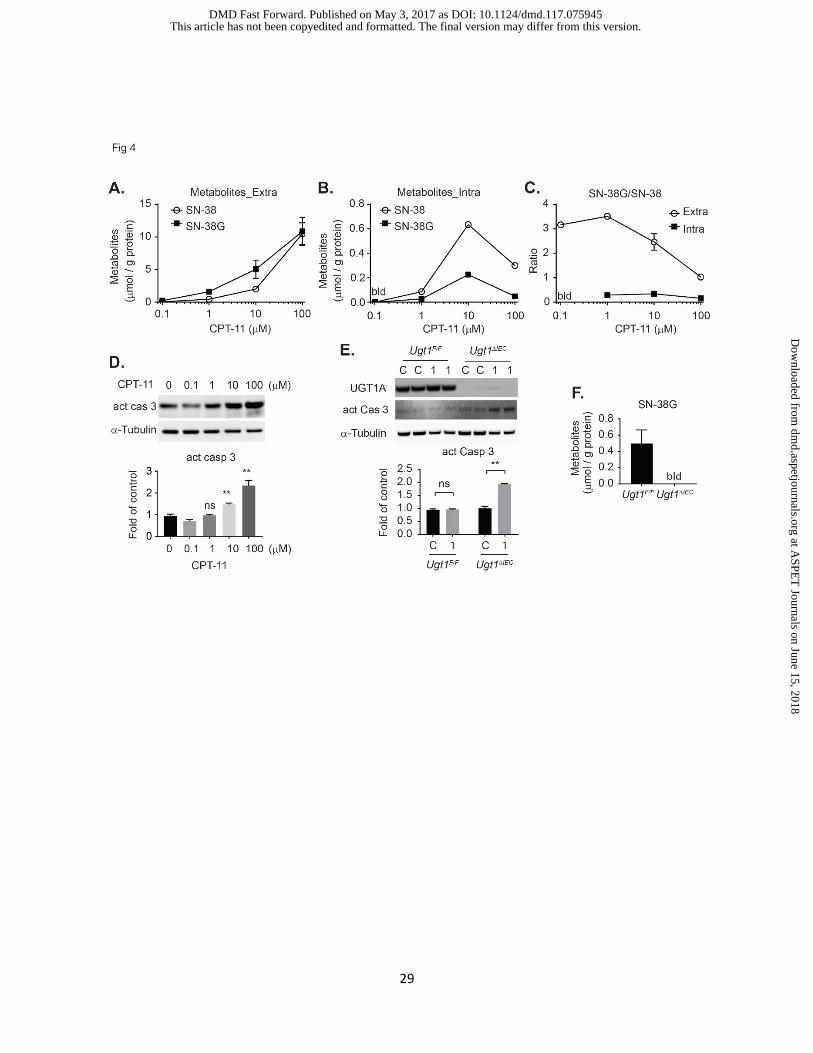
Intestinal crypt cultures metabolize CPT-11. Stable crypt organoids express CES1 & 2,
UGT1A1, and the transporters ABCC2 and ABCB1A based upon gene expression analysis. CPT-
11 enters intestinal epithelial cells and is cleaved by CES to the active topoisomerase inhibitor
SN-38, a product that is toxic and damaging to tissues if not metabolized further by
glucuronidation. To determine if intestinal crypt cultures could metabolize CPT-11 and culminate
with the formation of the SN-38 glucuronide (SN-38G), cells were first treated with a range of
CPT-11 concentrations (Fig 4A). After exposure for 24 h, culture media (extracellular) and cells
(intracellular) were harvested for quantitation of SN-38 and SN-38G by HPLC-MS analysis. In
culture media, as the concentration of CPT-11 was increased there was a concordant increase in
SN-38 and SN-38G (Fig 4A). The concentrations of SN-38 fit a linear model with R-squared at
0.99, but the SN-38G levels showed poor linearity with R-squared at 0.84 indicating enzymatic
saturation (Fig 4A). As the concentration of CPT-11 is increased, there is the rapid appearance
of intracellular SN-38 at 10 μM CPT-11 that is nearly triple the concentration of intracellular SN-
38G (Fig 4B), supporting the extracellular values that implicate UGT1A1 saturation. When the
This article has not been copyedited and formatted. The final version may differ from this version.DMD Fast Forward. Published on May 3, 2017 as DOI: 10.1124/dmd.117.075945
at ASPE
T Journals on June 15, 2018
dmd.aspetjournals.org
Dow
nloaded from

DMD Manuscript #75945
13
ratios of SN-38G/SN-38 were determined in both compartments, the ratios were much higher in
the extracellular compartment, indicating that both SN-38 and SN-38G are removed from the cell
by efflux transporters (Fig 4C). The highest concentration of CPT-11 used resulted in a sharp
decline in both the extracellular and intracellular SN-38G/SN-38 ratios, indicating that enzymatic
cleavage of CPT-11 to SN-38 and the resulting glucuronidation of SN-38 to SN-38G were
declining. Because SN-38 is toxic, this result corresponded to an increase in cell death, as
demonstrated by induction of cleaved caspase 3, a marker of cell apoptosis (Fig 4D).
In the absence of glucuronidation, the intestinal epithelium is sensitive to the toxic actions
of SN-38 (Chen et al., 2013). When we targeted the deletion the Ugt1 locus and the Ugt1a1 gene
in Ugt1ΔIEC mice and challenged Ugt1ΔIEC mice with CPT-11, they were highly susceptible to
diarrhea in comparison to wild type Ugt1F/F mice (Chen et al., 2013). Apoptosis was not observed
in Ugt1F/F crypt cells when treated with CPT-11 at 1 µM, but apoptosis was apparent in crypt
organoids from Ugt1ΔIEC mice at this concentration (Fig 4E), in which cells showed no expression
of UGT1 protein (Fig 4E) and failed to form the metabolite SN-38G (Fig 4F). Therefore, crypt
organoids from Ugt1ΔIEC mice were at least 10-fold more sensitive to the toxic actions of CPT-11
than Ugt1F/F mice. Crypt organoid cultures can be employed to examine the metabolic outcome
of drug and xenobiotic metabolism, the outcome of metabolism following XNR activation, and the
disposition of metabolism under conditions of altered pharmacogenetics.
This article has not been copyedited and formatted. The final version may differ from this version.DMD Fast Forward. Published on May 3, 2017 as DOI: 10.1124/dmd.117.075945
at ASPE
T Journals on June 15, 2018
dmd.aspetjournals.org
Dow
nloaded from
DMD Manuscript #75945
14
Discussion
Recent advances in establishing long-term culture conditions of crypt-villi that propagate
into functional organoids with intact luminal surfaces have made it possible to investigate intestinal
epithelial cell functionality in tissue culture (Sato et al., 2009; Sato and Clevers, 2013). As
intestinal epithelial cells turn over in adults, for example, every 5 days in mice, new epithelial cells
are formed from long-lived intestinal stem cells (ISCs) that sit at the base of the crypts (Barker et
al., 2008). These stem cells produce the rapidly proliferating progenitor cells called transit-
amplifying (TA) cells that migrate up the crypt-villus axis and eventually differentiate into mature
epithelial cells. With discovery of the signaling pathways that are necessary to maintain stemness
leading to epithelial cell proliferation, environmental cues have been employed to promote the
crypt ISCs to form mini-gut cultures that are composed of budding epithelial cells (Sato et al.,
2009; Sato et al., 2011; Sato and Clevers, 2013). These 3D mini-gut organoid cultures resemble
in vivo intestinal epithelial structures, with a single ISC producing the different types of
differentiated cells needed to support the development of the mature enterocytes (Sato et al.,
2009; Sato et al., 2011). It has also been demonstrated that the organoids maintain absorptive
and digestive functions making the tissue an excellent model to examine drug uptake and
metabolism and the impact of xenobiotic exposure on epithelial cell function. Taking advantage
of this unique physiology, we explored the use of 3D-mini-gut organoids to examine XPG
expression, inducibility and metabolism.
Within 8 days of culturing crypt stem cells, the expanding organoids show well-structured
epithelial cell projections. Constitutive expression of XPGs was abundant, with those encoding
the classically identified Phase 1, Phase II and Phase III (transporters) genes well represented.
The culture medium contains high concentrations of N-acetylcysteine, which is a substrate in the
synthesis of glutathione (Weinander et al., 1994). Elevated glutathione concentrations provide a
rich reductive capacity for the cell, indicating that organoid cultures may be an excellent model to
This article has not been copyedited and formatted. The final version may differ from this version.DMD Fast Forward. Published on May 3, 2017 as DOI: 10.1124/dmd.117.075945
at ASPE
T Journals on June 15, 2018
dmd.aspetjournals.org
Dow
nloaded from
DMD Manuscript #75945
15
examine receptor mediated induction of XPGs by drugs and xenobiotics without producing
excessive oxidative stress. The major XNRs that are targeted for activation by drugs and
xenobiotics are also expressed, and include the AhR, PXR, CAR, PPARα and LXR, which are
key modulators of gene expression that often control the balance between exposures resulting in
a toxic or efficacious outcome. Expression of the XNRs is functional in the proliferating organoids,
as we demonstrated by receptor mediated induction of key genes linked to drug transport and
metabolism. Similar results have recently been demonstrated (Bijsmans et al., 2017). There was
high specificity for activation of the AhR by TCDD and induction of Cyp1a1 gene expression
(Strom et al., 1992). While BNF is an agonist of the AhR, it also induces Cyp1a1, but the response
is not as potent as TCDD (Sinal et al., 1999). Interestingly, we did not notice any unusual growth
inhibition properties of our proliferating organoids following TCDD treatment, as was noted in a
recent publication examining growth properties following activation of the AhR (Park et al., 2016).
These differences may result from the concentrations of TCDD used; while we found excellent
induction using 50 nM, Park et al (Park et al., 2016). treated their organoid preparations with 0.1
to 1.0 μM. The mouse Cyp2b10 gene is selectively induced by mouse specific CAR ligands
(Honkakoski et al., 1998). Activation of PXR by PCN generates robust induction of Cyp3a11 gene
expression. Activation of CAR has been shown previously to induce Cyp3a11 (Hernandez et al.,
2009), corroborating our results with crypt organoids demonstrating induction of Cyp3a11 gene
expression by the CAR activator TCPOBOP. This is further supported when crypt organoids
deficient in PXR exhibit elevated Cyp3a11 gene expression when treated with TCPOBOP. In
addition, activation of LXR by T0901317, which targets activation of Abca1 gene expression
(Wagner et al., 2003), can also support induction of Cyp3a11 gene expression. These findings
indicate that Cyp3a11 can be regulated following activation of PXR, CAR and LXR. We have
shown previously that oral administration to mice with the PPARα agonist WY14643 induces
intestinal CYP4A protein expression (Senekeo-Effenberger et al., 2007). Significant activation of
This article has not been copyedited and formatted. The final version may differ from this version.DMD Fast Forward. Published on May 3, 2017 as DOI: 10.1124/dmd.117.075945
at ASPE
T Journals on June 15, 2018
dmd.aspetjournals.org
Dow
nloaded from
DMD Manuscript #75945
16
Cyp4a10 gene expression in organoid cultures is also documented following exposure with
WY14643, a response which is eliminated in cultures derived from PPARα-null mice.
The functionality of the crypt organoid cultures can be exploited for metabolism and
toxicology studies by implementing cultures derived from XNR and XPG knockout mice. We
demonstrated this capability using Ugt1ΔIEC mice, where we have targeted the specific deletion of
the Ugt1 locus in intestinal epithelial cells. These mice are deficient in the UGT1A proteins,
including UGT1A1. Characteristic of crypt organoids and intestinal epithelial cells, they express
CES2, necessary to cleave the prodrug CPT-11 to SN-38, the active topoisomerase 1 inhibitor.
The high expression levels of CES2 is an excellent example that documents the maintenance of
tissue specific expression markers that can be exploited to examine drug metabolism. It supports
previous reports from Clevers (Sato et al., 2009; Sato and Clevers, 2013) and more recently the
van Mills group (Bijsmans et al., 2017) that gene expression profiles in intestinal organoids
resemble patterns observed in normal tissue. We had shown previously in Ugt1ΔIEC mice that
treatment with CPT-11 led to severe intestinal damage, resulting from the efficient generation of
SN-38 coupled with the inability to generate UGT1A1 dependent SN-38G (Chen et al., 2013).
Organoids generated from Ugt1F/F mice efficiently generated SN-38 and SN-38G, with cell
apoptosis being evident at 10 to 100 μM CPT-11. When organoids from Ugt1ΔIEC mice were
treated with the same concentrations of CPT-11, remarkable cell apoptosis was evident at 1 μM
CPT-11. These findings provide evidence that examining drug metabolism in crypt organoids can
be efficiently linked to specific XPGs while confirming a mechanistic link between gene expression
and toxicity.
In conclusion, crypt organoids maintain functional, tissue specific gene expression
encoding DMEs, and can be further regulated following activation of XNRs. Crypt organoids
developed from stem cells isolated from genetically modified mice maintain their engineered
phenotypical features, as determined both at the gene expression and functional levels. It is
This article has not been copyedited and formatted. The final version may differ from this version.DMD Fast Forward. Published on May 3, 2017 as DOI: 10.1124/dmd.117.075945
at ASPE
T Journals on June 15, 2018
dmd.aspetjournals.org
Dow
nloaded from

DMD Manuscript #75945
17
argued that the use of organoid cultures may eventually reduce the need for animal models in
scientific research, since the cultures maintain tissue specific gene expression which can be
regulated following activation of XNRs (Clevers, 2013; Bijsmans et al., 2017). While the organoid
cultures are a superior model when compared to primary cells or long term differentiated cells,
there will always be the need for drug testing in vivo. An example of this need is to define the
mechanisms leading to regulation of intestinal UGT1A1 gene expression during development.
Neonatal jaundice and severe forms of neonatal hyperbilirubinemia develop because of delayed
expression of the UGT1A1 gene during development, and its expression in the GI tract is critical
in preventing bilirubin toxicity. Agents that activate many of the XNRs when given orally to
neonatal hUGT1 mice induce intestinal UGT1A1 and reduce serum bilirubin. These same agents
can induce the UGT1A1 gene in tissue culture. However, we have discovered that intestinal
UGT1A1 gene expression is very responsive to agents that induce mild oxidative stress (Liu et
al., 2016). This new mechanism, which can be replicated only in vivo, may lead to the identification
of new agents or therapeutics that may be considered as new therapy in the future to prevent the
substantial mortality and permanent morbidities resulting from severe neonatal hyperbilirubinemia,
which is estimated to impact over 1-million newborns every year (Wong et al., 2011; Bhutani et
al., 2013). Thus, crypt organoids could become a valuable tool in drug metabolism and toxicity
studies to complement those efforts being conducted with humanized and genetically modified
mice.
This article has not been copyedited and formatted. The final version may differ from this version.DMD Fast Forward. Published on May 3, 2017 as DOI: 10.1124/dmd.117.075945
at ASPE
T Journals on June 15, 2018
dmd.aspetjournals.org
Dow
nloaded from
DMD Manuscript #75945
18
Acknowledgments
We thank Dr. Kepeng Wang from Dr. Michael Karin’s laboratory at UCSD for helping with the
crypt organoid culture. We also want to thank Dr. Calvin Kuo, University of Stanford, for his
generous gift of the 293-HA-Rspol-Fc cell line for obtaining R-Spondin 1 conditioned medium.
Authorship Contribution
Participate in research design: Chen.
Conduct experiments: Lu, Rettenmeier, Paszek, Trottier, Barbier, Chen.
Contributed new reagents or analytic tools: Trottier, Barbier.
Performed data analysis: Lu, Rettenmeier, Trottier, Barbier, Chen.
Wrote and contributed to the writing of the manuscript: all authors.
This article has not been copyedited and formatted. The final version may differ from this version.DMD Fast Forward. Published on May 3, 2017 as DOI: 10.1124/dmd.117.075945
at ASPE
T Journals on June 15, 2018
dmd.aspetjournals.org
Dow
nloaded from

DMD Manuscript #75945
19
References
Alimonti A, Gelibter A, Pavese I, Satta F, Cognetti F, Ferretti G, Rasio D, Vecchione A, and Di Palma M (2004) New approaches to prevent intestinal toxicity of irinotecan-based regimens. Cancer Treat Rev 30:555-562.
Barker N, van de Wetering M, and Clevers H (2008) The intestinal stem cell. Genes Dev 22:1856-1864. Berge KE, Tian H, Graf GA, Yu L, Grishin NV, Schultz J, Kwiterovich P, Shan B, Barnes R, and Hobbs HH
(2000) Accumulation of dietary cholesterol in sitosterolemia caused by mutations in adjacent ABC transporters. Science 290:1771-1775.
Bhutani VK, Zipursky A, Blencowe H, Khanna R, Sgro M, Ebbesen F, Bell J, Mori R, Slusher TM, Fahmy N, Paul VK, Du L, Okolo AA, de Almeida MF, Olusanya BO, Kumar P, Cousens S, and Lawn JE (2013) Neonatal hyperbilirubinemia and Rhesus disease of the newborn: incidence and impairment estimates for 2010 at regional and global levels. Pediatr Res 74 Suppl 1:86-100.
Bijsmans IT, Milona A, Ijssennagger N, Willemsen EC, Ramos Pittol JM, Jonker JW, Lange K, Hooiveld GJ, and van Mil SW (2017) Characterization of stem cell-derived liver and intestinal organoids as a model system to study nuclear receptor biology. Biochim Biophys Acta 1863:687-700.
Chai X, Zeng S, and Xie W (2013) Nuclear receptors PXR and CAR: implications for drug metabolism regulation, pharmacogenomics and beyond. Expert Opin Drug Metab Toxicol 9:253-266.
Chen S, Yueh MF, Bigo C, Barbier O, Wang K, Karin M, Nguyen N, and Tukey RH (2013) Intestinal glucuronidation protects against chemotherapy-induced toxicity by irinotecan (CPT-11). Proc Natl Acad Sci U S A 110:19143-19148.
Clevers H (2013) The intestinal crypt, a prototype stem cell compartment. Cell 154:274-284. Gibbs JP, Yang JS, and Slattery JT (1998) Comparison of human liver and small intestinal glutathione S-
transferase-catalyzed busulfan conjugation in vitro. Drug Metab Dispos 26:52-55. Grabinger T, Luks L, Kostadinova F, Zimberlin C, Medema JP, Leist M, and Brunner T (2014) Ex vivo
culture of intestinal crypt organoids as a model system for assessing cell death induction in intestinal epithelial cells and enteropathy. Cell Death Dis 5:e1228.
Guo GL, Staudinger J, Ogura K, and Klaassen CD (2002) Induction of rat organic anion transporting polypeptide 2 by pregnenolone-16alpha-carbonitrile is via interaction with pregnane X receptor. Mol Pharmacol 61:832-839.
Hatfield MJ, Tsurkan L, Garrett M, Shaver TM, Hyatt JL, Edwards CC, Hicks LD, and Potter PM (2011) Organ-specific carboxylesterase profiling identifies the small intestine and kidney as major contributors of activation of the anticancer prodrug CPT-11. Biochem Pharmacol 81:24-31.
Hernandez JP, Mota LC, Huang W, Moore DD, and Baldwin WS (2009) Sexually dimorphic regulation and induction of P450s by the constitutive androstane receptor (CAR). Toxicology 256:53-64.
Hoffmann JM and Partridge L (2015) Nuclear hormone receptors: Roles of xenobiotic detoxification and sterol homeostasis in healthy aging. Crit Rev Biochem Mol Biol 50:380-392.
Honkakoski P, Zelko I, Sueyoshi T, and Negishi M (1998) The nuclear orphan receptor CAR-retinoid X receptor heterodimer activates the phenobarbital-responsive enhancer module of the CYP2B gene. Mol Cell Biol 18:5652-5658.
Huang EH, Hynes MJ, Zhang T, Ginestier C, Dontu G, Appelman H, Fields JZ, Wicha MS, and Boman BM (2009) Aldehyde dehydrogenase 1 is a marker for normal and malignant human colonic stem cells (SC) and tracks SC overpopulation during colon tumorigenesis. Cancer Res 69:3382-3389.
Iyer L, King CD, Whitington PF, Green MD, Roy SK, Tephly TR, Coffman BL, and Ratain MJ (1998) Genetic predisposition to the metabolism of irinotecan (CPT-11). Role of uridine diphosphate glucuronosyltransferase isoform 1A1 in the glucuronidation of its active metabolite (SN-38) in human liver microsomes. J Clin Invest 101:847-854.
This article has not been copyedited and formatted. The final version may differ from this version.DMD Fast Forward. Published on May 3, 2017 as DOI: 10.1124/dmd.117.075945
at ASPE
T Journals on June 15, 2018
dmd.aspetjournals.org
Dow
nloaded from
DMD Manuscript #75945
20
Kaneda N, Nagata H, Furuta T, and Yokokura T (1990) Metabolism and pharmacokinetics of the camptothecin analogue CPT-11 in the mouse. Cancer Res 50:1715-1720.
Lee SS, Pineau T, Drago J, Lee EJ, Owens JW, Kroetz DL, Fernandez-Salguero PM, Westphal H, and Gonzalez FJ (1995) Targeted disruption of the alpha isoform of the peroxisome proliferator-activated receptor gene in mice results in abolishment of the pleiotropic effects of peroxisome proliferators. Mol Cell Biol 15:3012-3022.
Liu M, Chen S, Yueh MF, Fujiwara R, Konopnicki C, Hao H, and Tukey RH (2016) Cadmium and arsenic override NF-kappaB developmental regulation of the intestinal UGT1A1 gene and control of hyperbilirubinemia. Biochem Pharmacol 110-111:37-46.
Marangon E, Posocco B, Mazzega E, and Toffoli G (2015) Development and validation of a high-performance liquid chromatography-tandem mass spectrometry method for the simultaneous determination of irinotecan and its main metabolites in human plasma and its application in a clinical pharmacokinetic study. PLoS One 10:e0118194.
Michael M, Thompson M, Hicks RJ, Mitchell PL, Ellis A, Milner AD, Di Iulio J, Scott AM, Gurtler V, Hoskins JM, Clarke SJ, Tebbut NC, Foo K, Jefford M, and Zalcberg JR (2006) Relationship of hepatic functional imaging to irinotecan pharmacokinetics and genetic parameters of drug elimination. J Clin Oncol 24:4228-4235.
Mottino AD, Hoffman T, Jennes L, and Vore M (2000) Expression and localization of multidrug resistant protein mrp2 in rat small intestine. J Pharmacol Exp Ther 293:717-723.
Ootani A, Li X, Sangiorgi E, Ho QT, Ueno H, Toda S, Sugihara H, Fujimoto K, Weissman IL, Capecchi MR, and Kuo CJ (2009) Sustained in vitro intestinal epithelial culture within a Wnt-dependent stem cell niche. Nat Med 15:701-706.
Park JH, Choi AJ, Kim SJ, Cheong SW, and Jeong SY (2016) AhR activation by 6-formylindolo[3,2-b]carbazole and 2,3,7,8-tetrachlorodibenzo-p-dioxin inhibit the development of mouse intestinal epithelial cells. Environ Toxicol Pharmacol 43:44-53.
Quattrochi LC and Tukey RH (1993) Nuclear uptake of the Ah (dioxin) receptor in response to omeprazole: transcriptional activation of the human CYP1A1 gene. Mol Pharmacol 43:504-508.
Ranga A, Gjorevski N, and Lutolf MP (2014) Drug discovery through stem cell-based organoid models. Adv Drug Deliv Rev 69-70:19-28.
Renaud HJ, Cui JY, Khan M, and Klaassen CD (2011) Tissue distribution and gender-divergent expression of 78 cytochrome P450 mRNAs in mice. Toxicol Sci 124:261-277.
Rushmore TH and Kong AN (2002) Pharmacogenomics, regulation and signaling pathways of phase I and II drug metabolizing enzymes. Curr Drug Metab 3:481-490.
Sato T and Clevers H (2013) Growing self-organizing mini-guts from a single intestinal stem cell: mechanism and applications. Science 340:1190-1194.
Sato T, van Es JH, Snippert HJ, Stange DE, Vries RG, van den Born M, Barker N, Shroyer NF, van de Wetering M, and Clevers H (2011) Paneth cells constitute the niche for Lgr5 stem cells in intestinal crypts. Nature 469:415-418.
Sato T, Vries RG, Snippert HJ, van de Wetering M, Barker N, Stange DE, van Es JH, Abo A, Kujala P, Peters PJ, and Clevers H (2009) Single Lgr5 stem cells build crypt-villus structures in vitro without a mesenchymal niche. Nature 459:262-265.
Senekeo-Effenberger K, Chen S, Brace-Sinnokrak E, Bonzo JA, Yueh MF, Argikar U, Kaeding J, Trottier J, Remmel RP, Ritter JK, Barbier O, and Tukey RH (2007) Expression of the human UGT1 locus in transgenic mice by 4-chloro-6-(2,3-xylidino)-2-pyrimidinylthioacetic acid (WY-14643) and implications on drug metabolism through peroxisome proliferator-activated receptor alpha activation. Drug Metab Dispos 35:419-427.
This article has not been copyedited and formatted. The final version may differ from this version.DMD Fast Forward. Published on May 3, 2017 as DOI: 10.1124/dmd.117.075945
at ASPE
T Journals on June 15, 2018
dmd.aspetjournals.org
Dow
nloaded from
DMD Manuscript #75945
21
Shin HC, Kim HR, Cho HJ, Yi H, Cho SM, Lee DG, Abd El-Aty AM, Kim JS, Sun D, and Amidon GL (2009) Comparative gene expression of intestinal metabolizing enzymes. Biopharm Drug Dispos 30:411-421.
Sinal CJ, Webb CD, and Bend JR (1999) Differential in vivo effects of alpha-naphthoflavone and beta-naphthoflavone on CYP1A1 and CYP2E1 in rat liver, lung, heart, and kidney. J Biochem Mol Toxicol 13:29-40.
Sparreboom A, van Asperen J, Mayer U, Schinkel AH, Smit JW, Meijer DK, Borst P, Nooijen WJ, Beijnen JH, and van Tellingen O (1997) Limited oral bioavailability and active epithelial excretion of paclitaxel (Taxol) caused by P-glycoprotein in the intestine. Proc Natl Acad Sci U S A 94:2031-2035.
Staudinger JL, Woody S, Sun M, and Cui W (2013) Nuclear-receptor-mediated regulation of drug- and bile-acid-transporter proteins in gut and liver. Drug Metab Rev 45:48-59.
Staudinger JL, Xu C, Cui YJ, and Klaassen CD (2010) Nuclear receptor-mediated regulation of carboxylesterase expression and activity. Expert Opin Drug Metab Toxicol 6:261-271.
Strassburg CP, Nguyen N, Manns MP, and Tukey RH (1999) UDP-glucuronosyltransferase activity in human liver and colon. Gastroenterology 116:149-160.
Strom DK, Postlind H, and Tukey RH (1992) Characterization of the rabbit CYP1A1 and CYP1A2 genes: developmental and dioxin-inducible expression of rabbit liver P4501A1 and P4501A2. Arch Biochem Biophys 294:707-716.
Sugimoto Y, Tsukahara S, Oh-hara T, Isoe T, and Tsuruo T (1990) Decreased expression of DNA topoisomerase I in camptothecin-resistant tumor cell lines as determined by a monoclonal antibody. Cancer Res 50:6925-6930.
Tzameli I, Pissios P, Schuetz EG, and Moore DD (2000) The xenobiotic compound 1,4-bis[2-(3,5-dichloropyridyloxy)]benzene is an agonist ligand for the nuclear receptor CAR. Mol Cell Biol 20:2951-2958.
Ueda A, Hamadeh HK, Webb HK, Yamamoto Y, Sueyoshi T, Afshari CA, Lehmann JM, and Negishi M (2002) Diverse roles of the nuclear orphan receptor CAR in regulating hepatic genes in response to phenobarbital. Mol Pharmacol 61:1-6.
Vaglenova J, Martinez SE, Porte S, Duester G, Farres J, and Pares X (2003) Expression, localization and potential physiological significance of alcohol dehydrogenase in the gastrointestinal tract. Eur J Biochem 270:2652-2662.
van Herwaarden AE, Wagenaar E, van der Kruijssen CM, van Waterschoot RA, Smit JW, Song JY, van der Valk MA, van Tellingen O, van der Hoorn JW, Rosing H, Beijnen JH, and Schinkel AH (2007) Knockout of cytochrome P450 3A yields new mouse models for understanding xenobiotic metabolism. J Clin Invest 117:3583-3592.
van Waterschoot RA, Rooswinkel RW, Wagenaar E, van der Kruijssen CM, van Herwaarden AE, and Schinkel AH (2009) Intestinal cytochrome P450 3A plays an important role in the regulation of detoxifying systems in the liver. FASEB J 23:224-231.
van Waterschoot RA, van Herwaarden AE, Lagas JS, Sparidans RW, Wagenaar E, van der Kruijssen CM, Goldstein JA, Zeldin DC, Beijnen JH, and Schinkel AH (2008) Midazolam metabolism in cytochrome P450 3A knockout mice can be attributed to up-regulated CYP2C enzymes. Mol Pharmacol 73:1029-1036.
Wagner BL, Valledor AF, Shao G, Daige CL, Bischoff ED, Petrowski M, Jepsen K, Baek SH, Heyman RA, Rosenfeld MG, Schulman IG, and Glass CK (2003) Promoter-specific roles for liver X receptor/corepressor complexes in the regulation of ABCA1 and SREBP1 gene expression. Mol Cell Biol 23:5780-5789.
This article has not been copyedited and formatted. The final version may differ from this version.DMD Fast Forward. Published on May 3, 2017 as DOI: 10.1124/dmd.117.075945
at ASPE
T Journals on June 15, 2018
dmd.aspetjournals.org
Dow
nloaded from
DMD Manuscript #75945
22
Weinander R, Anderson C, and Morgenstern R (1994) Identification of N-acetylcysteine as a new substrate for rat liver microsomal glutathione transferase. A study of thiol ligands. J Biol Chem 269:71-76.
Wong RJ, Vreman HJ, Schulz S, Kalish FS, Pierce NW, and Stevenson DK (2011) In vitro inhibition of heme oxygenase isoenzymes by metalloporphyrins. J Perinatol 31 Suppl 1:S35-S41.
Xie W, Barwick JL, Downes M, Blumberg B, Simon CM, Nelson MC, Neuschwander-Tetri BA, Brunt EM, Guzelian PS, and Evans RM (2000) Humanized xenobiotic response in mice expressing nuclear receptor SXR. Nature 406:435-439.
Yamazaki Y, Kakizaki S, Horiguchi N, Takagi H, Mori M, and Negishi M (2005) Role of nuclear receptor CAR in carbon tetrachloride-induced hepatotoxicity. World J Gastroenterol 11:5966-5972.
Zanger UM, Turpeinen M, Klein K, and Schwab M (2008) Functional pharmacogenetics/genomics of human cytochromes P450 involved in drug biotransformation. Anal Bioanal Chem 392:1093-1108.
This article has not been copyedited and formatted. The final version may differ from this version.DMD Fast Forward. Published on May 3, 2017 as DOI: 10.1124/dmd.117.075945
at ASPE
T Journals on June 15, 2018
dmd.aspetjournals.org
Dow
nloaded from
DMD Manuscript #75945
23
Footnotes:
Grant Support: This work was supported by U.S. Public Health Service Grants through National
Institute of Environmental Health Sciences [ES024818 and ES010337] and the National Institute
of General Medical Sciences [GM086713].
Conflict of Interest: The authors have no conflict of interests.
This article has not been copyedited and formatted. The final version may differ from this version.DMD Fast Forward. Published on May 3, 2017 as DOI: 10.1124/dmd.117.075945
at ASPE
T Journals on June 15, 2018
dmd.aspetjournals.org
Dow
nloaded from
DMD Manuscript #75945
24
Legends for Figures
Figure1. The growth of small intestinal crypt organoids and the expressions of XPGs in
mature crypt organoids.
(A) Shown are mouse small intestinal crypt organoid cultures at various times after primary culture.
Images were taken with a Nikon Eclipse Ts2 inverted microscope, with the scale bar shown at 90
µm. (B) Mature crypt organoids (in triplicate) were collected 8 days after plating and RNA prepared
for RT-qPCR analysis. Gene expression of Phase I and Phase II drug metabolism genes and
transporters are expressed as Target genes/mouse Cph and are described as average ± SEM
(n=3). Data represent multiple independent experiments with similar results.
Figure 2. Gene expression analysis of NRs and their function in mature crypt cells.
(A) Shown are nuclear receptor expression levels from mature crypt cells conducted by RT-qPCR
analysis, average ± SEM (n=3). (B-F) Crypts in exponential growth period were treated with
different nuclear receptor agonists, including TCDD (50 nM), βNF (20 µM), TCPOBOP (10 µM),
PCN (30 µM), T0901317 (100 µM), and WY14643 (100 µM). Twenty four hours after agonist
exposure, crypt cells were collected for RT-qPCR analysis (average ± SEM, experiments carried
out in triplicate).
Figure 3. RT-qPCR results of mature crypt cells from both wildtype and genetically
modified mice.
(A) Crypt cells were isolated from Pparα+/- (wild type) and Pparα-/- (Pparα deletion) mice. After
several passages, crypt cells were treated with vehicle control (Ctrl, DMSO at 0.1%), breast milk
(BMK) at 1:100 (v/v) dilution into the culture medium, DHA (5 µM), and WY14643 (100 µM) for 24
This article has not been copyedited and formatted. The final version may differ from this version.DMD Fast Forward. Published on May 3, 2017 as DOI: 10.1124/dmd.117.075945
at ASPE
T Journals on June 15, 2018
dmd.aspetjournals.org
Dow
nloaded from

DMD Manuscript #75945
25
h. Cells were collected for RT-qPCR analysis to measure Cyp4a10 gene expression. Results
were normalized with vehicle treated wild-type crypts and described as fold change (average ±
SEM, n=3). (B) Crypt cells were isolated from wild type Pxr+/- and Pxr-/- knockout mice. Cells were
treated with PCN at 30 µM and TCPOBOP at 10 µM for 24 h followed by Cyp3a11 gene
expression analyzed by RT-qPCR (n=3). ns: nonsignificant, * p<0.05, **p<0.01, student’s t-test.
Figure 4. Irinotecan metabolism and toxicity studies by utilizing crypt organoids from both
Ugt1F/F and Ugt1ΔIEC mice.
(A-D) Crypts were isolated from Ugt1F/F mice. Cells were treated with CPT-11 at 0, 0.1, 1, 10, and
100 µM (in triplicate). After 24 h, both media and cells were collected for HPLC-MS analysis. SN-
38 and SN-38G were determined in both (A) extracellular compartment, and (B) intracellular
compartment (cells from three wells were combined and then subjected to the bioanalysis), (C)
the ratio of SN-38G/SN-38. (D) Apoptosis was analyzed by Western blot of cleaved caspase 3,
which was further quantified by using the ChemiDoc Touch Imaging System and Image Lab
software (BioRad), and the data was calculated based on independent Western blots (average ±
SEM, n=3). (E-F) Crypts were isolated from both Ugt1F/F and Ugt1ΔIEC mice. After three days, cells
were treated with control or CPT-11 at 1 µM. (E) Twenty four hours later, whole cell extracts were
prepared for Western blot analysis using an anti-UGT1A, anti-active caspase 3, and anti α-Tubulin
antibodies. Western blots of cleaved caspase 3 were further quantified. (F) Cell culture media
was collected for bioanalysis of SN-38G (average ± SEM, n=3). bld, below limit of detection.
This article has not been copyedited and formatted. The final version may differ from this version.DMD Fast Forward. Published on May 3, 2017 as DOI: 10.1124/dmd.117.075945
at ASPE
T Journals on June 15, 2018
dmd.aspetjournals.org
Dow
nloaded from
26
This article has not been copyedited and formatted. The final version may differ from this version.DMD Fast Forward. Published on May 3, 2017 as DOI: 10.1124/dmd.117.075945
at ASPE
T Journals on June 15, 2018
dmd.aspetjournals.org
Dow
nloaded from

27
This article has not been copyedited and formatted. The final version may differ from this version.DMD Fast Forward. Published on May 3, 2017 as DOI: 10.1124/dmd.117.075945
at ASPE
T Journals on June 15, 2018
dmd.aspetjournals.org
Dow
nloaded from
28
This article has not been copyedited and formatted. The final version may differ from this version.DMD Fast Forward. Published on May 3, 2017 as DOI: 10.1124/dmd.117.075945
at ASPE
T Journals on June 15, 2018
dmd.aspetjournals.org
Dow
nloaded from
29
This article has not been copyedited and formatted. The final version may differ from this version.DMD Fast Forward. Published on May 3, 2017 as DOI: 10.1124/dmd.117.075945
at ASPE
T Journals on June 15, 2018
dmd.aspetjournals.org
Dow
nloaded from