CRYOTHERAPY AND CUTANEOUS BIOPSIES FOR … AND CUTANEOUS BIOPSIES FOR SOME COMMON DERMATOLOGIC...
66
CRYOTHERAPY AND CUTANEOUS BIOPSIES FOR SOME COMMON DERMATOLOGIC DIAGNOSES Daniel M. Peraza, MD Peraza Dermatology Group Adjunct Assistant Professor of Surgery— Dartmouth Medical School October 24, 2014
Transcript of CRYOTHERAPY AND CUTANEOUS BIOPSIES FOR … AND CUTANEOUS BIOPSIES FOR SOME COMMON DERMATOLOGIC...

CRYOTHERAPY AND CUTANEOUS
BIOPSIES FOR SOME COMMON
DERMATOLOGIC DIAGNOSES
Daniel M. Peraza, MD
Peraza Dermatology Group
Adjunct Assistant Professor of Surgery—Dartmouth Medical School
October 24, 2014

Macule and Patch
Macule
Small, flat, non-palpable lesion
Smaller than 5 mm in diameter (10
mm for others)
Lesion with color or subtle texture
change only
No elevation above skin surface
Patch
Flat, non-palpable lesion 5 mm in
diameter or larger (10mm for
others)
Some accept a slight amount of
scale as still acceptable in a patch

Papule
Small, superficial, circumscribed,
palpable lesion
Elevated above the skin surface
Less than 10 mm in diameter
(some use less than 5mm)

Plaque
Palpable lesion elevated above
the skin surface
10 mm or greater in diameter

Nodule
Firm (indurated) lesion that is thicker or
deeper than the average papule or
plaque
From the Latin word ‘nodulus’, meaning
‘knot’
If subcutaneous, may not elevate the
skin surface

Vesicle and Bullae
Vesicle
Elevated lesion that contains clear
fluid
Small blister less than 10 mm in
diameter
Bulla
Elevated lesion that contains clear
fluid
Blister more than 10 mm in
diameter
Pustule
Superficial elevated lesion
Contains yellow fluid (pus) within
or beneath the epidermis
Often protein-rich, containing
neutrophils

Special Descriptors
Annular
Serpiginous
Comedo
Cystic
Violaceous
Micaceous
Blah, blah, blah

NOW WHAT?

Cryosurgery (CS)
Very low temperatures applied to a
lesion resulting in local destruction
Extremely valuable alternative to
surgical options
Cost-effective with excellent
aesthetic results
Proper training EASY DELIVERY

Cryosurgery
Freeze with subzero temperature
Sloughing of damaged tissue
Depth of damage: technique and
freezing time
Structural changes due to heat loss
from cell (temperature flow from hot
to cold cell death)
GOAL: Low temperature in tissue by
freezing at a constant velocity with an
initial low temperature

Cryosurgery
Ice crystal formation extracellularly
Osmosis: water leaves cell
Intracellular dehydration
Water leaving cell eventually freezes
High freezing velocity: osmotic water movements
slower (internal crystallization)
Cell membrane destruction
If freezing not enough (low freezing velocity) only
ice formation extracellularly
Distortion and damage to cell, but sublethal

Cryosurgery
Ice crystals damage organelles and membranes
Slow thawing: extend time tissue at subzero temperature
Increase probability of intracellular ice formation and solute
damage

Cryosurgery
Total freeze time (30-60 sec), halo thaw
time, total thaw time
Useful indicators for measuring if freeze
adequate
Total freeze time < total thaw time
Golden cryosurgery rule: freeze fast, thaw
slowly
Freeze/thaw repeat cell is further
damaged because ice formation faster

Cryosurgery
General Rules of Thumb
1 freeze-thaw cycle: flat, benign
>1 freeze-thaw cycle: bulky benign or
malignant

Cryosurgery: Preoperative
MINIMAL
Useful and practical for treatment of older patients
Difficulty lying down on table
Wheelchair
Can’t leave home or nursing home
Safe for variety of medical conditions (bleeding issues, pacemaker, etc.)

Cryosurgery: Preoperative
KNOW WHAT YOU ARE
TREATING!
Skin biopsy to confirm, diagnose
type of lesion, and depth of
lesion
Guide treatment plan

Cryosurgery: Preoperative
Protect vital areas
Avoid metal coverings
Plastic, wood, or cotton

Cryosurgery: Technique
Open
Chamber
Closed technique
Open
Cryogen released through tips
Tip diameter
Intermittent use of cryogen
Distance from tip to target
Determine amount of cryogen to lesion

Cryosurgery: Technique
Open
Chamber
Closed technique
Open
Cryogen released through tips
Tip diameter
Intermittent use of cryogen
Distance from tip to target
Determine amount of cryogen to lesion

Cryosurgery
DIFFICULT to establish duration of
treatment! (APPROXIMATE)
Thicker lesions need to be treated longer
Keratin poor conductor

Cryosurgery
Cotton swabs should not be used!
Temperature control lost
Poor thermal capacity increase
risk of suboptimal temperature

Cryosurgery
Liquid nitrogen
Ideal cryogen
Safely transported, low cost, easy storage,
low temperature (-196°C)
Ideal temperature to destroy malignancies
-50°C to -60°C at periphery
Academic interest: tissue temperature
monitoring

Cryosurgery
Liquid nitrogen
Ideal cryogen
Safely transported, low cost, easy
storage, low temperature (-196°C)
Ideal temperature to destroy malignancies
-50°C to -60°C at periphery
Academic interest: tissue temperature
monitoring

Cryosurgery: What can be treated?

Cryosurgery
Papillomavirus
Keratin: poor cold conductor
Reduce lesion (keratolytic or shave off)
Freeze to several mm outside periphery

Cryosurgery
Papillomavirus
Keratin: poor cold conductor
Reduce lesion (keratolytic or shave off)
Freeze to several mm outside periphery

Cryosurgery
Papillomavirus
Keratin: poor cold conductor
Reduce lesion (keratolytic or shave off)
Freeze to several mm outside periphery

Cryosurgery
Papillomavirus
Keratin: poor cold conductor
Reduce lesion (keratolytic or shave off)
Freeze to several mm outside periphery

Cryosurgery Video
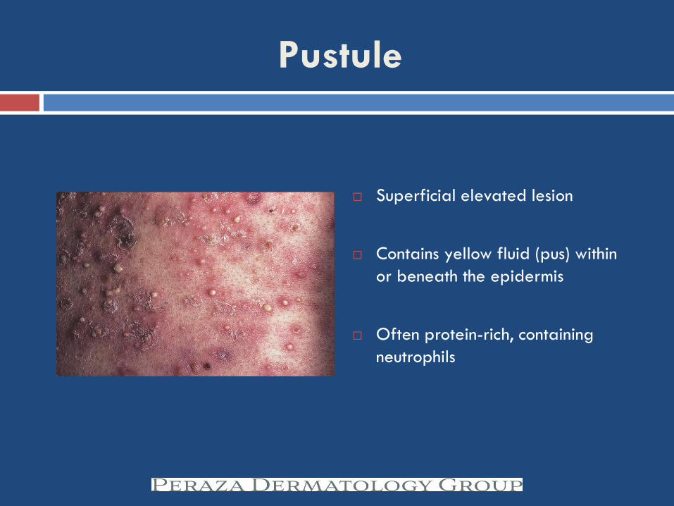
Cryosurgery
Molluscum
HIV - avoid bleeding
Aim at central dell

Cryosurgery
Seborrheic keratosis
Freeze: cover entire lesion plus 1-2mm
Thaw few seconds and then curette off

Cryosurgery
Seborrheic keratosis
Freeze: cover entire lesion plus 1-2mm
Thaw few seconds and then curette off

Cryosurgery
Lentigines and ephelides
Very cold sensitive
3-5 seconds from far away
Freeze halo barely advance outside edge
If not treated entirely residual pigment

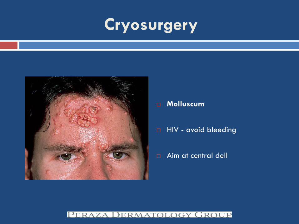
Cryosurgery
Actinic keratoses
5-10 seconds of intermittent freeze
Freeze: spread several mm past edge
Hyperkeratotic: increase freeze time
2 cycles!
Cryosurgery
Actinic keratoses
5-10 seconds of intermittent freeze
Freeze: spread several mm past edge
Hyperkeratotic: increase freeze time
2 cycles!

Cryosurgery
Actinic keratoses
5-10 seconds of intermittent freeze
Freeze: spread several mm past edge
Hyperkeratotic: increase freeze time
2 cycles!

Cryosurgery
Postoperative Care
MANAGE EXPECTATIONS
Edema (can be worse around apertures)
Pain: 45-60 minutes
Bacterial infection rare
Soap and water
Remove bullae/vesicle in several days
Exudate: may need gauze
3-10 days: dry eschar

Cryosurgery
Postoperative Care
MANAGE EXPECTATIONS
Erythema can last weeks
Sun protection: avoid hyperpigmentation
Hypopigmentation can develop
Weeks-years
Questions on Cryosurgery?

Skin Biopsy
Simple
Can confirm diagnosis
Remove cosmetically concerning lesions
Provide definitive treatment for number of diagnoses
Choice of technique
Location, depth, size, malignant potential

Skin Biopsy
Risk of blood borne infections
Hepatitis B vaccination
Universal precautions
Gloves
Eye guard
Used sharp objects: OSHA-approved containers

Skin Biopsy: Where to Biopsy?
Choosing site important
If generalized, avoid lower extremities, palms, soles
Chest and back: hypertrophic scars
Groin and axillae: secondary infection
Best: arms, upper legs, trunk
Choose most representative lesion

Skin Biopsy
When to Biopsy What?
Epidermal: shave
Dermal: punch, deep saucerization

Skin Biopsy: Bleeding Control
Minimal
Pressure
Chemical hemostasis
(20% aluminum chloride)
Monsel’s solution (ferric subsulfate)
TCA
Silver nitrate
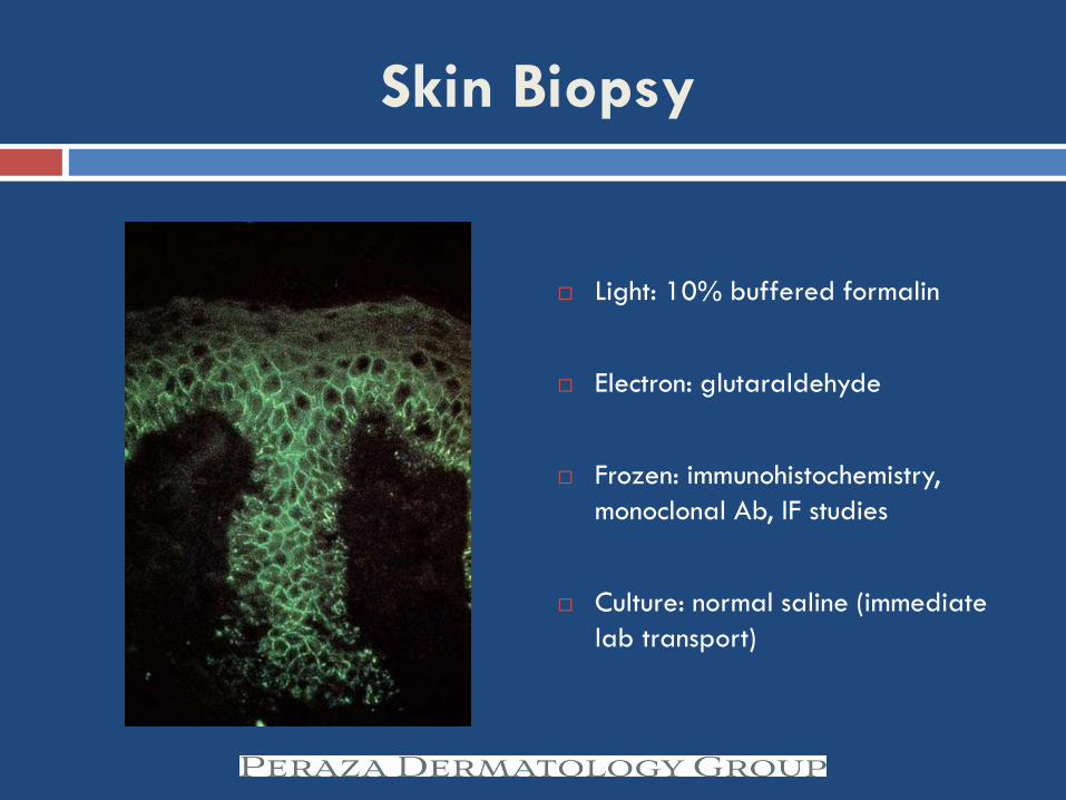
Skin Biopsy
Light: 10% buffered formalin
Electron: glutaraldehyde
Frozen: immunohistochemistry,
monoclonal Ab, IF studies
Culture: normal saline (immediate
lab transport)

Skin Biopsy: Type and Infection Risk
Shave, punch, scissor
Gowns, face masks, sterile field not strictly needed
Clean gloves
<1% risk of infection (poor technique)

Skin Biopsy: Infection Control
Preparation of site
Hand washing
Alcohols, chlorhexidine, iodophors
Prophylactic antibiotics usually not needed (uncontrolled DM,
alcoholism, morbid obesity, and malnutrition)

Skin Biopsy: Anesthesia
Lidocaine +/- epinephrine (1:100,000)
Affect hemostasis, absorption, duration
Buffered with 8.4% sodium bicarbonate (1:10 volume)
30 gauge needles
45° angle
Inject slowly
BE SAFE AND IN CONTROL
Alternatives

Shave Biopsy
Limited to epidermis and
papillary dermis
Quick
Little training
No sutures
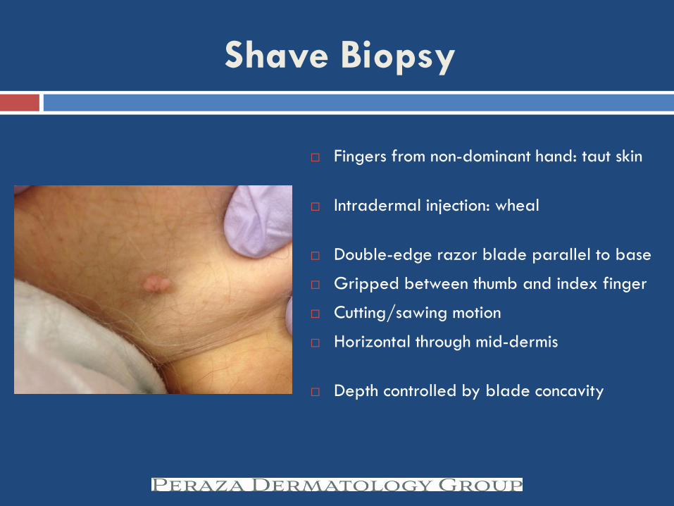
Shave Biopsy
Fingers from non-dominant hand: taut skin
Intradermal injection: wheal
Double-edge razor blade parallel to base
Gripped between thumb and index finger
Cutting/sawing motion
Horizontal through mid-dermis
Depth controlled by blade concavity

Shave Biopsy
Fingers from non-dominant hand: taut skin
Intradermal injection: wheal
Double-edge razor blade parallel to base
Gripped between thumb and index finger
Cutting/sawing motion
Horizontal through mid-dermis
Depth controlled by blade concavity
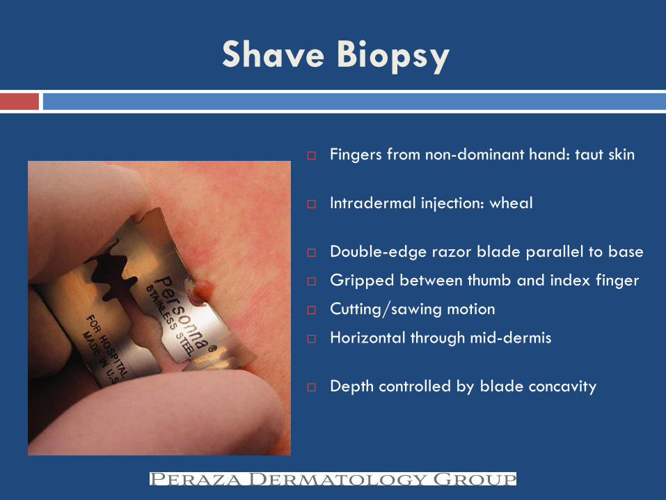
Shave Biopsy
Fingers from non-dominant hand: taut skin
Intradermal injection: wheal
Double-edge razor blade parallel to base
Gripped between thumb and index finger
Cutting/sawing motion
Horizontal through mid-dermis
Depth controlled by blade concavity

Shave Biopsy
Fingers from non-dominant hand: taut skin
Intradermal injection: wheal
Double-edge razor blade parallel to base
Gripped between thumb and index finger
Cutting/sawing motion
Horizontal through mid-dermis
Depth controlled by blade concavity

Shave Biopsy Hemostasis
Bleeding minimal
Pressure, aluminum chloride, electrodesiccation
Wounds heal in less than a week

Shave Biopsy Video

Saucerization Biopsy
Skin lesion plus portion of
surrounding skin
Includes subcutaneous fat
Lesions in epidermis and dermis
Nevi, BCC, SCC, melanoma, etc.

Saucerization Biopsy
Skin lesion plus portion of
surrounding skin
Includes subcutaneous fat
Lesions in epidermis and dermis
Nevi, BCC, SCC, melanoma, etc.

Scissor Biopsy
Pedunculated lesions
Nevi, filiform warts, skin tags
Cosmesis, itching, irritation,
catching on clothing
Iris or gradle scissor
Pressure or chemical hemostasis

Scissor Biopsy
Pedunculated lesions
Nevi, filiform warts, skin tags
Cosmesis, itching, irritation,
catching on clothing
Iris or gradle scissor
Pressure or chemical hemostasis

Punch Biopsy
Very common alternative
Sharp cylinders: punch or trephines
Disposable or reusable (2-8mm)
Easily mastered, quick, low incidence
for infection, minimal scarring
Dermal pathology
Punch Biopsy
≤3mm: second intention possible
≥4mm and face: sutures
Alcohol pad, local anesthesia, gloves, punch instrument, forceps, scissors, gauze

Punch Biopsy
Non-dominant hand: apply
pressure
Punch perpendicular to skin
Press down and twist bore out
Forceps and scissor for removal
Special sites: scalp, mucosa, nail

Punch Biopsy
Non-dominant hand: apply
pressure
Punch perpendicular to skin
Press down and twist bore out
Forceps and scissor for removal
Special sites: scalp, mucosa, nail

PRACTICE SESSIONS

THANK YOU
Please do not hesitate to email with questions
Most images from Peraza Dermatology Group
Content, tables, and some images:
Bolognia JL, Jorizzo JL, Rapini RP. Dermatology 2nd Edition. Spain: Elsevier 2008.
McKee PH, Calonje E, Granter SR. Pathology of the Skin. 3rd Edition. China: Elsevier Mosby 2005.
Robinson JK, Hanke CW, Sengelmann RD, Siegel DM. Surgery of the Skin. New York: Elsevier Mosby 2005.