
Criteria and case definitions for serious injury and death ...
36
DISEASES OF AQUATIC ORGANISMS Dis Aquat Org Vol. 103: 229–264, 2013 doi: 10.3354/dao02566 Published April 11 © Inter-Research 2013 · www.int-res.com *Email: [email protected] THEME SECTION Criteria and case definitions for serious injury and death of pinnipeds and cetaceans caused by anthropogenic trauma Editors: Michael J. Moore 1, *, Julie van der Hoop 1 , Susan G. Barco 2 , Alex M. Costidis 3,9 , Frances M. Gulland 4 , Paul D. Jepson 5 , Kathleen T. Moore 6 , Stephen Raverty 7 , William A. McLellan 8 1 Woods Hole Oceanographic Institution, Woods Hole, Massachusetts 02543, USA 2 Virginia Marine Aquarium and Marine Science Program, Virginia Beach, Virginia 23451, USA 3 Physiological Sciences, College of Veterinary Medicine, University of Florida, Gainesville, Florida 32610, USA 4 Marine Mammal Center, Fort Cronkhite, Sausalito, California 94965, USA 5 Institute of Zoology, Zoological Society of London, London NW1 4RY, UK 6 International Fund for Animal Welfare,Yarmouth Port, Massachusetts 02675, USA 7 British Columbia Ministry of Agriculture, Abbotsford, British Columbia V3G 2M3, Canada 8 Biology and Marine Biology, University of North Carolina Wilmington, Wilmington, North Carolina 28403, USA 9 Present address: Biology and Marine Biology, University of North Carolina Wilmington, Wilmington, North Carolina 28403, USA ABSTRACT: Post-mortem examination of dead and live stranded beach-cast pinnipeds and cetaceans for determination of a cause of death provides valuable information for the manage- ment, mitigation and prosecution of unintentional and sometimes malicious human impacts, such as vessel collision, fishing gear entanglement and gunshot. Delayed discovery, inaccessibility, logistics, human safety concerns, and weather make these events challenging. Over the past 3 decades, in response to public concern and federal and state or provincial regulations mandating such investigations to inform mitigation efforts, there has been an increasing effort to objectively and systematically investigate these strandings from a diagnostic and forensic perspective. This Theme Section provides basic investigative methods, and case definitions for each of the more commonly recognized case presentations of human interactions in pinnipeds and cetaceans. Wild animals are often adversely affected by factors such as parasitism, anthropogenic contaminants, biotoxins, subclinical microbial infections and competing habitat uses, such as prey depletion and elevated background and episodic noise. Understanding the potential contribution of these sub- clinical factors in predisposing or contributing to a particular case of trauma of human origin is hampered, especially where putrefaction is significant and resources as well as expertise are lim- ited. These case criteria descriptions attempt to acknowledge those confounding factors to enable an appreciation of the significance of the observed human-derived trauma in that broader context where possible. KEY WORDS: Seal · Dolphin · Whale · Marine mammal · Entrapment · Entanglement · Vessel strike · Gunshot Resale or republication not permitted without written consent of the publisher FREE REE ACCESS CCESS
Transcript of Criteria and case definitions for serious injury and death ...
DISEASES OF AQUATIC ORGANISMSDis Aquat Org
Vol. 103: 229–264, 2013doi: 10.3354/dao02566
Published April 11
© Inter-Research 2013 · www.int-res.com*Email: [email protected]
THEME SECTION
Criteria and case definitions for serious injury and death of pinnipeds and cetaceans caused by
anthropogenic trauma
Editors: Michael J. Moore1,*, Julie van der Hoop1, Susan G. Barco2, Alex M. Costidis3,9, Frances M. Gulland4, Paul D. Jepson5, Kathleen T. Moore6,
Stephen Raverty7, William A. McLellan8
1Woods Hole Oceanographic Institution, Woods Hole, Massachusetts 02543, USA2Virginia Marine Aquarium and Marine Science Program, Virginia Beach, Virginia 23451, USA
3Physiological Sciences, College of Veterinary Medicine, University of Florida, Gainesville, Florida 32610, USA4Marine Mammal Center, Fort Cronkhite, Sausalito, California 94965, USA5Institute of Zoology, Zoological Society of London, London NW1 4RY, UK
6International Fund for Animal Welfare,Yarmouth Port, Massachusetts 02675, USA7British Columbia Ministry of Agriculture, Abbotsford, British Columbia V3G 2M3, Canada
8Biology and Marine Biology, University of North Carolina Wilmington, Wilmington, North Carolina 28403, USA
9Present address: Biology and Marine Biology, University of North Carolina Wilmington, Wilmington, North Carolina 28403, USA
ABSTRACT: Post-mortem examination of dead and live stranded beach-cast pinnipeds andcetaceans for determination of a cause of death provides valuable information for the manage-ment, mitigation and prosecution of unintentional and sometimes malicious human impacts, suchas vessel collision, fishing gear entanglement and gunshot. Delayed discovery, inaccessibility,logistics, human safety concerns, and weather make these events challenging. Over the past 3decades, in response to public concern and federal and state or provincial regulations mandatingsuch investigations to inform mitigation efforts, there has been an increasing effort to objectivelyand systematically investigate these strandings from a diagnostic and forensic perspective. ThisTheme Section provides basic investigative methods, and case definitions for each of the morecommonly recognized case presentations of human interactions in pinnipeds and cetaceans. Wildanimals are often adversely affected by factors such as parasitism, anthropogenic contaminants,biotoxins, subclinical microbial infections and competing habitat uses, such as prey depletion andelevated background and episodic noise. Understanding the potential contribution of these sub-clinical factors in predisposing or contributing to a particular case of trauma of human origin ishampered, especially where putrefaction is significant and resources as well as expertise are lim-ited. These case criteria descriptions attempt to acknowledge those confounding factors to enablean appreciation of the significance of the observed human-derived trauma in that broader contextwhere possible.
KEY WORDS: Seal · Dolphin · Whale · Marine mammal · Entrapment · Entanglement · Vesselstrike · Gunshot
Resale or republication not permitted without written consent of the publisher
FREEREE ACCESSCCESS

Dis Aquat Org 103: 229–264, 2013
INTRODUCTION AND OVERVIEW
Michael J. Moore, Julie van der Hoop, Susan G. Barco, Alex M. Costidis, Frances M.Gulland, Paul D. Jepson, Kathleen T. Moore,
Stephen Raverty, William A. McLellan
Background
The Marine Mammal Protection Act (MMPA) wasenacted to conserve and manage all marine mammalpopulations under US jurisdiction. It prohibits theunauthorized ‘take’ of marine mammals (defined ascapture, harassment, or killing of any individual) andrequires federal agencies to monitor the status ofpopulations, including the extent to which humanactivities contribute to marine mammal morbidityand mortality. Section 117 directs the National Mar-ine Fisheries Service (NMFS) and the US Fish andWildlife Service to prepare stock assessment reports,in which human-caused marine mammal mortalitiesand serious injuries must be enumerated. In addition,the Marine Mammal Health and Stranding ResponseAct of the MMPA requires the evaluation of causes ofmorbidity and mortality in marine mammals, as wellas the monitoring of trends in population health.
The nature of serious injury (SI) was reviewedthrough NMFS workshops in 1997 (Angliss &DeMaster 1998) and more recently in 2008 (Ander-sen et al. 2008). The goal of the workshops was toenhance consistency in the assessment and report-ing of human interaction (HI) injuries within theNational Oceanic and Atmospheric Administration(NOAA) by refining criteria used in human interac-tion determinations. The SI criteria that were devel-oped were with respect to HI injuries sustained by
large cetaceans that did not result in an immediatemortality but would likely result in subsequent mor-tality. In these cases, the animals were seriouslyinjured but still alive. Draft guidelines (Moore &Merrick 2011) and procedures (NMFS 2012a,b) forthis were re cently published. There has been anexcellent attempt to standardize how HI is recog-nized grossly by personnel responding to marinemammal strandings (Moore & Barco in press). Thisincludes the development of a training manual(Moore & Barco in press), standardized data collec-tion form (see Supplement 1, available at www. int-res. com/ articles/ suppl/ d103 p229 _ supp/), and theprovision of training workshops to members of theUS Marine Mammal Stranding Network. The proto-col presented to stranding re sponse personnel isdesigned to provide (1) an objective gross evalua-tion of an animal or carcass that determines whetherany signs of HI are present (regardless of whetherthey may have contributed to the stranding or deathof the animal, are pre- or post-mortem, healed orrecent); and (2) a subjective finding in which exam-iners use all available information and their experi-ence to evaluate the likelihood that any observedevidence of HI contributed to the stranding event.Ideally, to reach a conclusion as to whether HIcaused the death (or was a contributing cause of thedeath) requires review of all sample results and vet-erinary pathological information (Moore & Barco inpress). Such information could include history, grossnecropsy, histopathology, microbiology, radiology,molecular tests, toxicology, hematology and otherdiagnostic disciplines. As such the manual fallsshort of a goal of establishing objective criteria todetermine the proximate cause of death (COD),
CONTENTS
M. Moore et al.Introduction and overview......................................230P. Jepson et al.Peracute underwater entrapment of pinnipedsand cetaceans...........................................................235M. Moore et al.Chronic entanglement trauma of pinnipedsand ceta ceans...........................................................240W. McLellan et al.Blunt force trauma induced by vessel collisionswith large whales ....................................................245A. Costidis et al.Sharp trauma by vessel collisions with pinni-peds and and cetaceans ..........................................251
K. Moore et al.Gunshot injuries of pinnipeds and cetaceans........256
LITERATURE CITED...............................................259Appendix 1: Glossary ..............................................263Appendix 2: Abbreviations .....................................264
Online supplemental material (available at www. int-res.com/ articles/ suppl/ d103 p229 _supp/):Supplement 1: Human Interaction (HI)
Documentation FormSupplement 2: Entanglement Response FormSupplement 3: Level A Data Collection SheetSupplement 4: Chain of Custody FormSupplement 5: Additional author affiliations
230

Moore et al.: Pinniped and cetacean anthropogenic trauma case criteria
whether it be by HI or not. More rigorous scientificexamination of all available information is importantfor the scientific community and re quires the coop-eration and collaboration of the veterinary andstranding network communities.
This Theme Section aims to establish case state-ments for each common class of HI to enable examin-ers of cetaceans and pinniped cases to generate andinterpret gross, histological and other analytical datato conclude if an animal died or was seriously injuredas a result of a specific human activity, and to qualifysuch conclusions in terms of degrees of confidence. Itidentifies knowledge gaps to complete the neededcriteria whereby each casual factor can be recog-nized as causative, or not, in a particular case. It iscritical to recognize that there is a spectrum of theamount and type of data available for the analysis ofspecific cases that ranges from a single at-sea photo-graph of a dead or seriously injured animal to a fullcase history involving necropsy, histology and ancil-lary investigations. It is important to use the availableprecedents from all similar documented cases toinform the interpretation of data-poor cases to arriveat the best assessment. On 1 and 2 February 2012, agroup of biologists, government scientists and veteri-narians convened in a workshop to define evidence
necessary to attribute observations of ceta cean andpinniped mortality or SI from proximate anthro-pogenic events that initiated their ultimate demise. Itwas in no way designed to advise or modify NOAApolicy and procedure for the determination of causesof SI and mortality, but the hope remains that thisTheme Section will enable such determinations(Fig. 1) to be made on the basis of the best availablecriteria as summarized herein. While this reviewworks within the relevant legal framework in theUnited States of America, it should have global rele-vance in principle.
Methods for investigation of mortality and SI cases
Clinical signs
Investigators should observe and record, prefer-ably with a camera, the behavior of any live animal(see Supplement 2, available at www.int-res. com/articles/ suppl/d103 p229 _supp/). The nature of res -piration, response to stimuli, external body condition,and extent, distribution and severity of the trauma,and skin color or discoloration should also berecorded.
231
Fig. 1. Potential flow of mortality and serious injury (SI) observations through the generation and review process in the USA in the context of the Marine Mammal Protection Act (MMPA)

Dis Aquat Org 103: 229–264, 2013
Assignment of decomposition condition code
A condition code should be assigned to a de -ceased individual based on the level of decomposi-tion of a carcass. As sample quality diminishes withdecomposition (with higher condition codes), assess-ing condition can help determine the samples thatwill be of greatest value. Carcasses can be classifiedinto one of 5 categories, as described by Geraci &Lounsbury (2005). Live animals are Code 1. Code 2carcasses are those in good (fresh) condition, char-acterized by having normal appearance, little scav-enger damage, fresh odor, lack of bloating, and firmmuscle and blubber, among other features (Geraci& Lounsbury 2005). Carcasses with moderate de -com position are Code 3, typically with organs moreor less intact, bloating, cracked and sloughing skin,potential scavenger damage, mild odor, blood-tinged oily blubber, soft muscles and intestinal dis-tention. Code 4 carcasses are in poor condition, withadvanced decomposition, and are often collapsedand displaying sloughing skin, severe scavengerdamage, strong odor, soft blubber, liquefied mus-cles, and gas-filled intestines. Mummified or skele-tal remains are Code 5.
Level A data and first response
Researchers should collect Level A data (species,age, sex, length, location, date stranded) (Supple-ment 3, available at www.int-res. com/ articles/ suppl/d103 p229 _ supp/) (Geraci & Lounsbury 2005), andde scribe and photograph the entire animal and anysurface lesions and attached fishing gear if present.Photographs should capture flukes, callosities, orother distinguishing features used for photo-identifi-cation of individuals. This can aid in matching an ani-mal with known sighting and history information thatcan in turn aid in determining the timing of the
injury. A skin sample should be collected for geneticanalyses. Complete a HI form (Moore & Barco inpress).
If a case is floating at sea, towing ashore is a prior-ity. If any attached fishing gear is likely to fall off dur-ing towing, it should be removed before moving theanimal. Similarly, in the event of a severely impactedcarcass, investigators should anticipate the potentialseparation of body parts. The body part to which anytowline is attached should be documented prior toattachment.
Level B data
Detailed data (measurements, patterns, diagramsand photographs from multiple angles) and sampleson all injuries and wounds should be collected, andbody condition (emaciated, thin or robust) assessed.
Sample collection
Clinical pathology and histopathology
Sample collection from various condition code animals can be guided by Table 1. Blood from liveanimals and vitreous from fresh dead (Code 2 or 3)animals should be collected for baseline serumchemistry and hematology (from blood) and potas-sium and creatine kinase (from vitreous); surplusserum should be banked at −70°C. Clinical chemistrytests of important note include creatine phospho -kinase, lactate dehydrogenase and aspartate trans -aminase for muscle damage (capture myopathy/acute rhabdomyolysis).
Thorough tissue sampling should be conductedwhen practical for all cases according to conditioncode (Table 1); major organs, lesion, and woundmargins and subtending tissue including bone on
232
Code Animal status Samples of value
Code 1 Live animal Morphometrics, blood, urine, biopsies, genetics, toxicologyCode 2 Fresh carcass Morphometrics, blood, urine, histology, cytology, virology (tissue), microbio-
logy (swabs or tissue for culture, tissue for polymer chain reaction (PCR), para sitology, contaminants, biotoxins, life history, genetics
Code 3 Moderate decomposition Morphometrics, histology (limited), virology (PCR), microbiology (PCR), par a -si tol ogy, contaminants, biotoxins, life history, genetics
Code 4 Advanced decomposition Morphometrics, histology (limited), virology (PCR), life history, geneticsCode 5 Mummified or skeletal remains Morphometrics, life history, genetics
Table 1. Valuable sample analysis according to carcass condition code (adapted from Geraci & Lounsbury 2005, Pugliares et al. 2007)

Moore et al.: Pinniped and cetacean anthropogenic trauma case criteria
all carcasses should be sampled as practical. For-eign objects should be handled with appropriateinstruments (e.g. plastic forceps) to avoid artifactualtool-marks. Histologic sampling of wound marginsshould be conducted with care to avoid artifactualdestruction of physical (e.g. muscle fiber curling orcontracture) and cellular (e.g. vital response) indica-tors of ante-mortem injury.
All lesions should be labeled with a unique identi-fier, and photographed in situ, with a scale and CaseID marker. Lesion identifiers should accompany allsubsequent samples derived from each lesion, withthe anatomic site of trauma recorded.
In cases with traumatic bone injury, evaluation ofthe cortical defect or bone fracture margins is recom-mended. If possible, concurrent collection of urine forurinalysis is recommended for evaluating the pres-ence of muscle and blood breakdown products (myo-globinuria and hemaglobinuria).
Necropsy
Gross necropsies should be performed by experi-enced prosectors on all carcasses following pre-scribed pinniped and cetacean protocols (McLellanet al. 2004, Pugliares et al. 2007). Ensure appropriatelife history and individual identification charactersare collected. Collect detailed documentation (Fig. 2)(e.g. at impact sites, photograph from several angles,measure and diagram, scar/wound patterns, sketchwith scale) and histology samples of soft and hard tis-sue wound margins at various depths to account fordifferential resolution of peri-mortem and chronic
wounds as well as to help rule out exposure artifacts(e.g. salt water or scavenging) (DiMaio & DiMaio2001). The texture (e.g. curling/ fiber bunching, bridg-ing) and histopathology of wound margin and mus-cle can often provide in sights into ante-mortem/peri-mortem vs. post-mortem injury or scavenging(Shkrum & Ramsay 2007). Presence, volume andlocation of loose and/or adherent well-formed bloodclots and frank hemorrhage associated with incisedwounds should be noted. Additionally, documenta-tion of wound location may help with ante-mortemvs. post-mortem injury determination; for example,blunt trauma injuries to the dorsum may indicate thata whale was alive when it was struck, as most deadwhales float with either the ventral or the lateral mid-line facing upward (Campbell-Malone et al. 2008).
Abdominal displacements, herniation and visceralruptures should be carefully documented, as thesecan be post-mortem changes. The pulmonary systemshould be examined for evidence of froth, or waterand/ or blood aspiration. Measuring the specific grav-ity and chemical constituents of airway fluids mayhelp to discriminate between body fluid and sea water.
Stomach chambers should be examined for foreigningesta, and for presence of blood, as maxillofascialand upper respiratory tract injuries can result inhemoptysis and subsequent ingestion of blood,which provide strong support for ante-mortem tim-ing of injuries.
All major organs should be examined and sampledto exclude other possible causes of death. Focusedsampling of liver, kidneys, and lymph nodes shouldbe conducted for evidence of ante-mortem blooddrainage from the site of injury and erythrocyte
degradation. Regional and globallymph node examination and histolog-ical sampling may help narrow downextent of damage and/or post-injurysurvival time based on presence anddegree of erythrocyte turn-over/degradation.
Histological staining
Examining all samples, especiallyrelating to gross images and descrip-tions if practical, is primarily of use indiagnosing intercurrent conditions.Conventional histologic staining, suchas hematoxylin and eosin (H&E), isrecommended with additional specialstains undertaken at the discretion of
233
Fig. 2. Image of the fractured vertebral elements discovered during necropsyof MJM 9406 Eg. Fractures (shown in red) were recorded on necropsy data
sheets described in McLellan et al. (2004)

Dis Aquat Org 103: 229–264, 2013
the pathologist. Oil Red O or osmium tetroxide post-fixation may detect the presence of fat embolization(Fernández et al. 2005), which has been associatedwith traumatic injury to lipid-rich tissues (e.g. longbone fractures with marrow exposure or adipose tis-sue injury) (Shkrum & Ramsay 2007). Note that sam-ples must be collected for adipose staining and thatthese cannot be put through routine processing. Inthe case of bone fractures, histological evaluation offracture margins should also be undertaken, althoughno changes will be detected if death is peracute.Okajima staining for hemaglobin may better defineprotein casts in renal tubules, if there is a myoglobin-uria or hemoglobinuria.
Microscopic evaluation of regional lymph nodes toassess draining hemorrhage and of kidney to evalu-ate proteinuria associated with myoglobin or hemo -globin is recommended. Electron microscopy, specialstains for pathogens, phosphotungstic acid hema-toxylin-fibrin, and Trichrome-collagen for fibrosismay also be considered.
Report
Reporting of gross necropsy findings should followa standardized protocol (McLellan et al. 2004,Pugliares et al. 2007). A gross report should includeas detailed a life history (from catalog ID if applica-ble) and case history as possible, and sketches, meas-urements, descriptions and samples taken and theirdisposition. Diagrams and photographs of significantlesions should be supplied with the gross report. Useof HI forms (Supplement 1) and/or injury-specificforms (Supplement 2) is recommended. Once ancil-lary diagnostic findings (e.g. histology) are available,a final case report and interpretation should be pre-pared with conclusions as to likely COD. COD fromanthropogenic trauma should be rated as confirmed,probable or suspect.
Case criteria
The subsequent sections of this Theme Sectioninclude criteria for the following diagnostic traumacategories: peracute underwater entrapment; chronicentanglement; blunt vessel; sharp vessel; and gun-shot. For each trauma type described, case definitionmaterial is mostly presented as follows: background,signalment, epidemiology, clinical signs, host re -sponse, injury-specific gross necropsy, toxicology,and histology findings, and COD assignation.
Degrees of confidence
Scoring ‘confirmed, probable and suspect’ cases asdefined in the Glossary (Appendix 1) remains aninexact science for cetacean and pinniped traumamortality investigations. The traumatic event is rarelywitnessed or documented, the time from death isgenerally not known and for large whales the inves-tigation is often hampered by the inability for largeequipment to reach the site to aid the necropsy. Con-straints also include loss of tissue from scavenging,combinations of chronic entanglement and re solvedpast lesions, additional post-mortem insults from ves-sel collision, tissue degradation and dissolution fromputrefaction, beach abrasion and finally, animal con-dition prior to the insult. Each category includes sep-arate well-defined forensic findings and providescase reports that best illustrate each of these find-ings. As more large whale necropsy findings arepublished world-wide these categories can becomemore codified. These problems of size are less ex -treme for smaller pinnipeds and cetaceans, but over-all the same limitations apply.
General recommendations
(1) A periodic retrospective peer analysis should beundertaken of the prevalence of each lesion type foreach trauma type in past necropsy cases based onCOD confidence levels, including information onlocation and depth of wounds and entanglements;trauma dorsal or ventral to the center of gravity ofthe animal; organ displacement or herniation;whether there was an acute, subacute or chronicmortality; and the review of pathology cases inorder to determine the frequency of observing eachtype of injury/ gross lesion. Such an analysis wouldconfirm as sump tions on past cases, strengthen theconclusions from specific observations and increaseconfidence with enhanced sample sizes for catego-rizing mortalities.(2) Cross-correlation of injury data from live and deadanimals should be used to evaluate lethality criteria.(3) All necropsy teams should adopt standardizedprotocols of examination and reporting (e.g. necropsyteam leader [NTL] / logistics coordinator / offsite coor-dinator) (McLellan et al. 2004) to maximize data comparability.(4) Increased effort should be made for systematicarchiving, reporting, and sharing of data, such asopen/ limited access online researcher databases forarchiving of trauma determinations.
234

Moore et al.: Pinniped and cetacean anthropogenic trauma case criteria
(5) Greater transparency and communication shouldoccur between managers, NTLs, and interest groupsin order to enhance future diagnostic quality, andconsequent implementation of suitable conservationmeasures to mitigate impacts.
(6) Communication should occur between east andwest coast stranding programs and managers regard-ing cases, to increase the learning curve based onnational caseload rather than regional loads.
PERACUTE UNDERWATER ENTRAPMENT OFPINNIPEDS AND CETACEANS
Paul D. Jepson1, Michelle Barbieri, Susan G. Barco,Yara Bernaldo de Quiros, Andrea Bogomolni, Kerri
Danil, Teri Rowles
Background
By-catch has been identified as a major cause ofmortality in individual marine mammals (Kuiken etal. 1994, Kuiken 1996, Kirkwood et al. 1997, North-ridge & Hofman 1999). More importantly, there is in -creasing evidence of the negative ecological impactof by-catch from specific commercial fisheries onmany cetacean populations (Northridge & Hofman1999). For example, several observer-based studieshave reported high or unsustainable mortality in har-bor porpoises Phocoena phocoena due to by-catch incommercial gill-net fisheries (Tregenza et al. 1997,Moore & Barco in press) and in trawl-caught commondolphin Delphinus delphis (Peltier et al. 2012).
Case definition
Clinical signs
Clinical signs of peracute underwater entrapmentin fishing gear are unlikely to be seen in most casesdue to the essentially cryptic nature of immersion influid. However, pathological findings in peracutedrowning due to underwater entrapment often sug-gest some degree of physical struggle associatedwith often marked muscular exertion.
Signalment
For cetaceans, any age and sex can be affected byaccidental entanglement in commercial fishing gear(by-catch) although biases have been found for par-
ticular species and fisheries. For example, juvenileswere over-represented in some studies of by-caughtanimals (stranded and retrieved directly from fishingvessels) (Kirkwood et al. 1997, Siebert et al. 2001, Jau-niaux et al. 2002, Jepson 2006), while in other studiesthey were under-represented (Delphinus delphis andStenella longirostris by-catch in tuna purse-seine fish-eries) (Danil & Chivers 2007, Larese & Chivers 2009).Male D. delphis have been over-represented in bothPacific and Atlantic fisheries (Ferrero & Walker 1995,Murphy 2004, Westgate & Read 2007, Danil et al.2010) and there are anecdotal reports that juvenilemale bottlenose dolphins Tursiops truncatus on theUS east coast are also over-represented.
Epidemiology
All types of fishing gear pose significant globalthreats to welfare and conservation status of exposedpinnipeds and cetaceans (Read et al. 2006, Moore &Barco in press). Pinnipeds, odontocetes and mysticetescan be affected (Jepson 2006, Moore & Barco in press,Cassoff et al. 2011). Gillnet entrapment (Figs. 3 & 4)seems to occur in all regions of the globe where gill-nets are employed (Read et al. 2006). Underwater en-trapment in mobile gear also occurs widely but maybe rather more cryptic (Northridge & Hofman 1999).
Injury-specific gross necropsy findings
These gross necropsy findings are based on a num-ber of sources (Kuiken et al. 1994, Kuiken 1996, Moore& Barco in press). The diagnosis of peracute under -water entrapment (by-catch) when circumstances ofdeath/ discovery or external findings are obvious canusually be made on gross examination alone withoutthe need for histopathology. Histo pathology andmicro biology are useful investigative tools to help ruleout other causes of death. In addition, assays for algaltoxins (e.g. domoic acid and others), may also be use-ful investigative tools to rule out underlying or pre- existing conditions that may predispose animals to HI.
Contact with fishing gear
Evidence of contact with fishing gear is not re -quired to confirm an underwater entrapment mortal-
235
1For author affiliations, see Supplement 5 at www. int-res.com/ articles/suppl/d103p229_supp/

Dis Aquat Org 103: 229–264, 2013
ity. Entrapment can occur without direct contact withfishing gear, such as when surrounded by fish in theend of a mobile trawl. Evidence includes (1) fresh lin-ear skin lesions and furrows (e.g. net marks; Figs. 3& 4) in or around the mouth, fin or tail (Moore &Barco in press) or encircling one or more extremities(Fig. 5); (2) bruises in body regions consistent withentanglement such as peri-mandibular (Fig. 6), peri-
scapular and thoracic rete mirabile; and (3) fractures(in pinnipeds and small odontocetes) and associatedhemorrhage and soft tissue maceration in the man -dible, other parts of the cranium, and ribs. Further,whether or not strandings occurred in the vicinity offishing activity may assist in making a determination.
Evidence of hypoxia
Lack of oxygen (hypoxia) can be evidenced by wet,moist, glistening and heavy lungs (edematous lungs).Persistent froth in the airways (Fig. 7) may be red-tinged, but can also be white and clear; some frothmay be stable, likely depending on protein content,surfactants, or mucus. Generalized congestion (darkred lungs) can also suggest lack of oxygen. Fluid inthe primary and secondary airways may or may notbe evident grossly. Occasional hyperinflation oflungs and emphysema have been observed (Jepsonet al. 2000, Jepson 2006).
236
Fig. 3. Linear impressions from net entanglement on the(a) rostrum and (b) fluke of a bottlenose dolphin Tursiopstruncatus. VAq cases (a) VAQS20081081 Tt and (b)
VAQS20091093 Tt
Fig. 4. Impressions from entanglement of a harbor porpoise Phocoena phocoena in gillnet. WHOI case DO8760Pp
Fig. 5. Large mesh net marks on the fur of a grey seal Halichoerus grypus. IFAW case CCSN04-153Hg

Moore et al.: Pinniped and cetacean anthropogenic trauma case criteria
Although histology would not be required to con-firm the diagnosis of pulmonary edema, microscopicassessment of the tissues would be critical to excludeother underlying or pre-existing conditions that mayhave contributed to entanglement or even death,such as septicemia, parasitemia, cardiomyopathy, orforms of vasculopathy.
Physical trauma
Evidence of physical trauma during the releasefrom a net would include the amputation of fins,flukes, or tail (Fig. 8) (Cox et al. 1998), a penetratingincision into a body cavity (Fig. 9), or rope around thetail stock that was added to enable removal from anet (Fig. 10).
There are no pathognomonic clinical signs orlesions for an animal that died from peracute under-water entrapment. However, there are a number offindings on necropsy that are consistent with this
cause of mortality. By-caught harbor porpoises (usu-ally caught in bottom set gill-nets or tangle nets usingwide-meshed monofilament nylon nets) typicallyexhibit characteristic cuts on the edge of the mouth,fin or tail and sometimes have encircling lesion(s)around the head (e.g. Fig. 4). In contrast, commondolphins (mostly suspected to be caught in mid-waterpelagic trawl fisheries) typically present with fewexternal cutaneous lesions, although amputatedfins/flukes, fractured beaks and broken teeth aresometimes identified (Kuiken et al. 1994, Jepson2006, Deaville & Jepson 2011).
237
Fig. 6. Skin and blubber removed and pre mortem subcuta-neous and fascial hemorrhage apparent around the left man -dible of bottlenose dolphin. VAq case VMSM10021035 HEM
Fig. 7. Incised harbor porpoise lung showing copiousfroth exuding from cut surfaces and airways. VAq case
VMSM20041006
Fig. 8. Amputated flukes in a net-entrapped bottlenose dolphin. VAq case VMSM20011123
Fig. 9. Post-mortem abdominal wall incision in net- entangled bottlenose dolphin. VAq case VAQS 20121008
Fig. 10. Tooth rake marks (oval) and penducle line impres-sion (arrow) in bottlenose dolphin. VAq case VAQS20111014
Dis Aquat Org 103: 229–264, 2013
Pulmonary lesions are similar to those seen indeath due to asphyxia in terrestrial mammals andinclude pulmonary congestion, edematous lungs,and presence of a fine persistent whitish (or blood-tinged) froth within the airways. Pleural or pericar-dial petechial hemorrhages and bullous emphysemahave also been proposed as criteria for diagnosis ofcetacean by-catch in the UK (Kuiken et al. 1994,Kuiken 1996) but are very rarely seen (Jepson et al.2000). Edematous lungs and the presence of persist-ent blood-tinged or white stable foam in the airwaysare thought to result from asphyxia associated withhypoxic damage to the integrity of alveolar mem-branes leading to leakage of erythrocytes and pro-teinaceous fluid into the alveoli (Modell 1981, Davis& Bowerman 1990, Lunetta & Modell 2005). Howeverlung edema can also occur post mortem, followingintrinsic contractions of the atrio-ventricular andsino-atrial nodes in the heart; contractions may per-sist for up to 5−10 min. As blood is ejected, it in -creases the hydrostatic pressure in the pulmonarymicrovasculature with seepage or suffusion of fibrinand edema into the alveolar spaces. Subsequent mix-ing with residual air may then result in stable frothformation. The role of possible traumatic asphyxia isnot understood, but should be considered given thecompression and physical restraint involved in net-entanglement events (Shkrum & Ramsay 2007).
Intravascular gas bubbles have been found in fresh(Code 2) odontocetes that were by-caught at consid-erable depth (>100 m) and probably relate to nitro-gen off-gassing post mortem from supersaturated tis-sues as the by-caught carcass returns to the surface(Moore et al. 2009). Bubbles may not be seen in Code 2carcasses which have been by-caught at shallowerdepths.
Health status must be considered in confirming anunderwater entrapment mortality. Nutritional condi-tion of by-caught small cetaceans is often good tomoderate, or not emaciated in the case of pinnipeds.Evidence of recent feeding is commonly presentedeither by whole prey within the stomach, partiallydigested ingesta mixed with hard parts (skeleton,squid pen, squid beaks, or otoliths) or chyle withinintestinal lymphatics.
Histological findings
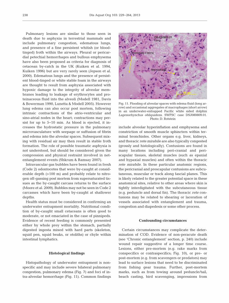
Histopathology of underwater entrapment is non-specific and may include severe bilateral pulmonarycongestion, pulmonary edema (Fig. 7) and foci of in-tra-alveolar hemorrhage (Fig. 11). Common findings
include alveolar hyperinflation and emphysema andconstriction of smooth muscle sphincters within ter-minal bronchioles. Other organs e.g. liver, kidneys,and thoracic rete mirabile are also typically congested(grossly and histologically). Contusions are found inmany locations including peri-cranial and peri-scapular tissues, skeletal muscles (such as epaxialand hypaxial muscles) and often within the thoracicrete mirabile. In these particular anatomic regions,the pericranial and prescapular contusions are subcu-taneous, muscular or track along fascial planes. Thisis likely related to the greater potential space in theseanatomical sites, relative to other areas where skin istightly interdigitated with the subcutaneous tissue(e.g. peduncle and dorsal fin). The thoracic rete con-tusions may be related to shearing or laceration ofvessels associated with entanglement and trauma,congestion and diapedesis or some other process.
Confounding circumstances
Certain circumstances may complicate the deter-mination of COD. Evidence of non-peracute death(see ‘Chronic entanglement’ section, p. 240) includewound repair suggestive of a longer time course.Lesions, either pre-mortem (e.g. rake marks fromconspecifics or contraspecifics; Fig. 10), or pre- orpost-mortem (e.g. from scavengers or predators) maylead to surface lesions that need to be discriminatedfrom fishing gear trauma. Further, post-mortemmarks, such as from towing around peduncle/tail,beach casting, bird scavenging, impressions from
238
Fig. 11. Flooding of alveolar spaces with edema fluid (long ar-row) and occasional aggregates of macrophages (short arrow)in an underwater-entrapped Pacific white sided dolphinLagenorhynchus obliquidens. SWFSC case DSJ080809.01.
Photo: D. Rotstein

transport bag (some body bags have a weave patternthat may be artifactually imprinted on the surface ofthe carcass during freezing and transport), andimpressions from lying on other uneven surfacesmust be recognized. Alternative causes of fatality,such as traumatic injury (e.g. propeller injury; see‘Sharp trauma’ section, p. 251) or due to poor nutri-tional status, must also be considered. Finally, de -composition state can lead to loss of critical informa-tion and can preclude COD determination.
Moderate to low levels of some persistent organicpollutants such as polychlorinated biphenyls (PCBs)and pesticides such as DDTs and dieldrin have beenassociated with cases of entrapment (by-catches) inpinnipeds and cetaceans, including cases of UK-stranded harbor porpoises (Jepson et al. 2005, Hall etal. 2006) in contrast to higher levels in diseased ani-mals. Levels of exposure to biotoxins are also likely tobe low or absent in tissues or stomach contents.
Examination of frozen and then thawed carcassesis often useful; although net marks should ideally beexamined prior to freezing, most by-catch relatedinjuries and lesions will survive the freezing process(e.g. subcutaneous bruising, foam in the trachea,fin/fluke amputations). Some additional decomposi-tion may occur and tissue will be less suitable forhistopathology following freeze or thaw.
Data gaps and research needs
Gross and histological examination of mysticetesdrowned in fishing gear should be undertaken. Further, the gross and histological appearance ofdrowned cetacean lungs should be compared be -tween different depths of drowning to establish ifphenomena such as hyperinflation are characteristicof a particular depth. Finally, we suggest that thespecific gravity and chemical consti tuents of airwayfluids should be more frequently measured to dis-criminate between body fluid and seawater.
COD assignation
Table 2 lists conditions necessary and sufficient toassign a case to a category of confirmed, probable orsuspect. To assign a case to a particular level of con-fidence, a case must conform to one of the sets of con-ditions given in the various columns for each level.For instance, entanglement would be ‘confirmed’ asCOD if a dead cetacean was (1) reported by a fish-eries observer; or (2) showed net marks; or (3) wasentangled in gear and Code 3 or less; or (4) wasentangled in gear, had food in its stomach and was ingood nutritional condition.
Moore et al.: Pinniped and cetacean anthropogenic trauma case criteria 239
Criterion Confirmed Probablea Suspect
CetaceansReported by fisheries observer Entangled in gear Code 2 or 3 Froth in lungs Whole or partially digested prey in stomach Most parsimoniousBruising around appendages/neck conclusion based onNo other significant gross pathology observer experienceGood nutritional status Net marks Rope/line marks Amputation/body slit Rostral/mandibular fractures
PinnipedsReported by fisheries observer Entangled in gear Code 2 or 3 Most parsimonious Most parsimoniousCode 2 conclusion based on conclusion based onBruising around appendages/neck observer experience observer experienceRedness in eyes (Code 2) Net marks Gas bubbles in blood vessels/heart (Code 2) aFor cetaceans, fewer criteria than those shown may be sufficient for a ‘Probable’ diagnosis, according to the most parsimo-nious conclusion based on observer experience
Table 2. Criteria sets for diagnosis of underwater entrapment in pinnipeds and cetaceans. For explanation of Codes and scorings ‘Confirmed’, ‘Probable’ and ‘Suspect’ see ‘Introduction and overview’ and Appendix 1

Dis Aquat Org 103: 229–264, 2013
CHRONIC ENTANGLEMENT TRAUMA OFPINNIPEDS AND CETACEANS
Michael J. Moore1, Mendy Garron, Lanni Hall,Allison Henry, Scott Landry, Heather Pettis,
Jooke Robbins, David Rotstein, Julie van der Hoop,David Mattila
Background
Wherever fishing gear/marine debris and marineanimals overlap there is potential for entanglement.Although entanglement frequency is not well-under-stood in most areas of the world, it is known to beprevalent in some species and populations (Hender-son 2001, Hofmeyr et al. 2006, Robbins et al. 2007,Moore et al. 2009, Neilson et al. 2009, Raum-Suryanet al. 2009, Robbins 2011, Henry et al. 2012, Knowl-ton et al. 2012). Many whales are repeatedly entan-gled (Knowlton et al. 2012). Therefore it is critical todiscern potential causal relationships in any case ofmortality or SI. When an entanglement event occursthat is not immediately lethal through underwaterentrapment, the incident can be acute or chronic.This usually occurs when the strength of the animalexceeds that of the gear, enabling a return to the sur-face to breathe. Chronic cases may involve severewounds from the entanglement trauma, persistententanglement or both, whenever the trauma exceedsthe capacity for first intention healing.
Case definition
Signalment
Juveniles show a higher risk of becoming entangled(Pemberton et al. 1992, Lien 1994, Arnould & Croxall1995, Moore et al. 2009, Knowlton et al. 2012, Robbins2011), but all age classes and genders can be affected.
Epidemiology
Entanglement can persist if fishing gear or marinedebris remain attached to the animal without causingimmediate death through peracute underwater en -trapment (see previous section, p. 235). However,even if gear/ debris detaches, severe wounds canremain (Fowler 1985, Heyning & Lewis 1990, Alzuetaet al. 2001, Jepson 2006).
Injury-specific response protocol
In addition to the data and sample collection meth-ods described in the ‘Introduction and over view’ sec-tion, the following steps should be considered to fullydocument a chronic entanglement case. Describeand photograph gear and any associated wounds(see Supplement 2 for a standardized datasheet).Sketch the best understanding of entanglement. If acase is floating at sea, towing ashore is a priority. Ifthe gear is in any way likely to fall off during towing,then document it in place and remove it before mov-ing animal. Document the body part to which anytowline is attached prior to towing. Collect anyentangled body parts if practical and examine in lab-oratory if possible, imaging with CT or MRI if avail-able, and undertake further gross dissection. Tag,collect and submit all gear removed to gear special-ists for identification.
Clinical signs
A cetacean or pinniped that comes ashore, or isfound floating at sea with signs of entanglement mayhave had a prior (likely healed) entanglement unre-lated to the stranding event, a recent (unhealed)entanglement event that led directly to its death, or aprior entanglement that has contributed to death dueto a recurring impairment or trauma. Acute deathsfrom entanglement may result from drowning orsevere trauma (i.e. bleeding out from a traumaticincision, laceration or amputation) during the anchor-ing phase of the event, when the animal is tethered toa fixed point by the netting or line. Death fromchronic entanglement may result from physical in -juries of entanglement (described in ‘Host re sponse’,p. 244) and/or the impairment and energetic burdenof the entanglement on the animal.
Evidence of a recent entanglement event is deter-mined by one or more of the following: the presenceof gear; gear impressions and/or unhealed injuriessuch as abrasions, lacerations and contusions at multi-ple sites on the body; and damaged baleen or teeth.No single mark is likely to be conclusive of an entan-glement but cumulatively, these injuries will show ev-idence of wrapping of at least one anatomic site, orconnections of gear between multiple parts of the body.The most common attachment sites are the mouth/head, the flippers and the tail insertion. In juries asso-ciated with an entanglement should be most prevalentat and behind the primary entanglement attachmentsite(s), as linear marks tend to lead aft due to the
240
1For author affiliations, see Supplement 5 at www. int-res.com/ articles/suppl/d103p229_supp/

Moore et al.: Pinniped and cetacean anthropogenic trauma case criteria
forces of drag in free-swimming cases (Cassoff et al.2011). Cases in which there are linear (or other) markson the body should be considered consistent with en-tanglement, but without a clear pattern of wrappingaround at least one appendage, protuberance orsnout, are not necessarily diag nostic. Single linearmarks can also be suggestive of blunt trauma.
Signs of an acute event include the signs above,but the outward conditions of the injuries are fresh/uninfected and do not yet show any healing re -sponse. Body condition, skin condition, and cyamidloads are not necessarily altered. There may also beevidence of peracute underwater entrapment (seeprevious section, p. 235).
Subacutely, the animal may be weak, listing ormotionless on the water surface, vocalizing, unre-sponsive to human approach, and with poor exhala-tions, appendages or segments of torso submergeddue to weight of gear or line, restricted range ofmotion of flippers, or frank hemorrhage. The animalmay show suboptimal body condition, cyamid spreadand discoloration, dull skin, or grey or white cuta-neous mottles (Pettis et al. 2004).
Signs of a chronic entanglement event include therecent entanglement evidence as defined above, butthe lesions appear infected and or damaged with sig-nificant host response. There may be expansion of thedefect through abrasion or excoriation or incision,with a host response characterized by remodeling,epidermal proliferation, or depigmentation (Fig. 12).If the wound margins are apposed, repair may occurwith both first and second intention healing, or if thedefect is too large or the area too mobile, the woundmay persist with exposed granulation tissue and pos-sible contraction and stricture formation. In some ani-mals, there may be a loss of range of mobility andfunction, as well as possible atrophy secondary to is-
chemia. The entangling gear may persist and remainembedded in lacerations (Fig. 13; Moore et al. 2013).Alternatively an acute entanglement may have re-solved via disentanglement or shedding of gear, butresidual tissue damage, such as structural damageto developing bones, may lead to chronic lesions(Fig. 14). The whale may exhibit emaciation (Figs. 15& 16a), abnormal skin condition (overall or just atcon stricted appendages; Fig. 16b), and higher thanaverage cyamid loads often at wound sites and bodydepressions (Figs. 17 & 18). Large whales commonlycarry rope and nets (Figs. 16b & 19). Ingested debrissuch as nets and other products can be found, espe-cially in sperm whales (Jacobsen et al. 2010). Pin-nipeds can have neck-encircling debris such as gill-net, toys (e.g. frisbees), packing strips, and salmonflashers (Figs. 20 & 21; Raum-Suryan et al. 2009).Small odontocetes and pinnipeds can carry and/orconsume recreational fishing gear, monofilament andbraid, longline, fishing lures and debris such as cloth-ing. Factors contributing to SI and mortality from en-tanglement include drag-induced negative energybalance and consequent emaciation (Fig. 15), infec-tion, amputation of fluke or flipper, hemorrhage,baleen malocclusion and loss of ram filtration, tissue(Fig. 22), vessel (Fig. 23), and bone damage and con-striction (Figs. 12 & 14), ischemia (Fig. 23), atrophy(Fig. 24), and reduction in range of motion (Fig. 14).
It is important to recognize that some large whalespecies such as the North Atlantic right whale(Eubalaena glacialis; NARW) usually float after
241
Fig. 12. Dissection of left flipper recovered from an entan-glement resulting in fibrocartilageneous proliferations of aNorth Atlantic right whale Eubalena glacialis. 1: scapula; 2:shoulder joint; 3: mass of fibro-cartilaginous and partially os-sified repair tis sue. Catalog: NEAq Eg #2301; VAq case
VAQS2005-1008Eg
Fig. 13. Section through lip of a North Atlantic right whale,with rope embedded in the left lip (arrow) with severe scardevelopment. This animal was sighted gear-free in Febru-ary 2010 and then with chronic entanglements in December2010. It died in January 2011. Catalog: NEAq Eg #3911;FFWC case EgNEFL1103 (Fig. 6 from Moore et al. 2013)
Dis Aquat Org 103: 229–264, 2013242
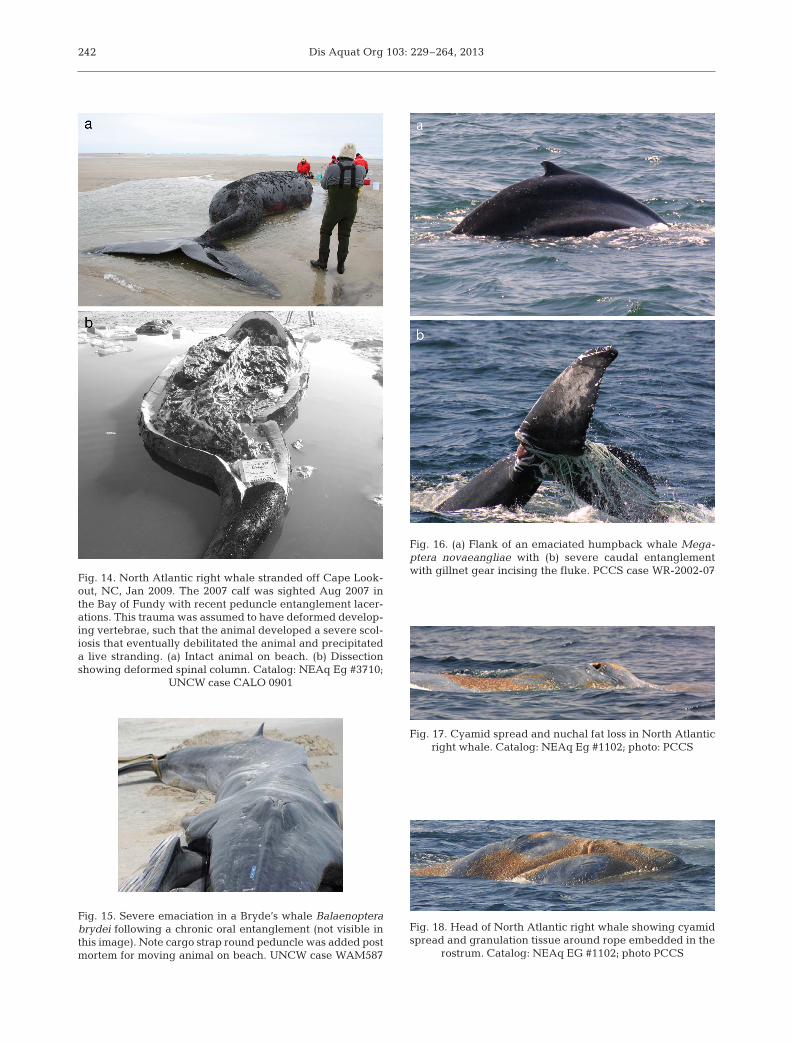
Fig. 17. Cyamid spread and nuchal fat loss in North Atlantic right whale. Catalog: NEAq Eg #1102; photo: PCCS
Fig. 18. Head of North Atlantic right whale showing cyamidspread and granulation tissue around rope embedded in the
rostrum. Catalog: NEAq EG #1102; photo PCCS
Fig. 16. (a) Flank of an emaciated humpback whale Mega -ptera novaeangliae with (b) severe caudal entanglementwith gillnet gear incising the fluke. PCCS case WR-2002-07
Fig. 15. Severe emaciation in a Bryde’s whale Balaenopterabrydei following a chronic oral entanglement (not visible inthis image). Note cargo strap round peduncle was added postmortem for moving animal on beach. UNCW case WAM587
Fig. 14. North Atlantic right whale stranded off Cape Look-out, NC, Jan 2009. The 2007 calf was sighted Aug 2007 inthe Bay of Fundy with recent peduncle entanglement lacer-ations. This trauma was as sumed to have deformed develop-ing vertebrae, such that the animal developed a severe scol-iosis that eventually debilitated the animal and precipitateda live stranding. (a) Intact animal on beach. (b) Dissectionshowing deformed spinal column. Catalog: NEAq Eg #3710;
UNCW case CALO 0901

Moore et al.: Pinniped and cetacean anthropogenic trauma case criteria 243
Fig. 20. California sea lion Zalophus californianus chroni-cally entangled in gillnet. TMMC case CSL 4909 (Chelsea)
Fig. 21. Northern elephant seal Mirounga angustirostriswith a chronic entanglement in a toilet seat. Ano Nuevo, CA.
Photo: TMMC
Fig. 22. Damage to North Atlantic right whale blowhole.Rope was entwined around the left baleen plates, passedacross the mouth, exiting the right side and then taughtlyembedded over the left nares before tightly constrictingaround the left flipper. Catalog: NEAq Eg #2301 VAq;
VAQS2005-1008Eg
Fig. 23. Dissection of North Atlantic right whale peduncle.Chronic stricture around the ventral fluke insertion severedthe 2 lateral superficial veins draining the fluke. These thenhealed, as seen in this image. The walls of the veins are dis-sected to show their blind ending at left (black ovals). Thecourse of the lacerating rope is shown with a dashed white
line. Catalog: NEAq Eg #3107; photo: WHOI
Fig. 19. North Atlantic right whale May 25, 2008. Firstsighted with entangled right flipper (upper left of image)March 17 2004, gear still attached March 2010, last seen Jan16, 2012 in moderate condition, but unclear if still entangled.
Catalog: NEAq Eg #3346; photo: NEFSC

Dis Aquat Org 103: 229–264, 2013
death, but are more likely to sink if they havebecome emaciated and thus less buoyant due todepletion of lipid stores (Allison et al. 1991, Reisdorfet al. 2012). Also, not all persistent entanglements arefatal (Fig. 19), although the long-term sub-lethaleffects of these persistent entanglements are notclear.
Host responses
Host responses include emaciation and, in rightwhales, parallel white lines radiating from the blowhole (also called ‘rake’ marks; Fig. 25) (Pettis et al.2004). The timing of these changes appears to bevariable. Skin discoloration (mottling from extremesloughing), swath lesions (Fig. 25), and flipper discol-oration are also observed. Note the ability to detectskin color change depends on normal skin color:white humpback whale flippers and right whale bellypatches show bruises, whereas black skin does not. Incontrast, skin edema and sloughing is better seen inblack skin. Persistent spinal deviation, altered respi-ration and lethargy have also been observed. Thereare numerous other causes of these host responses,but in the context of persistent entanglement, thesechanges are commonly related to trauma (Fig. 14).
Injury-specific gross necropsy findings
These can include hematoma, edema, and shock(multi-systemic congestion and disseminated intra -vascular coagulation). Ingested material and debrismay be found. Incompletely healed scars with orwithout associated abscessation, fractures, and serousatrophy of fat are also present at times. Disuseosteopenia; muscular and skeletal damage such asjaw, flipper, spine fracture; disuse atrophy; and for-mation of pseudoarthroses in the jaw have beendescribed (Moore et al. 2004, Cassoff et al. 2011).
Lesions can also include granulation, abrasion, lac-eration, impression, incision, and constriction (Mooreet al. 2004, Cassoff et al. 2011). Fishing gear may beembedded to various depths (Figs. 13, 16b, 18 & 20)and may be accompanied by massive fibro-osseousperiostitis (Fig. 12) (Moore et al. 2004, Cassoff et al.2011).
Toxicology
Biotoxins may impair mental awareness, changebehavior and precipitate anthropogenic trauma,although this has not been documented in largewhales.
Histological findings
Proximate effects can include fibrosis (early, late),acute to chronic inflammation (osteomyelitis, derma -titis, or cheilitis), hemorrhage (acute/chronic-macro-phages, hemoglobin pigments such as hematoidin),fibrin, vascular thrombosis, localized infection (openwound) (acute and chronic), myofiber degeneration/fragmentation/necrosis/mineralization, bony response,and cardiac and skeletal muscle contraction bands.
Host response effects can include serous atrophyof fat/edema, muscle atrophy, osteopenia (also re -quires gross information such as flipper weight),metabolic/stress-adrenal cortical hypertrophy, me -dullary hyperplasia, and adrenal cortical lipoidaldegeneration.
COD assignation
Confirmed
Sufficient evidence to conclude that entanglementwas the proximate COD requires a combination of
244
Fig. 24. Chronic entanglement resulting in severe necrosisand tissue defects in a live entangled humpback whale.
PCCS case WR-2006-15
Fig. 25. Rake marks (linear white marks below blowhole)and swath lesions anterior to the rake marks (Hamilton &Marx 2005) in North Atlantic right whale after severe entan-glement injury. Catalog: NEAq Eg #1608; photo: PCCS

Moore et al.: Pinniped and cetacean anthropogenic trauma case criteria
the above factors that logically lead to a major declinein health, resulting in death from consequent factors,such as inanition from emaciation, metabolic exhaus-tion from increased drag, exertional myopathy, over-whelming infection or starvation, or amputation, sec-ondary to the chronic effects of ischemic necrosis andloss. The entangling material need not still be presentat the time of death. The post-mortem condition ofcases examined to date has rarely allowed diagnosisof ultimate COD, thus inference from the above at-sea observations, gross necropsy data, and histologi-cal information as available, need to be assimilatedinto an assessment of the most parsimonious interpre-tation of the reason(s) for the demise of the animal.
Probable
A finding of probable would arise if some or all ofthe above factors were present, but carcass qualitycould not allow confident linkage of entanglementevidence with observed condition of the mortality.
Suspect
Suspect cases would have evidence of current orpast entanglement, without sufficient findings to linkthe entanglement to major consequent changes inthe animal, but that still had a suggestion of linkage.
BLUNT FORCE TRAUMA INDUCED BY VESSELCOLLISIONS WITH LARGE WHALES
William A. McLellan1, Michelle Berman, Tim Cole,Alex M. Costidis, Amy Knowlton, Janet Neilson,
D. Ann Pabst, Stephen Raverty
Background
Collisions between watercraft and pinnipeds andcetaceans can have adverse effects on the health ofindividual animals as well as the population status ofsome endangered species (e.g. NARW) (Kraus et al.2005). The severity and type of trauma resulting froma collision depends on a number of factors, includingvessel speed and size (Fig. 26) (Laist et al. 2001, Van-derlaan & Taggart 2007), the angle of impact, theanatomic site of contact with the body, and whether
the strike occurred ante- or post-mortem. In contrastto sharp traumatic injuries to large whales typicallyassociated with propeller strike, blunt force traumamay be attributed to a number of physical insults. Asmost necropsy data on large whales are collectedfrom carcasses hauled up onto beaches, care shouldbe taken interpreting pre- and post- mortem traumathat could be caused by the necropsy itself (Fig. 27).Here, we will focus on blunt trauma injuries associatedwith ante-mortem vessel collisions with large whales.
Broadly, blunt trauma injuries fall into 4 categories,defined by DiMaio & DiMaio (2001) as (1) abrasions,(2) contusions, (3) lacerations, and (4) fractures of the
245
Fig. 26. Humpback whale and cruise ship in Glacier Bay,Alaska. Photo taken under NMFS Scientific Research Permit
No. 945-1776-00
Fig. 27. Manipulating North Atlantic right whale up to ahigh position on the beach for necropsy. The left fluke wasmissing when initially discovered, thus rolling the carcass ismore practical than the usual attachment of a hawser to thepeduncle, despite the fact that rolling with machinery in-creases the risk of post-mortem bone damage. Catalog:
NEAq Eg #1909; photo: VAq
1For author affiliations, see Supplement 5 at www. int-res.com/ articles/suppl/d103p229_supp/
Dis Aquat Org 103: 229–264, 2013
skeletal system. Campbell-Malone et al. (2008, p. 51)characterized blunt force trauma as follows: ‘mechan-ical stress applied to a body causes blunt forcetrauma. To cause a blunt force injury, stress appliedto the tissue must be great enough to deform theelastic or viscoelastic tissue beyond its ability torecover or maintain integrity. This can occur in situa-tions where (1) the magnitude of the applied stress isgreater than the ultimate strength of the tissue; (2)the stress is imparted in an unnatural direction, load-ing the tissue in a direction with weaker materialproperties; or (3) the stress is applied to mechanicallyinferior pathologic tissue.’ As a result, blunt forcetrauma disrupts the inte grity of tissues and can resultin impact abrasions, contusions (hematoma), tears,shears, and crush injuries.
Blunt force trauma injuries in large whales maybe attributed to a number of physical insults, includ-
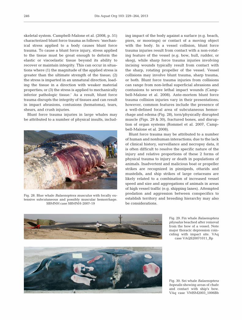
ing impact of the body against a surface (e.g. beach,piers, or moorings) or contact of a moving objectwith the body. In a vessel collision, blunt forcetrauma injuries result from contact with a non-rotat-ing feature of the vessel (e.g. bow, hull, rudder, orskeg), while sharp force trauma injuries involvingincising wounds typically result from contact withthe sharp, rotating propeller of the vessel. Vesselcollisions may involve blunt trauma, sharp trauma,or both. Blunt force trauma injuries from collisionscan range from non-lethal superficial abrasions andcontusions to severe lethal impact wounds (Camp-bell-Malone et al. 2008). Ante-mortem blunt forcetrauma collision injuries vary in their presentations;however, common features include the presence ofa well-defined focal area of subcutaneous hemor-rhage and edema (Fig. 28), torn/physically disruptedmuscle (Figs. 29 & 30), fractured bones, and disrup-tion of organ systems (Rommel et al. 2007, Camp-bell-Malone et al. 2008).
Blunt force trauma may be attributed to a num berof human and nonhuman interactions; due to the lackof clinical history, surveillance and necropsy data, itis often difficult to resolve the specific nature of theinjury and relative proportions of these 2 forms ofphysical trauma to injury or death in populations ofanimals. Inadvertent and malicious boat or propellerstrikes are recognized in pinnipeds, ottarids andmustelids, and ship strikes of large cetaceans arelikely related to a combination of increased vesselspeed and size and aggregations of animals in areasof high vessel traffic (e.g. shipping lanes). Attemptedpredation and ag gression between conspecifics toestablish territory and breeding hierarchy may alsobe considerations.
246
Fig. 29. Fin whale Balaenopteraphysalus beached after removalfrom the bow of a vessel. Notemajor thoracic depression coin-ciding with impact site. VAq
case VAQS20071011_Bp
Fig. 30. Sei whale Balaenopterabopealis showing areas of chafeand contact with ship’s bow.VAq case VMSM2003_ 1006Bb
Fig. 28. Blue whale Balaenoptera musculus with focally ex-tensive subcutaneous and possibly muscular hemorrhage.
SBMNH case SBMNH-2007-19

Moore et al.: Pinniped and cetacean anthropogenic trauma case criteria
Although entanglement injuries may also be con-sidered sharp or blunt force trauma, they will not beconsidered in this section.
Blunt force trauma of natural origins
Non-anthropogenic blunt force injuries, such asthose associated with intra- and inter-specific aggres-sion can lead to mortality, are well-described else-where for small cetaceans (Ross & Wilson 1996, Pat-terson et al. 1998, Dunn et al. 2002) but not for largewhales as yet.
An age-related injury is specifically recognized inneonatal bottlenose dolphins with infanticide (Dunnet al. 2002) and harbor porpoises, killed by bottlenosedolphins (Ross & Wilson 1996). No external lesionsmay be apparent on necropsy and reflection of theskin may reveal focally extensive subcutaneous hem-orrhage within the throat, thoracic and abdominalregions. Massive thoracic and abdominal hemor-rhage with occasional lung and liver ruptures arerecognized. Physical trauma has also increasinglybeen documented in walruses; due to receding icelevels, increasing numbers of adult, subadult andjuvenile animals haul out on shore and when star-tled, stampede and trample primarily young animals(Fay & Kelly 1980). Attempted predation and aggres-sion with conspecifics may also be considerations.Impacted animals may present with protruding andoccasional hemorrhagic eyes, swollen heads, ap -pendages and random regions throughout the trunk.Involvement of appendages or the facial region mayimpede normal locomotion (loss of function) andinterfere with foraging and predation. In cetaceans,animals may present with no apparent externallesions, superficial poorly circumscribed transverseto oblique linear white depressions, focally extensivepallor, subcutaneous swelling or asymmetry of thetorso (vertebral dislocation).
Case definition
Signalment
In large whales all age classes and both sexes aresusceptible to blunt force trauma induced by vesselcollisions. Stranding data indicate that in some spe-cies calves and juveniles may be at a higher risk ofvessel collisions than adults (Wiley et al. 1995, Knowl-ton & Kraus 2001, Laist et al. 2001, Panigada et al.2006, Douglas et al. 2008, Carrillo & Ritter 2010, Neil-
son et al. 2012), although in blue whales vessel strikeshave primarily involved adults. However, it is un-known if young animals are more likely than adults tobe struck by vessels (based on differences in their be-havior, sightability, or other factors) and/or if younganimals are more likely to suffer lethal injuries fromblunt force trauma because of their smaller body size.
Epidemiology
Most commonly, anthropogenic blunt force in juriesare associated with vessel strikes, although impactwith piers, moorings and other submerged structuresmay also be considerations. Blunt force trauma ispossible whenever vessels and animals are in closespatial proximity. Blunt force trauma wounds havebeen observed in an array of small and largecetaceans, ranging from coastal delphinids to mys-ticetes (Wells & Scott 1997, Moore et al. 2004, VanWaerebeek et al. 2007, Wells et al. 2008). Vessel col-lisions, including those involving fatal vessel strikes,have been documented during all months of the year(Laist et al. 2001, Panigada et al. 2006). Mapping ofdocumented blunt force injury trauma cases mayprovide valuable insights into additional contributingfactors (e.g. NARW calving grounds) (Neilson et al.2012, van der Hoop et al. 2013).
Fourteen of the 30 NARW for which necropsyreports were examined by Moore et al. (2004) weredetermined to have been killed by vessel collisions.This represents at least 3% of the population, and isprobably an underestimate given the uncertaintiesregarding carcass recovery rates. Similarly, Camp-bell-Malone et al. (2008) found that 21 of 40 NARWnecropsies identified vessel collision as the mostprobable COD, with 9 (22.5%) resulting specificallyfrom blunt trauma. Van der Hoop et al. (2013) sum-marized data from 1762 mortalities (all known) andserious injuries (likely fatal) involving 8 species oflarge whales in the Northwest Atlantic (23.5°N to48.0°N), from 1970 through 2009. Vessel strike wasthe third leading determined COD (171/750) for allspecies combined, and the leading determined CODfor fin (59/116) and right (38/87) whales.
Injury-specific response protocol
In addition to the data and sample collection meth-ods described in the ‘Introduction and overview’ sec-tion (e.g. Fig. 31), the following steps should be con-sidered to fully document a blunt-trauma case.
247

Dis Aquat Org 103: 229–264, 2013
The carcass should be fully flensed to examineskeletal elements for evidence of blunt trauma andeither fracture, subluxation or luxation with hemor-rhage (Fig. 2; see ‘Introduction and overview’).
Ancillary diagnostic samples (e.g. for biotoxin test-ing, bacteriology, virology, parasitology, hormonaltesting) should be collected when practical to ex -clude or confirm the possible contribution of otherpathologic factors to ante-mortem morbidity and pos-sible predisposition to vessel collision. Such diagnos-tics can include diagnostic evaluation of fluids orswabs from pleural and abdominal cavities, as wellas tissue cultures and bacteriology of major internalorgans. If there is evidence of material transfer (e.g.hull paint), sample the tissue or object and preserve.
If blunt trauma is found, diagrams and photographsof the lesions should be supplied with the grossreport (Figs. 2, 28 to 30).
Clinical signs
Typical signs include impaired locomotion, ab -normal body posture/positioning, lethargy, sub cuta -neous swelling, external discoloration of the skin atthe site of impact, impaired or forced respirations,disarticulation or malocclusion of the mandible, pro-lapsed eyes, periorbital swelling and conjunctivalhemorrhage, hemorrhage from the nares or blow-hole, shock, unresponsiveness, inappetence or ano -rexia. Localized to regional asymmetric subcuta-neous swelling with elevation of the affected areaabove the plane of adjoining, normal skin may occur.Conversely, impacted areas can show marked de -pression at the site of the injury related to destructionof underlying tissue (Figs. 29 & 32). The externalaspect of the impact site may be de-pigmented with
white transverse to oblique poorly circumscribedbands or depressed furrows (Fig. 30) or focally exten-sive pallor (Fig. 33) related to more superficial abra-sions, erosions, and lacerations. Visual health assess-ment (cyamid load, color) can provide information ongeneral health status (Pettis et al. 2004). Circulatoryassessment via infrared thermography can also bevery instructive in relation to hemorrhage and edemain the acute phase of injury to inflammation, woundhealing, and possible resolution with more chronicnon-lethal progression of the injury. Varying degreesof wound resolution with primary and/or secondaryintention healing may be observed.
Clinical diagnostic data are often unavailable;however, depending on the time between impact andsubsequent clinical evaluation or post-mortem exam-ination, an inflammatory leukogram, electrolyte im -balances, possible anemia, and elevated serum amy-loid A (SAA) and creatinine kinase may be observed.
Injury-specific gross necropsy findings
Field observations of floating or beach-cast animalsmay include localized to regional asymmetric subcu-
248
Fig. 32. Live humpback whale (southeastern Alaska ID #954in 2008) The origin of the injury, first seen in 1989, is un-known, however the lesion closely resembles confirmedbow strike lesions. Photo: Steve Lewis (taken under NMFS
Scientific Research Permit No. 14122)
Fig. 33. Focal pallor (arrow) on the left flank of a live hump-back whale approximately 1 h after the whale was struck atthat point by the bow of a 72 ft (~22.2 m) catamaran in GlacierBay on Aug 31, 2011. NMFS AK Region accession #2011142
Fig. 31. Field response team collecting external morphomet-rics on the fetus of North Atlantic right whale on 14 January,
2005. Catalog: NEAq Eg #2143 ‘Lucky’; photo: UNCW

Moore et al.: Pinniped and cetacean anthropogenic trauma case criteria
taneous swelling with elevation of the affected areaabove the plane of adjoining, normal skin. The exter-nal aspect of the impact site may be depigmentedwith white transverse to oblique poorly circum-scribed bands or depressed furrows or focally exten-sive pallor related to more superficial abrasions, ero-sions, and lacerations (Fig. 29).
However, in cases of impact with smooth objects,there may be no externally visible signs of blunttrauma, or signs may only be observed followinginternal examination and retrospective correlation ofexternal lesions. On reflection of the skin, variablyextensive subcutaneous hemorrhage (Fig. 34), whichmay track dependently as well as extend deepinto the adjoining skeletal musculature (Fig. 35), is typically observed. Evaluation of the impact site forassociated subcutaneous edema and hemorrhage is
imperative. Should the animal survive impact/previ-ous injury, resolution of the hemorrhage should beapparent on gross exam and histopathology. In moreseverely affected animals, physical impact may resultin muscle shredding, especially surrounding bones,and rupture of the liver or spleen, or lung with mas-sive hemoperitoneum or hemothorax, respectively.
Histological samples should be collected and thelocation and extent of the defect described.
Large volumes of dark red serous fluid are com-monly observed in the abdominal cavity, and to amuch lesser extent, thoracic cavities; it is important todifferentiate this fluid from acute hemorrhage bydetection in the latter of fibrin strands on serosal sur-faces, spontaneous clot formation on exposure to air,and other criteria. Herniation, transposition and pro-lapse of internal viscera may also be apparent. If theinjury is localized to the head, careful dissection ofthe cranium and assessment of the superficial brainfor coup contre coup lesions is recommended. Brainswelling and occasional herniation of the brain stemand posterior cerebellum through the foramen mag-num may also occur. Cranial, mandibular, scapularand rib fractures and luxation and subluxation of ver-tebrae at the point of impact with occasional com-minuted fractures of lateral or dorsal processes mayalso be apparent (Figs. 36 to 38).
Histological findings
Histologic findings in whales with blunt traumavary considerably based on anatomic site of impact,
249
Fig. 34. North Atlantic right whale focal hemorrhage in lat-eral blubber with sample location identified by 2 incisions.Note epidermis has been shed post mortem in this region of
the animal. WHOI case MJM0906Eg
Fig. 35. Deep hemorrhage on the lateral surface of the leftribs of a North Atlantic right whale. UNCW case KLC 022 Eg
Fig. 36. North Atlantic right whale axial skeleton laid out insequence to locate multiple fracture of the left transverseprocesses, and a shattering of the lumbar vertebra in the
green gloved hand. WHOI case MJM 9406Eg

Dis Aquat Org 103: 229–264, 2013
deep visceral versus more superficial subcutaneousinvolvement, carcass condition code, and temporalassociation between wound injury and death (e.g.peracute, acute, chronic) (Campbell-Malone et al.2008). In peracute lethal situations, no microscopiccellular responses may be apparent. In the earlystages of injury, hemorrhage (Fig. 39) admixed withvariable amounts of edema and fibrin deposition maybe apparent in the subcutis and subjacent muscula-ture. If the injury is non-lethal, the hemorrhage mayresolve in 14 to 21 d with a progression from red orblue (2 to 4 d) to green (4 to 7 d) to yellow (7 to 14 d)discoloration of the area associated with metabolismof hemoglobin, hema toidin and hemosiderin. Thesecolor changes may not be easily identifiable onwhales with very dark epidermis or carcasses only
photographed floating at sea. Myocel-lular degeneration and necrosis maybe apparent.
Over time, inflammatory cells are recruited to the affected area with fibro plasia, neovascularisation andeventual granulation tissue formation.Secondary microbial involvement isunlikely in acute cases; however, if apre-existing bacterial infection waspresent, localization to the hematomaand hemorrhage with abscessationmay occur. Clostridial myositis hasbeen documented (Greenwood & Tay-lor 1978); however, this is uncommon.Detailed microscopic review of select
live beach-cast animals with prolonged recumbencyhas not demonstrated any indication of compartmen-tal type syndrome (Mabee & Bostwick 1993) or myo -globinuria. Abdominal or thoracic visceral fractureswith hemorrhage and fibrin deposition, as well asdraining hemorrhage in regional lymph nodes maybe evident.
Toxicology
At present, anecdotal evidence suggests a possiblecorrelation of elevated domoic acid and vessel inter-actions with select pinniped and cetacean species.Biotoxins (domoic acid, brevetoxin, saxitoxin) havebeen documented in some instances of large whale
250
Fig. 38. Oral rete in North Atlantic right whale, with evi-dence of soft tissue tearing and coagulated blood in thebaleen racks (arrow). Deep to the tears was a complete frac-ture of the premaxillary and maxillary rostral elements. Catalog: NEAq Eg #1004 ‘Stumpy’; VMSM 2004 1004 Eg
Fig. 39. Extravasation of erythrocytes in the fibroadipose of aBryde’s whale. Considered to be most suggestive of an ante-mortem occurrence, though hypostatic congestion could alsooccur if the abrasions were located in a region that would be
considered ‘dependent’. MMPL0906. Photo: D. Rotstein
Fig. 37. Humpback whale with (a) the right mandible protruding laterally be-yond the contour of the skull and (b) the compound fracture in the right
mandible. VAq case VMSM 961010

Moore et al.: Pinniped and cetacean anthropogenic trauma case criteria
strandings (Geraci et al. 1989). More comprehensivepost-mortem examinations of suspect animals andscreening for harmful algal blooms may providevaluable insights into the potential contributing rolesof these compounds (see case report V10-137 Mn,7/30/ 2010 from Douglas Island, Juneau, Alaska;NMFS AK Region accession #2010089).
Confirmed versus suspect cases
Confirmed
Witnessed and documented strikes or mortalitywith corroborating lesions are the most confident factors to confirm a case (e.g. Whale 68, NMFSAK Region accession #2011038; SBMNH caseSBMNH2007-20 Mn). The sum of the observationsshould lead to death by blunt trauma being the mostparsimonious explanation for the case to be con-firmed. Such cases should include a number of thefollowing: frank hemorrhage with edematous fluid inthe subcutaneous tissue; hematoma formation; lacer-ation or rupture with hemorrhage within the skeletalmusculature; hemothorax; hemoperitoneum; visceraldisplacement, herniation or rupture; skeletal frac-tures, luxations or subluxations with associated hem-orrhage; microscopic fat emboli; acute hemorrhage;edema; rhabdomyocytolysis; subcapsular and me dul -lary draining hemorrhage in regional lymph nodes;history of animal on bow of vessel. Evidence ofchronic lesions (pyothorax, abscessation) may ac -company findings.
Probable
This conclusion requires a necropsy (as practical),blunt-trauma sequelae, and histopathology support-ive of gross findings of trauma if available to be col-lected (e.g. MMPL0906 Bb, VAQS 20051017 Bp,KMS 374 NEAq Eg #1308 UNCW). A ‘probable’ casewill have similar gross necropsy and histopathologyfindings to a ‘confirmed’ case but insufficient infor-mation to conclude that other interpretations of CODare not as likely.
Suspect
Cases where necropsy was limited or no necropsywas conducted, or cases with advanced decomposi-tion and blunt-trauma sequelae, but limited or no
histopathology findings of trauma, cannot be ratedmore confidently than ‘suspect’. Unilateral and local-ized abrasions with epidermal pallor, depressedbands or furrows of depigmented epidermis and sub-cutaneous swelling may be considerations. Abnor-mal body posture/positioning, vertebral fracturesand distension of the abdominal cavity may also sup-port blunt trauma. Prolapse of the colon, reproduc-tive tract or stomach in fresh dead animals may alsosuggest deep blunt force trauma. Blunt-traumasequelae and/or bony lesions consistent with blunttrauma may be present; the carcass may or may nothave other signs of pathology from entanglement ordisease (e.g. UNCW case NCARI 006 Eg).
SHARP TRAUMA INDUCED BY VESSEL COLLISIONS WITH PINNIPEDS
AND CETACEANS
Alex M. Costidis1, Michelle Berman, Tim Cole,Amy Knowlton, William A. McLellan, Janet
Neilson, D. Ann Pabst, Stephen Raverty
Background
Watercraft collisions can take the form of blunt-force impacts involving contact between the animaland some non-rotating feature of the vessel (e.g. hull,rudder, skeg), sharp-force injuries such as incisingand/or chopping wounds typically generated by thesharp, rotating propeller of the vessel, or a combina-tion of the 2 types of injuries (Lightsey et al. 2006,Rommel et al. 2007, Byard et al. 2012). Additionally, aless distinct category is occasionally observed inwhich a relatively sharp but non-rotating feature of avessel (e.g. skeg or rudder) has enough speed andsharpness to penetrate the skin, causing a combina-tion of blunt and sharp-force injuries likely bestdefined as a chop wound. Sharp-trauma injuries tocetaceans and pinnipeds range from mild nonfatalsuperficial nicks to severe amputations and otherlethal wounds (Kreuder et al. 2003, Campbell- Malone et al. 2008).
Injuries resulting from sharp force trauma in pin-nipeds and cetaceans vary in their presentation;however, a common feature is the presence of morethan one approximately parallel and roughly equi-distant laminar incising wound ranging from simplelinear to abruptly curvilinear or sigmoid (S- or anti-S
251
1For author affiliations, see Supplement 5 at www. int-res.com/ articles/suppl/d103p229_supp/

Dis Aquat Org 103: 229–264, 2013
shaped) defects (Rommel et al. 2007). This presenta-tion is typically associated with propeller-inducedtrauma. Vessel-related sharp-force trauma can alsopresent as a single wound composed of a linear orcurved incision or chop wound. This is more commonin cases involving contact with a large propeller, rel-ative to the animal’s size, along appendages andexternal protuberances, or along regions of signifi-cant convexity of the animal’s surface where only onepropeller blade makes contact. The depth of penetra-tion of the propeller is related to the proximity of thepropeller to the animal as well as the propeller radiusand the type of propeller (e.g. exhaust-through hubvs. narrower shaft-driven hub without exhaust-through hub). The type of propeller (e.g. perform-ance propeller with high rake vs. working propellerwith low rake) can dictate the degree of curvature ofthe incising wounds. The distance between succes-sive propeller wounds (i.e. cut span) is sometimesrepresentative of the propeller pitch but can varysubstantially due to operational characteristics of thevessel. Although not definitively diagnostic of pro-peller or vessel type, cut span can sometimes providevaluable information when coupled with otherwound features such as wound shape, chord lengthand chord depth (Rommel et al. 2007). It should benoted that the distance between successive propellerwounds is also affected by the gape of the wound.Since incising wounds have a tendency to gape dueto intrinsic (e.g. Langer’s lines due to skin/blubberfiber orientation) and extrinsic forces (e.g. shearingand stretching forces generated by locomotory move-ments) it can hinder interpretation of cut span(DiMaio & DiMaio 2001, Rommel et al. 2007). Staband puncture wounds are also considered to be sharptrauma wounds but are not typically associated withwatercraft-induced wounds.
Non-anthropogenic sharp-force injuries can lead tomortality but are often sporadic (e.g. infanticide,attempted predation) or not associated with signifi-cant trauma (e.g. conspecific rake marks) and aretherefore not discussed.
Case definition
Signalment
There is no gender bias known at the present time,though there could be bias due to ship strikes in calv-ing grounds where females are more common (e.g.NARW) (van der Hoop et al. 2013). Wounds havebeen documented to heal, only to break down years
later during pregnancy (Fig. 40). Age biases appearto exist with respect to likelihood of ship strike, withjuveniles showing a greater tendency to be struck(Wiley et al. 1995, Hamilton et al. 1998, Knowlton &Kraus 2001, Laist et al. 2001, McLellan et al. 2004,Panigada et al. 2006, Lammers et al. 2007, Douglas etal. 2008, Berman-Kowalewski et al. 2010, Carrillo &Ritter 2010, Neilson et al. 2012). Biases related torecovery rates of perinates/juveniles are not known.Nonetheless, there could be reduced recovery of pro-peller-struck neonates/juveniles due to smaller size,lesser positive buoyancy due to modest fat reserves,and greater percentage of tissue loss due to preda-tion. It is unknown whether injured perinates/juve-niles are more likely to be predated upon or scav-enged, and hence less likely to be recovered.
Epidemiology
Most commonly, sharp-force injuries in pinnipedsand cetaceans are associated with watercraft vesselinteractions. Sharp-force trauma is possible when-
252
Fig. 40. North Atlantic right whale presenting large pro-peller wounds and resolving scars along the dorsolateral as-pect of the torso. Injuries were incurred 14 yr previously as acalf. Note the variation in pathologic presentation in thiscase. In the more cranial region, there is second intentionhealing, whereas the most caudal incision features partialretraction of the wound margin and exposure of the underly-ing dermis. There is patchy loss of epidermis due to post-mortem exfoliation. Histopathology of the wound marginsconfirmed healing skin, and the cause of death was attrib-uted to long-past propeller-induced sharp trauma likelyopened up by her first full-term pregnancy, with presump-tive secondary sepsis/bacteremia. Expansion of the bodycavity by the uterus containing the fetus, fetal membranes,and fluids was thought to have resulted in increased abdom-inal pressure and stretching of the abdomen resulting instructural tension greater than the tensile strength of thescarred propeller wounds. Catalog: NEAq Eg #2143 ‘Lucky’;
photo: UNCW

Moore et al.: Pinniped and cetacean anthropogenic trauma case criteria
ever vessels and animals are in close spatial and temporal proximity, as is seen in areas of highvessel traffic, particularly along the continentalshelf (Fonnesbeck et al. 2008, Berman-Kowalewskiet al. 2010). Propeller wounds have been observedin an array of small and large cetaceans, rangingfrom coastal delphinids to mysticetes (Wells & Scott1997, Moore et al. 2004, Van Waerebeek et al. 2007,Wells et al. 2008), as well as sea otters and anumber of pinnipeds (Goldstein et al. 1999, Kreuderet al. 2003). Campbell-Malone et al. (2008) foundthat 21 of 40 NARW necropsies identified vessel col-lision as the most probable COD, with 11 (27.5%)resulting specifically from sharp trauma. Propellerinjuries have been observed during all months ofthe year; however, specific temporal and spatialexceptions may occur in some species, particularlywhen feeding or breeding ranges intersect withshipping routes or boating channels (e.g. bluewhales and NARW) (Fonnesbeck et al. 2008,Berman-Kowalewski et al. 2010). In some cases (M.J. Moore unpubl. data), drift analyses have beenuseful for assessing the possible origin of mortalityand transiting of shipping lanes. Retrospective stud-ies to identify more frequented shipping lanes maybe indicated.
Clinical signs of sharp trauma
Clinical signs
Depending on the location, distribution and sever-ity of the trauma, typical signs may include impaired locomotion, with logging or listing at the water sur-face; single or multiple approximately parallel, linearto curvilinear, roughly equidistant, shallow todeeply-penetrating incising or chop-ping wounds with associated hemor-rhage or exposed red and/or pinkmuscle (if acute), or gray to tan/whitemuscle (if more chronic). In thosecases where large diameter blood ves-sels have been incised, extensive hem-orrhage and lethargy may be appar-ent. In more chronic lesions, woundrepair around the margins of thelesions with focal or multifocal, irregu-lar and/or excessive cyamid distribu-tion along body may occur. In thosecases of traumatic lethal amputations,portions or entire appendages, trunkor head may be lost.
Diagnosis of sharp trauma is based upon the fol-lowing: incisions of skin (Figs. 41 to 44) and possiblydeeper structures that may vary in size, depth, extentand location; presence or absence of hemorrhage,amputation, pneumothorax, extrusion of thoracic orabdominal viscera; buoyancy abnormalities; second-ary infections; or epidermal color changes (e.g. gen-
253
Fig. 41. Flukes of a North Atlantic right whale incised by apropeller: (a) March 10, 2005, (b) September 3, 2005. Notethe substantial cyamid spread and tissue failure. The animalhas not been sighted since. Catalog: NEAq Eg #2425;
photos: (a) NEAq, (b) Tim Voorheis
Fig. 42. Sublethal propeller incisions in the dorsal aspect of a humpback whale calf. Photo: PCCS
Fig. 43. North Atlantic right whale. Extending from the rostrum along the leftlateral aspect of the torso, there are 20 curvilinear propeller incisions of vary-ing depth and length with more pronounced ‘gaping’ along the thoracic wall.Note the absence of visible hemorrhage associated with wound margins (pre-sumptive washout), but blood pouring out of the ventral aspect of cut 8. Thewhite areas of shark scavenging can be seen along many of the propellerwounds; this is a common finding that can affect evaluation and interpretation
of sharp-trauma wounds. FWC case GA2006025

Dis Aquat Org 103: 229–264, 2013
eralized pallor) associated with wound sites. Visualhealth assessment (cyamid load, skin or fur color,body condition) can provide information on generalhealth status of impacted animals (Pettis et al. 2004).Similar to blunt-trauma cases, circulatory assessmentvia infrared thermography can also be instructive inrelation to wound healing or inflammation. Varyingdegrees of healing may be present, with primaryand/or secondary intention healing visible.
Even shallow open wounds represent an entry pointfor opportunistic or secondary microbial colonization,proliferation and deeper tissue invasion, which mayinterfere with wound resolution. Additionally, scar tis-sue can affect the resilience and function of affectedtissue, potentially contributing to reduced tensilestrength, as evidenced by right whale ‘Lucky’ (NEAq#2143) (Fig. 40) or possible restriction in range of mo-tion, with involvement of an appendage.
Clinical observations and diagnostic data are oftenunavailable; however, inflammatory leukograms,elec trolyte imbalances, inflammatory response, ane-mia, elevated serum amyloid A and creatinine kinasehave been observed in live stranded animals andmay provide further support regarding prognosis forrecovery or immediate COD (Dierauf & Gulland2001).
Gross necropsy findings
Gross findings of sharp trauma range from smallincising wounds to large transecting and/or bisectingchop wounds (Figs. 41 to 43) or amputations (Figs. 44& 45) (DiMaio & DiMaio 2001, Lightsey et al. 2006,Rommel et al. 2007). Soft tissue damage that pene-trates the blubber and deeper muscle layers is often
associated with shearing and/or shredding of musclefibers, severing of large caliber blood vessels andpotential hypovolemic shock (Lightsey et al. 2006,Campbell-Malone et al. 2008). Deeper penetratingincisions may extend to and involve internal organs(Fig. 43). Depending on anatomical location, the mus -culoskeletal and integumentary systems can be dif-ferentially affected by shallow and/or deeply pene-trating wounds.
The time between initial insult and death may beso abrupt that death may ensue with few microscopiclesions (peracute death). In animals surviving the ini-tial trauma, gross and microscopic changes mayrange from a defect in the skin with varying degreesof hemorrhage and edema (e.g. Fig. 42), to resolvingwounds with primary and second intention healing(Fig. 40). The absence of blood in an incised or chopwound does not discount an antemortem event, asprolonged wound exposure to water results inhemolysis and leaching of blood from wounds.Depending on the anatomic location, such as moremobile regions of skin in the axillae or oral cavity,complete resolution of the injury and scar formationmay be compromised due to repetitive motion or lackof apposition of the wound margins. When a largecaliber blood vessel is incised or transected massivehemorrhage and exsanguination occurs (e.g. Fig. 45).In many cases, prolonged time between death andnecropsy of pinnipeds and cetaceans may result intissue decomposition and hamper pathologic assess-ment of wounds.
254
Fig. 45. North Atlantic right whale showing unilateral ampu-tation of the left fluke with degloving of the skin to the mi-dlevel of the peduncle. Note the angulated and abrupt mar-gin of intact skin, cranial to this defect. The cause of deathwas attributed to acute propeller-sharp trauma, with hypo -volemic shock. The macerated subcutaneous tissue is due toprolonged immersion in salt water and putrefaction. Catalog:
NEAq #1909 KMS374; UNCW case
Fig. 44. North Atlantic right whale exhibiting fluke amputa-tion at the mid-peduncle and a large gaping propeller inci-sion through the abdominal musculature, partially exposingloops of intestines and providing a portal for salt water to
enter the abdomen. UNCW case EgNEFL0602

Moore et al.: Pinniped and cetacean anthropogenic trauma case criteria
Histological findings
The microscopic features of sharp trauma vary con-siderably and depend on carcass condition code, ex-tent of environmental wound exposure, secondarymicrobial invasion or cyamid infestation, and stage ofpathogenesis of the wound (e.g. peracute, acute,chronic resolving). The histopathological presentationof wound exposure (to salt water, heat, or air drying)in pinnipeds and cetaceans is not well characterized.Typical findings of acute sharp trauma may includesubcutaneous edema and hemorrhage with myofiberdegeneration, necrosis, and contracture. Wound-as-sociated hemorrhage and myocellular de generationand necrosis (Fig. 46) can be important determinantsof ante- versus peri- or post-mortem injury; however,artifactual loss (e.g. imbibed water due to disruptionof the epidermis or autolysis) and/ or peracute deathbefore manifestation of microscopic lesions may con-found interpretation. Sequelae to sharp injury traumaare typically primary or secondary intention injury-re-pair (e.g. healing propeller wounds), although sec-ondary microbial involvement, pre-existing systemicdisease, and nutritional status of the affected animalmay impede or disrupt this process (Fig. 47). Gas in-troduction via embolization from the wound, as wellas pneumothorax may also be present.
Toxicology
Biotoxins (domoic acid, brevetoxin, saxitoxin) mayinterfere with normal mentation and possibly predis-
pose an animal to traumatic injury. Toxicants andcontaminants (e.g. PAHs) may disrupt normal im -mune function and impede inflammation and normalhealing. Gas embolization may also result from sharpwound injury, and if air can be aspirated, chemicalanalysis to determine ante versus post-mortem gasintroduction may be considered (Bernaldo de QuirósMiranda 2011).
Injury-specific response protocol
In addition to the data and sample collection meth-ods described in the ‘Introduction and over view’ sec-tion, the following steps should be considered to fullydocument a sharp trauma case. Collect wound meas-urements (e.g. chord length & chord depth) on allpropeller wounds to aid in propeller diameter andsubsequent vessel size estimation. Additional woundmeasurements as described in Rommel et al. (2007)can also inform the forensic interpretation.
255
Fig. 46. Photomicrograph of muscle beneath propeller lacer-ation in North Atlantic right whale (see Fig. 44) showing endomysial hemorrhage and muscle degeneration charac-terized by loss of sarcoplasmic striations, cytoplasmic vacuo-lation and contraction bands. UNCW case EgNEFL0602
Fig. 47. Dorsal tip of caudal scar, with neutrophils packingvascular channels and infiltrating the supporting stromaof North Atlantic right whale Catalog: NEAq Eg #2143
EgNEFL0501 ‘Lucky’. See Fig. 40 for case details

Dis Aquat Org 103: 229–264, 2013
Although sharp trauma is easily recognizable,assessment of wound injury and differentiation be -tween ante- and post-mortem trauma can be morecomplex determinations. Examination of sharp traumalesions should include detailed documentation (e.g.photo, scar/wound sketch/measurements), gross in -spection and histologic sampling of soft and hard tis-sue wound margins at various depths, to differentiatebetween peri-mortem and chronic wounds as well asassess exposure artifacts, such as salt water, bacterialovergrowth or scavenging (DiMaio & DiMaio 2001).Gross texture of wound margins (e.g. wound margincurling/fiber bunching), and histopathology of woundmargin and adjoining or exposed skeletal musclecan often provide insights into ante-mortem, peri-mortem or post-mortem injury (Shkrum & Ramsay2007). Presence of foreign bodies within the defect(e.g. propeller piece, hull paint transfer) should bewell-documented, sampled and maintained througha chain of custody (Supplement 4, available at www.int-res. com/ articles/ suppl/ d103 p229 _ supp/ ).
In cases of sharp injury to cetaceans, the skeletonshould be flensed and bones examined for evidenceof sharp- (e.g. bone nicks or shearing) and blunt-force trauma (e.g. fractures, suture separations, dis-placements). The airways should be examined forwater and/or blood aspiration and stomach contentsevaluated for blood, as cranial and pulmonarytrauma can result in regurgitation and ingestion ofblood, respectively, which provide strong supportfor ante- versus post-mortem injury. If the incisedwounds communicate with the thoracic or abdomi-nal cavity, pneumothorax and pulmonary collapse(atelectasis) should be assessed and if indicated,peritoneal/ pleural lining sampled for histology andbacteriology.
COD assignation
Due to the gross appearance of lesions associatedwith sharp trauma, suspect cases are generally notassigned. Post-mortem condition or position of theanimal in the water column or on the beach mayhamper or preclude diagnostic evaluation. Clinicalsigns or gross pathology with associated hemorrhagein Code 2 and early Code 3 carcasses are confirma-tive. In those individuals that present with moreadvanced autolysis, interpretation of gross findingsmay be hampered and microscopic assessment of tis-sues at the margins and beneath the injuries mayprovide some indication of whether trauma occurredante- or post mortem.
Confirmed
Cases can be confirmed when they include (1)necropsy findings supporting ante- or peri-mortemship strike as COD (e.g. Fig. 43, GA2006025); (2)thorough necropsy, open wounds with sharp (incis-ing) or sharp- and blunt-trauma (chop wounds)sequelae, histopathology supportive of gross find-ings of antemortem sharp trauma (e.g. Fig. 46,EqNEFL0602) or (3) a reported, well documentedvessel collision and resultant mortality with carcasspresent, where a necropsy may not be practical.
Probable
If a carcass is available for study, and necropsy hasbeen carried out as far as practical despite advanceddecomposition, open incised wounds with sharp orsharp- and blunt-trauma sequelae are present, buthistopathology findings of trauma are limited or notpresent, then a confidence level of ‘probable’ may beassigned.
Suspect
Cases which consist of a report/documentation ofcar cass, no carcass in hand or minimal examination,should be deemed ‘suspect’. If necropsy is limited ornot done, but open wounds and/or bony lesions con-sistent with sharp trauma are present, with or withoutother signs of pathology (e.g. entanglement or disease),a confidence level of ‘suspect’ could be assigned.
GUNSHOT INJURIES OF PINNIPEDS AND CETACEANS
Katie Moore1, Frances Gulland, Michelle Berman,Elsa Cabrera, Kerri Danil, Lanni Hall
Background
Gunshot wounds are types of ballistic trauma pro-duced by projectiles or other missiles launched froma firearm. These wounds may be glancing or pene-trating, and vary from incidental to lethal. Gunshotwounds will have different characteristics based onthe type of firearm, type of ammunition (projectile,
256
1For author affiliations, see Supplement 5 at www. int-res.com/ articles/suppl/d103p229_supp/

Moore et al.: Pinniped and cetacean anthropogenic trauma case criteria
shotgun pellet), angle of the shot and the distancebetween the muzzle of the firearm and the animal’sbody (Fisher & Petty 1977, Goldstein et al. 1999).Gunshot wounds inflicted on pinnipeds and ceta -ceans are most likely to be inflicted from a distance atwhich muzzle imprints and gunshot residue will notbe present (>1 ft, 30 cm).
Detailed clinical and histological examinations oflive and freshly dead small pinnipeds and ceta ceans,especially California sea lions Zalophus california -nus, have demonstrated that animals with gunshotinjuries often have multiple active disease processes,such as leptospirosis or domoic acid toxicosis. Thus,determination of the final COD and relative impor-tance of a gunshot injury in causing death can be dif-ficult. The designation of a case as a gunshot case isrelatively clear, whereas assigning COD often re -quires interpretation of all clinical and pathologydata by an experienced pathologist.
Case definition
Signalment and epidemiology
There is no consistent presentation to gunshotcases. Although more common in pinnipeds (Gold-stein et al. 1999), gunshot trauma can be found in anyspecies, age, or sex, with no specific geographic areaor season. Gunshot trauma may occur in clustersassociated with overlap of inshore migration andfishery activity specific human activities (Goldstein etal. 1999).
Clinical signs
Clinical presentation varies with the extent andduration of injury, weapon used, body part affected,and presence or absence of infection. Injuries may beassociated with external lesions (holes, fistulae, sinustracts, Figs. 48 to 50), but the skin may be healed overat the site of projectile entry and/or exit (Figs. 51& 52). Exit wounds may be more obvious thanentrance wounds and typically have extruded mar-gins. Bird peck holes are commonly identified as gunshot injuries by the public. Holes do not usually havedistinguishing features to conclusively identify themas caused by guns.
Projectiles (such as lead, copper jacket and alloyshots, among others) may be in a different body partthan the obvious site of injury. For example a projectilein the thoracic spine can result in paresis of the hind
257
Fig. 49. Skin hole in a harbor seal Phoca vitulina associated with projectile entry. Photo: MLML
Fig. 50. Skull of long beaked common dolphin Delphinuscapensis with hole associated with 0.22 caliber projectile
entry. SWFSC case KXD0123
Fig. 48. Two different holes in a common dolphin Delphinusdelphis, one with hem orrhage (left), one without (right), as-sociated with projectile entry. Zip-ties were used to probe
tracts. SWFSC case KXD0124

Dis Aquat Org 103: 229–264, 2013
flippers and perineal edema. Thus, attempts at detect-ing a projectile should consider the entire body.
Diagnosis
Detection of projectiles associated with tissue dam-age is the ideal diagnosis of gunshot injury. To deter-
mine if the gunshot was the cause of death or SIrequires examination of organ and tissues and estab-lishing an association between the projectile and thecompromised tissue.
Detection of a projectile may be by radiography(Figs. 53 & 54) or direct observation. Finding a pro-jectile may be problematic as many X-ray machinescannot penetrate thick tissues over about 60 cm;however, it is possible to take radiographs of sepa-rated body parts to detect projectiles. Probing tractswith blunt probes can facilitate detection of a projec-tile and extent of injuries, but samples should betaken first to avoid artifactual damage.
Metal detectors can be useful in some cases todetect some types of projectile, but results are incon-sistent and require confirmation by radiography ordirect observation.
Perforating holes may also be caused by birdbeaks, mammalian canine teeth, gaffs, spears, andcrossbow arrows.
258
Fig. 53. Radiograph of sea lion head showing lead pellets (red) and trace of gunshot tract (yellow). Photo: TMMC
Fig. 54. Radiograph of typical fatal gunshot to a sea lion head. Photo: TMMC
Fig. 51. Sinus tract in nasal chambers of California sea lion associated with projectile wound. TMMC case ‘Chippy’
Fig. 52 (a) Soft tissue swelling in California sea lion with noskin hole associated with projectile. (b) Soft tissue swellingin (a) lanced to expose projectile. TMMC case ‘Chippy’

Moore et al.: Pinniped and cetacean anthropogenic trauma case criteria
Gross necropsy and histological findings
Trauma characterized by hemorrhage, fracture, orsecondary infection of any body part may occur inassociation with a gunshot injury. However, post-mortem gunshot wounds can ooze bloody liquid. Different projectiles fragment or pass through tovarying degrees.
Response protocol
Live animals
If clinical signs are present, observe potential casesfor neurological deficits, general listlessness, and fis-tulas, which may have associated discharge andswelling. Routine blood chemistry and hematologymay show signs of inflammation and or infection.
Level B data
Level B data should include X-ray or CT scanresults if available. The animal should be examinedfor fragments of projectiles to inform gross necropsy.
Necropsy
Investigators should undertake a very careful ex -ternal examination, labelling, photographing, meas-uring and describing each lesion. Representativesections should be sampled for histology. Exploreall penetrating tracts for the presence of ballisticfragments (Fig. 55). The entire carcass should be
skinned in suspected gunshot cases, as projectilesor fragments often lodge under the skin oppositethe entry wound. A thorough necropsy is required,to ensure no pre-existing lesions or disease pro-cesses which may have adversely affected the ani-mal and might have resulted in a decision to eutha-nize. The gastrointestinal tract should be examinedfor any foreign ingesta, and all necropsy findingsshould be re corded on standardized samplingworksheets.
COD assignation
Confirmed
This level of confidence is warranted if projectile(s)or projectile fragment(s) are present and associatedwith massive trauma or organ damage: specifically,skull, vertebral or long bone fracture; hemorrhage;soft tissue or pulmonary abscess; pleuritis; peritonitis;liver, spleen, kidney or intestinal rupture.
Probable
For an injury where projectile or projectile frag-ments are recovered, but severity cannot be confi-dently evaluated due to postmortem condition, CODshould be assigned as ‘probable gunshot wound’. Adefined wound tract associated with severe traumabut no projectile/fragment(s) detected/recovered (es -pe cially if no X-ray is available), should also receivethis level of COD confidence.
Suspect
This confidence level should be assigned to caseswhere projectile(s) or projectile fragments are pres-ent, but tissue damage or compromised organ func-tion is not confirmed.
Acknowledgements. This Theme Section resulted from aworkshop on this topic held at the Woods Hole Oceano-graphic Institution, Woods Hole, MA 02543, USA, February1/2, 2012. Such a document would not have been possiblewithout the extraordinary efforts of a multitude of dedicatedmarine mammal stranding response personnel, and we sin-cerely thank them. Funded by NOAA Cooperative Agree-ment NA09OAR4320129. All images were taken underNOAA Permit No. 932-1905-001MA-009526, unless other-wise noted.
259
Fig. 55. Use of probes to assess gunshot injury tract in a grey seal. Photo: IFAW/WHOI

Dis Aquat Org 103: 229–264, 2013
LITERATURE CITED
Allison PA, Smith C, Kukert H, Deming J, Bennett J (1991)Deep-water taphonomy of vertebrate carcasses: a whaleskeleton in the bathyal Santa Catalina Basin. Paleobiol-ogy 17:78−89
Alzueta J, Florez-Gonzalez L, Fernandez P (2001) Mortalityand anthropogenic harassment of humpback whalesalong the Pacific Coast of Colombia. Mem Queensl Mus47:547−553
Andersen M, Forney K, Cole T, Eagle T and others (2008)Differentiating serious and non-serious injury of marinemammals: report of the serious injury technical work-shop 10−13 September 2007, Seattle, WA. NOAA TechMemo NMFS-OPR-39. Available at http:// 137. 110. 142. 7/uploadedFiles/ Divisions/ PRD/ Programs/ Coastal _ Marine_ Mammal/ serious_injury_techmemo2008.pdf
Angliss RP, DeMaster DP (1998) Differentiating serious andnon-serious injury of marine mammals taken incidentalto commercial fishing operations: Report of the SeriousInjury Workshop, 1–2 April 1997, Silver Spring, MD. USDept. Comm., NOAA Tech Memo NMFS-OPR-13. Avail-able at www.nmfs.noaa.gov/pr/pdfs/nmfsopr13.pdf
Arnould JPY, Croxall JP (1995) Trends in entanglement ofAntarctic fur seals (Arctocephalus gazella) in man-madedebris at South Georgia. Mar Pollut Bull 30:707−712
Berman-Kowalewski M, Gulland FMD, Wilkin S, Calam-bokidis J and others (2010) Association between bluewhale (Balaenoptera musculus) mortality and ship strikesalong the California coast. Aquat Mamm 36: 59−66
Bernaldo de Quirós Miranda Y (2011) Methodology andanalysis of gas embolism: experimental models andstranded cetaceans. PhD thesis, Institute of AnimalHealth, University of Las Palmas de Gran Canaria
Byard RW, Winskog C, Machado A, Boardman W (2012) Theassessment of lethal propeller strike injuries in sea mam-mals. J Forensic Leg Med 19:158−161
Campbell-Malone R, Barco SG, Daoust PY, Knowlton AR,McLellan WA, Rotstein DS, Moore MJ (2008) Gross andhistologic evidence of sharp and blunt trauma in NorthAtlantic right whales (Eubalaena glacialis) killed byships. J Zoo Wildl Med 39:37−55
Carrillo M, Ritter F (2010) Increasing numbers of ship strikesin the Canary Islands: proposals for immediate action toreduce risk of vessel−whale collisions. J Cetacean ResManag 11:131−138
Cassoff RM, Moore KM, McLellan WA, Barco SG, RotsteinDS, Moore MJ (2011) Lethal entanglement in baleenwhales. Dis Aquat Org 96:175−185
Cox TM, Read AJ, Barco SG, Evans J and others (1998) Doc-umenting the bycatch of harbor porpoises in coastal gillnet fisheries from strandings. Fish Bull 96:727−734
Danil K, Chivers SJ (2007) Growth and reproduction offemale short-beaked common dolphins, Delphinus del-phis, in the eastern tropical Pacific. Can J Zool 85:108–121
Danil K, Chivers S, Henshaw M, Thieleking J, Daniels R, St.Leger J (2010) Cetacean strandings in San Diego County,California, USA: 1851−2008. J Cetacean Res Manag 11:163−184
Davis JH, Bowerman DL (1990) Bodies found in water. In:Froede RC (ed) Handbook of forensic pathology. Collegeof American Pathologists, Northfield, IL, p 139−147
Deaville R, Jepson PD (eds) (2011) CSIP Final Report for theperiod 1st January 2005–31st December 2010. Institute
of Zoology, London Dierauf LA, Gulland F (2001) Marine mammal medicine.
CRC Press, Boca Raton, FLDiMaio VJ, DiMaio D (2001) Forensic pathology, 2nd edn.:
Practical aspects of criminal and forensic investigation.CRC Press, Boca Raton, FL
Douglas AB, Calambokidis J, Raverty S, Jeffries SJ, Lam-bourn DL, Norman SA (2008) Incidence of ship strikes oflarge whales in Washington State. J Mar Biol Assoc UK88:1121−1132
Dunn DG, Barco SG, Pabst DA, McLellan WA (2002) Evi-dence for infanticide in bottlenose dolphins of the west-ern North Atlantic. J Wildl Dis 38:505−510
Fay FH, Kelly B (1980) Mass natural mortality of walruses(Odobenus rosmarus) at St. Lawrence Island, Bering Sea,autumn 1978. Arctic 33:226−245
Fernández A, Edwards JF, Rodríguez F, Espinosa De LosMonteros A and others (2005) Gas and fat embolic syn-drome involving a mass stranding of beaked whales(Family Ziphiidae) exposed to anthropogenic sonar sig-nals. Vet Pathol 42:446−457
Ferrero RC, Walker WA (1995) Growth and reproduction ofthe common dolphin, Delphinus delphis Linnaeus, in theoffshore waters of the North Pacific Ocean. Fish Bull 93:483−494
Fisher RS, Petty CS (1977) Forensic pathology: a handbookfor pathologists. Department of Justice Doc No. 027-000-00541-1, Washington, DC
Fonnesbeck CJ, Garrison LP, Ward-Geiger LI, BaumstarkRD (2008) Bayesian hierarchichal model for evaluatingthe risk of vessel strikes on North Atlantic right whales inthe SE United States. Endang Species Res 6:87−94
Fowler CW (1985) An evaluation of the role of entanglementin the population dynamics of northern fur seals on thePribilof Islands. In: Shomura RS, Yoshida HO (eds) Pro-ceedings of the Workshop on the Fate and Impact of Mar-ine Debris, 26−29 November 1984, Honolulu, HI. NOAATech Memo NOAA-TM-NMFS-SWFC-54, p 291−307
Geraci JR, Lounsbury VJ (2005) Marine mammals ashore: afield guide for strandings. National Aquarium in Balti-more, Baltimore, MD
Geraci JR, Anderson DM, Timperi RJ, St.-Aubin DJ, EarlyGA, Prescott JH, Mayo CA (1989) Humpback whales(Megaptera novaeangliae) fatally poisoned by dinofla-gellate toxin. Can J Fish Aquat Sci 46:1895−1898
Goldstein T, Johnson SP, Phillips AV, Hanni KD, FauquierDA, Gulland FMD (1999) Human-related injuries ob -served in live stranded pinnipeds along the central Cali-fornia coast 1986−1998. Aquat Mamm 25:43−51
Greenwood AG, Taylor DC (1978) Clostridial myositis inmarine mammals. Vet Rec 103:54−55
Hall AJ, Hugunin K, Deaville R, Law RJ, Allchin CR, JepsonPD (2006) The risk of infection from polychlorinatedbiphenyl exposure in harbour porpoise (Phocoena pho-coena): a case−control approach. Environ Health Per-spect 114:704−711
Hamilton PK, Marx MK (2005) Skin lesions on NorthAtlantic right whales: categories, prevalence and changein occurrence in the 1990s. Dis Aquat Org 68:71−82
Hamilton PK, Marx MK, Kraus SD (1998) Scarification ana -lysis of North Atlantic right whales (Eubalaena glacialis)as a method of assessing human impacts. Paper SC/ M98/RW28 presented to the IWC Special Meeting of the Sci-entific Committee of the International Whaling Commis-sion towards a Comprehensive Assessment of Right
260
Moore et al.: Pinniped and cetacean anthropogenic trauma case criteria
Whales Worldwide, 16−25 March 1998, Cape Town.International Whaling Commission, Cambridge
Henderson JR (2001) A pre- and post-MARPOL Annex Vsummary of Hawaiian monk seal entanglements andmarine debris accumulation in the Northwestern Hawai-ian Islands, 1982–1998. Mar Pollut Bull 42:584−589
Henry AG, Cole TVN, Garron M, Hall L, Ledwell W, Reid A(2012) Mortality and serious injury determinations forbaleen whale stocks along the Gulf of Mexico, UnitedStates east coast and Atlantic Canadian provinces, 2006–2010. US Dept Comm, Northeast Fish Sci Cent Red Doc12–11. Available at www. nefsc. noaa.gov/ publications/crd/ crd1211/
Heyning JE, Lewis TD (1990) Entanglements of baleenwhales in fishing gear off southern California. Rep IntWhal Comm 40:427−431
Hofmeyr GJG, Bester MN, Kirkman SP, Lydersen C, KovacsKM (2006) Entanglement of Antarctic fur seals at Bou-vetøya, Southern Ocean. Mar Pollut Bull 52: 1077−1080
IWC (2011) Report of the second workshop on welfare issuesassociated with the entanglement of large whales, with afocus on entanglement response. IWC/64/WKM&AWIREP1. International Whaling Commission, Cambridge
Jacobsen JK, Massey L, Gulland F (2010) Fatal ingestion offloating net debris by two sperm whales (Physetermacrocephalus). Mar Pollut Bull 60:765−767
Jauniaux T, Petitjean D, Brenez C, Borrens M and others(2002) Post-mortem findings and causes of death of har-bour porpoises (Phocoena phocoena) stranded from 1990to 2000 along the coastlines of Belgium and NorthernFrance. J Comp Pathol 126:243−253
Jepson PD (ed) (2006) Cetaceans strandings investigationand coordination in the UK: Final Report to Defra for theperiod 1st January 2000−31st December 2004. Defra,London. Available at http://randd.defra.gov.uk/Docu-ment.aspx?Document=WP01011_8244_FRP.pdf
Jepson PD, Baker JR, Kuiken T, Simpson VR, Kennedy S,Bennett PM (2000) Pulmonary pathology of harbour por-poises stranded in England and Wales between 1990 and1996. Vet Rec 146:721−728
Jepson PD, Bennett PM, Deaville R, Allchin CR, Baker JR,Law RJ (2005) Relationships between PCBs and healthstatus in UK-stranded harbour porpoises (Phocoena pho-coena). Environ Toxicol Chem 24:238−248
Kirkwood JK, Bennett PM, Jepson PD, Kuiken T, SimpsonVR, Baker JR (1997) Entanglement in fishing gear andother causes of death in cetaceans stranded on the coastsof England and Wales. Vet Rec 141:94−98
Knowlton AR, Kraus SD (2001) Mortality and serious injuryof northern right whales (Eubalaena glacialis) in thewestern North Atlantic Ocean. J Cetacean Res Manag2(Spec Issue):193−208
Knowlton AR, Hamilton PK, Marx MK, Pettis HM, Kraus SD(2012) Monitoring North Atlantic right whale Eubalaenaglacialis entanglement rates: a 30 yr retrospective. MarEcol Prog Ser 466:293−302
Kraus SD, Brown MW, Caswell H, Clark CW and others(2005) North Atlantic right whales in crisis. Science 309:561−562
Kreuder C, Miller MA, Jessup DA, Lowenstine LJ and others(2003) Patterns of mortality in southern sea otters (Enhy-dra lutris nereis) from 1998-2001. J Wildl Dis 39:495−509
Kuiken T (1996) Review of the criteria for the diagnosis ofby-catch in cetaceans. In: Kuiken T (ed) Proc 2nd ECSworkshop on cetacean pathology: Diagnosis of by-catch
in cetaceans, Montpellier, France, 2 March 1994. Euro-pean Cetacean Society Newsletter (Spec Issue) 26:38−43
Kuiken T, Simpson VR, Allchin CR, Bennett PM and others(1994) Mass mortality of common dolphins (Delphinusdelphis) in south west England due to incidental capturein fishing gear. Vet Rec 134:81−89
Laist DW, Knowlton AR, Mead JG, Collet A, Podesta M(2001) Collisions between ships and whales. Mar MammSci 17:35−75
Lammers MO, Pack AA, Davis L (2007) Trends in whale/vessel collisions in Hawaiian waters. Paper SC/59/BC14presented to the International Whaling Commission Sci-entific Committee, Cambridge
Larese JP, Chivers SJ (2009) Growth and reproduction offemale eastern and whitebelly spinner dolphins inciden-tally killed in the eastern tropical Pacific tuna purse-seine fishery. Can J Zool 87:537−552
Lien J (1994) Entrapments of large cetaceans in passiveinshore fishing gear in Newfoundland and Labrador(1979−1990). Rep Int Whal Comm Spec Issue 15:149−157
Lightsey JD, Rommel SA, Costidis AM, Pitchford TD (2006)Methods used during gross necropsy to determine water-craft-related mortality in the Florida manatee (Tri che -chus manatus latirostris). J Zoo Wildl Med 37: 262−275
Lunetta P, Modell JH (2005) Macroscopical, microscopicaland laboratory findings in drowning victims. ForensicPathol Rev 3:3−77
Mabee JR, Bostwick TL (1993) Pathophysiology and mecha-nisms of compartment syndrome. Orthop Rev 22:175−181
McLellan WA, Rommel SA, Moore MJ, Pabst DA (2004)Right Whale Necropsy Protocol. Final Report to NOAAFisheries for contract # 40AANF112525, US Departmentof Commerce, National Oceanic and Atmospheric Admin-istration, Silver Spring, MD. Available at www. nmfs. noaa.gov/ pr/ pdfs/ health/ right whale _ necropsy _ protocol .pdf
Modell J (1981) Drown or near drown: a discussion of defini-tions. Crit Care Med 9:351−352
Moore E, Lyday S, Roletto J, Litle K and others (2009) Entan-glements of marine mammals and seabirds in centralCalifornia and the north-west coast of the United States2001–2005. Mar Pollut Bull 58:1045−1051
Moore J, Merrick R (2011) DRAFT Guidelines for AssessingMarine Mammal Stocks: Report of the GAMMS III Work-shop, February 15−18, 2011, La Jolla, California. NOAATech Memo NMFS-OPR-47. Available at www. nmfs.noaa. gov/ pr/ pdfs/ sars/ gamms3 _ nmfsopr 47 .pdf
Moore KT, Barco S (in press) Protocol for examining mar-ine mammals for signs of human interaction. NOAASWFSC Tech Memo. Available at http:// swfsc. noaa. gov/publications/ PubBIN/qrypublications.asp?Parent MenuId=32
Moore M, Knowlton A, Kraus S, McLellan W, Bonde R (2004)Morphometry, gross morphology and available histo -pathology in Northwest Atlantic right whale (Eubalaenaglacialis) mortalities (1970 to 2002). J Cetacean ResManag 6:199−214
Moore M, Andrews R, Austin T, Bailey J and others (2012)Rope trauma, sedation, disentanglement, and monitor-ing-tag associated lesions in a terminally entangledNorth Atlantic right whale (Eubalaena glacialis). MarMamm Sci 29: E98–E113
Murphy S (2004) The biology and ecology of the commondolphin Delphinus delphis in the northeast Atlantic. PhDthesis, University College Cork
261

Dis Aquat Org 103: 229–264, 2013
Neilson JL, Straley JM, Gabriele CM, Hills S (2009) Non-lethal entanglement of humpback whales (Megapteranovaeangliae) in fishing gear in northern southeastAlaska. J Biogeogr 36:452−464
Neilson JL, Gabriele CM, Jensen AS, Jackson K, Straley JM(2012) Summary of reported whale−vessel collisions inAlaskan waters. J Mar Biol 2012:106282
NMFS (2012a) Process for distinguishing serious from non-serious injury of marine mammals. Available at www.nmfs. noaa. gov/ op/ pds/ documents/02/02-238.pdf
NMFS (2012b) Process for distinguishing serious from non-serious injury of marine mammals: Process for injurydetermination. Available at www. nmfs. noaa. gov/ op/pds/ documents/02/238/02-238-01.pdf
Northridge SP, Hofman RJ (1999) Marine mammal interac-tions with fisheries. In: Twiss J, Reeves R (eds) Conserva-tion and management of marine mammals. SmithsonianInstitution, Washington, DC, p 99−119
Panigada S, Pesante G, Zanardelli M, Capoulade F, GannierA, Weinrich MT (2006) Mediterranean fin whales at riskfrom fatal ship strikes. Mar Pollut Bull 52:1287−1298
Patterson IAP, Reid RJ, Wilson B, Grellier K, Ross HM,Thompson PM (1998) Evidence for infanticide in bottle-nose dolphins: an explanation for violent interactionswith harbour porpoises? Proc R Soc Lond B Biol Sci265:1167−1170
Peltier H, Dabin W, Daniel P, Van Canneyt O, Dorémus G,Huon M, Ridoux V (2012) The significance of strandingdata as indicators of cetacean populations at sea: Model-ling the drift of cetacean carcasses. Ecol Indic 18:278−290
Pemberton D, Brothers NP, Kirkwood R (1992) Entangle-ment of Australian fur seals in man-made debris in Tas-manian waters. Wildl Res 19:151−159
Pettis H, Rolland R, Hamilton P, Knowlton A, Kraus S, BraultS (2004) Visual health assessment of North Atlantic rightwhales (Eubalaena glacialis) using photographs. Can JZool 82:8−19
Pugliares K, Herzig S, Bogomolni A, Harry C, Touhey K,Moore M (2007) Marine mammal necropsy: an introduc-tory guide for stranding responders and field biologists.Woods Hole Oceanographic Institution Tech Doc 2007-06. Available at http://hdl.handle.net/1912/1823
Raum-Suryan KL, Jemison LA, Pitcher KW (2009) Entangle-ment of Steller sea lions (Eumetopias jubatus) in marinedebris: identifying causes and finding solutions. Mar Pol-lut Bull 58:1487−1495
Read AJ, Drinker P, Northridge S (2006) Bycatch of marinemammals in U.S. and global fisheries. Conserv Biol 20:163−169
Reisdorf A, Bux R, Wyler D, Benecke M and others (2012)Float, explode or sink: postmortem fate of lung-breathingmarine vertebrates. Palaeobiodivers Palaeoenviron92:67−81
Robbins J (2011) Scar-based inference into Gulf of Mainehumpback whale entanglement: 2009, Report to the
National Marine Fisheries Service. Order numberEA133F09CN0253
Robbins J, Barlow J, Burdin A, Calambokidis J and others(2007) Comparison of humpback whale entanglementacross the North Pacific Ocean based on scar evidence.Report SC/59/Bc15 to the Scientific Committee of theInternational Whaling Commission, Cambridge
Rommel SA, Costidis AM, Pitchford TD, Lightsey JD, SnyderRH, Haubold EM (2007) Forensic methods for character-izing watercraft from watercraft-induced wounds on theFlorida manatee (Trichechus manatus latirostris). MarMamm Sci 23:110−132
Ross HM, Wilson B (1996) Violent interactions between bot-tlenose dolphins and harbour porpoises. Proc R Soc LondB Biol Sci 263:283−286
Shkrum MJ, Ramsay DA (2007) Forensic pathology oftrauma: common problems for the pathologist. HumanaPress, Totawa, NJ
Siebert U, Wünschmann A, Weiss R, Frank H, Benke H,Friese K (2001) Post-mortem findings in harbour por-poises (Phocoena phocoena) from the German North andBaltic Seas. J Comp Pathol 124:102−114
Tregenza NJC, Berrow SD, Hammond PS, Leaper R (1997)Harbour porpoise (Phocoena phocoena L.) by-catch inset gillnets in the Celtic Sea. ICES J Mar Sci 54:896−904
van Beeck EF, Branche CM, Szpilman D, Modell JH, BierensJJ (2005) A new definition of drowning: towards docu-mentation and prevention of a global public health con-cern. Bull World Health Organ 83:853−856
van der Hoop JM, Moore MJ, Barco SG, Cole TVN and others (2013) Assessment of management to mitigateanthropogenic effects on large whales. Conserv Biol 27:121−133
Van Waerebeek K, Baker AN, Félix F, Gedamke J and oth-ers (2007) Vessel collisions with small cetaceans world-wide and with large whales in the Southern Hemisphere,an initial assessment. Latin Am J Aquat Mamm 6:43−69
Vanderlaan ASM, Taggart CT (2007) Vessel collisions withwhales: the probability of lethal injury based on vesselspeed. Mar Mamm Sci 23:144−156
Wells RS, Scott MD (1997) Seasonal incidence of boat strikeson bottlenose dolphins near Sarasota, Florida. MarMamm Sci 13:475−480
Wells RS, Allen JB, Hoffman S, Bassos-Hull K and others(2008) Consequences of injuries on survival and repro-duction of common bottlenose dolphins (Tursiops trunca-tus) along the west coast of Florida. Mar Mamm Sci 24:774−794
Westgate A, Read A (2007) Reproduction in short-beakedcommon dolphins (Delphinus delphis) from the westernNorth Atlantic. Mar Biol 150:1011−1024
Wiley DN, Asmutis RA, Pitchford TD, Gannon DP (1995)Stranding and mortality of humpback whales, Mega -ptera novaeangliae, in the mid-Atlantic and southeastUnited States, 1985–1992. Fish Bull 93:196−205
262
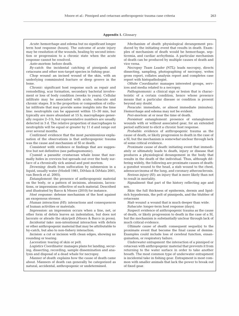
Moore et al.: Pinniped and cetacean anthropogenic trauma case criteria 263
Appendix 1. Glossary
Acute: hemorrhage and edema but no significant longerterm host response (hours). The outcome of acute injurymay be resolution of the wounds, healing by second inten-tion or progression to a chronic state when the acuteresponse cannot be resolved.
Ante-mortem: before death.By-catch: the incidental catching of pinnipeds and
cetaceans and other non-target species in fishing gear.Chop wound: an incised wound of the skin, with an
underlying comminuted fracture or deep groove in thebone.
Chronic: significant host response such as repair andremodelling, scar formation, secondary bacterial involve-ment or loss of body condition (weeks to years). Cellularinfiltrate may be associated with acute, subacute andchronic stages. It is the proportion or composition of cellu-lar infiltrate that may provide some insights into the timeline: neutrophils can be apparent within 15−30 min, buttypically are more abundant at 15 h; macrophages gener-ally require 2−3 h, but representative numbers are usuallydetected in 3 d. The relative proportion of macrophages toneutrophils will be equal or greater by 11 d and range outover several months.
Confirmed: evidence that the most parsimonious expla-nation of the observations is that anthropogenic traumawas the cause and mechanism of SI or death.
Consistent with: evidence or findings that are sugges-tive but not definitive (see pathognomonic).
Cyamid: a parasitic crustacean whale louse that nor-mally hides in crevices but spreads out over the body sur-face of a chronically sick animal and post mortem.
Drowning: death from suffocation by submersion in aliquid, usually water (Modell 1981, DiMaio & DiMaio 2001,van Beeck et al. 2005).
Entanglement: the presence of anthropogenic materialon the body, or a pattern of incisions, abrasions, lacera-tions, or impressions reflective of such material. Describedand illustrated by Barco & Moore (2010) for instance.
Host response: defense mechanism of the host againstan exogenous stressor.
Human interaction (HI): interactions and consequencesof human activities or materials.
Impression: an impression occurs when a line, net, orother form of debris leaves an indentation, but does notlacerate or abrade the skin/pelt (Moore & Barco in press).
Incidental take: non-intentional interaction with debrisor other anthropogenic material that may be attributable toby-catch, but also to non-fishery interaction.
Incision: a cut or incision with clean edges, showing norounding or tearing.
Laceration: tearing of skin or pelt.Logistics Coordinator: manages plans for landing, secur-
ing, dissecting, recording, sample dissemination and ana -lysis and disposal of a dead whale for necropsy.
Manner of death: explains how the cause of death cameabout. Manners of death can generally be categorized asnatural, accidental, anthropogenic or undetermined.
Mechanism of death: physiological derangement pro-duced by the initiating event that results in death. Exam-ples of mechanism of death would be hemorrhage, sep-ticemia, and cardiac arrhythmia. A particular mechanismof death can be produced by multiple causes of death andvice versa.
Necropsy Team Leader (NTL): leads necropsy, directsdissecting, sampling, photographing of necropsy, writesgross report, collates analysis report and completes casereport with histopathologist.
Offsite Coordinator: manages interested groups, serv-ices and media related to a necropsy.
Pathognomonic: a clinical sign or lesion that is charac-teristic of a certain condition, hence whose presencemeans that a particular disease or condition is presentbeyond any doubt.
Peracute: immediate, or almost immediate (minutes).Hemorrhage and edema may not be evident.
Peri-mortem: at or near the time of death.Persistent entanglement: presence of entanglement
wounds with or without associated gear, for an extendedperiod sufficient to elicit a chronic host response.
Probable: evidence of anthropogenic trauma as thecause of death, or likely progression to death in the case ofa SI, but the mechanism is somewhat unclear through lackof some critical evidence.
Proximate cause of death: initiating event that immedi-ately or ultimately leads to death, injury or disease thatproduces a physiological derangement in the body thatresults in the death of the individual. Thus, although dif-fering widely, the following are proximate causes of death:a gunshot wound to the head, a stab wound to the chest,adenocarcinoma of the lung, and coronary atherosclerosis.
Serious injury (SI): an injury that is more likely than notto result in mortality.
Signalment: that part of the history reflecting age andsex.
Skin: the full thickness of epidermis, dermis and lipid-rich hypodermis, the pelt of pinnipeds, and the blubber ofcetaceans
Stab wound: a wound that is much deeper than wide.Subacute: longer-term host response (days).Suspect: evidence of anthropogenic trauma as the cause
of death, or likely progression to death in the case of a SI,but the mechanism is substantially unclear through lack ofmuch critical evidence.
Ultimate cause of death: consequent sequel(s) to theproximate event that become the final cause of demise.Examples could include loss of cerebral function, exsan-guination, or respiratory failure.
Underwater entrapment: the interaction of a pinniped orcetacean with anthropogenic material that prevents it fromreturning to the water surface in order to take anotherbreath. The most common type of underwater entrapmentis incidental take in fishing gear. Entrapment is most com-mon with smaller animals that lack the power to break outof fixed gear.

Dis Aquat Org 103: 229–264, 2013264
Appendix 2. Abbreviations used in the Theme Section, including institutional abbreviations used in table and figure legends
CCSN Cape Cod Stranding Network (now IFAW MarineMammal Rescue Research)
COD Cause of deathFFWC Florida Fish & Wildlife CommissionHI Human interactionIFAW International Fund for Animal WelfareNARW North Atlantic right whale Eubalaena glacialisMLML Moss Landing Marine LabMMPA Marine Mammal Protection ActNEAq New England AquariumNEFSC NOAA Northeast Fisheries Science CenterNMFS NOAA National Marine Fisheries ServiceNOAA National Oceanographic Atmospheric AdminsitrationPCCS Provincetown Center for Coastal StudiesSAR Stock Assessment ReportSBMNH Santa Barbara Museum of Natural HistorySI Serious injurySWFSC NOAA Southwest Fisheries Science CenterTMMC The Marine Mammal CenterUNCW University North Carolina WilmingtonVAq Virginia AquariumWHOI Woods Hole Oceanographic Institution
Editorial responsibility: John Austin, Oldendorf/Luhe, Germany
Submitted: July 5, 2012; Accepted: December 3, 2012Proofs received from author(s): March 29, 2013