Crimean-Congo Hemorrhagic Fever Virus Endemicity in United ... · Crimean-Congo Hemorrhagic Fever...
6
We conducted a cross-sectional survey of Crimean- Congo hemorrhagic fever virus (CCHFV) in dromedary camels and attached ticks at 3 locations in the United Arab Emirates. Results revealed a high prevalence of CCHFV-reactive antibodies in camels and viral RNA in ticks and camel serum, suggesting the virus is endemic in this country. Crimean-Congo Hemorrhagic Fever Virus Endemicity in United Arab Emirates, 2019 Jeremy V. Camp, Dafalla O. Kannan, Babiker Mohammed Osman, Moayyed Sher Shah, Brigitte Howarth, Tamer Khafaga, Pia Weidinger, Noushad Karuvantevida, Jolanta Kolodziejek, Hessa Mazrooei, Nadine Wolf, Tom Loney, Norbert Nowotny Author affiliations: University of Veterinary Medicine Vienna, Vienna, Austria (J.V. Camp, P. Weidinger, J. Kolodziejek, N. Wolf, N. Nowotny); Al Ain City Municipality, Al Ain, United Arab Emirates (D.O. Kannan, B.M. Osman); Dubai Desert Conservation Reserve, Dubai, United Arab Emirates (M. Sher Shah, T. Khafaga); Zayed University, Dubai (B. Howarth); Mohammed Bin Rashid University of Medicine and Health Sciences, Dubai (N. Karuvantevida, H. Mazrooei, T. Loney, N. Nowotny) DOI: https://doi.org/10.3201/eid2605.191414 C rimean-Congo hemorrhagic fever virus (CCHFV; order Bunyavirales, family Nairoviridae, genus Or- thonairovirus) is a geographically widespread species of tickborne virus. Enzootic transmission cycles in- volve livestock (cattle, sheep, goats) and tick species of the genus Hyalomma (Acari: Ixodidae) (1). Spillover into humans typically occurs through tick bites; however, some severe (and even fatal) CCHFV infections have occurred as a result of exposure to blood or tissue from infected animals. The virus is genetically diverse, and evidence indicates that frequent reassortment of viral gene segments occurs, potentially as a result of animal trade between regions of Africa and Asia (1,2). Reports of infections in humans during 2016– 2019 (3–5), the outbreak in the United Arab Emirates (UAE) in 1979 (6), and the outbreak in Oman during 1994–1995 (7) suggest that CCHFV is present in the Arabian Peninsula. However, little is known about enzootic transmission and the frequency of importa- tion into this region. Therefore, we conducted a cross- sectional survey of ticks and dromedary camels in the UAE to determine exposure status and detect active CCHFV infections. We collected whole blood samples from camels at 3 sites within the UAE that differed by frequency of camel use: a family farm, a desert conservation reserve with multiple tour operators, and a large livestock market (Appendix, https://wwwnc.cdc.gov/EID/ article/26/5/19-1414-App1.pdf). We found CCHFV antibodies in the serum samples of 67% (84/125) of camels. CCHFV antibody prevalence was highest in older camels (96% in camels >10 years of age), and no difference in antibody prevalence was detected between sexes (68% [51/75] male, 71% [29/41] fe- male) (Appendix Table 1). The prevalence of reactive antibodies differed between sampling locations, po- tentially because of differences in animal ages at the respective sites. We removed 314 adult ticks and 33 tick nymphs (0–5 ticks/camel) from camels and identified the species under a stereomicroscope. Most (99%, 311/314) adults were Hyalomma dromedarii ticks, and 3 were H. scupense ticks. Two pools of adult H. dromedarii ticks (1 containing 3 males and the other containing 1 male) from 2 separate camels (both 6-year-old females, one of which was anti- body positive) and serum samples from 2 camels (a 3-year-old female and 2-year-old male, both an- tibody negative) were positive for CCHFV nucleic acid (Appendix Table 2). These 4 camels were all from the livestock market but originated from dif- ferent regions of the UAE. The 2 camels with CCH- FV RNA–positive serum were only briefly at the livestock market (for 1 and 2 days), and the 2 with CCHFV RNA–positive ticks were housed at the market for 7 and 41 days. We performed 2 conventional reverse transcrip- tion PCRs on the RNA-positive serum samples and on each tick from the 2 RNA-positive pools, 1 am- plifying a 492-bp portion of the viral small (S) seg- ment and 1 amplifying a 672-bp portion of the viral medium (M) segment (Appendix). We then subject- ed these PCR products to Sanger sequencing (Gen- Bank accession nos. MN516481–8; Appendix Table 3). The S segment sequences from 3 ticks (from 2 camels) and 2 serum samples were all identical to each other, except for a single synonymous substi- tution in the sequence from 1 serum sample; these sequences were genetically similar to sequences of isolates from West and South Africa (group III; Figure, panel A). We obtained the M segment se- quences from only 3 ticks from 2 camels. These se- quences were 85% identical to available sequences in GenBank, and the isolate with the closest iden- tity (AP92, GenBank accession no. DQ211625) was from Greece (Figure panel B). Thus, the 2019 UAE 1019 Emerging Infectious Diseases • www.cdc.gov/eid • Vol. 26, No. 5, May 2020 RESEARCH LETTERS
Transcript of Crimean-Congo Hemorrhagic Fever Virus Endemicity in United ... · Crimean-Congo Hemorrhagic Fever...
We conducted a cross-sectional survey of Crimean-Congo hemorrhagic fever virus (CCHFV) in dromedary camels and attached ticks at 3 locations in the United Arab Emirates. Results revealed a high prevalence of CCHFV-reactive antibodies in camels and viral RNA in ticks and camel serum, suggesting the virus is endemic in this country.
Crimean-Congo Hemorrhagic Fever Virus Endemicity in United Arab Emirates, 2019
Jeremy V. Camp, Dafalla O. Kannan, Babiker Mohammed Osman, Moayyed Sher Shah, Brigitte Howarth, Tamer Khafaga, Pia Weidinger, Noushad Karuvantevida, Jolanta Kolodziejek, Hessa Mazrooei, Nadine Wolf, Tom Loney, Norbert NowotnyAuthor affiliations: University of Veterinary Medicine Vienna, Vienna, Austria (J.V. Camp, P. Weidinger, J. Kolodziejek, N. Wolf, N. Nowotny); Al Ain City Municipality, Al Ain, United Arab Emirates (D.O. Kannan, B.M. Osman); Dubai Desert Conservation Reserve, Dubai, United Arab Emirates (M. Sher Shah, T. Khafaga); Zayed University, Dubai (B. Howarth); Mohammed Bin Rashid University of Medicine and Health Sciences, Dubai (N. Karuvantevida, H. Mazrooei, T. Loney, N. Nowotny)
DOI: https://doi.org/10.3201/eid2605.191414
Crimean-Congo hemorrhagic fever virus (CCHFV; order Bunyavirales, family Nairoviridae, genus Or-
thonairovirus) is a geographically widespread species of tickborne virus. Enzootic transmission cycles in-volve livestock (cattle, sheep, goats) and tick species of the genus Hyalomma (Acari: Ixodidae) (1). Spillover into humans typically occurs through tick bites; however, some severe (and even fatal) CCHFV infections have occurred as a result of exposure to blood or tissue from infected animals. The virus is genetically diverse, and evidence indicates that frequent reassortment of viral gene segments occurs, potentially as a result of animal trade between regions of Africa and Asia (1,2).
Reports of infections in humans during 2016–2019 (3–5), the outbreak in the United Arab Emirates (UAE) in 1979 (6), and the outbreak in Oman during 1994–1995 (7) suggest that CCHFV is present in the Arabian Peninsula. However, little is known about enzootic transmission and the frequency of importa-tion into this region. Therefore, we conducted a cross-sectional survey of ticks and dromedary camels in the UAE to determine exposure status and detect active CCHFV infections.
We collected whole blood samples from camels at 3 sites within the UAE that differed by frequency of camel use: a family farm, a desert conservation reserve with multiple tour operators, and a large livestock market (Appendix, https://wwwnc.cdc.gov/EID/article/26/5/19-1414-App1.pdf). We found CCHFV antibodies in the serum samples of 67% (84/125) of camels. CCHFV antibody prevalence was highest in older camels (96% in camels >10 years of age), and no difference in antibody prevalence was detected between sexes (68% [51/75] male, 71% [29/41] fe-male) (Appendix Table 1). The prevalence of reactive antibodies differed between sampling locations, po-tentially because of differences in animal ages at the respective sites.
We removed 314 adult ticks and 33 tick nymphs (0–5 ticks/camel) from camels and identified the species under a stereomicroscope. Most (99%, 311/314) adults were Hyalomma dromedarii ticks, and 3 were H. scupense ticks. Two pools of adult H. dromedarii ticks (1 containing 3 males and the other containing 1 male) from 2 separate camels (both 6-year-old females, one of which was anti-body positive) and serum samples from 2 camels (a 3-year-old female and 2-year-old male, both an-tibody negative) were positive for CCHFV nucleic acid (Appendix Table 2). These 4 camels were all from the livestock market but originated from dif-ferent regions of the UAE. The 2 camels with CCH-FV RNA–positive serum were only briefly at the livestock market (for 1 and 2 days), and the 2 with CCHFV RNA–positive ticks were housed at the market for 7 and 41 days.
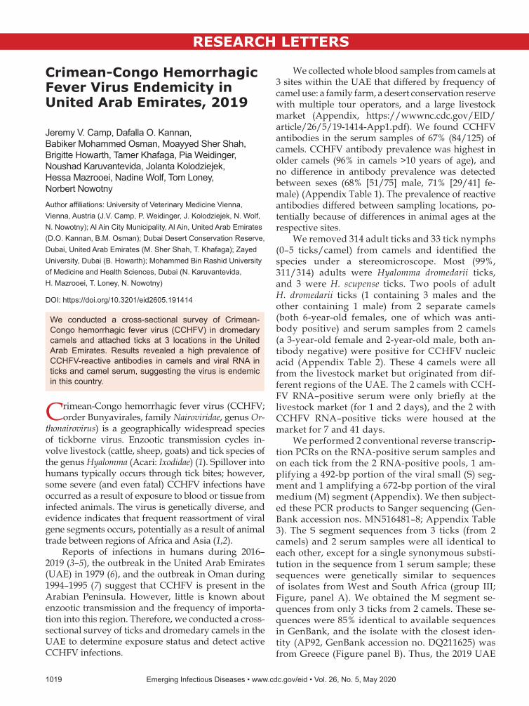
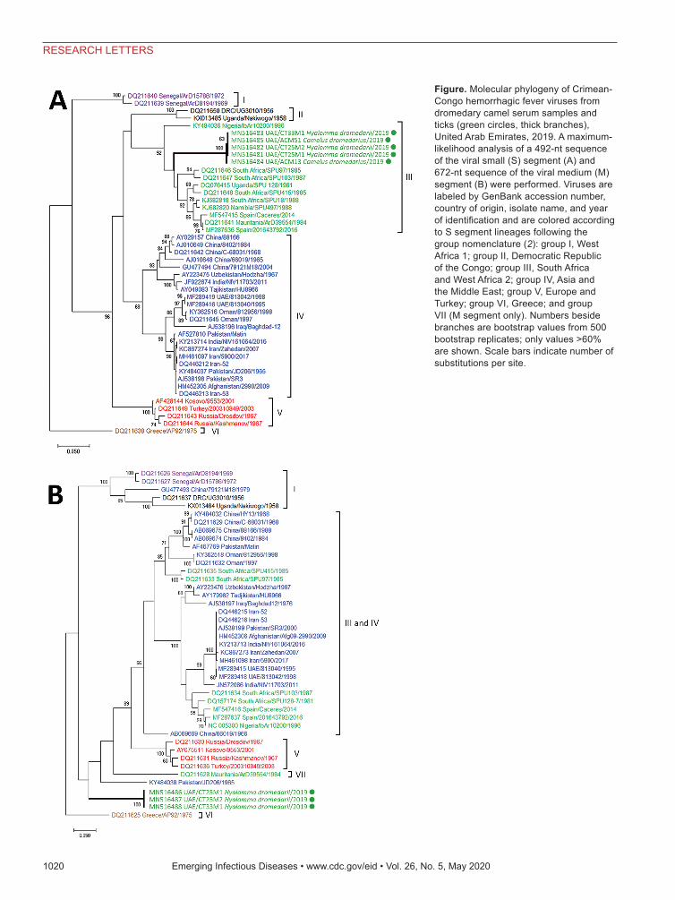
We performed 2 conventional reverse transcrip-tion PCRs on the RNA-positive serum samples and on each tick from the 2 RNA-positive pools, 1 am-plifying a 492-bp portion of the viral small (S) seg-ment and 1 amplifying a 672-bp portion of the viral medium (M) segment (Appendix). We then subject-ed these PCR products to Sanger sequencing (Gen-Bank accession nos. MN516481–8; Appendix Table 3). The S segment sequences from 3 ticks (from 2 camels) and 2 serum samples were all identical to each other, except for a single synonymous substi-tution in the sequence from 1 serum sample; these sequences were genetically similar to sequences of isolates from West and South Africa (group III; Figure, panel A). We obtained the M segment se-quences from only 3 ticks from 2 camels. These se-quences were 85% identical to available sequences in GenBank, and the isolate with the closest iden-tity (AP92, GenBank accession no. DQ211625) was from Greece (Figure panel B). Thus, the 2019 UAE
1019 Emerging Infectious Diseases • www.cdc.gov/eid • Vol. 26, No. 5, May 2020
RESEARCH LETTERS
1020 Emerging Infectious Diseases • www.cdc.gov/eid • Vol. 26, No. 5, May 2020
RESEARCH LETTERS
Figure. Molecular phylogeny of Crimean-Congo hemorrhagic fever viruses from dromedary camel serum samples and ticks (green circles, thick branches), United Arab Emirates, 2019. A maximum-likelihood analysis of a 492-nt sequence of the viral small (S) segment (A) and 672-nt sequence of the viral medium (M) segment (B) were performed. Viruses are labeled by GenBank accession number, country of origin, isolate name, and year of identification and are colored according to S segment lineages following the group nomenclature (2): group I, West Africa 1; group II, Democratic Republic of the Congo; group III, South Africa and West Africa 2; group IV, Asia and the Middle East; group V, Europe and Turkey; group VI, Greece; and group VII (M segment only). Numbers beside branches are bootstrap values from 500 bootstrap replicates; only values >60% are shown. Scale bars indicate number of substitutions per site.
isolates did not fall within previously defined phy-logenetic groups (2).
Our data indicate that exposure to CCHFV is com-mon among camels in the UAE, and transmission to camels might be occurring via native infected H. drom-edarii ticks. A previous survey of UAE livestock that occurred shortly after the 1994–1995 outbreak ruled out camels and camel ticks as CCHFV reservoirs (7). Our data might indicate increased transmission activ-ity in the region, potentially explaining the human case in Sharjah, UAE, in August 2019 associated with han-dling infected meat (5). The largest outbreak of CCH-FV infection in the UAE (1994–1995) was associated with a high case-fatality ratio (73%) and was limited to abattoir workers (8,9); however, hospital outbreaks have also previously occurred in the UAE (6).
All previously characterized CCHFV iso-lates from the Arabian Peninsula and the Middle East (including viruses from the UAE and Oman) were genetically similar to each other, clustering to-gether according to the S segment (group IV, Figure panel A). The M segments of the isolates from UAE and Oman were similar to those of viruses from Asia, the Middle East, West Africa, and South Africa (Figure panel B) (2,3,7). Overall, the data suggest that CCHFV is endemic in the UAE, where enzootic transmission cycles involve camels and camel ticks.
AcknowledgmentsThe authors thank Matter Mohammed Saif Alnuaimi (General Manager of Al Ain City Municipality) and his team for supporting the study; Hashim Ahmed Saeed and Md. Helal Ahmed for assistance with sampling at the livestock market; Greg Simkins and the Dubai Desert Conservation Reserve staff for access and support during our sampling of the reserve; staff of the camel tour providers Al Maha, Arabian Adventures, Desert Star, Alpha Tours, and Travco Tours; and the Mazrooei family for their support and generosity during the sampling of their farm. We are grateful for the assistance of Athiq Ahmed Wahab and Abubakkar Babuhan in facilitating the study.
This work was supported by a research grant of the College of Medicine, Mohammed Bin Rashid University of Medicine and Health Sciences, Dubai, United Arab Emirates (to N.N., grant no. MBRU-CM-RG2018-14).
About the AuthorDr. Camp is a research scientist at the Institute of Virology of the University of Veterinary Medicine Vienna, Austria. He uses his backgrounds in entomology and virology to pursue his primary research interest, the ecology of zoonotic and vectorborne viruses.
References 1. Spengler JR, Bergeron É, Spiropoulou CF. Crimean-Congo
hemorrhagic fever and expansion from endemic regions. Curr Opin Virol. 2019;34:70–8. http://dx.doi.org/10.1016/ j.coviro.2018.12.002
2. Deyde VM, Khristova ML, Rollin PE, Ksiazek TG, Nichol ST. Crimean-Congo hemorrhagic fever virus genomics and global diversity. J Virol. 2006;80:8834–42. http://dx.doi.org/ 10.1128/JVI.00752-06
3. Al-Abri SS, Hewson R, Al-Kindi H, Al-Abaidani I, Al-Jardani A, Al-Maani A, et al. Clinical and molecular epidemiology of Crimean-Congo hemorrhagic fever in Oman. PLoS Negl Trop Dis. 2019;13:e0007100. http://dx.doi.org/10.1371/journal.pntd.0007100
4. Yadav PD, Thacker S, Patil DY, Jain R, Mourya DT. Crimean-Congo hemorrhagic fever in migrant worker returning from Oman to India, 2016. Emerg Infect Dis. 2017;23:1005–8. http://dx.doi.org/10.3201/eid2306.161950
5. ProMED-mail. Crimean-Congo hemorrhagic fever—Asia (17): United Arab Emirates (Sharjah). ProMED. 2019 Aug 24 [cited 2019 Oct 2]. http://www.promedmail.org, archive no. 20190829.6641849.
6. Suleiman MNH, Muscat-Baron JM, Harries JR, Satti AGO, Platt GS, Bowen ETW, et al. Congo/Crimean haemorrhagic fever in Dubai. An outbreak at the Rashid Hospital. Lancet. 1980;316:939–41. http://dx.doi.org/10.1016/ S0140-6736(80)92103-0
7. Rodriguez LL, Maupin GO, Ksiazek TG, Rollin PE, Khan AS, Schwarz TF, et al. Molecular investigation of a multisource outbreak of Crimean-Congo hemorrhagic fever in the United Arab Emirates. Am J Trop Med Hyg. 1997;57:512–8. http://dx.doi.org/10.4269/ajtmh.1997.57.512
8. Schwarz TF, Nsanze H, Ameen AM. Clinical features of Crimean-Congo haemorrhagic fever in the United Arab Emirates. Infection. 1997;25:364–7. http://dx.doi.org/ 10.1007/BF01740819
9. Schwarz TF, Nitschko H, Jäger G, Nsanze H, Longson M, Pugh RN, et al. Crimean-Congo haemorrhagic fever in Oman. Lancet. 1995;346:1230. http://dx.doi.org/10.1016/S0140-6736(95)92936-3
Address for correspondence: Jeremy V. Camp or Norbert Nowotny, Institute of Virology, University of Veterinary Medicine Vienna, Veterinaerplatz 1, 1210 Vienna, Austria; email: [email protected] or [email protected]
1021
RESEARCH LETTERS
Page 1 of 3
Article DOI: https://doi.org/10.3201/eid2605.191414
Crimean-Congo Hemorrhagic Fever Virus Endemicity in United Arab Emirates, 2019
Appendix
Supplemental Methods
Three sites were specifically selected within the United Arab Emirates (UAE) that
differed in camel use: (i) a family farm with camels primarily raised for racing, breeding, and
trading; (ii) tour operators at a desert conservation reserve with camels used for tourism; and (iii)
a large high-turnover livestock market with camels used for trading and meat. Whole blood was
drawn from camels into serum tubes as part of mandated brucellosis testing. Camel age and sex
were recorded. All camels originated within the UAE. Sera were separated from whole blood by
centrifugation, and then tested for the presence of Crimean-Congo hemorrhagic fever virus
(CCHFV)-reactive antibodies using a commercial ELISA kit (ID Screen® CCHF Double Antigen
Multi-species, IDVet).
During blood-taking, ticks were removed from the camels by hand and frozen at -80°C.
Adult ticks were later identified to species on dry ice under a stereomicroscope using species
descriptions and dichotomous keys (1–4). Molecular barcoding was used to confirm selected
voucher specimens to species, using PCR to amplify a portion of cytochrome oxidase I (5).
Parasagittal sections were taken from individual ticks with a sterile scalpel blade, the halves were
pooled by camel and stadium, and homogenized in DNA/RNA Shield (ZymoResearch) with
metal beads using a TissueLyzer (Qiagen). RNA was extracted from tick homogenates and camel
serum with a commercial kit (QIAamp viral RNA kit, Qiagen) and tested for the presence of
CCHFV nucleic acid using a commercial reverse transcription quantitative PCR (RT-qPCR)
assay (RealStar® CCHFV RT-PCR Kit, Altona).
Putative-positive samples were then tested by conventional RT-PCR to amplify a 492
base portion of the viral small (S) segment (using primers F2 and R3 from (6)) and a 672 base
portion of the viral medium (M) segment (primers CCHFV_3605F, 5’-
Page 2 of 3
CAGAAAGATGTGGCTGCACA; CCHFV_4316R, 5’-TCTCCRTGTGCWGTRACCCT) and
subjected to Sanger sequencing. The resulting sequences were aligned to published sequences
found in GenBank using Mega7 (version 7.0.26) that were representative of the various genetic
lineages of CCHFV (7) and for which both S and M segment sequences were available.
References
1. Apanaskevich DA, Schuster AL, Horak IG. The genus Hyalomma: VII. Redescription of all parasitic
stages of H. (Euhyalomma) dromedarii and H. (E.) schulzei (Acari: Ixodidae). J Med Entomol.
2008;45:817–31. PubMed http://dx.doi.org/10.1093/jmedent/45.5.817
2. Apanaskevich DA, Filippova NA, Horak IG. The genus Hyalomma Koch, 1844. X. Redescription of all
parasitic stages of H. (Euhyalomma) scupense Schulze, 1919 (= H. detritum Schulze) (Acari:
Ixodidae) and notes on its biology. Folia Parasitol (Praha). 2010;57:69–78. PubMed
http://dx.doi.org/10.14411/fp.2010.009
3. Asadollah HC, Hosseini R, Tavakoli M, Telmadarraiy Z, Abdigoudarzi M. The Iranian Hyalomma
(Acari: Ixodidae) with a key to the identification of male species. Persian J Acarol. 2013;2:503–
29.
4. Estrada-Pena AL. Ticks of domestic animals in the Mediterranean region: a guide to identification of
species. Zaragoza, Spain: University of Zaragoza; 2004.
5. Ivanova NV, Zemlak TS, Hanner RH, Hebert PDN. Universal primer cocktails for fish DNA
barcoding. Mol Ecol Notes. 2007;7:544–8. http://dx.doi.org/10.1111/j.1471-8286.2007.01748.x
6. Schwarz TF, Nsanze H, Longson M, Nitschko H, Gilch S, Shurie H, et al. Polymerase chain reaction
for diagnosis and identification of distinct variants of Crimean-Congo hemorrhagic fever virus in
the United Arab Emirates. Am J Trop Med Hyg. 1996;55:190–6. PubMed
http://dx.doi.org/10.4269/ajtmh.1996.55.190
7. Deyde VM, Khristova ML, Rollin PE, Ksiazek TG, Nichol ST. Crimean-Congo hemorrhagic fever
virus genomics and global diversity. J Virol. 2006;80:8834–42. PubMed
http://dx.doi.org/10.1128/JVI.00752-06
Page 3 of 3
Appendix Table 1. Camel sera with antibodies reactive to CCHFV in the United Arab Emirates, 2019 (number positive / number tested, %) measured by commercial ELISA*
Age class (yr) CCHFV-reactive antibodies (no. positive/total tested, %)
Livestock Market Family Farm Desert Reserve TOTAL < 2 4 / 10 1 / 1 1 / 1 6 / 12, 50% 2-5 11 / 29 1 / 4 2 / 6 14 / 39, 36% 6-10 3 / 4 3 / 3 12 / 15 18 / 22, 82% > 10 0 / 0 7 / 7 36 / 38 43 / 45, 96% Unknown 3 / 7 0 / 0 0 / 0 3 / 7, 4% TOTAL 21 / 50, 42% 12 / 15, 80% 51 / 60, 85% 84 / 125, 67% *CCHFV, Crimean-Congo hemorrhagic fever virus.
Appendix Table 2. Demographic information and results of serum antibody ELISA (CCHFV-Ab) for samples that tested positive for CCHFV by RT-qPCR* ID Sample Type Sex Age Days at Market CCHFV-Ab CT25 Tick pool (3 ♂) F 6 7 Negative CT33 Tick pool (1 ♂) F 6 41 Positive ACM13 Camel serum F 3 1 Negative ACM51 Camel serum M 2 2 Negative *Information for tick pools refers to host status. Ab, antibody; CCHFV, Crimean-Congo hemorrhagic fever virus; RT-qPCR, reverse transcription quantitative PCR.
Appendix Table 3. Results of molecular diagnostics for positive camel serum and camel tick samples* ID Altona RT-qPCR RT-PCR S segment RT-PCR M segment CT25 22.5 MN516481,
MN516482 MN516486, MN516487
CT33 23.9 MN516483 MN516488 ACM13 >40 MN516484 Negative ACM51 36.7 MN516485 Negative *For screening, RT-qPCR kit was used and Ct values are given. The products from follow-up conventional RT-PCR of putative positive samples were sequenced and deposited in GenBank (accession numbers given). Ct, cycle threshold; ID, identification; M, medium; RT-PCR, reverse transcription PCR; RT-qPCR, reverse transcription quantitative PCR; S, small.