validação farmacológica da esquiva inibitória do danio rerio

UNIVERSIDADE DE LISBOA
FACULDADE DE CIÊNCIAS
DEPARTAMENTO DE BIOLOGIA VEGETAL
Creation of a Danio rerio mutant
using CRISPR-Cas9
as a model system to study Primary Ciliary Dyskinesia
(PCD)
Margarida Oliveira Almeida Rasteiro
Mestrado em Biologia Molecular e Genética
Dissertação orientada por:
Doutora Susana Lopes
Professora Doutora Rita Zilhão
2017
II
1.1. Abstract
On the surface, vertebrates seem to have a bilateral symmetry. However, the disposition of the
internal organs, such as the heart and liver says otherwise. The left-right axis differentiation is preceded
by asymmetries in the gene expression pattern in the tissues around the left-right organiser (LRO).
The LRO (called Kupffer’s vesicle in zebrafish) is a transient structure localized at the end of the
notochord that is formed by cells contain a cilium protruding from its apical membrane. Most of these
cilia are motile and their beating movement creates a fluid flow towards the left side of the LRO. In a
way not yet fully understood, the cells surrounding the LRO sense the fluid directionality and trigger
asymmetric expression of nodal and cerl2. The expression of nodal on the left side of the LRO triggers
the Nodal-Lefty-Pitx2 pathway that leads to the typical positioning of the internal organs recognised as
situs solitus.
Besides the laterality establishment, the respiratory epithelium, the ependymal cells lining the brain
ventricles, the oviducts epithelium and the sperm cells also relay on motile cilia to function properly.
When motile cilia function is impaired, this sequence of events is not guaranteed and patients may
suffer from heart congenital malformations bronchiectasis and infertility that characterize primary
ciliary dyskinesia (PCD). PCD is caused by mutations in a vast number of genes that codify for cilia
components. One of those genes is called CCDC40 and codifies for a protein necessary for the assembly
of the inner dynein arms (IDA) and the nexin-dynein regulatory complexes (N-DRC).
In this work, we generated a ccdc40-/- zebrafish mutant (116 aa) using a CRISPR-Cas9 approach.
Due to the long zebrafish maturation period, it was not possible to study the mutant phenotype.
Instead, we studied the phenotype of embryos injected with a translation-blocking morpholino (MO).
We noted that the organ situs was altered in 40 to 70% of the cases, (depending on the amount of MO-
injected) and the developing of tail malformations (not related to the injected MO amount).
The ccdc40-/- zebrafish mutant was designed to replicate a human mutation and can be used to test
gene editing approaches in an attempt to develop a PCD treatment.
Keywords: Motile cilia, Primary ciliary dyskinesia (PCD); CCDC40, CRISPR-Cas9, Zebrafish
1.2. Resumo alargado
Os vertebrados apresentam uma organização corporal complexa com três eixos: eixo antero-
posterior, o eixo dorso-ventral e o eixo esquerda-direita. As assimetrias nos eixos antero-posterior e
dorso-ventral são facilmente identificáveis exteriormente. No entanto, no que diz respeito ao eixo
esquerda direita, as diferenças são notadas unicamente na disposição interna dos órgãos. No Homem, o
coração está deslocado para o lado esquerdo, assim como o estômago e o baço, enquanto que o fígado
e a vesícula biliar ficam do lado direito. Também os pulmões, devido à posição do coração, apresentam
assimetria no número de lóbulos. Esta disposição (situs solitus) é determinada cedo durante o
desenvolvimento embrionário e mantem-se conservada nos vertebrados. A diferenciação do eixo
esquerda-direita é precedida por assimetrias na expressão de genes no organizador esquerda-direita
(LRO, na sigla em inglês). O LRO é uma estrutura transiente localizada no final da notocorda, que no
peixe-zebra é chamada de vesícula de Kupffer (KV).
Esta vesícula é formada por células, cada uma com um cílio diferenciado na sua membrana apical.
A maioria destes cílios são móveis e com o seu movimento coordenado produzem um fluxo direcionado
para o lado esquerdo do LRO. Esta assimetria no fluxo é depois transferida para uma assimetria na
expressão de genes nos tecidos à volta do LRO.

III
Inicialmente há dois genes que são expressos de forma simétrica à volta do LRO: nodal e cerl2. A
forma como o fluxo consegue controlar a expressão de nodal e cerl2 ainda não é conhecida, mas foram
propostas duas hipóteses que a tentam explicar. A primeira propõe que o fluxo gerado pelos cílios
móveis transporta substâncias secretadas pelo LRO e as concentra do lado esquerdo, levando à
assimetria na expressão nodal e cerl2 verificada posteriormente. A outra hipótese propõe que os cílios
móveis criam o fluxo de fluído que, no lado esquerdo, vai causar a deflação dos cílios imóveis,
estimulando os seus mecanorecetores e induzindo uma via de ativação dependente de Ca2+ que culmina
na expressão assimétrica de nodal e cerl2. No entanto, apesar dos esforços, ambas as hipóteses têm sido
questionadas e o exacto mecanismo de transdução da informação permanece desconhecido.
Após a ação do fluxo, que é sentido pelas células que rodeiam o LRO, a expressão de cerl2 é inibida
no lado esquerdo. A inibição da expressão de cerl2 (inibidor de nodal) permite uma maior expressão de
nodal e consequentemente a ativação da cascata de sinalização Nodal-Lefty-Pitx2. Pitx2 é a proteína
efetora da cascata iniciada por Nodal, sendo apontada como a responsável por induzir o fenótipo
“esquerdo” nas células do lado esquerdo da mesoderme lateral (L-LPM).
Os cílios, que podem ser móveis ou imóveis (primários), estão presentes em quase todos os tipos
células nos vertebrados e por isso, são determinantes quer no período de desenvolvimento embrionário
quer na idade adulta. Quando o seu funcionamento fica comprometido pode desencadear várias doenças,
como por exemplo a doença do rim poliquístico (PKD, na sigla em inglês) ou síndromes somáticas
associadas a polidactilia, malformações neurológicas ou obesidade, no caso de alterações associadas aos
cílios primários. Quando são os cílios móveis afetados, as alterações manifestam-se através de
infertilidade, infeções respiratórias recorrentes e situs inversus que caracterizam a uma condição
chamada discinesia ciliar primaria (PCD). A PCD afecta 1 em cada 10 000 nascimentos e é uma das
causas de defeitos cardíacos congénitos. A presença de heterotaxia, condição na qual, pelo menos um
órgão está no lado oposto ao que seria de esperar, aumenta 200 vezes a prevalência destes defeitos
congénitos.
A PC pode ser causada por mutações que alterem ou inibam a produção de qualquer um dos
componentes necessários à montagem das estruturas essenciais para o movimento dos cílios, como o
par central, os raios, o complexo regulador da nexina, os braços interno e externo de dineínas ou outras
proteínas citoplasmáticas que participam na montagem de componentes ciliares.
Este trabalho foca-se na proteína ccdc40, que é um dos componentes do braço interno de dineínas.
Esta proteína é também responsável pela poliglutamilação dos microtúbulos contribuindo para a sua
estabilização. Além disso, a proteína Ccdc40 forma um complexo com outra, a Ccdc39, e atua como
uma régua molecular. Este complexo permite que os raios dos cílios apenas se liguem no local devido
ao longo dos microtúbulos, isto é, a cada 96 nm. Na sua ausência, estes raios continuam a ligar-se aos
microtúbulos, mas em locais onde isso não era suposto acontecer.
Em pacientes com PCD portadores de alterações neste gene, foram identificados locais da proteína
onde as mutações são mais frequentemente encontradas: as mutações c.248delC e c.3129delC alteram a
grelha de leitura enquanto que as outras (c.2440 C>T, c.961 C>T e c.1345C>T) produzem proteínas
truncadas.
Neste trabalho, pretendeu-se criar um peixe-zebra mutante que produza a proteína Ccdc40 truncada
e que possa servir como modelo de doença para estudar o impacto das mutações mais perto do terminal
N da proteína (c.248delC e c.961 C>T). Estas mutações próximas da extremidade N representam cerca
de 50% dos casos de mutações identificadas neste gene e produzem proteínas sem o domínio helicoidal
(coiled-coil).
Para produzir este mutante usou-se o sistema CRISPR-Cas9. Este sistema foi adaptado a partir de
um mecanismo de defesa descoberto em bactérias e permite direcionar a Cas9 (DNase) para um local
específico do genoma, usando uma sequência guia de RNA complementar ao local escolhido. Uma vez
clivado o DNA, a célula inicia o processo de reparação através da junção das extremidades não

IV
homólogas (NHEJ), processo este que é propenso à inserção ou remoção de bases azotadas, levando à
ocorrência de mutações que podem inativar a proteína.
Neste caso, o local escolhido como alvo da Cas9 foi a zona do genoma que codifica para o
aminoácido 116 da proteína ccdc40 no peixe zebra. Esta região é homóloga da região do genoma
humano onde ocorre a mutação c.961 C>T. Uma mutação nonesense ou uma alteração da grelha na
leitura neste local impede totalmente a tradução do domínio em hélice, o que causa alterações na
montagem da estrutura interna dos cílios e consequentemente na sua mobilidade.
De forma a poder estudar e futuramente validar o fenótipo resultante da injeção do morfolino
bloqueador da tradução, foram analisados a posição do coração, fígado e pâncreas assim como defeitos
na cauda e aparecimento de edema cardíaco, em embriões injetados com o morfolino. Os resultados
mostraram que, independentemente da concentração injetada, os embriões apresentam defeitos relativos
à posição do coração (coração à direita ou ao centro), fígado e pâncreas, assim como curvaturas na cauda
e aparecimento de edema por volta dos 3dpf. Estes resultados, irão futuramente ser comparados com o
fenótipo observado no mutante produzido, validando ou não este morfolino.
Além disso, este mutante permitirá estudar as alterações estruturais e funcionais dos cílios em que a
proteína Ccdc40 apresenta uma mutação próxima da extremidade N e compará-las com a estrutura dos
cílios de outa linha mutante já existente, lok, em que a proteína é mutada próximo da extremidade C e
ainda possui parte do domínio helicoidal.
Futuramente, este mutante poderá ser útil para testar terapia génica direcionada à correção desta
mutação que poderá mais tarde ser reproduzida em células humanas recolhidas de pacientes com PCD
com o intuito de serem reimplantadas, possibilitando uma forma de tratamento que possa minimizar os
efeitos da PCD no aparelho respiratório.
Palavras-chave: Cílios móveis, Discinesia ciliar primária, Ccdc40, CRISPR-Cas9, Peixe-zebra

V
1.3. Contents
1.1. ABSTRACT ___________________________________________________________________________ II
1.2. RESUMO ALARGADO __________________________________________________________________ II
1.3. CONTENTS __________________________________________________________________________ V
1.4. LIST OF FIGURES _____________________________________________________________________ VII
1.5. LIST OF TABLES ______________________________________________________________________ VII
1.6. ABBREVIATIONS LIST ________________________________________________________________ VIII
1. INTRODUCTION ________________________________________________________________________ 1
1.1. ASYMMETRY AXIS FORMATION DURING EMBRYOGENESIS ___________________________________________ 1 1.2. L-R ASYMMETRY: NODAL-LEFTY-PITX2 PATHWAY _______________________________________________ 1 1.3. HOW FLOW-RECEIVING CELLS SENSE FLOW: TWO-CILIA VS MORPHOGEN MODEL ___________________________ 2 1.4. MORPHOGEN GRADIENT MODEL ___________________________________________________________ 3 1.5. TWO-CILIA MODEL ____________________________________________________________________ 3 1.6. CILIARY STRUCTURE ____________________________________________________________________ 4 1.7. CILIARY STRUCTURE: MOTILE VS IMMOTILE CILIA _________________________________________________ 5 1.8. CILIOPATHIES ________________________________________________________________________ 6 1.9. PRIMARY CILIARY DYSKINESIA (PCD) (MIM 244400) ____________________________________________ 6 1.10. COILED-COIL DOMAIN-CONTAINING PROTEIN 40 (CCDC40) _______________________________________ 8 1.11. CRISPR-CAS9 SYSTEM AS A TOOL FOR GENE EDITING _____________________________________________ 9 1.12. ZEBRAFISH AS ANIMAL MODEL FOR PCD _____________________________________________________ 10 1.13. PROJECT GOAL ______________________________________________________________________ 11
2. METHODS ____________________________________________________________________________ 12
2.1. ZEBRAFISH MAINTENANCE ______________________________________________________________ 12 2.2. ZEBRAFISH EUTHANASIA ________________________________________________________________ 12 2.3. CRISPR-CAS9 TARGET SEQUENCES - DESIGN OF SGRNA _________________________________________ 12 2.4. SGRNA ANNEALING AND CLONING INTO PDR274 USING BSAI RESTRICTION SITE __________________________ 13 2.5. E. COLI (DHΑ5) TRANSFORMATION AND IDENTIFICATION OF POSITIVE COLONIES __________________________ 14 2.6. IN VITRO TRANSCRIPTION OF SGRNAS USING T7 PROMOTER _______________________________________ 15 2.7. CAS9 MRNA PRODUCTION ______________________________________________________________ 15 2.8. MICRO CO-INJECTION OF CAS9 MRNA AND SGRNA INTO ONE-CELL STAGE ZEBRAFISH EMBRYOS _______________ 15 2.9. PRIMERS’ DESIGN AND OPTIMIZATION ______________________________________________________ 16 2.10. GENOMIC DNA EXTRACTION ____________________________________________________________ 17 2.11. HETERODUPLEXES DETECTION USING PAGE ANALYSIS ____________________________________________ 17 2.12. MICROINJECTION OF ZEBRAFISH EMBRYOS WITH MORPHOLINO _____________________________________ 18 2.13. HEART AND GUT SCREENING _____________________________________________________________ 18
3. RESULTS _____________________________________________________________________________ 19
3.1. CRISPR-CAS9 PREPARATION ____________________________________________________________ 19 3.2. SGRNA CLONING INTO PDR274 AND SGRNA SYNTHESIS _________________________________________ 19 3.3. PRIMERS OPTIMIZATION ________________________________________________________________ 20 3.4. CO-INJECTION OF CAS9 MRNA AND SGRNA AND CRISPR-CAS9 EFFECTIVENESS ANALYSIS BY PAGE ASSAY _______ 21 3.5. SEARCH FOR FOUNDERS: ZEBRAFISH SCREENING AND GENOTYPING ___________________________________ 22 3.6. MORPHOLINO ANTISENSE TECHNOLOGY - PHENOTYPE CHARACTERIZATION ______________________________ 25
4. DISCUSSION __________________________________________________________________________ 27
5. CONCLUSION AND PROSPECTS ___________________________________________________________ 29
6. BIBLIOGRAPHY ________________________________________________________________________ 29

VI
7. APPENDICES __________________________________________________________________________ 35
7.1. HUMAN- ZEBRAFISH CCDC40 ALIGNMENT ____________________________________________________ 35 7.2. PDR274 STRUCTURE __________________________________________________________________ 37 7.3. PLASMID SEQUENCING RESULTS SHOWING THE DETAILED SEQUENCE __________________________________ 38 7.4. CRISPANTS SEQUENCING _______________________________________________________________ 39 7.5. CCDC40 CLONING PRIMERS ______________________________________________________________ 40
VII
1.4. List of Figures FIGURE 1.1 – TEMPORAL AND SPATIAL DISTRIBUTION OF NODAL, PITX2 AND LEFTY2. ______________________ 2 FIGURE 1.2 – MORPHOGEN GRADIENT MODEL AND TWO CILIA MODEL. __________________________________ 3 FIGURE 1.3 – TRANSVERSAL VIEWS OF THE AXONEME ORGANISATION. __________________________________ 5 FIGURE 1.4 – CILIA ULTRASTRUCTURE AND DYNEIN PLACEMENT IN THE AXONEME IN A TRANSVERSAL AND
LONGITUDINAL VIEW. ____________________________________________________________________ 6 FIGURE 1.5 – NORMAL SITUS AND SITUS ABNORMALITIES. ____________________________________________ 7 FIGURE 1.6 – TYPICAL AXONEME DEFECTS IN DIFFERENT MUTATED GENES IN PCD. ________________________ 7 FIGURE 1.7 – PROTEINS KNOWN TO BE ASSOCIATED TO EACH AXONEME COMPONENT IN HUMANS. ____________ 8 FIGURE 1.8 – RELATIVE POSITION OF THE CCDC40 PROTEIN WITHIN THE MOTILE CILIA. ____________________ 8 FIGURE 1.9 – CCDC40 LOCATION INSIDE THE CELL. _________________________________________________ 9 FIGURE 1.10 – KNOCKOUT GENERATING PROCESS USING CRISPR-CAS9 SYSTEM. _________________________ 9 FIGURE 1.11 – HUMAN AND ZEBRAFISH CCDC40 CDNA SEQUENCE ALIGNMENT. _________________________ 11 FIGURE 3.1 – COLONY PCR RESULTS. _______________________________________________________________ 19 FIGURE 3.2 – PDR274 LINEARIZATION WITH HINDIII. ______________________________________________ 19 FIGURE 3.3 – SEQUENCING RESULTS FROM COLONY 10 (SGRNA#2) AND COLONY 14 (SGRNA#1). ___________ 20 FIGURE 3.4 – PRIMER-PAIRS OPTIMIZATION FOR ZEBRAFISH GENOMIC DNA. ____________________________ 20 FIGURE 3.5 – DEFORMITIES FOUND IN ZEBRAFISH LARVAE (3DPF) AFTER CO-INJECTION WITH CAS9 MRNA AND SGRNA. _______ 21 FIGURE 3.6 – HETERODUPLEXES DETECTED IN PAGE-BASED ANALYSIS OF CRISPR-CAS9 SYSTEM EFFICACY IN
TGBAC(CFTR-GFP) EMBRYOS. ____________________________________________________________ 21 FIGURE 3.7 – HETERODUPLEXES DETECTED IN PAGE-BASED ASSAY FOR TESTING CRISPR-CAS9 EFFICACY IN AB,
TG(FOXJ1A:GFP) AND TG(SOX17-GFP) EMBRYOS INJECTED WITH CAS9 MRNA (75 NG/ΜL) AND SGRNA#1
(37.5 NG/ΜL).__________________________________________________________________________ 22
FIGURE 3.8 – HETERODUPLEXES DETECTED IN A PAGE-BASED ASSAY USED TO TEST TGBAC(CFTR-GFP) ♂ 23
AND FOX:GFP ♂ 3 OFFSPRING. ___________________________________________________________ 23 FIGURE 3.9 – SANGER SEQUENCING RESULTS FOR WILDTYPE (WT) AND HETERODUPLEX (HTD) BAND ISOLATED
FROM ♂ 23 B TGBAC(CFTR-GFP) AND ♂ 3 B TG(FOXJ1A:GFP). _______________________________ 24 FIGURE 3.10 – MORPHOLOGICAL DEFECTS OBSERVED IN CCDC40 TRANSLATION-BLOCKING MO INJECTED
EMBRYOS. ____________________________________________________________________________ 25 FIGURE 3.11.-. PHENOTYPE OF LAVAE (3DPF) INJECTED WITH CCDC40 TRANSLATION-BLOCKING MO (6 NG)
SHOWING CARDIAC OEDEMA AND TAIL CROOKS. ______________________________________________ 25 FIGURE 3.12 – GUT SITUS FROM TG(SOX17-GFP) EMBRYOS INJECTED WITH TRANSLATION-BLOCKING CCDC40 MO __________ 25 FIGURE 3.13 – HEART AND GUT SITUS COMBINED. _________________________________________________ 26 FIGURE 3.14 – DIAGRAM SHOWING THE PREDICTED ZEBRAFISH CCDC40 PROTEIN DOMAINS IN WT, LOK MUTANTS
AND CCDC40 GENERATED MUTANT USING CRISPR-CAS9 SYSTEM. _______________________________ 29 FIGURE 7.1 – PDR274 PLASMID STRUCTURE ______________________________________________________ 37 FIGURE 7.2– SEQUENCING RESULTS FOR COLONY 10 (SGRNA2) ______________________________________ 38 FIGURE 7.3 – SEQUENCING RESULTS FOR COLONY 14 (SGRNA2) ______________________________________ 38 FIGURE 7.4 – COMPLETED SANGER SEQUENCING RESULTS FOR WILDTYPE (WT) AND HETERODUPLEX (HTD)
BANDS ISOLATED FROM ♂ 23 TGBAC(CFTR-GFP) AND ♂ 3 TG(FOXJ1A:GFP). ____________________ 39
1.5. List of Tables
TABLE 2.1 – SGRNA SEQUENCES _______________________________________________________________ 13 TABLE 3.1 – SGRNA1 AND 2 NANODROP QUANTIFICATION __________________________________________ 20 TABLE 7.1 – PRIMERS TESTED TO AMPLIFY ZEBRAFISH CCDC40 CDNA _________________________________ 40
VIII
1.6. Abbreviations list aa Amino acid
ATP Adenosine triphosphate
Ca2+ Calcium ion
CCDC39/40/65 Coiled-coil domain-
containing protein 39/40/65
CHD Chronic heart disease
CRISPR-Cas9 Clustered regularly
interspaced palindromic
repeats associated with
caspase 9
Dand5/Cerl-2 DAN domain family
member 5/Cerberus-like
protein 2
DNAH5 Dynein axonemal heavy
chain 5
DNAI Dynein intermediate chain
DNAL Dynein light chain
dNTP Deoxynucleotide
triphosphate
dpf Days post fertilization
GRP Gastrocoel roof plate
h Hour(s)
hpf Hours post fertilization
IDA Inner dynein arm
iv Inversus viscerum
Kif3A/B Kinesin-Associated Protein
3A/B
KO Knockout
KS Kartagener syndrome
KV Kupffer’s vesicle
LB medium Luria-Bertani medium
LPM Lateral plate mesoderm
L-R Left-right
lrd Left-right dynein
LRO Left-right organiser
min Minute(s)
MO Morpholino
MTOC Microtubule organising
centre
N-DRC Nexin-dynein regulatory
complex
O/N Overnight
ODA Outer dynein arm
PAM Protospacer adjacent motif
PCD Primary ciliary dyskinesia
PCR Polymerase chain reaction
Pitx2 Pituitary homeobox 2
PKD Polycystic kidney disease
PKD1L1 Polycystin 1 Like 1
PKD2 Polycystin 2
RA Retinoic acid
rNTP Ribonucleotide triphosphate
rpm Rotations per min
RS Radial spoke
RT Room temperature
Shh Sonic hedgehog
TGF Transforming growth factor
V Volts
WT Wildtype
1
1. Introduction
1.1. Asymmetry axis formation during embryogenesis
Most of the living beings, specifically the vertebrates, have a complex body organization. The axis
differentiation is crucial in the process of shaping multicellular organisms. Several existing pathways,
spatially and temporally coordinated, cooperate to establish antero-posterior, dorso-ventral, and L-R
(left-right) axis, breaking the embryo’s initial radial symmetry.
The embryo’s initial radial symmetry is only apparent, the ovule goes through several maturation
steps that result in an asymmetrical environment inside this cell. The dorso-ventral axis formation is
dependent on the establishment of animal and vegetal poles during oogenesis. The maternal dorsalizing
factors are located in the vegetal pole and immediately after fertilization, together with zygotic factors,
induce the dorso-ventral axis establishment by acting on Wnt /β-catenin, BMP (Bone morphogenic
protein), Nodal, and FGF (fibroblast growing factor) pathways (reviewed by Langdon and Mullins 2011
[1]).
The formation of the anterior-posterior axis ultimately establishes the tail, trunk and head. This body
segmentation results from the interaction of three signalling pathways: BMP, Nodal and Wnt [2]. The
tail forms when all these three are activated, if only Nodal is on, that region will be the trunk, and the
head develops at the end where all three pathways are off [3].
The third and last asymmetry axis formed is the one that establishes sidedness (L-R). This
asymmetry is first noticed in gene expression in the left-right organiser (LRO) and then spreads to the
LPM (lateral plate mesoderm). Ultimately, this information is translated into an asymmetric organ
disposition [4].
1.2. L-R asymmetry: Nodal-Lefty-Pitx2 pathway
At first glance, most vertebrates show bilateral symmetry. However, there is an asymmetry within
the body. This asymmetry, referred to as situs solitus, is highly conserved across species: the heart,
stomach, pancreas and spleen lay on the left side of the longitudinal axis, while the liver and gallbladder
are on the right. Additionally, the lungs also show asymmetries in the number of lobes. These
morphological asymmetries are preceded by asymmetries in gene expression on the LRO during early
embryogenesis [5].
The LRO is a transient embryonic organ (cavity) positioned at the end of the notochord and
accordingly to the animal model it is given a different designation: in zebrafish is called Kupffer’s
vesicle, in mouse is referred to as the node, in chick as Hensen’s nodea and in Xenopus as gastrocoel
roof plate [6]. Even though these structures are morphologically diverse, the mechanisms behind the L-
R asymmetry establishment seem to be conserved across vertebrates [7].
The exact mechanisms that led to the onset of the L-R asymmetry axis are not fully understood, but
it is known that a directional fluid flow on the LRO is essential in its establishment [8]. The cells
surrounding the LRO have motile cilia protruding from their apical membranes that rotate generating a
leftward flow [9]. This asymmetry in the flow is then transcribed into asymmetries in gene expression
in the LRO surrounding cells.
a The Hensen’s node does not have ciliated epithelium. Instead, in chick, the left-side expression of Nodal is achieved by the
LRO displacement to the left side of the L-R axis during early gastrulation. The notochord forms on the right side of the LRO
restricting Nodal expression only to the left side without requiring flow [113].

2
There are two specific genes, nodal and
dand5 (nodal inhibitor also known as cerl2), that
are initially symmetrically expressed on the
LRO. When the flow downregulates the
expression of dand5 on the left side of the LRO,
it promotes the Nodal-signalling activation on
this side [10]. As a consequence nodal
expression becomes limited to the left side lateral
plate mesoderm (Figure 1.1 A), the same side
towards which the embryonic heart loops [5]
while its antagonist, Dand5 is restricted to the
right side of the LRO [11].
Nodal is a member of the TGFβ
(transforming growth factor beta) family that,
when lateralized on the LRO, spreads throughout
the left-LPM where it induces its own expression
as well as Lefty1, Lefty2 and Pitx2. Lefty1 and
Lefty2, which are also members of the TGFβ
family, compete with Nodal for the receptors
preventing its action. Additionally, Nodal is only
active as a dimmer while Lefty1 and Lefty2 are
monomers, this lets them diffuse faster and
farther, inhibiting the Nodal activity on the
surrounding tissues. The lefty1 expression is
restricted to the left side of the ventral neural tube, where it acts as a midline barrier preventing the left-
side establishing factors from crossing to the right side [12], while lefty2 is expressed in the left LPM
[13] (Figure 1.1 B).
Meno et al. demonstrated that, in lefty2 mutants, Nodal expression lasts longer and can diffuse over
to the right side [14]. Therefore, Lefty2 on itself does not decide which is going to be the left side, it
functions as a regulator that restricts the duration of Nodal expression.
Finally, Pitx2 is the effector of Nodal signalling and is the most likely candidate for “informing” the
cells on the left LPM that they are to adopt a leftward morphology [4]. However, there are some
asymmetries such as the visceral looping that seem to be independent of Pitx2-signalling as observed in
zebrafish [15].
1.3. How flow-receiving cells sense flow: two-cilia vs morphogen
model
The asymmetric expression of Nodal is established when the motile cilia rotate and generate a fluid
flow in the LRO [16]. The crown cells sense this leftward flow and trigger the Nodal-Lefty-Pitx2
pathway in a Ca2+ induced way [17]. This principle is supported by studies showing that mutants lacking
motile cilia do not generate the required flow and thus have defects regarding gene expression and organ
laterality [18]. Also, mutants having cilia with motility impairments display similar phenotype [19,20]
making it clear that the defects are due to lack of movement and not because of the lack of cilia. The
unequivocal connection between the cilia-generated flow in the LRO and the L-R asymmetry
establishment was done by Nonaka et al. when they proved that the Nodal-Lefty-Pitx2 pathway
asymmetry could be disturbed in WT mouse embryos or normalized in mutants showing laterality
defects by inducing an artificially generated flow [16].
Figure 1.1 – Temporal and spatial distribution of Nodal, Pitx2
and Lefty2.
Lefty2 limits nodal diffusion by migrating faster and competing
for the same receptors. Lefty1 is expressed in the midline (green)
preventing Nodal from crossing to the right side. Pitx2 is
expressed were Nodal is.
Horizontal aligned blocks stand for the LPM, the vertical
brown line stands for the midline and blue, pink and yellow
indicate the presence of Nodal, Lefty2 and Pitx2, respectively,
along the LPM. Adapted from Meno et al. 2001 and Peeters et
al. 2006 .
A B

3
However, the exact mechanism on how the LRO senses the flow and transcribes it into gene
expression is not yet clear. In an attempt to understand how this cilia-generated flow is translated into
an asymmetry in molecular markers, two hypotheses were put forward.
In a summarized way, according to the mechanosensory hypothesis, cilia react to the flow’s
mechanical force. When the flow passes through the cilia, they bend and trigger the Nodal-Lefty-Pitx2
pathway [21]. On the other hand, the chemosensory hypothesis proposes that the Nodal-Lefty-Pitx2
pathway is triggered by a morphogen gradient created by the flow.
1.4. Morphogen gradient model
This model assumes that when beating, the motile cilia create a unidirectional flow towards the left
side carrying secreted morphogens along with it. This assures that the left side of the organiser has a
higher morphogen concentration than the right side and consequently the gene expression on the left
side will diverge from that on the right [22].
However, these hypothetic free secreted morphogens capable of directly inducing the Nodal-Lefty-
Pitx2 pathway have not yet been identified [9].
Along with this, it has also been proposed that some of these potential morphogens (such as Shh,
RA) could instead be encapsulated inside vesicles (nodal vesicular parcels) that once caught in the fluid
flow are transported towards the left side where they eventually break down near the LRO wall setting
the morphogens free (Figure 1.2A) [23].
However, unlike expected, slowing or accelerating the flow does not alter the situs [16].
Furthermore, mutants lacking motile cilia (kif3A/B)
on the LRO show bilateral expression or absence of
the Nodal-Lefty-Pitx2 pathway [18] while in
mutants with motionless cilia (lrd, iv) the Nodal-
Lefty-Pitx2 pathway can be expressed on the left
side, on the right side, bilaterally or be totally absent
[24].
According to the morphogen model, it was
expected that both types of mutants presented the
same phenotype, as they both have no fluid flow in
the LRO [21]. Additionally, some other factors such
as PKD2 [25] that are not involved in generating the
flow, but seem to be important in sensing it, have
been described to have a role in the gene asymmetric
expression on the LRO.
1.5. Two-cilia model
In an attempt to justify the phenotype differences
between mutants lacking motile cilia on the LRO
(Kif3A/B) and mutants with cilia but immotile (iv
and Lrd), it was demonstrated that the LRO has two
types of cilia: motile cilia, those which generate the
flow, and immotile cilia, likely those that may sense it (Mcgrath and Brueckner 2003).
This model assumes that immotile cilia on the left side of the LRO encounter a flow stronger than
those on the right. The model advocates that the flow can bend the cilia stimulating the
A
Figure 1.2 – Morphogen gradient model and two cilia
model.
A: The morphogen model predicts that secreted
morphogens, free or encapsulated in vesicles (NVP), are
transported within the flux crating a concentration gradient
that is sensed by the cilia in the left side triggering the Ca2+
release on the left side. B: The two -cilia model proposes
that motile cilia create the flow while immotile cilia sense
it. The flow detection occurs when the fluid pressure bends
the sensory cilia triggering the L-R asymmetry breaking in
a Ca2+ dependent way. Adapted from Fliegauf et al. 2007
B
A

4
mechanoreceptors to induce a Ca2+-dependent signalling pathway (Figure 1.2B) leading to the
asymmetric release of signalling molecules [22].
Mutants with motionless cilia are incapable of generating the flow but preserve the sensing capacity,
meaning that external stimuli can randomly activate the mechanosensors. On the other hand, mutants
lacking motile cilia loose both abilities rendering them unable to generate or sense the flow [21]. This
offers one explanation to the different phenotypes observed.
The explanation backing the mechanosensation connects PKD1 and PKD2 to the early L-R
signalling. PKD1 and PKD2 were first identified in polycystic kidney disease. These are membrane
proteins present in the kidney cell’s primary cilia [26] that physically interact to change the conformation
of PKD2, a Ca2+ channel, and allow it to open [27].
PKD1L1 (PKD1-related) was found to be the important protein, being restricted to the LRO cilia
both in mouse [28] and in medaka[25].
Praetorius & Spring showed that bending the primary cilia in kidney culture cells led to an increase
of Ca2+ into the cytoplasm that spreads to the nearby cells through gap-junctions [29] bringing up the
possible role of the PKD1L1-PKD2 complex as a mechanosensor in the LRO cilia. Furthermore, both
mutants in PKD2 and PKD1L1 do not succeed in establishing asymmetric gene expression at the LRO
and LPM, displaying right lung isomerism regardless of the fact that both LRO morphology and cilia
motility are apparently normal [28] [25]. PKD2 mice mutants also show several laterality defects [30]
similar to those seen in mutants lacking motile cilia on the LRO [18].
These results connect the PKD1L1-PKD2 complex (located in all cilia) and Ca2+ levels to the L-R
axis establishment [17][28]. In addition, Ca2+ signalling levels are typically higher on the left side of the
LRO and become randomized in PKD2 mutants [30].
It has recently been described that PKD1L1 is not necessary to Nodal activation, instead, it acts as
PKD2 regulator limiting Nodal expression to the left side [31]. Therefore, PKD1L1 acts as an
intermediate between the flow and the Ca2+ signalling cascade activated by PKD2 [31].
In spite of Ca2+ asymmetries and flow response likely being connected, the exact mechanisms how
they induce the signalling cascade that leads to gene expression asymmetries still needs to be clarified
[32]. Furthermore, recently there has been some evidence against this hypothesis. The mechanosensation
was recently questioned by measuring the Ca2+ influx in response to the flow in primary cilia in vivo
[33]. The results showed that no Ca2+influxes were observed in flow stimulated nodal cilia, suggesting
that, if cilia are to act as mechanosensors, they must induce a non-Ca2+ related signalling pathway.
Nevertheless, the calcium indicator used by these researchers [33] was also questioned at subsequent
commentaries and meetings and other alternatives and better calcium probes are being developed in labs
from different countries. We shall have to wait for the new results to conclude if the mechanosensation
is still feasible in the LRO.
1.6. Ciliary structure
The majority of cells only have one cilium, while others have several, such as the multiciliated
respiratory epithelium [34].
Cilia are cellular hair-like structures projected from the plasma membrane that are built when cells
get into a differentiated form and reabsorbed when they re-enter the cell cycle [35]. Cilia can be divided
into sub-compartments consisting of basal body, transition zone, axoneme and ciliary tip.
Two similar proteins mainly form cilia: α-tubulin and β-tubulin [36]. These two proteins come
together to form dimers that eventually aggregate into a polymer. This polymer rolls itself up to create
a tube that serves as rails for the intraflagellar transport (IFT) [37]. The basal end of the microtubule is
anchored to the microtubule organising centre (MTOC), which is derived from the centriole. The other
end is let free so tubulin dimers can be added or removed accordingly to the cell’s state.

5
In cells that have cilia or flagella, the MTOCb is called basal body. Cilia and flagella have similar
structures, with the difference that cilia are shorter while flagella tend to be longer.
A cilium is not in continuity with the cytoplasm. All the building blocks necessary to its structure
are produced in the cytoplasm and selectively transported through the transition zone (minus end) to the
ciliary tip (plus end). Also, when the cilia need to be removed, all the components must be brought back
to the cytoplasm.
Aside from building blocks, the motor proteins can also transport signalling molecules. This
transport is done by kinesins (or KIFs) (anterograde) and cytoplasmic dyneins (retrograde) that shuttle
in and out of the cilia respectively [38]. In humans, there are 46 kinesins [39] and 17 dyneins [40]
identified with different motor domains handling different types of transport.
The distal tail domains of the motor proteins bind selectively to cargo while the motor domains
interact with the microtubules. These motor domains have the ability to hydrolyze ATP using the
generated energy to move forward along the microtubule [41].
In the absence of these motor proteins such as the KIF3 complex (anterograde transporter), the cilia
are not correctly assembled. As a result, even though the basal bodies are present, Kif3B [18] and Kif3A
[19] mice mutants have no cilia in the LRO leading to the randomization of the L-R asymmetry breaking.
1.7. Ciliary structure: motile vs
immotile cilia
Cilia can be categorised as motile or immotile
accordingly to their morphological structure.
Immotile cilia (also called primary), present in
almost every cell type in vertebrates, are usually
short and motionless. Motile cilia are longer and
have the ability to beat thanks to the presence of
dynein arms in their ultrastructure.
Most motile cilia’s axonemes exhibit a 9+2
arrangement (Figure 1.3A) with 9 doublets hold
together by nexin fibres (N-DRC) and a central
pair, as those generating the flow in zebrafish KV
[42]. Each doublet is composed of two tubules, A (complete) and B (incomplete) where tubule A has
two dynein arms attached: inner dynein arm (IDA) and outer dynein arm (ODA) [34]. Apart from
holding the microtubules together, N-DRC also influences the axonemal bending by controlling the
IDA’s attachment to the A-tubule and mediating the communication between the central pair and the
dynein arms through the RS [43].
Cilia with 9+0 arrangement (Figure 1.3 B) lack the central pair and dynein arms and are mostly
found in immotile cilia. However, there are 9+0 cilia that have dynein arms and are capable of beating
(such as those inducing nodal flow in mouse [44] or medaka [45]).
bThe centrosome is also a type of MTOC and acts as an anchor to microtubules during mitosis.
Figure 1.3 – Transversal views of the axoneme organisation.
A: The 9+2 structure is commonly found in motile cilia,
showing 9 microtubule doublets, a central pair, radial spokes
and dynein arms. B: The 9+0 structure is often associated with
immotile cilia that have no dynein arms. From Dawe, et
al.2006) [116].
B
6
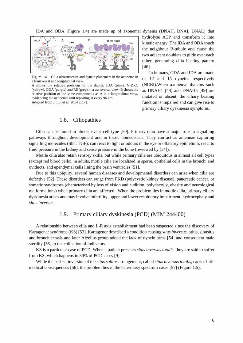
IDA and ODA (Figure 1.4) are made up of axonemal dyneins (DNAH, DNAI, DNAL) that
hydrolyse ATP and transform it into
kinetic energy. The IDA and ODA touch
the neighbour B-tubule and cause the
two adjacent doublets to glide over each
other, generating cilia beating pattern
[46].
In humans, ODA and IDA are made
of 12 and 15 dyneins respectively
(NCBI).When axonemal dyneins such
as DNAH1 [48] and DNAH5 [49] are
mutated or absent, the ciliary beating
function is impaired and can give rise to
primary ciliary dyskinesia symptoms.
1.8. Ciliopathies
Cilia can be found in almost every cell type [50]. Primary cilia have a major role in signalling
pathways throughout development and in tissue homeostasis. They can act as antennae capturing
signalling molecules (Shh, TGF), can react to light or odours in the eye or olfactory epithelium, react to
fluid pressure in the kidney and sense pressure in the bone (reviewed by [34]).
Motile cilia also retain sensory skills, but while primary cilia are ubiquitous in almost all cell types
(except red blood cells), in adults, motile cilia are localized in sperm, epithelial cells in the bronchi and
oviducts, and ependymal cells lining the brain ventricles [51].
Due to this ubiquity, several human diseases and developmental disorders can arise when cilia are
defective [52]. These disorders can range from PKD (polycystic kidney disease), pancreatic cancer, or
somatic syndromes (characterised by loss of vision and audition, polydactyly, obesity and neurological
malformations) when primary cilia are affected. When the problem lies in motile cilia, primary ciliary
dyskinesia arises and may involve infertility, upper and lower respiratory impairment, hydrocephaly and
situs inversus.
1.9. Primary ciliary dyskinesia (PCD) (MIM 244400)
A relationship between cilia and L-R axis establishment had been suspected since the discovery of
Kartagener syndrome (KS) [53]. Kartagener described a condition causing situs inversus, otitis, sinusitis
and bronchiectasis and later Afzelius group added the lack of dynein arms [54] and consequent male
sterility [55] to the collection of indicators.
KS is a particular case of PCD. When a patient presents situs inversus totalis, they are said to suffer
from KS, which happens in 50% of PCD cases [9].
While the perfect inversion of the situs solitus arrangement, called situs inversus totalis, carries little
medical consequences [56], the problem lies in the heterotaxy spectrum cases [57] (Figure 1.5).
Figure 1.4 – Cilia ultrastructure and dynein placement in the axoneme in
a transversal and longitudinal view.
A shows the relative positions of the duplet, IDA (pink), N-DRC
(yellow), ODA (purple) and RS (grey).in a transversal view. B shows the
relative position of the same components as A in a longitudinal view,
evidencing the axonemal unit repeating at every 96 nm.
Adapted from J. Lin et al. 2014 [117].
B A
7
In humans, laterality defects can be manifested in distinct ways. All organs can be placed in their
normal relative position (situs solitus) but in a mirrored image (situs inversus), what is estimated to
occur in 1 out of 6 000 – 8 000 newborns [56]. Another
situation, known as heterotaxy or situs ambiguous, occurs
when at least one organ is misplaced along the L-R axis.
Heterotaxy syndrome is responsible for several
congenital cardiac and gastrointestinal malformations [58]
found in PCD patients [59] and it is thought to affect about
1 in 10 000 newborns or 1 in every 5 000 – 7 000 of live
births with a chronic heart disease (CHD) [57].
According to Kennedy et al., 45% of patients diagnosed
with PCD had situs solitus, 47.7% situs inversus, and 6.3%
showed heterotaxy. Among this 6.3 %, the majority had
cardiovascular malformations [60]. The same authors also
reported that the prevalence of congenital heart diseases
among patients diagnosed with heterotaxy is 200-fold
higher in PCD-suffering patients than in the general
population (1:50 vs.1:10 000) [60].
To complicate matters, heterotaxy often comes associated with isomerism. When isomerism is
present, one organ that
normally would be asymmetric
presents itself symmetrically
(Figure 1.5). Left isomerism is
linked to polysplenia while
right isomerism is associated
with asplenia [61] but its
prevalence in PCD patients is
not known [60]. However,
sometimes patients present
anatomical uncommon
arrangements, making it
difficult to classify it as left or
right isomerism [62].
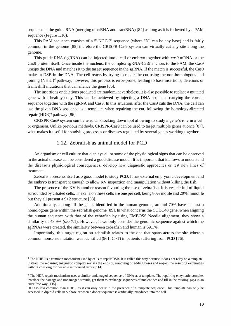
PCD can arise as the result of several inherited mutations in the cilia-motor machinery such as ODA
[49] IDA, radial spokes or central pair components [59].
Mutations in DNAH1, DNAH5 and DNAH11 make up for 25% of all PCD cases [56], but so far
more than 35 genes have been linked to PCD (with 70% of patients testing positive for biallelic
mutations) [63]. However, a defect in any other one of the about 120 proteins that make up the cilia or
in the cytoplasmic proteins [64] responsible for transporting or assembling cilia components can
potentially affect the cilia’s ultrastructure and cause PCD. Figure 1.6 shows the most common mutated
genes and the consequent effect on cilia’s axoneme structure.
Figure 1.6 – Typical axoneme defects in different mutated genes in PCD.
It shows the list of PCD-related genes that when mutated result in normal axoneme
structure, absence of ODA, IDA, or central pair are absent, or axoneme
disorganisation. There are still two identified genes that when mutated impair the cilia
formation. Adapted from Ferkol 2017 [63].
Figure 1.5 – Normal situs and situs abnormalities.
GB, gallbladder; IVC: inferior vena cava; PA:
pulmonary artery; SVC: superior vena cava
From: https://radiologykey.com/ultrasound-
evaluation-of-the-fetal-hear

8
1.10. Coiled-Coil Domain-Containing Protein 40 (CCDC40)
Among the genes responsible for PCD there is
Coiled-Coil Domain-Containing Protein 40 (CCDC40)
(HGNC: 26090) [65–67]. When mutated, CCDC40
does not completely render the cilia immotile, however,
they were found to beat in a fast, flickering way within
a reduced amplitude [68].
CCDC40 stands for Coiled-Coil Domain-
Containing Protein 40. These mentioned coiled-coil
domains are common in proteins [69], they are made of
two to five amphipathic α-helices that twist around each
other to form a supercoil. Coiled-coil domains are
implicated in homodimerization and are present in
proteins involved in intracellular transport, molecular
recognition, signal transduction and movement
regulation [70].
CCDC40 (also known as FAP172, KIAA1640 or CILD15) is necessary for cilia motility, as it is
needed for the correct assembly of the N-DRC and IDA [71,72] (Figure 1.7).
It localizes to the area where IDA and RS attach to the
A-tubule [73] (Figure 1.8) and is also essential for tubulin
polyglutamylation at the proximal extremity of the cilium
[74], a post-translational modification that regulates the
microtubule’s stability [75].
CCDC40 does not act alone, it has been shown that
mutations in CCDC39 or CCDC40 result in
indistinguishable phenotypes [67]. Also, in a 2013 study,
CCDC40, together with CCDC39, when mutated were
found to be the cause of PCD in 69% of the identified
patients, especially among those previously diagnosed
with “radial spoke defect” (loss of IDAs and axonemal
disorganization) [76]. Furthermore, there are clues that the
destabilization of CCDC40 (along with CCDC65) leads to
the disassembly of the major structural components of the
axoneme [73].
CCDC40, together with CCDC39, form a complex
that was identified as a molecular ruler in cilia, responsible
for forming a 96 nm-length gap between RS [77]. When
the complex CCDC39/40 is absent, there is an inconsistent
number of RS that bind along the A-tubule, meaning that
the binding of the RS to the tubule does not depend on the CCDC39/40 complex. On the contrary, in
normal cilia, it is thought that the complex blocks the radial spoke binding regions leaving available
only the suitable ones at every 96 nm [77].
Antony et al. noted that RS components are detected in cilia from patients carrying CCDC40/39
mutation however, there is no indication that they are correctly assembled or localized [76]. Therefore,
the so-called “radial spoke defect” may not be due to the loss of RS, but instead due to its mislocalization
or inability to attach to the microtubules.
Figure 1.7 – Proteins known to be associated with each
axoneme component in humans.
Ccdc40 encodes a protein necessary for the assembly of
dynein regulatory complex (DRC) and inner dynein arm
(IDA) complexes.
From Pereira et al. 2015 [118].
Figure 1.8 – Relative position of the CCDC40 protein
within the motile cilia.
CCDC40 locates where the IDA, RS and N-DRC
attach to the A-tubule.
Adapted from Werner-Peterson & Sloboda 2013 [73].
RS

9
Additionally, CCDC39/40 also regulates IDA and the N-DRC attachment to the A-tubule [77]
making it a crucial element in the assembly of axonemal structures and spatial integrity.
Several studies seem to attest this notion by linking CCDC40 mutations with IDA absence and
axonemal disorganisation [65,67,76,78].
CCDC40 has been identified in several organismsc. According to
AmiGO2 database, it is located on cilia (respiratory epithelia, sperm
flagellum, LRO) and also in the cytoplasm as shown in Figure 1.9.
In humans, it is located in the chromosome 17 (ENSG00000141519) and
has 16 transcripts identified with at least 7 producing a protein.
It has two annotated domains: BRE1 (E3 ubiquitin ligase) [79] and SMC
N terminal domain. The BRE1domain role is, in this case, not yet known
[76], the SMC domain is larger and is believed to be involved in microtube
transporting [72]. In zebrafish, ccdc40 is located on chromosome 6
(ENSDARG00000100584), has only two transcripts annotated with only
one resulting in a protein.
In PCD patients, some of this gene’s (CCDC40) locations seem to be
more prone to mutations (“hot spots”). The most frequent mutations are
c.248delC and c.3129delC, that modify the reading frame [71] and c.2440
C>T, c.961 C>T and c.1345C>T that produce truncated proteins [76].
1.11. CRISPR-Cas9 system as a tool for gene editing
The CRISPR/Cas9 (clustered regularly-interspaced
palindromic repeats associated with Cas9) is based on a
defence system evolved by bacteria to protect themselves
against viral infections [80,81].
When a virus infects a bacterium, it stores a fragment
of the viral DNA (spacer) in between PAM (Protospacer
adjacent motif) sequences [80].
When the virus strikes again, the bacterium produces
two types of RNA: crRNA (CRISPR RNA) and tracrRNA
(trans-activating crRNA) [82]. The crRNA holds a
sequence that is complementary to the spacer, while the
tracrRNA facilitates the connection between crRNA and
Cas9.
crRNA and tracrRNA form a complex with the Cas9
which acts as a helicase and nuclease. When the complex
finds the matching sequence to the crRNA, the Cas9 makes
a double-stranded break (DSB) in the DNA neutralising the
virus.
There are at least three types of CRISPR/Cas systems
[83] but type II is most used in genome editing [84]. This
system was engineered to allow scientists to cut any DNA
strand at a particularly chosen location by changing the
c http://amigo.geneontology.org/amigo/search/annotation?q=ccdc40
Figure 1.9 – CCDC40 location
inside the cell.
CCDC40 is found in
throughout the cytoplasm,
were it co-localizes with
microtubules, and in cilia.
White triangle shows a cilium.
Image available at: www.proteinatlas.org/ENSG00000141519-
CCDC40/cell
Figure 1.10 – Knockout generating process using
CRISPR-Cas9 system.
After the Cas9 makes a DSB and the cell repairs
the cut using the NHEJ pathway. This pathway is
susceptible to insert or remove bases what can
result in protein truncation or alterations in the
reading frame. From https://www.addgene.org/crispr/guide/
10
sequence in the guide RNA (merging of crRNA and tracrRNA) [84] as long as it is followed by a PAM
sequence (Figure 1.10).
This PAM sequence consists of a 5'-NGG-3' sequence (where "N" can be any base) and is fairly
common in the genome [85] therefore the CRISPR-Cas9 system can virtually cut any site along the
genome.
This guide RNA (sgRNA) can be injected into a cell or embryo together with cas9 mRNA or the
Cas9 protein itself. Once inside the nucleus, the complex sgRNA-Cas9 anchors to the PAM, the Cas9
unzips the DNA and matches it to the target sequence in the sgRNA. If the match is successful, the Cas9
makes a DSB in the DNA. The cell reacts by trying to repair the cut using the non-homologous end
joining (NHEJ)d pathway, however, this process is error-prone, leading to base insertions, deletions or
frameshift mutations that can silence the gene [86].
The insertions or deletions produced are random, nevertheless, it is also possible to replace a mutated
gene with a healthy copy. This can be achieved by injecting a DNA sequence carrying the correct
sequence together with the sgRNA and Cas9. In this situation, after the Cas9 cuts the DNA, the cell can
use the given DNA sequence as a template, when repairing the cut, following the homology-directed
repair (HDR)e pathway [86].
CRISPR-Cas9 system can be used as knocking down tool allowing to study a gene’s role in a cell
or organism. Unlike previous methods, CRISPR-Cas9 can be used to target multiple genes at once [87],
what makes it useful for studying processes or diseases regulated by several genes working together.
1.12. Zebrafish as animal model for PCD
An organism or cell culture that displays all or some of the physiological signs that can be observed
in the actual disease can be considered a good disease model. It is important that it allows to understand
the disease’s physiological consequences, develop new diagnostic approaches or test new lines of
treatment.
Zebrafish presents itself as o good model to study PCD. It has external embryonic development and
the embryo is transparent enough to allow KV inspection and manipulation without killing the fish.
The presence of the KV is another reason favouring the use of zebrafish. It is vesicle full of liquid
surrounded by ciliated cells. The cilia on these cells are one per cell, being 80% motile and 20% immotile
but they all present a 9+2 structure [88].
Additionally, among all the genes identified in the human genome, around 70% have at least a
homologous gene within the zebrafish genome [89]. In what concerns the CCDC40 gene, when aligning
the human sequence with that of the zebrafish by using EMBOSS Needle alignment, they show a
similarity of 43.9% (see 7.1). However, if we only consider the genomic sequence against which the
sgRNAs were created, the similarity between zebrafish and human is 59.1%.
Importantly, this target region on zebrafish relates to the one that spans across the site where a
common nonsense mutation was identified (961, C>T) in patients suffering from PCD [76].
d The NHEJ is a common mechanism used by cells to repair DSB. It is called this way because it does not relay on a template.
Instead, the repairing enzymatic complex revises the ends by removing or adding bases and re-join the resulting extremities
without checking for possible introduced errors [114].
e The HDR repair mechanism uses a similar undamaged sequence of DNA as a template. The repairing enzymatic complex
interlace the damage and undamaged strands, get them to exchange sequences of nucleotides and fill in the missing gaps in an
error-free way [115].
HDR is less common than NHEJ, as it can only occur in the presence of a template sequence. This template can only be
accessed in diploid cells in S phase or when a donor sequence is artificially introduced into the cell.
11
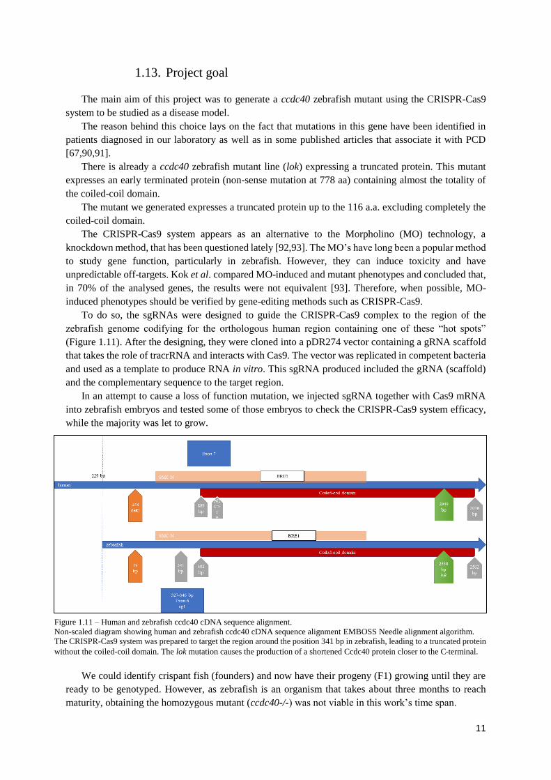
1.13. Project goal
The main aim of this project was to generate a ccdc40 zebrafish mutant using the CRISPR-Cas9
system to be studied as a disease model.
The reason behind this choice lays on the fact that mutations in this gene have been identified in
patients diagnosed in our laboratory as well as in some published articles that associate it with PCD
[67,90,91].
There is already a ccdc40 zebrafish mutant line (lok) expressing a truncated protein. This mutant
expresses an early terminated protein (non-sense mutation at 778 aa) containing almost the totality of
the coiled-coil domain.
The mutant we generated expresses a truncated protein up to the 116 a.a. excluding completely the
coiled-coil domain.
The CRISPR-Cas9 system appears as an alternative to the Morpholino (MO) technology, a
knockdown method, that has been questioned lately [92,93]. The MO’s have long been a popular method
to study gene function, particularly in zebrafish. However, they can induce toxicity and have
unpredictable off-targets. Kok et al. compared MO-induced and mutant phenotypes and concluded that,
in 70% of the analysed genes, the results were not equivalent [93]. Therefore, when possible, MO-
induced phenotypes should be verified by gene-editing methods such as CRISPR-Cas9.
To do so, the sgRNAs were designed to guide the CRISPR-Cas9 complex to the region of the
zebrafish genome codifying for the orthologous human region containing one of these “hot spots”
(Figure 1.11). After the designing, they were cloned into a pDR274 vector containing a gRNA scaffold
that takes the role of tracrRNA and interacts with Cas9. The vector was replicated in competent bacteria
and used as a template to produce RNA in vitro. This sgRNA produced included the gRNA (scaffold)
and the complementary sequence to the target region.
In an attempt to cause a loss of function mutation, we injected sgRNA together with Cas9 mRNA
into zebrafish embryos and tested some of those embryos to check the CRISPR-Cas9 system efficacy,
while the majority was let to grow.
We could identify crispant fish (founders) and now have their progeny (F1) growing until they are
ready to be genotyped. However, as zebrafish is an organism that takes about three months to reach
maturity, obtaining the homozygous mutant (ccdc40-/-) was not viable in this work’s time span.
Figure 1.11 – Human and zebrafish ccdc40 cDNA sequence alignment.
Non-scaled diagram showing human and zebrafish ccdc40 cDNA sequence alignment EMBOSS Needle alignment algorithm.
The CRISPR-Cas9 system was prepared to target the region around the position 341 bp in zebrafish, leading to a truncated protein
without the coiled-coil domain. The lok mutation causes the production of a shortened Ccdc40 protein closer to the C-terminal.

12
So, the second aim was to compare the CRISPR-Cas9 to the MO-induced phenotype. I, therefore,
knocked down ccdc40 by using a transcription blocking MO. The MO was tested by injecting several
concentrations (as explained in 2.12 in page 18). After the MO injection, parameters such as mortality,
heart and gut situs, cardiac oedema and tail defects were evaluated to future comparison with ccdc40-/-
mutant zebrafish.
2. Methods
2.1. Zebrafish maintenance
In this work were used wild-type AB lines (ZFIN ID: ZDB-GENO-960809-7), Tg(sox17:GFP)
(ZFIN ID: ZDB-TGCONSTRCT-070117-57) Tg(foxj1a:GFP) (ZFIN ID: ZDB-TGCONSTRCT-
131120-3) and TgBac(cftr-GFP) (ZFIN ID: ZFIN ID: ZDB-TGCONSTRCT-130423-1). These fish
were kept in the CEDOC fish facility under a controlled environment approved by the Direção Geral de
Alimentação e Veterinária (DGAV).
Adult fish were kept in fresh water (28o C, 700 µS, pH 7) under a 14:10 h light:dark cycle. Embryos
were obtained by natural mating and incubated at 25oC or 28ºC up to five days post-fertilization (dpf)
after which they were transferred to a tank in the main system or sacrificed.
The natural mating was stimulated by placing a male and one or two females inside a mating box
with a transparent divider allowing visual contact. The fish were removed from the main system and
placed in the mentioned mating boxes in the evening and left separated, as described, during the night.
In the morning, the dividers were removed and the water level lowered allowing the courtship behaviour
between male and female and consequently the external fertilization of the eggs. This method allows for
the synchronization of the embryonic development.
In each mating box, there was a sieve preventing the fish from eating the eggs. After the eggs were
laid the adult fish were again placed in the same main system tank. The eggs were collected by filtering
the water remaining in the mating box and then were placed in a Petri dish immersed in embryonic
medium with methylene blue (5mM NaCl, 0.2mM KCl, 0.3mM CaCl2, 0.3mM MgSO4 and distilled
water H2O, pH 6.5). The embryonic medium renewal and the removal of dead embryos were done daily
to prevent bacterial and fungal infections.
The embryonic staging was done according to what is described in Kimmel et al. [94]
2.2. Zebrafish euthanasia
Larvae up to 5dpf were sacrificed by being placed in a bleach solution. Larvae older than 5dpf and
adult fish were euthanized using an overdose of tricaine (MS-222) solution in a 250 mg/L concentration
[95,96].
2.3. CRISPR-Cas9 target sequences - Design of sgRNA
To study the functionality of ccdc40 gene (ENSDARG00000100584) in zebrafish, the CRISPR-
Cas9 technique was used to induce DNA breaks. The CRISPR-Cas9 target sequences inside the ccdc40
gene were determined using the crisprscan tool available online (crisprscan.org). This tool scans the
gene-of-interest coding sequence (ENSDART00000169752) and gives back a list of potential targets
sites ranked accordingly to its predicted efficacy. All the 20 nucleotide-long sequences presented are
followed by an NGG motif also called PAM (protospacer adjacent motif) that is required for the Cas9
13
recognition. The designed sgRNA oligonucleotides are expected to induce the cutting at any sequence
following the formula 5′-GG- N18 -NGG-3’. The GG-N18 (underlined) is included in the transcribed
sgRNA while the NGG integrates the genomic DNA only (not present in the sgRNA). If the sequence
has a mismatch in one of the first two GG’s it is called non-canonical but the Cas9 can still work properly
[97].
The provided list shows information concerning the score assessed by the tool, the sequence’s
position in the chromosome, its canonical state and the number of off-targets.
From the referred list, we chose two oligonucleotides that along with having the highest score
(higher scores mean higher predicted efficacy), were closer to the initial ATG (to create a mutation as
early as possible), these had no off-targets (those with no off targets minimize induced cuts in other
genome sites) and were considered canonical (no mismatches in the first GG providing a stronger
binding to the target site). The capital letters shown indicate the sequence that must be present in the
sgRNA and transcribed in vitro.
To allow the sgRNA cloning into a vector, for each target site were designed two complementary
oligonucleotides exhibiting single-stranded overhanging ends (bold) (ordered from Stab Vida; Oeiras,
Portugal) (Table 2.1).
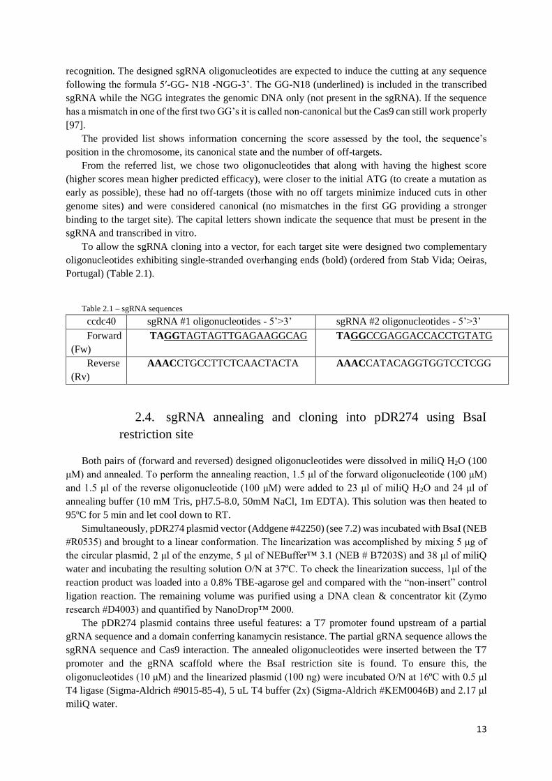
Table 2.1 – sgRNA sequences
ccdc40 sgRNA #1 oligonucleotides - 5’>3’ sgRNA #2 oligonucleotides - 5’>3’
Forward
(Fw)
TAGGTAGTAGTTGAGAAGGCAG TAGGCCGAGGACCACCTGTATG
Reverse
(Rv)
AAACCTGCCTTCTCAACTACTA AAACCATACAGGTGGTCCTCGG
2.4. sgRNA annealing and cloning into pDR274 using BsaI
restriction site
Both pairs of (forward and reversed) designed oligonucleotides were dissolved in miliQ H2O (100
μM) and annealed. To perform the annealing reaction, 1.5 μl of the forward oligonucleotide (100 μM)
and 1.5 μl of the reverse oligonucleotide (100 μM) were added to 23 μl of miliQ H2O and 24 μl of
annealing buffer (10 mM Tris, pH7.5-8.0, 50mM NaCl, 1m EDTA). This solution was then heated to
95ºC for 5 min and let cool down to RT.
Simultaneously, pDR274 plasmid vector (Addgene #42250) (see 7.2) was incubated with BsaI (NEB
#R0535) and brought to a linear conformation. The linearization was accomplished by mixing 5 μg of
the circular plasmid, 2 μl of the enzyme, 5 μl of NEBuffer™ 3.1 (NEB # B7203S) and 38 μl of miliQ
water and incubating the resulting solution O/N at 37ºC. To check the linearization success, 1μl of the
reaction product was loaded into a 0.8% TBE-agarose gel and compared with the “non-insert” control
ligation reaction. The remaining volume was purified using a DNA clean & concentrator kit (Zymo
research #D4003) and quantified by NanoDrop™ 2000.
The pDR274 plasmid contains three useful features: a T7 promoter found upstream of a partial
gRNA sequence and a domain conferring kanamycin resistance. The partial gRNA sequence allows the
sgRNA sequence and Cas9 interaction. The annealed oligonucleotides were inserted between the T7
promoter and the gRNA scaffold where the BsaI restriction site is found. To ensure this, the
oligonucleotides (10 μM) and the linearized plasmid (100 ng) were incubated O/N at 16ºC with 0.5 μl
T4 ligase (Sigma-Aldrich #9015-85-4), 5 uL T4 buffer (2x) (Sigma-Aldrich #KEM0046B) and 2.17 μl
miliQ water.
14
In the forward strand, BsaI cuts immediately after the recognition sequence 5’-GGTCTCN-3’
whereas in the reverse strand it cuts only four nucleotides ahead (3’-CCAGAGN(N)4-5’) creating non-
compatible sticky ends that match the overhanging ends added to the designed oligonucleotides.
This directional restriction cut by BsaI not only forces the correct insertion of the annealed
oligonucleotides into the plasmid backbone but it also prevents the plasmid from self-ligating.
2.5. E. coli (DHα5) transformation and identification of positive
colonies
The ligation product was used to transform competent E. coli bacteria. In this case, DHα5 was the
elected strain, rendering a transformation efficiency >1x10⁶ colony forming units for every mg (Thermo
Fisher #18265017).
The bacteria, stored at -80ºC, were thawed on ice for 20 min. Then, a 50 μl aliquot of DHα5 together
with 10 μl of the ligation product were subjected to a thermal shock (30 min on ice, 1 min at 42ºC, 2
min on ice) to boost the plasmid uptake. Next, followed an incubation at 37ºC and 250 rpm for 1h 30
min maximizing air-liquid interface. The incubation step was intended to let the bacteria recover from
the thermal shock and assure the plasmid stability inside the cells. Subsequently, the culture was
centrifuged (5000x g, 10min, RT), to increase cell density. These transformed bacteria were streaked in
a LB-agar plate treated with kanamycin (30μg/μl) and incubated once again at 37ºC O/N with the agar
layer facing upwards.
Because the cloning plasmid has a domain conferring resistance to kanamycin, only the bacteria that
up-took it were able to subsist. However, some bacteria could have assimilated the plasmid without the
oligonucleotides inserted. This oligonucleotide chain is too small to be resolved on an agarose gel, so to
avoid this predicament, colonies were screened by PCR (colony PCR). This technique relied on the use
of M13R primer (5’- TGTAAAACGACGGCCAGT-3’), that anneals with the plasmid backbone, and
the forward designed oligonucleotide for the corresponding Cas9 decided-target. When amplification
occurs, it meant the colony-inducing bacterium integrated the plasmid with the insert. However, when
no amplification was detected it could be because the cloning process was unsuccessful or the PCR
failed.
To detect the true positive colonies, we picked around six colonies for each sgRNA-dedicated agar
plate were directly used as template in a colony PCR reaction and streaked in a new agarose plate. Each
of these colony PCR reactions was prepared by combining 19.55μl of miliQ water, 2.5μl of buffer (10x,
provided with the enzyme), 1.25μl of MgCl2 (50mM, provided with the enzyme), 0.5μl of dNTPs
(10mM),0.2μl of NZYTaq DNA polymerase (Nzytech MB00101) and 0.5μl of each primer: M13R (10
μM) and forward sgRNA oligonucleotide (10 μM), adding up to a total volume of 25μl. The colony PCR
followed the steps from a common PCR i.e. initial denaturation (95ºC, 10min), denaturation (95ºC,
1min), annealing (51ºC for sgRNA#1 and #2, 1min), elongation (72ºC, 1min) and final elongation (72ºC,
10min) for 30 cycles. The M13R primer is tolerant to a wide range of temperatures, therefore the
annealing conditions were adjusted to the designed oligonucleotides structure.
The positive colonies were inoculated in primary liquid cultures (LB medium, kanamycin 30μg/μl)
and grown O/N at 37ºC and 250 rpm. These cultures sustain high bacteria densities, essential to a good
plasmid yield.
The plasmids were isolated using a commercial kit from Zymo Research (ZR Plasmid Miniprep™
#D4015) and then Sanger sequenced by Stab Vida using M13R primer.
All the described bacteria manipulations, including agar plate preparation, were done in a sterile
environment.
15
2.6. In vitro transcription of sgRNAs using T7 promoter
The plasmids (for sgRNA #1 and sgRNA #2) isolated from the positive colonies and confirmed by
Sanger sequencing were linearized downstream of the cloning site using a “unique cutter” enzyme. The
linearization was achieved by incubating together 1.25 μl of HindIII (NEB # R0104), 5 μl of NEBuffer
TM 2.1 (NEB # B7202S), 5μg of plasmidic DNA and miliQ water (up to 50 μl) for 10h at 37ºC followed
by storage at 4ºC. The then linear DNA was purified using a Zymo Research commercial kit (Zymo
Research #D4003) and since it contained a T7 promoter, it was used as a template to produce RNA.
Besides the purified DNA template with a promoter, the RNA in vitro transcription reaction required
triphosphate ribonucleotides (rNTP) (mix containing 10 mM of ATP, UTP, CTP and GTP), transcription
buffer 5x (MgCl2, 1 M; NaCl, 5 M; Tris-HCl, 1 M pH8), dithiothreitol (DTT) (50 mM), T7 RNA
polymerase (NEB #M0251S) and miliQ water adding up to a total volume of 50 μl.
DTT is a redox reagent that stabilizes the sulfhydryl groups within the T7 protein and without it, the
enzyme efficiency drops considerably.
First, the water, 10μl of transcription buffer, 5 μl of DTT and 5 μl of the rNTP mix were added
together and incubated for 5 min. Secondly, the DNA template was added (1.5 μg) and after a 1-minute
incubation, 1 μl of RNA inhibitor was joint to the solution, followed by another 1-minute incubation.
Ultimately, T7 RNA polymerase was joined. First, 2 μl preceded a 2h-incubation and then another 1 μl
was followed by a 1h-incubation.
Lastly, 1 μl of DNase (provided with RNA clean & concentrator kit by Zymo Research #R1013)
was incubated for 30 min with the solution to remove the template DNA initially added and make sure
just RNA remained. All the incubation periods were performed at 37ºC.
The generated RNA was purified with an RNA clean & concentrator kit by Zymo Research
(#R1013) and stored at -20ºC. The produced RNA was not 5′-capped or 3′-polyadenylated as it was not
to be translated. This protocol makes the RNA more prone to degradation, however as it is to be promptly
used, the quality decay is minimal.
This resulting RNA was injected together with Cas9 mRNA into one-cell stage zebrafish embryos.
2.7. Cas9 mRNA production
For Cas9 mRNA synthesis, the plasmid pCS2-ncas9n (Addgene, #47929) was linearized using NotI,
a “unique cutter) enzyme.
The in vitro transcription reaction done here was very similar to the one done to synthesize the
sgRNAs with the exception that here the reaction also included 5ul of G(5')ppp(5')G RNA Cap Structure
Analog (20mM)(NEB #S1407S). Therefore, as expected from a mRNA, the Cas9 mRNA is 5′-capped.
This feature is crucial as the Cas9 protein needs to be produced by the cell after co-injection with sgRNA
for the CRISPR-Cas9 system to work.
Raquel Jacinto, a lab’s PhD student performed the synthesis of this mRNA.
2.8. Micro co-injection of Cas9 mRNA and sgRNA into one-cell
stage zebrafish embryos
After the collection, the fertilized eggs were lined up against a glass slide inside a petri dish, without
embryonic medium and injected using a thin glass needle attached to a Narishigi pico-pump injector.
The needle was calibrated using a graticule (10mm/0.1mm Graticule Ltd., Tonbridge, Kent) in a way
that each pulse delivered 1.4 nL into the yolk.
16
The solution filling the needle contained Cas9 mRNA and sgRNA. Two different proportions of
these components were injected: 1) 100ng/μl Cas9 mRNA and 50ng/μl sgRNA, 2) 75ng/μl Cas9 mRNA
and 37.5ng/μl sgRNA.
Around 200 one-cell stage embryos were injected with each corresponding Cas9 mRNA/sgRNA
proportion and allowed to develop at 28ºC. At 24 hpf, each petri dish was sampled by picking 10
embryos used to extract genomic DNA. This DNA was amplified and a 15% polyacrylamide-agarose
gel electrophoresis (PAGE) was performed. Each gel contained 6.17 ml of Milli-Q water, 1.2 ml of TBE
buffer (10x) (Invitrogen™ 15581044), 4.5 ml of acrylamide/bis-acrylamide (29:1 Nzytech MB04501),
120μl of ammonium persulfate (10%), 9.6μl of TEMED and was let to polymerize between two glass
plates with 1.5mM spacer. After 3 h of electrophoresis at 150 V, the polyacrylamide gel was stained
with GreenSafe Premium (Nzytech MB13201) (2ul) diluted in TBE buffer (1x) (50ml) for 10 min,
washed twice in Milli-Q water and then visualised using ChemiDoc™ XRS+ System (Bio-Rad).
The aim was to detect heteroduplexes that migrate at a different rate due to the existence of
mismatches between the WT and the mutated sequence.
Whenever the experiment identified heteroduplexes, the fish were put in a separate tank and
transferred to the main system to grow, if not they were euthanized before 5 dpf.
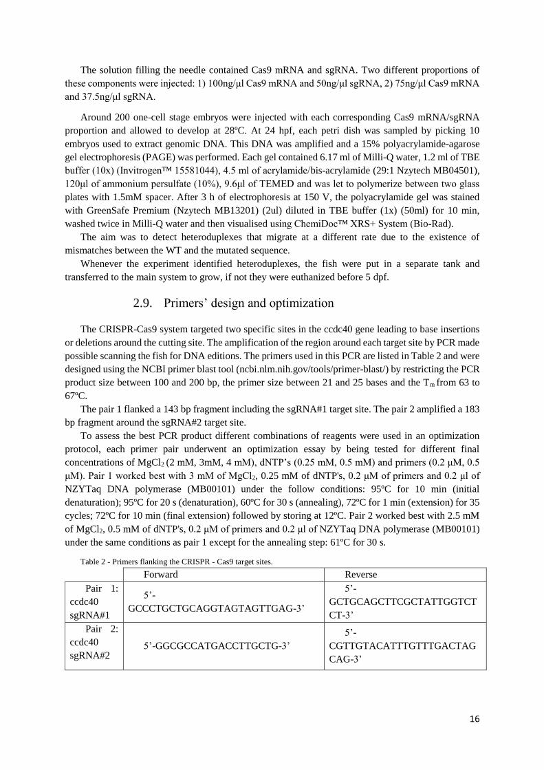
2.9. Primers’ design and optimization
The CRISPR-Cas9 system targeted two specific sites in the ccdc40 gene leading to base insertions
or deletions around the cutting site. The amplification of the region around each target site by PCR made
possible scanning the fish for DNA editions. The primers used in this PCR are listed in Table 2 and were
designed using the NCBI primer blast tool (ncbi.nlm.nih.gov/tools/primer-blast/) by restricting the PCR
product size between 100 and 200 bp, the primer size between 21 and 25 bases and the Tm from 63 to
67ºC.
The pair 1 flanked a 143 bp fragment including the sgRNA#1 target site. The pair 2 amplified a 183
bp fragment around the sgRNA#2 target site.
To assess the best PCR product different combinations of reagents were used in an optimization
protocol, each primer pair underwent an optimization essay by being tested for different final
concentrations of MgCl2 (2 mM, 3mM, 4 mM), dNTP’s (0.25 mM, 0.5 mM) and primers (0.2 μM, 0.5
μM). Pair 1 worked best with 3 mM of MgCl2, 0.25 mM of dNTP's, 0.2 μM of primers and 0.2 μl of
NZYTaq DNA polymerase (MB00101) under the follow conditions: 95ºC for 10 min (initial
denaturation); 95ºC for 20 s (denaturation), 60ºC for 30 s (annealing), 72ºC for 1 min (extension) for 35
cycles; 72ºC for 10 min (final extension) followed by storing at 12ºC. Pair 2 worked best with 2.5 mM
of MgCl2, 0.5 mM of dNTP's, 0.2 μM of primers and 0.2 μl of NZYTaq DNA polymerase (MB00101)
under the same conditions as pair 1 except for the annealing step: 61ºC for 30 s.
Table 2 - Primers flanking the CRISPR - Cas9 target sites.
Forward Reverse
Pair 1:
ccdc40
sgRNA#1
5’-
GCCCTGCTGCAGGTAGTAGTTGAG-3’
5’-
GCTGCAGCTTCGCTATTGGTCT
CT-3’
Pair 2:
ccdc40
sgRNA#2 5’-GGCGCCATGACCTTGCTG-3’
5’-
CGTTGTACATTTGTTTGACTAG
CAG-3’
17
2.10. Genomic DNA extraction
Genomic DNA was extracted from 24 hpf embryos. In each case, we collected 3 batches of 10
embryos that were dechorionated, placed in a 1.5 ml Eppendorf tube with 20μl of NaOH (5mM) and
incubated for 20 min at 96ºC. The embryos were then smashed with a pipette tip and kept at 4ºC for
another 20 min. Lastly, the remaining extract was centrifuged at 12 500g for 10 min with 2 ul of Tris-
HCl (1 M, pH8). The supernatant was transferred to a new Eppendorf tube and frozen at -20ºC or used
straight away as a template.
2.11. Heteroduplexes detection using PAGE analysis
When two single-stranded DNA or RNA molecules deriving from different sources anneal together
they form a heteroduplex. In this case, the different sources are the WT embryos and the crispant
embryos where the CRISPR-Cas9 system made a double-stranded cut. The repair (NHEJ) of this cut is
prone to errors and therefore may insert or remove several base pairs in the vicinity of the cut. When
amplifying this region by PCR using DNA from a pool of embryos as a template, WT (non-altered)
fragments can hybridize with repaired fragments (with base insertions or deletions), forming
heteroduplexes. Due to the alterations carried out by the CRISPR-Cas9 system, the annealing with WT
fragments is not perfect. The existence of mismatches alters the DNA double-stranded conformation
and as a result, it changes its migration rate.
On a PAGE experiment (polyacrylamide gel electrophoresis), heteroduplexes generally migrate
slower than homoduplexes [98] making its visualization a good method to screen for crispants
(potentially modified embryos).
The search for heteroduplexes was done at two different time-points. First, right after the co-
injection of Cas9 mRNA and sgRNA and then again when the injected fish reached maturity.
The analysis at the first time-point included randomly picking ten injected embryos from each petri
dish to guarantee that the Cas9 was cutting at the targeted genome region. The analysis at the second
time-point involved screening each individual adult fish to find crispants. At this point, the potential
crispant fish were mated with wild-type fish and their progeny was analysed to access the mutation
presence in the germline. In this case, the analysis included three batches of ten embryos from each
couple (potential crispant + WT).
In both cases, the selected embryos were dechorionated and processed to extract genomic DNA.
This DNA was then amplified by PCR using the primers flanking the corresponding Cas9-targeting
region.
PCR followed the conditions specified in “Primers design and optimization”.
The annealed PCR products were then loaded and resolved by electrophoresis on a non-denaturing
PAGE.
In the lanes with positive heteroduplexes, the bands were isolated and the DNA extracted by
incubating the acrylamide with 20ul of TE (supplied with DNA clean & concentrator kit, Zymo research
#D4003) at 37ºC O/N.
The isolated DNA was further amplified using Xpert HighFidelity DNA Polymerase (GRiSP
#GE07.0250) accordingly to the manufacturer’s instructions and a Ta= 60.5ºC. After the PCR product
purification (DNA clean & concentrator kit, Zymo Research #D4003). Then, the DNA was sequenced
(Stab Vida) for confirmation. Overlapping sequences in the chromatograms downstream to the PAM
region were interpreted as a sign of heterozygosity and are expected in the heteroduplexes’ samples.

18
2.12. Microinjection of zebrafish embryos with Morpholino
A morpholino is a synthetic molecule with a structure similar to the natural nucleic acids. It acts as
an antisense agent by binding to the RNA and preventing its translation.
The translation-blocking morpholino (ccdc40 MO) was purchased from Gene Tools LLC
(Philomath, OR, USA) and received in a lyophilized form. Upon arriving, it was suspended in Milli-Q
water, achieving a final concentration of 1mM, heated up to 65ºC for 10 min and then aliquoted.
This ccdc40 MO was designed against the 5’UTR region of the ccdc40 gene, excluding the start
codon (to allow future rescue experiences): 5’-TTTTAATTCACAGTCCTTTAGCGGA- 3’.
The ccdc40 MO was injected into the yolk in each one cell-stage embryo to maximize the
morpholino uptake by the cells. To do so, right after the eggs were laid, they were lined up against a
glass slide and injected with a previously calibrated borosilicate glass needle loaded with a ccdc40 MO
solution. This needle, attached to the air injector, which allows the standardization of the volume injected
in each pulse 1.4nl. The solutions loaded into the needle held several MO concentrations: 4 ng, 4.5 ng,
5ng, 5.5 ng, 6 ng, 6.5 ng, 7 ng, and 7.5 ng.
In order to characterize the MO effects, were taken into consideration, heart laterality, liver and
pancreas relative position and defects such as oedema and tail crooks.
Heart laterality was screened at 30hpf, liver and pancreas relative position at 50hpf and oedema and
tail crooks at 5dpf in fish incubated at 28ºC.
2.13. Heart and gut screening
The heart and gut situs were screened to assess the impact of the morpholino injection in the
zebrafish left-right development.
In wild-type embryos, the heart can be seen ventrally on the right side of the body under the right-
sided eye. The heart tube starts to gain its form around 21hpf and at 36hpf it becomes possible to
distinguish the atrium from the ventricle. The S-shape looping occurs between 26 and 48hpf [99],
therefore the heart situs screening was done at 30 hph. In wildtype embryos, this looping
characteristically occurs towards the left side but it can also be formed towards the right side or the loop
can be absent (central heart). The screening of non-injected and MO-injected embryos was done in vivo
under a stereoscope using a 60x magnification. The three different phenotypes were placed in separate
Petri dishes and later fixed at 50 hpf for further gut situs analysis.
The pancreas is formed as a result of the fusion of the dorsal and ventral bud that happens at 44hpf
[100]. The liver morphogenesis starts at 28 hpf with the thickening of the endoderm rod under the first
somite and is completed around 50 hpf when the hepatic duct is formed [101]. To assess the positions
of these two organs we used the 50 hpf tg(sox17:GFP) larvae and fixed them in 4% PFA-PBS O/N.
These larvae and then evaluated under the Zeiss Lumar V12 stereoscope’s fluorescence after the yolk
removal by careful dissection.

19
3. Results
3.1. CRISPR-Cas9 preparation
CRISPR-Cas9 is a useful method to create mutants. For it to work, first, it is necessary to produce
cas9 mRNA (or the protein itself) and the sgRNA holding a complementary sequence to the genomic
region we want to disrupt. The cas9 mRNA was already available in the laboratory, however, the specific
sgRNAs had to be produced. Besides that, in order
to screen the region of interest for CRISPR-Cas9
induced ‘indels’, we had to design and optimise a
pair of primers for each target region.
3.2. SgRNA cloning into pDR274
and sgRNA synthesis
The sgRNAs sequences were inserted into a
pDR274 plasmid using the restriction enzyme
BsaI. This plasmid containing the sgRNAs
template was replicated in E. coli (DHα5)
incubated in a solid medium with kanamycin.
To verify if the bacteria had, in fact, the
plasmid with the insert we performed a colony PCR (cPCR) and run the products on a 1% TBE-agarose
gel. Colonies 2, 3, 7, 10 and 12 showed
amplification (~300 bp) (Figure 1.1) and therefore, were sent to sequence.
The Sanger sequencing results showed that
only colony 10 (sgRNA#2) had the plasmid with
the insert (Figure 3.3 A). To save time and
reagents, colonies 13, 14 and 15 (for sgRNA#1)
were promptly sequenced without undergoing a
cPCR beforehand. The results showed that
bacteria in colony 14 had integrated the plasmid
with the sgRNA#1 template (Figure 3.3 B).
Figure 3.2 – pDR274 linearization with HindIII.
The plasmid has approximately 2100 bp, but when in a
circular conformation, it coils slowing its migration rate.
1% TAE-agarose gel, 100 V, 30 min
Figure 3.1 – Colony PCR results.
Colonies 2, 3, 7, 10 and 12 were positive showing amplification
around 300 bp.
1% TBE-agarose gel, 100 V, 30 min
300 bp
300 bp

20
Colonies 10 and 14 were inoculated into a primary culture. When they reached confluence, we
isolated the plasmids and linearized them using the HindIII restriction enzyme. The linearization was
tested by running a sample on an agarose gel (Figure 3.2). The linearized plasmids were used as a
template to produce the sgRNAs. Table 3.1 shows the sgRNAs quantification.
Table 3.1 – sgRNA#1 and#2 nanodrop quantification
Sample Mean of RNA
concentration
(ng/μl)
Total
volume (μl)
Amount of
sgRNA (ng)
sgRNA#1 (colony 14) 873 9 7857
sgRNA#2 (colony 10) 829 9 7461
3.3. Primers optimization
To test CRISPR-Cas9 efficacy after injection and later screen for crispants, we designed two primer
pairs, each flanking sgRNA#1 and sgRNA#2 target-sites. The optimization was done as explained in
2.9.
Primer pair 1 and 2 specifically amplified
only one band with 143 bp and 184 bp
respectively (Figure 3.4). At this point, primers
were ready to be used in testing CRISPR-Cas9
efficacy after injection and later to screen for
crispant fish.
Figure 3.3 – Sequencing results from colony 10 (sgRNA#2) and colony 14 (sgRNA#1).
Both sgRNA templates were inserted between the T7 promoter and the gRNA scaffold. In colony 10 the sgRNA template
seems to have been partially duplicated (feature A). For detailed sequence see Figure 7.2 (colony 10) and Figure 7.3
(colony 14).
A: Colony 10 – sgRNA#2
B: Colony 14 – sgRNA#1
Figure 3.4 – Primer-pairs optimization for zebrafish
genomic DNA.
Primer pair 1 and 2 show amplified a band with 143 and
184 bp respectively.
0.8 % TBE-agarose gel, 120 V, 45 min

21
3.4. Co-injection of cas9 mRNA and sgRNA and CRISPR-Cas9
effectiveness analysis by PAGE assay
In our first attempt, we injected cas9 mRNA, in a concentration of 100 ng/μl, together with each
sgRNA, in a concentration of 50 ng/μl in one-cell stage zebrafish embryos. Most of the injected embryos
developed into larvae with malformations such as curly-up, curly-down, tail atrophy, cyclopia, and
cardiac oedema (Figure 3.5 A). Those with severe deformities were not viable and were not picked when
testing for cas9-induced mutations. When analysing normal-looking 24 hpf-embryos, no heteroduplexes
were found, meaning that the CRISPR-Cas9 system was not working in those embryos and that could
be the reason for their normal appearance.
The deformities could be linked to the
toxicity of the injected components,
therefore, the amounts of Cas9 mRNA and
sgRNA were lowered to 75 ng/μl and 37.5
ng/μl respectively.
This time around, the embryos injected
with lower concentrations of cas9 mRNA
and sgRNA developed into larvae with fewer
deformities (Figure 3.5 B). The PAGE-based
analyses showed heteroduplexes in the
sample from the TgBac(cftr-GFP) embryos
injected with sgRNA#1 (Figure 3.6). Since
only sgRNA#1 yield positive results, the
following attempts focused on it rather than
sgRNA#2. sgRNA#2 presents itself partially
duplicated (Figure 3.3 A) what can one
reason for its ineffectiveness.
To verify if the CRISPR-Cas9 system had successfully cut the target region in defective larvae, a
group of them was tested independently. As expected, samples extracted from pools of defective
embryos showed a larger collection of heteroduplexes (Figure 3.7). Opportunely, samples coming from
apparently non-defective embryos also presented several heteroduplexes (white boxes in Figure 3.7).
Neg
ativ
e
contr
ol
Posi
tive
contr
ol
AB
Non-
inje
cted
embry
os
CF
TR
sgR
NA
#1
200 bp
100 bp
CF
TR
Non
-
inje
cted
AB
sgR
NA
#2
Figure 3.6 – Heteroduplexes detected in PAGE-based analysis of
CRISPR-Cas9 system efficacy in TgBac(cftr-GFP) embryos.
Here we can see heteroduplexes in the sample extracted from
TgBac(cftr-GFP) embryos injected with Cas9 mRNA (75 ng/μl)
and sgRNA#1 (37.5 ng/μl). (white box).
A B
Figure 3.5 – Deformities found in zebrafish larvae (3dpf) after co-injection with Cas9 mRNA and sgRNA.
A: Embryos injected with 100 ng/μl of Cas9 mRNA and 50 ng/μl of sgRNAs. The malformations included curly-up and curly-
down tails, cyclopia, cardiac oedema and tail atrophy.
B: Embryos injected with 75 ng/μl of Cas9 mRNA and 37.5 ng/μl of sgRNAs. The malformations included cardiac oedema
and tail kinks.

22
These results confirmed that CRISPR-Cas9 system was working, thus the remaining batch of
injected fish (possible founders) was transferred to the fish facility main system to grow until reaching
maturity.
3.5. Search for founders: zebrafish screening and genotyping
This is the most time-consuming task when generating mutants using CRISPR-Cas9 method [102].
It is not only necessary to assure that a certain fish carries a mutation, but, because these can present
mosaicism, it is also necessary to guarantee that that mutation is present in the germline cells. This can
be done by directly analysing the gametes or by screening the progeny of each individual founder fish.
In this case, the fish, that developed from the CRISPR-Cas9 injected embryos, were screened
through the analysis of their progeny in a PAGE-based assay.
The first to be screened were the TgBAC(cftr-GFP) zebrafish injected with ccdc40 sgRNA#1. This
aquarium only had four females and none of them was a crispant. From the remaining fish (30 males),
male 23 analysis (Figure 3.8) showed a similar heteroduplex pattern as before in all three pools of ten
embryos(see Figure 3.6).
The next aquarium to be screened was the one with Tg(foxj1a:GFP) zebrafish injected with ccdc40
sgRNA#1. From the ten fish (all male) screened in this aquarium, only male 3 produced positive results.
In replica A, heteroduplexes appeared slightly above the 300 bp marker (not coincident with the
observations in Figure 3.6), while in replica B, a band was detected around the 200 bp mark, consistent
with the pattern observed in Figure 3.6.
Nine of the AB fish injected with sgRNA#1 were also screened, but none yield positive results. The
remaining fish (tg(sox17-GFP)) had not yet reached full maturity at the end of this project’s time limit.
Figure 3.7 – Heteroduplexes detected in PAGE-based assay for testing CRISPR-Cas9 efficacy in AB, Tg(foxj1a:GFP) and
Tg(sox17-GFP) embryos injected with Cas9 mRNA (75 ng/μl) and sgRNA#1 (37.5 ng/μl).
Heteroduplexes are highlighted in white boxes.

23
The DNA from the revealed heteroduplex bands, as well as from the WT bands, was isolated and
sequenced. The Sanger sequencing results showed a chromatogram overlapping in heteroduplex-
isolated DNA from male 23 TgBac(cftr-GFP) (all three polls of embryos) (Figure 3.9) (for complete
results, see appendix 7.4 ). In male 3 Tg(foxj1a:GFP) samples, the overlapping was only detected in
replica B. The bands in replica A, around 300 bp, were probably due to some unspecific PCR
amplification or contamination.
The band slightly above 200 bp in
replica A, already had appeared in
the PAGE-based assay when
testing CRISPR-Cas9 efficacy
(Figure 3.7).
The chromatogram
overlapping is a tell-tale sign of
heterozygosity [103], therefore
these results allowed the
identification of male 23
TgBAC(cftr-GFP) and male 3
Tg(foxj1a:GFP) as founders (F0).
The analysis of the founders
through their offspring is an
effective way of assuring that the
CRISPR-Cas9 induced indels are
passed on through the germline,
however, as the sequenced DNA
derives from a batch of ten
embryos, at this point was not yet
possible to determine the exact
induced mutation. Among this batch of embryos, it is possible to find a maximum of three different
alleles (and therefore peaks). Each embryo can carry the WT allele, inherited from the AB mother, and
one of the two paternal alleles, that, in theory, can both carry induced indels. The exact induced mutation
can only be determined when analysing a single fish. Aiming for that, the offspring resulting from the
mating between male 23 TgBAC(cftr-GFP) and male 3 Tg(foxj1a:GFP) with AB females (F1), were
kept in the fish facility main system to be screened when reaching maturity.
Figure 3.8 – Heteroduplexes detected in a PAGE-based assay used to test
TgBac(cftr-GFP) ♂ 23 and Fox:GFP ♂ 3 offspring.
The analysis was run in triplicate (A, B, C): in ♂ 23 the results were uniform
while in ♂3 were detected two different sets of heteroduplexes (lane C showed
a very faint WT band with no heteroduplexes detectable). The bands marked
with a * probably resulted from a contamination with ♂ 23 samples.

24
Assuming that only one paternal allele was mutated, 50% of these fish should carry the mutation.
However, if both paternal alleles were mutated, the DSBs were resolved differently in each allele,
resulting in two different mutations. Therefore, we will have 50% of the fish carrying one mutation and
50% carrying the other. Fish carrying a certain mutation are to be in crossed among each other, and 25%
of the resulting offspring (F3) should be the sought-after ccdc40 homozygous mutants.
Figure 3.9 – Sanger sequencing results for wildtype (WT) and heteroduplex (HTD) band isolated from ♂ 23 B TgBAC(cftr-
GFP) and ♂ 3 B Tg(foxj1a:GFP).
The referred bands were isolated from the gel shown in Figure 3.8 .
The WT sequences showed individualized peaks, while HTD sequences exhibited overlapping in the chromatogram
immediately after the PAM sequence 3’ edge.
This sequencing was performed using a reverse primer, therefore the PAM sequence (CCT) highlight in blue is the reverse
complement sequence of the canonical PAM sequence 5'-NGG-3'.

25
3.6. Morpholino antisense technology - phenotype
characterization
To study the ccdc40 loss-of-function phenotype and later compare it with the ccdc40 -/- mutant, and also to access MO toxicity and effects on laterality axis establishment, we injected several
Figure 3.11.-. Phenotype of lavae (3dpf) injected
with ccdc40 translation-blocking MO (6 ng)
showing cardiac oedema and tail crooks.
A
Figure 3.10 – Morphological defects observed in ccdc40
translation-blocking MO injected embryos.
A: Percentage of mortality, tail defects, oedema and abnormal
heart and gut situs accordingly to the injected amount of MO.
B: Heart laterality phenotype amid MO injected embryos.
Black, grey and white bars represent the percentage of embryos
with heart positioned on the left side, in the middle and on the
right side respectively.
B
Figure 3.12 – Gut situs from tg(sox17-GFP) embryos injected with translation-blocking ccdc40 MO
A: Pictures showing the gut situs of MO-injected tg(sox17-GFP) embryos (50 hpf) taken under the fluorescence steroscope.
B and C: The bar graphs represent the gut phenotype of tg(sox17-GFP) embryos injected with the translation-blocking ccdc40
MO. The gut situs was evaluated separately in embryos with the heart on the left (B) and embryos with right or central heart (C).
L-liver; P-pancreas
Normal-liver on the left and pancreas on the right; Midline-both liver and pancreas centred; Reversed-liver on the right and
pancreas on the left.

26
quantities of ccdc40 translation-blocking MO into one-cell stage embryos. We evaluated mortality rates
(at 24 hpf), external morphological defects such as cardiac oedema and tail crooks (4 dpf), and internal
morphological features such as heart (at 30 hpf), liver and pancreas (at 50 hpf) placement along the L-
R axis.
All the evaluated parameters seemed to stabilize from 6 ng onwards, suggesting that these defects were
not a direct cause of toxicity. Furthermore, the development of cardiac oedema, tail crooks and lethargy
were consistent throughout all the MO amounts injected, being present in almost all of the embryos
(Figure 3.11).
Particularly, heart situs abnormalities presented themselves in about 20% (6 ng) to 30% (7 ng) of all the
injected embryos analysed, in contrast with non-injected ones. (Figure 3.10 B). Among those with heart
misplacement, it was noticeable a higher proportion of central hearts, with 70% (6 ng) to 95% (7 ng) of
the embryos having a heart with no loop.
Besides the heart situs, the injected embryos also were screened for the liver and pancreas
positioning (Figure 3.12A). This evaluation was done separately for embryos with a left heart and for
embryos with right or central heart (Figure 3.12 B-C) and in both cases, the gut situs was affected.
Embryos with a left-sided heart presented between 23% (6.5 ng) and 50% (6 ng) of gut positioning
defects, while in embryos with a central or right-sided heart the gut situs defects were present in 32% (7
ng) to 77% (6 ng). The fact that these numbers do not correlate directly to the amount of MO-injected
can be due to the randomization of the situs establishment.
Also, in the case of central and right-hearted embryos, the number considered was low, what,
together with the morpholino variability, adds to this discrepancy. Interestingly, when the gut was
mispositioned the prevalent phenotype was the midline situs. This happened despite the heart position
but was more evident in embryos with a central or right-sided heart.
Figure 3.13 shows the merge of heart and gut situs results. Since the embryos with central and right-
side hearts were not separated, it is not possible to distinguish situs inversus from heterotaxy. Still, we
can say that in 40% (6.5 ng) to 70% (7.5 ng) of the cases presented situs defects (Figure 3.13) as well as
tail crooks, oedema and lethargy.
Figure 3.13 – Heart and gut situs combined.
The gut situs assessment for embryos with central and right hearts was done together. Therefore, cases of situs inversus and
heterotaxy cannot be separated.
RCH: central or right heart, LH: left heart, N: normal gut (left liver and right pancreas), R: reversed gut.
27
4. Discussion
In this project, our goal was to create a ccdc40 -/- zebrafish mutant line (position 116 aa). As the
chosen animal model takes three months to reach maturity and the desired mutant can only be obtained
after three generations, we studied MO-injected embryos instead.
Our results in MO-injected embryos showed situs defects in up to 70% (7.5 ng) of the cases as well
as tail crooks, oedema and lethargy.
In studies with lok mutants, known to have a recessive mutation in ccdc40, embryos are shown to
develop a similar phenotype with curly down tails, organ situs problems in 50% of the cases (situs
inversus and heterotaxy) [90], lethargy and cardiac oedema [104].
In both cases, it makes sense that the organ situs is randomly established because when cilia are
immotile or beat in an abnormal way, nodal expression does not become asymmetric to the left on the
LPM and instead it can become either absent, bilateral, expressed on the right or on the left side of the
LPM.
Although both phenotypes represent a Ccdc40 knockdown, the MO-induced phenotype appeared
stronger. MO-induced situs defects seemed accordant with lok phenotype, ranging from 40% to 70%,
however, the cardiac oedema is more severe and the tail curvatures more convoluted Figure 3.11. in the
morphants than in the lok homozygous mutants.
At 3 dpf WT embryos break out of the chorion and straighten their tails. In lok homozygous mutants,
as well as in our MO-injected embryos this does not occur. They kept their curvatures even after
hatching. lok mutants have their tails bent ventrally and in MO-injected embryos, the tail can bend
downwards, upwards or to the sides and present more than one curvature. lok mutants [71,104], as well
as the ccdc40 MO-injected embryos in this study, died at 7 dpf.
In zebrafish, ccdc40 gene has two transcripts annotated. One encodes for the CCDC40 protein and
the other is a non-coding retained intron transcriptf.
In humans, there are six transcripts producing protein and other six transcripts with premature stop
codonsg. Based on the literature, around half of the homozygous patients with a CCDC40 mutation carry
the frameshift mutation c.248delC [76,90] This mutation inserts a stop codon upstream the region
codifying the coiled-coil domain. The other half of the mutations take place much closer to the end of
the codifying region (such as c.3129delC and c.2824-2825insTGT) allowing the integral transcription
of smaller CCDC40 isoforms, containing a truncated coiled-coil domain, and non-coding transcripts.
The axonemal ultrastructures shown by Antony et al. (2013) [76] and by Becker-Heck et al. (2011)
[90] are from nasal cilia collected from patients carrying early termination mutations and are similar to
the ciliary ultrastructure reported for the lok homozygous mutants. We were not able to find reported
TEM images from cilia collected from PCD patients with early mutations such as c.248delC (closer to
the N terminal) to compare the phenotypes. Both events, mutations closer to the N extremity or
mutations closer to the C terminal of the protein, cause PCD, meaning that the complete CCDC40 variant
is necessary for the correct IDA assembly in the ciliary axoneme. The other protein variants may play a
regulatory role. It was described that transcripts with premature stop codons can have a role in gene
regulation [105,106]. In the lok mutants, only the protein-coding transcript is affected, with the non-
coding transcript still being produced normally. In the MO-injected embryos, both transcripts were
fhttp://www.ensembl.org/Danio_rerio/Gene/Summary?db=core;g=ENSDARG00000100584;r=6:21779819-21821089
ghttp://www.ensembl.org/Homo_sapiens/Transcript/ProteinSummary?db=core;g=ENSG00000141519;r=17:80036632-
80100613;t=ENST00000269318

28
knocked down, offering an explanation to the more severe defects, that in this situation can result from
both IDA assembly problems and absence of regulatory elements.
Apart from being structurally necessary for correct assembly of IDA, Ccdc40 is also needed for the
recruitment of tubulin polyglutamilases, [74] that promote tubulin stabilization. This was found in
Chlamydomonas, that only expresses the protein-coding transcripth, not clarifying if proteins with
truncated coiled-coil domains can still do the recruitment of the tubulin polyglutamilases. However, in
case they still preserve that ability, the truncated Ccdc40 protein can be able to promote tubulin
stabilization, even if with less efficiency in both lok zebrafish mutants and PCD patients carrying early
termination mutations near the C terminal.
To verify this, the ciliary structure organization of the CCDC40 -/- mutant line obtained by CRISPR-
Cas9 must be analyzed using an antibody for glutamylated tubulin and compared with that of lok
mutants.
It would also be interesting to analyse the cilia beat frequency of both mutants to verify if higher
polyglutamylation levels are reflected in increased the cilia motility control as suggested in [107]
The analysis of cilia beat frequency in lok mutants revealed that cilia beat within a reduced amplitude
and with a more rigid movement [108]. This stiffer movement can be due to the portion of the coiled-
coil domain that is present and still allows IDA assembly (but in lower numbers as found in Becker-
heck et al. 2011 [90]) that together with an intact ODA results in the presence of some residual
movement.
In the case of MO-injected embryos, the ccdc40 protein is not present at all and the IDA assembly
should not be possible. We did not measure the cilia beat frequency in MO-injected embryos but, if this
assumption is right, no movement is expected in MO-injected or ccdc40 -/- mutants because in both
cases the coiled-coil domain necessary for movent regulation [70] is not present.
According to Grimes et al. the spinal curvatures in lok mutants appeared as a result of abnormalities
in CSF flow and vertebrae morphogenesis [109]. The resulting phenotype is not viable, however, if lok
embryos are injected with WT ccdc40 mRNA at one cell stage, they survive to develop spinal curvatures
that resemble human idiopathic scoliosis [109]. The mRNA injected only counteracts the absence of the
protein-coding transcript and the non-coding transcript is not compensated for. If the co-injection of
mRNA for both coding and non-coding transcripts leads to a complete phenotype rescue resulting in
fish with not spinal curvatures, then the non-coding transcript may play an important role in osteogenesis
regulation.
One possible explanation for the spinal defects detected in our knockdown of ccdc40 is in the
evidence connecting cilia and osteogenesis in a PKD1-dependent signalling pathway. PKD1 (present in
the cilia membrane of the KV cilia) is also expressed in cartilage and somites and Pkd1 mutant mice
develop problems in bones and cartilage, exhibiting spinal curvatures and vertebra deformities [110].
We know from previously unpublished work in the lab that the KV cells will later integrate the tail tissue
[111]. In particular, they will become part of the notochord, hypochord and somites. So, it is possible
that defects in Pkd1 could lead to tail deformities. Nevertheless, a link between the lack of Ccdc40 and
abnormal expression of Pkd1 would need to be first established.
hhttp://plants.ensembl.org/Chlamydomonas_reinhardtii/Transcript/Domains?db=core;g=CHLREDRAFT_170513;r=DS496120:1834038-1840048;t=EDP04735

29
5. Conclusion and Prospects
Based on the MO phenotype, CCDC40 is essential for correct organ situs. It seems that the
phenotype obtained when injecting translation-blocking MO is more severe than that obtained in lok
mutants.
lok mutants assemble a ccdc40 protein that still has part of the coiled-coil domain, while the
homozygous ccdc40 -/- mutant produced expresses only the first 346 of the total 2826 bp, which
excludes the all the extension of the coiled-coil domain (Figure 5.1). Similar mutations causing PCD
have been identified in patients and up to date, no one investigated if a mutation truncating a part of the
protein C-terminal (lok-like) and a mutation closer to the N-terminal, excluding the total extension of
the coiled-coil domain, result in similar phenotypes such as ultrastructural defects and tubulin post-
translational modification patterners. The comparative analysis of both phenotypes will help to clarify
these questions.
Since this MO was designed to exclude the initial ATG codon to make possible rescue experiences,
we attempted to clone the ccdc40 gene into a PCS2+ mcherry vector but, with the tested pair of primers
(appendix 7.5), it was not possible to obtain the desired amplification. However, with the production of
mRNA for the protein coding sequence and for the non-coding transcript, it will be possible to study the
effected of each one alone in the cilia motility and development of spinal cord in zebrafish.
To conclude this project, in the immediate future, it is necessary to incross the heterozygotes (F1)
to achieve a homozygous mutant and identify the exact mutation introduced. When getting this ccdc40
-/- mutant, its phenotype can be compared with the MO phenotype validating it or not.
The mutation induced in ccdc40 -/- mutant lies in a region homologous to the sequence in the human
genome where the mutation c.961 C>T has been identified. This mutation appears to be in a region
regarded as a “hot spot” where at least other two mutations have been identified [76]. Therefore, with
the exact mutation identified, the ccdc40 -/- mutant line can be used to test gene therapy approaches to
try to correct it back to normal. These same approaches can then be applied in patient-derived cells,
leading to a way of alleviating the respiratory symptoms present in PCD in a similar way to what has
been done to other diseases [112].
6. Bibliography 1. Langdon YG, Mullins MC. 2011 Maternal and Zygotic Control of Zebrafish Dorsoventral Axial Patterning. Annu.
Rev. Genet. 45, 357–377. (doi:10.1146/annurev-genet-110410-132517)
2. Piccolo S, Agius E, Leyns L, Bhattacharyya S, Grunz H, Bouwmeester T, De Robertis EM. 1999 The head inducer
Cerberus is a multifunctional antagonist of Nodal, BMP and Wnt signals. Nature 397, 707–710.
(doi:10.1038/17820)
3. Agathon A, Thisse C, Thisse B. 2003 The molecular nature of the zebrafish tail organizer. Nature 424, 448–452.
Figure 5.1 – Diagram showing the predicted zebrafish Ccdc40 protein domains in WT, lok mutants and Ccdc40 generated
mutant using CRISPR-Cas9 system.
In WT zebrafish, the coiled-coil domain ranges from the 134 aa to the 854 aa. In lok mutants, this domain is truncated at the
778 aa position. The mutant line generated in this work (Cccdc40 -/-) synthesize a protein truncated at the 116 aa, with no
coiled-coil domain.
30
(doi:10.1038/nature01822)
4. Hamada H, Meno C, Watanabe D, Saijoh Y. 2002 Establishment of vertebrate left–right asymmetry. Nat. Rev.
Genet. 3, 103–113. (doi:10.1038/nrg732)
5. Collignon J, Varlet I, Robertson EJ. 1996 Relationship between asymmetric nodal expression and the direction of
embryonic turning. Nature 381, 155–158. (doi:10.1038/381155a0)
6. Nakamura T, Hamada H. 2012 Left-right patterning: conserved and divergent mechanisms. Development 139, 3257–
3262. (doi:10.1242/dev.061606)
7. Blum M, Weber T, Beyer T, Vick P. 2009 Evolution of leftward flow. Semin. Cell Dev. Biol. 20, 464–471.
(doi:10.1016/j.semcdb.2008.11.005)
8. Levin M, Palmer AR. 2007 Left-right patterning from the inside out: Widespread evidence for intracellular control.
BioEssays 29, 271–287. (doi:10.1002/bies.20545)
9. Dasgupta A, Amack JD. 2016 Cilia in vertebrate left-right patterning. Philos. Trans. R. Soc. Lond. B. Biol. Sci. 371,
20150410. (doi:10.1098/rstb.2015.0410)
10. Nakamura T et al. 2012 Fluid flow and interlinked feedback loops establish left-right asymmetric decay of Cerl2
mRNA. Nat. Commun. 3, 1322. (doi:10.1038/ncomms2319)
11. Marques S, Borges AC, Silva AC, Freitas S, Cordenonsi M. 2004 The activity of the Nodal antagonist. Genes Dev. ,
2342–2347. (doi:10.1101/gad.306504.2342)
12. Tsukui T et al. 1999 Multiple left-right asymmetry defects in Shh-/- mutant mice unveil a convergence of the Shh
and retinoic acid pathways in the control of Lefty-1. Proc. Natl. Acad. Sci. 96, 11376–11381.
(doi:10.1073/pnas.96.20.11376)
13. Meno C, Ito Y, Saijoh Y, Matsuda Y, Tashiro K. 1997 Two closely-related left-right asymmetrically expressed
genes, and domains, chromosomal linkage and direct neuralizing lefty-2 : their distinct expression activity in lefty-1
Xenopus embryos. Genes to Cells 2, 513–524. (doi:10.1046/j.1365-2443.1997.1400338.x)
14. Meno C et al. 2001 Diffusion of Nodal Signaling Activity in the Absence of the Feedback Inhibitor Lefty2. Dev.
Cell 1, 127–138. (doi:10.1016/S1534-5807(01)00006-5)
15. Ji Y, Buel SM, Amack JD. 2016 Mutations in zebrafish pitx2 model congenital malformations in Axenfeld-Rieger
syndrome but do not disrupt left-right placement of visceral organs. Dev. Biol. 416, 69–81.
(doi:10.1016/j.ydbio.2016.06.010)
16. Nonaka S, Shiratori H, Saijoh Y, Hamada H. 2002 Determination of left–right patterning of the mouse embryo by
artificial nodal flow. Nature 418, 96–99. (doi:10.1038/nature00849)
17. McGrath J, Somlo S, Makova S, Tian X, Brueckner M. 2003 Two populations of node monocilia initiate left-right
asymmetry in the mouse. Cell 114, 61–73. (doi:10.1016/S0092-8674(03)00511-7)
18. Nonaka S, Tanaka Y, Okada Y, Takeda S, Harada A, Kanai Y, Kido M, Hirokawa N. 1998 Randomization of left-
right asymmetry due to loss of nodal cilia generating leftward flow of extraembryonic fluid in mice lacking KIF3B
motor protein. Cell 95, 829–837. (doi:10.1016/S0092-8674(00)81705-5)
19. Takeda S, Yonekawa Y, Tanaka Y, Okada Y, Nonaka S, Hirokawa N. 1999 Left-right asymmetry and kinesin
superfamily protein KIF3a: New insights in determination of laterality and mesoderm induction by KIF3A(-/-) mice
analysis. J. Cell Biol. 145, 825–836. (doi:10.1083/jcb.145.4.825)
20. Supp DM, Brueckner M, Kuehn MR, Witte DP, Lowe LA, McGrath J, Corrales J, Potter SS. 1999 Targeted deletion
of the ATP binding domain of left-right dynein confirms its role in specifying development of left-right
asymmetries. Development 23, 83–88. (doi:10.1016/j.jdiacomp.2008.01.002.Postural)
21. Tabin CJ, Vogan KJ. 2003 A two-cilia model for vertebrate left-right axis specification. Genes Dev. 17, 1–6.
(doi:10.1101/gad.1053803)
22. Okada Y, Nonaka S, Tanaka Y, Saijoh Y, Hamada H, Hirokawa N. 1999 Abnormal Nodal Flow Precedes Situs
Inversus in iv and inv mice. Mol. Cell 4, 459–468. (doi:10.1016/S1097-2765(00)80197-5)
23. Tanaka Y, Okada Y, Hirokawa N. 2005 FGF-induced vesicular release of Sonic hedgehog and retinoic acid in
leftward nodal flow is critical for left–right determination. Nature 435, 172–177. (doi:10.1038/nature03494)
24. Lowe LA, Supp DM, Sampath K, Yokoyama T, Wright CVE, Potter SS, Overbeek P, Kuehn MR. 1996 Conserved
left–right asymmetry of nodal expression and alterations in murine situs inversus. Nature 381, 158–161.
(doi:10.1038/381158a0)

31
25. Kamura K, Kobayashi D, Uehara Y, Koshida S, Iijima N, Kudo A, Yokoyama T, Takeda H. 2011 Pkd1l1 complexes
with Pkd2 on motile cilia and functions to establish the left-right axis. Development 138, 1121–1129.
(doi:10.1242/dev.058271)
26. Pazour GJ, San Agustin JT, Follit JA, Rosenbaum JL, Witman GB. 2002 Polycystin-2 localizes to kidney cilia and
the ciliary level is elevated in orpk mice with polycystic kidney disease. Curr. Biol. 12, R378–R380.
(doi:10.1016/S0960-9822(02)00877-1)
27. Hanaoka K, Qian F, Boletta a, Bhunia a K, Piontek K, Tsiokas L, Sukhatme VP, Guggino WB, Germino GG. 2000
Co-assembly of polycystin-1 and -2 produces unique cation-permeable currents. Nature 408, 990–994.
(doi:10.1038/35050128)
28. Field S et al. 2011 Pkd1l1 establishes left-right asymmetry and physically interacts with Pkd2. Development 138,
1131–1142. (doi:10.1242/dev.058149)
29. Praetorius HA, Spring KR. 2003 Removal of the MDCK cell primary cilium abolishes flow sensing. J. Membr. Biol.
191, 69–76. (doi:10.1007/s00232-002-1042-4)
30. Pennekamp P, Karcher C, Fischer A, Schweickert A, Skryabin B, Blum M, Dworniczak B. 2002 The Ion Channel
Polycystin-2 Is Required for Left-Right Axis Determination in Mice. 12, 938–943.
31. Grimes DT et al. 2016 Genetic Analysis Reveals a Hierarchy of Interactions between Polycystin-Encoding Genes
and Genes Controlling Cilia Function during Left-Right Determination. PLoS Genet. 12, 1–25.
(doi:10.1371/journal.pgen.1006070)
32. Grimes DT, Burdine RD. 2017 Left–Right Patterning: Breaking Symmetry to Asymmetric Morphogenesis. Trends
Genet. 33, 616–628. (doi:10.1016/j.tig.2017.06.004)
33. Delling M et al. 2016 Primary cilia are not calcium-responsive mechanosensors. Nature 531, 656–660.
(doi:10.1038/nature17426)
34. Fliegauf M, Benzing T, Omran H. 2007 When cilia go bad: cilia defects and ciliopathies. Nat. Rev. Mol. Cell Biol. 8,
880–893. (doi:10.1038/nrm2278)
35. Quarmby LM, Parker JDK. 2005 Cilia and the cell cycle?: Figure 1. J. Cell Biol. 169, 707–710.
(doi:10.1083/jcb.200503053)
36. Horani A, Brody SL, Ferkol TW. 2014 Picking up speed: advances in the genetics of primary ciliary dyskinesia.
Pediatr Res 75, 158–164. (doi:10.1038/pr.2013.200)
37. Rosenbaum JL, Witman GB. 2002 Intraflagellar transport. 3. (doi:10.1038/nrm952)
38. Hirokawa N. 1998 Superfamily and Dynein Kinesin of and the Mechanism Proteins Organelle Transport. Science
(80-. ). 279, 519–526. (doi:10.1126/science.279.5350.519)
39. Miki H, Setou M, Kaneshiro K, Hirokawa N. 2001 All kinesin superfamily protein, KIF, genes in mouse and human.
Proc. Natl. Acad. Sci. U. S. A. 98, 7004–11. (doi:10.1073/pnas.111145398)
40. Gray KA, Yates B, Seal RL, Wright MW, Bruford EA. 2015 Genenames . org : the HGNC resources in 2015. 43,
1079–1085. (doi:10.1093/nar/gku1071)
41. Roossien DH, Miller KE, Gallo G. 2015 Ciliobrevins as tools for studying dynein motor function. Front. Cell.
Neurosci. 9, 252. (doi:10.3389/fncel.2015.00252)
42. Kramer-Zucker AG. 2005 Cilia-driven fluid flow in the zebrafish pronephros, brain and Kupffer’s vesicle is required
for normal organogenesis. Development 132, 1907–1921. (doi:10.1242/dev.01772)
43. Lin J, Tritschler D, Song K, Barber CF, Cobb JS, Porter ME, Nicastro D. 2011 Building blocks of the nexin-dynein
regulatory complex in chlamydomonas flagella. J. Biol. Chem. 286, 29175–29191. (doi:10.1074/jbc.M111.241760)
44. Lee JD, Anderson K V. 2008 Morphogenesis of the node and notochord: the cellular basis for the establishment and
maintenance of left-right asymmetry in the mouse. Dev. Biol. 237, 3464–3476.
(doi:10.1002/dvdy.21598.Morphogenesis)
45. Okada Y, Takeda S, Tanaka Y, Belmonte JCI, Hirokawa N. 2005 Mechanism of nodal flow: A conserved symmetry
breaking event in left-right axis determination. Cell 121, 633–644. (doi:10.1016/j.cell.2005.04.008)
46. Porter ME, Sale WS. 2000 The 9 + 2 axoneme anchors multiple inner arm dyneins and a network of kinases and
phosphatases that control motility. J. Cell Biol. 151, 37–42. (doi:10.1083/jcb.151.5.F37)
47. NCBI. In press. (outer dynein arm) AND "Homo sapiens"[porgn:__txid9606] - Gene - NCBI. See
https://www.ncbi.nlm.nih.gov/gene/?term=(outer+dynein+arm)+AND+%22Homo+sapiens%22%5Bporgn%3A__txi
32
d9606%5D (accessed on 10 August 2017).
48. Ben Khelifa M et al. 2014 Mutations in DNAH1, which encodes an inner arm heavy chain dynein, lead to male
infertility from multiple morphological abnormalities of the sperm flagella. Am. J. Hum. Genet. 94, 95–104.
(doi:10.1016/j.ajhg.2013.11.017)
49. Olbrich H et al. 2002 Mutations in DNAH5 cause primary ciliary dyskinesia and randomization of left–right
asymmetry. Nat. Genet. 30, 143–144. (doi:10.1038/ng817)
50. Wheatley DN, Wang AM, Strugnell GE. 1996 Expression of primary cilia in mammalian cells. Cell Biol. Int. 20,
73–81. (doi:10.1006/cbir.1996.0011)
51. Davy BE, Robinson ML. 2003 Congenital hydrocephalus in hy3 mice is caused by a frameshift mutation in Hydin, a
large novel gene. Hum. Mol. Genet. 12, 1163–1170. (doi:10.1093/hmg/ddg122)
52. Badano JL, Mitsuma N, Beales PL, Katsanis N. 2006 The Ciliopathies: An Emerging Class of Human Genetic
Disorders. Annu. Rev. Genomics Hum. Genet. 7, 125–148. (doi:10.1146/annurev.genom.7.080505.115610)
53. Afzelius B a. 1976 A human syndrome caused by immotile cilia. Science (80-. ). 193, 317–319.
(doi:10.1126/science.1084576)
54. Camner P, Mossberg B, Afzelius BA. 1975 Evidence of congenitally nonfunctioning cilia in the tracheobronchial
tract in two subjects. Am. Rev. Respir. Dis. 112, 807–809. (doi:10.1164/arrd.1975.112.6.807)
55. Eliasson R, Mossberg B, Camner P, Afzelius BA. 1977 The Immotile-Cilia Syndrome. N. Engl. J. Med. 297, 1–6.
(doi:10.1056/NEJM197707072970101)
56. Peeters H, Devriendt K. 2006 Human laterality disorders. Eur. J. Med. Genet. 49, 349–362.
(doi:10.1016/j.ejmg.2005.12.003)
57. Shiraishi I, Ichikawa H. 2012 Human Heterotaxy Syndrome. Circ. J. 76, 2066–2075. (doi:10.1253/circj.CJ-12-0957)
58. Sutherland MJ, Ware SM. 2009 Disorders of left-right asymmetry: Heterotaxy and situs inversus. Am. J. Med.
Genet. Part C Semin. Med. Genet. 151, 307–317. (doi:10.1002/ajmg.c.30228)
59. Shapiro AJ et al. 2014 Laterality Defects Other Than Situs Inversus Totalis in Primary Ciliary Dyskinesia. Chest
146, 1176–1186. (doi:10.1378/chest.13-1704)
60. Kennedy MP et al. 2007 Congenital heart disease and other heterotaxic defects in a large cohort of patients with
primary ciliary dyskinesia. Circulation 115, 2814–2821. (doi:10.1161/CIRCULATIONAHA.106.649038)
61. Brueckner M. 2007 Heterotaxia, congenital heart disease, and primary ciliary dyskinesia. Circulation 115, 2793–
2795. (doi:10.1161/CIRCULATIONAHA.107.699256)
62. Bartram U, Wirbelauer J, Speer CP. 2005 Heterotaxy syndrome - Asplenia and polysplenia as indicators of visceral
malposition and complex congenital heart disease. Biol. Neonate 88, 278–290. (doi:10.1159/000087625)
63. Ferkol T. 2017 Movement. Paediatr. Respir. Rev. , 16–17. (doi:10.1016/j.prrv.2017.06.005)
64. Omran H et al. 2008 Ktu/PF13 is required for cytoplasmic pre-assembly of axonemal dyneins. Nature 456, 611–616.
(doi:10.1038/nature07471)
65. Sui W et al. 2015 CCDC40 mutation as a cause of primary ciliary dyskinesia: a case report and review of literature.
Clin. Respir. J. , n/a-n/a. (doi:10.1111/crj.12268)
66. Warner WA, Sanchez R, Dawoodian A, Li E, Momand J. 2013 NIH Public Access. 80, 631–637.
(doi:10.1111/j.1747-0285.2012.01428.x.Identification)
67. Blanchon S et al. 2012 Delineation of CCDC39/CCDC40 mutation spectrum and associated phenotypes in primary
ciliary dyskinesia. J. Med. Genet. 49, 410–416. (doi:10.1136/jmedgenet-2012-100867)
68. Casey JP, McGettigan PA, Healy F, Hogg C, Reynolds A, Kennedy BN, Ennis S, Slattery D, Lynch SA. 2015
Unexpected genetic heterogeneity for primary ciliary dyskinesia in the Irish Traveller population. Eur. J. Hum.
Genet. 23, 210–217. (doi:10.1038/ejhg.2014.79)
69. Reichlin S. 1969 Handbook of Experimental Pharmacology. Am. J. Med. Sci. 258, 366. (doi:10.1097/00000441-
196911000-00008)
70. Burkhard P, Stetefeld J, Strelkov S V. 2001 Coiled coils: A highly versatile protein folding motif. Trends Cell Biol.
11, 82–88. (doi:10.1016/S0962-8924(00)01898-5)
71. Becker-heck A et al. 2011 The coiled-coik domain containing protein CCDC40 is essentioal for morile cilia function
and left-right axis formation. 43, 79–84. (doi:10.1038/ng.727.The)

33
72. Merveille A-C et al. 2011 CCDC39 is required for assembly of inner dynein arms and the dynein regulatory
complex and for normal ciliary motility in humans and dogs. Nat. Genet. 43, 72–8. (doi:10.1038/ng.726)
73. Werner-Peterson R, Sloboda RD. 2013 Methylation of structural components of the axoneme occurs during flagellar
disassembly. Biochemistry 52, 8501–8509. (doi:10.1021/bi4011623)
74. Lin H, Zhang Z, Guo S, Chen F, Kessler JM, Wang YM, Dutcher SK. 2015 A NIMA-Related Kinase Suppresses the
Flagellar Instability Associated with the Loss of Multiple Axonemal Structures. PLoS Genet. 11, 1–25.
(doi:10.1371/journal.pgen.1005508)
75. Kubo T, Yagi T, Kamiya R. 2012 Tubulin polyglutamylation regulates flagellar motility by controlling a specific
inner-arm dynein that interacts with the dynein regulatory complex. Cytoskeleton 69, 1059–1068.
(doi:10.1002/cm.21075)
76. Antony D et al. 2013 Mutations in CCDC39 and CCDC40 are the Major Cause of Primary Ciliary Dyskinesia with
Axonemal Disorganization and Absent Inner Dynein Arms. Hum. Mutat. 34, 462–472. (doi:10.1002/humu.22261)
77. Oda T;, Yanagisawa H, Kamiya R, Kikkawa M. 2014 A molecular ruler determines the repeat length in eukaryotic
cilia and flagella. Science (80-. ). 346, 857–860. (doi:10.1126/science.1260214)
78. Lobo LJ, Zariwala MA, Noone PG. 2014 Primary ciliary dyskinesia. Qjm 107, 691–699.
(doi:10.1093/qjmed/hcu063)
79. Kim J, Hake SB, Roeder RG. 2005 The human homolog of yeast BRE1 functions as a transcriptional coactivator
through direct activator interactions. Mol. Cell 20, 759–770. (doi:10.1016/j.molcel.2005.11.012)
80. Bolotin A, Quinquis B, Sorokin A, Dusko Ehrlich S. 2005 Clustered regularly interspaced short palindrome repeats
(CRISPRs) have spacers of extrachromosomal origin. Microbiology 151, 2551–2561. (doi:10.1099/mic.0.28048-0)
81. Mojica FJM, Díez-Villaseñor C, García-Martínez J, Soria E. 2005 Intervening sequences of regularly spaced
prokaryotic repeats derive from foreign genetic elements. J. Mol. Evol. 60, 174–182. (doi:10.1007/s00239-004-
0046-3)
82. Deltcheva E, Chylinski K, Sharma CM, Gonzales K. 2011 CRISPR RNA maturation by trans -encoded small RNA
and host factor RNase III. Nature 471, 602–607. (doi:10.1038/nature09886.CRISPR)
83. Makarova KS, Brouns SJJ, Horvath P, Sas DF, Wolf YI. 2012 Evolution and classification of the CRISPR-Cas
systems. Nat. Rev. … 9, 467–477. (doi:10.1038/nrmicro2577.Evolution)
84. Jinek M, Chylinski K, Fonfara I, Hauer M, Doudna JA, Charpentier E. 2012 A Programmable Dual-RNA – Guided.
337, 816–822. (doi:10.1126/science.1225829)
85. Tanentzapf G, Devenport D, Godt D, Brown NH. 2007 Integrin-Dependent Anchoring of a Stem Cell Niche. Nat
Cell Biol 9, 1413–1418. (doi:10.1038/nature13579.Structural)
86. Iliakis G et al. 2004 Mechanisms of DNA double strand break repair and chromosome aberration formation.
Cytogenet. Genome Res. 104, 14–20. (doi:10.1159/000077461)
87. Smith et al. 2006. 2014 NIH Public Access. 153, 910–918. (doi:10.1016/j.cell.2013.04.025.One-Step)
88. Tavares B et al. 2017 Notch/Her12 signalling modulates, motile/immotile cilia ratio downstream of Foxj1a in
zebrafish left-right organizer. Elife 6. (doi:10.7554/eLife.25165)
89. Howe K et al. 2013 The zebrafish reference genome sequence and its relationship to the human genome. Nature
496, 498–503. (doi:10.1038/nature12111)
90. Becker-Heck A et al. 2011 The coiled-coil domain containing protein CCDC40 is essential for motile cilia function
and left-right axis formation. Nat. Genet. 43, 79–84. (doi:10.1038/ng.727)
91. Sugrue KF, Zohn IE. 2017 Mechanism for generation of left isomerism in Ccdc40 mutant embryos. , 1–17.
(doi:10.1371/journal.pone.0171180)
92. Place ES, Smith JC. 2017 Zebrafish atoh8 mutants do not recapitulate morpholino phenotypes. PLoS One 12, 1–12.
(doi:10.1371/journal.pone.0171143)
93. Kok FO et al. 2016 Reverse genetic screening reveals poor correlation between Morpholino-induced and mutant
phenotypes in zebrafish F. Dev Cell. 32, 97–108. (doi:10.1016/j.devcel.2014.11.018.Reverse)
94. Kimmel CB, Ballard WW, Kimmel SR, Ullmann B, Schilling TF. 1995 Stages of embryonic development of the
zebrafish. Dev. Dyn. an Off. public 203, 253–310. (doi:10.1002/aja.1002030302)
95. Matthews M, Varga ZM. 2012 Anesthesia and euthanasia in zebrafish. ILAR J. 53, 192–204.
34
(doi:10.1093/ilar.53.2.192)
96. Leary S, Underwood W, Anthony R, Cartner S. 2013 AVMA Guidelines for the Euthanasia of Animals: 2013
Edition. (doi:10.1016/B978-012088449-0.50009-1)
97. Zhang Y, Ge X, Yang F, Zhang L, Zheng J, Tan X, Jin Z, Qu J, Gu F. 2015 Comparison of non-canonical PAMs for
CRISPR/Cas9-mediated DNA cleavage in human cells. Sci. Rep. 4, 5405. (doi:10.1038/srep05405)
98. Zhu X et al. 2014 An efficient genotyping method for genome-modified animals and human cells generated with
CRISPR/Cas9 system. Sci Rep 4, 6420. (doi:10.1038/srep06420)
99. Keßler M, Just S, Rottbauer W. 2012 Ion flux dependent and independent functions of ion channels in the vertebrate
heart: Lessons learned from zebrafish. Stem Cells Int. 2012, 1–9. (doi:10.1155/2012/462161)
100. Nandy D, Mukhopadhyay D. 2011 Growth Factor Mediated Signaling in Pancreatic Pathogenesis.
(doi:10.3390/cancers3010841)
101. Field HA, Ober EA, Roeser T, Stainier DY. 2003 Formation of the digestive system in zebrafish. Liver
morphogenesis. Dev. Biol. 253, 279–290. (doi:10.1016/S0012-1606(02)00017-9)
102. Brocal I, White RJ, Dooley CM, Carruthers SN, Clark R, Hall A, Busch-Nentwich EM, Stemple DL, Kettleborough
RNW. 2016 Efficient identification of CRISPR/Cas9-induced insertions/deletions by direct germline screening in
zebrafish. BMC Genomics 17, 259. (doi:10.1186/s12864-016-2563-z)
103. Gu J-Y, Xu J-H, Yu H, Yang Y-Q. 2012 Novel GATA5 loss-of-function mutations underlie familial atrial
fibrillation. Clinics 67, 1393–9. (doi:10.6061/clinics/2012(12)08)
104. Zhao C, Malicki J. 2007 Genetic defects of pronephric cilia in zebrafish. Mech. Dev. 124, 605–616.
(doi:10.1016/j.mod.2007.04.004)
105. Lewis BP, Green RE, Brenner SE. 2003 Evidence for the widespread coupling of alternative splicing and nonsense-
mediated mRNA decay in humans. Proc. Natl. Acad. Sci. U. S. A. 100, 189–92. (doi:10.1073/pnas.0136770100)
106. Lareau LF, Inada M, Green RE, Wengrod JC, Brenner SE. 2007 Unproductive splicing of SR genes associated with
highly conserved and ultraconserved DNA elements. Nature 446, 926–929. (doi:10.1038/nature05676)
107. Suryavanshi S et al. 2014 NIH Public Access. 20, 1–20. (doi:10.1158/2326-6066.CIR-13-0034.PD-L1)
108. Sullivan-Brown J, Schottenfeld J, Okabe N, Hostetter CL, Serluca FC, Thiberge SY, Burdine RD. 2008 Zebrafish
mutations affecting cilia motility share similar cystic phenotypes and suggest a mechanism of cyst formation that
differs from pkd2 morphants. Dev. Biol. 314, 261–275. (doi:10.1016/j.ydbio.2007.11.025)
109. Grimes DT, Boswell CW, Morante NFC, Henkelman RM, Burdine RD, Ciruna B. 2016 Zebrafish models of
idiopathic scoliosis link cerebrospinal fluid flow defects to spine curvature. Science (80-. ). 352, 1341–1344.
110. Boulter C, Mulroy S, Webb S, Fleming S, Brindle K, Sandford R. 2001 Cardiovascular, skeletal, and renal defects in
mice with a targeted disruption of the Pkd1 gene. Proc. Natl. Acad. Sci. U. S. A. 98, 12174–12179.
(doi:10.1073/pnas.211191098)
111. Bordalo DM. 2015 Fate map of zebrafish left-right organizer. See https://run.unl.pt/handle/10362/15790 (accessed
on 27 September 2017).
112. Schwank G et al. 2013 Functional repair of CFTR by CRISPR/Cas9 in intestinal stem cell organoids of cystic
fibrosis patients. Cell Stem Cell 13, 653–658. (doi:10.1016/j.stem.2013.11.002)
113. Blum M, Feistel K, Thumberger T, Schweickert A. 2014 The evolution and conservation of left-right patterning
mechanisms. Development 141, 1603–13. (doi:10.1242/dev.100560)
114. Lieber MR. 2011 The Mechanism of Double-Strand DNA Break Repair by the Nonhomologous DNA End Joining
Pathway. Annu. Rev. Biochem. 79, 181–211. (doi:10.1146/annurev.biochem.052308.093131.The)
115. Brandsma I, Gent DC. 2012 Pathway choice in DNA double strand break repair: observations of a balancing act.
Genome Integr. 3, 9. (doi:10.1186/2041-9414-3-9)
116. Dawe HR, Farr H, Gull K. 2006 Centriole/basal body morphogenesis and migration during ciliogenesis in animal
cells. J. Cell Sci. 120, 7–15. (doi:10.1242/jcs.03305)
117. Lin J, Okada K, Raytchev M, Smith MC, Nicastro D. 2014 Structural mechanism of the dynein powerstroke. Pmc
16, 479–485. (doi:10.1038/ncb2939.Structural)
118. Pereira R, Sá R, Barros A, Sousa M. 2015 Major regulatory mechanisms involved in sperm motility. Asian J.
Androl. 19, 5–14. (doi:10.4103/1008-682X.167716)
35
7. Appendices
7.1. Human- zebrafish ccdc40 alignment
########################################
# Program: needle
# Rundate: Tue 23 May 2017 16:10:03
# Commandline: needle
# -auto
# -stdout
# -asequence emboss_needle-I20170924-232552-0667-57819030-pg.asequence
# -bsequence emboss_needle-I20170924-232552-0667-57819030-pg.bsequence
# -datafile EDNAFULL
# -gapopen 10.0
# -gapextend 0.5
# -endopen 10.0
# -endextend 0.5
# -aformat3 pair
# -snucleotide1
# -snucleotide2
# Align_format: pair
# Report_file: stdout
########################################
#=======================================
# Aligned_sequences: 2
# 1: D.rerio
# 2: H.sapiens
# Matrix: EDNAFULL
# Gap_penalty: 10.0
# Extend_penalty: 0.5
#
# Length: 3934
# Identity: 1723/3934 (43.9%)
# Similarity: 1723/3934 (43.9%)
# Gaps: 1613/3934 (40.9%)
# Score: 3461.0
#=======================================
D.rerio 1 -------------------------------------------------- 0
H.sapiens 1 ATGGCGGAACCGGGCGGCGCGGCGGGCCGGTCCCATCCGGAAGATGGATC 50
D.rerio 1 -------------------------------------------------- 0
H.sapiens 51 GGCTTCTGAGGGAGAGAAGGAAGGGAATAATGAAAGCCACATGGTGTCAC 100
D.rerio 1 -------------------------------------------------- 0
H.sapiens 101 CACCAGAGAAGGATGATGGCCAGAAAGGTGAAGAAGCTGTCGGTAGCACA 150
D.rerio 1 -------------------------------------------------- 0
H.sapiens 151 GAGCATCCTGAGGAAGTCACAACCCAAGCGGAAGCTGCAATTGAAGAGGG 200
D.rerio 1 -------------------------------------------------- 0
H.sapiens 201 GGAGGTGGAGACAGAAGGGGAAGCAGCAGTGGAAGGGGAAGAGGAGGCTG 250
D.rerio 1 -------------------------------------------------- 0
H.sapiens 251 TGTCCTATGGAGATGCTGAAAGCGAAGAGGAATATTACTATACAGAAACT 300
D.rerio 1 -------------------------------------------------- 0
H.sapiens 301 TCATCCCCGGAAGGGCAAATCAGTGCTGCAGATACGACTTACCCGTATTT 350
D.rerio 1 -------------------------------------------------- 0
H.sapiens 351 CAGTCCTCCTCAGGAACTGCCTGGAGAGGAGGCATACGATAGTGTTAGCG 400
D.rerio 1 ------ATGG-----AAGG---TCGGCAAGAGG----------ATCAG-A 25
.||| |||| .|.|||||||| |.||| |
H.sapiens 401 GGGAGGCTGGTCTCCAAGGCTTCCAGCAAGAGGCCACCGGTCCACCAGAA 450
D.rerio 26 TGAACAGACAGGAAGAGGT---------GGAGC----------AGTCTAA 56
|..|.|||.|||| .||| .|||| ||||
H.sapiens 451 TCCAGAGAAAGGA--GGGTCACCTCCCCAGAGCCATCCCACGGAGTC--- 495
D.rerio 57 TAATTATGACAGAAGCGT-TGATCA----TGCCAGG-----ACTGGTAGA 96
|||.|.| ||| .||.|| .|||||| .||||...|
H.sapiens 496 ---TTAGGCC-----CGTCGGAGCAAATGGGCCAGGTCACCTCTGGGCCA 537
D.rerio 97 TCAAGTTG---GATAGACA--ATTCTGAAATAAACA-------------- 127
.| |||.| |||.|||| ||.| |||
H.sapiens 538 GC-AGTGGGCAGATTGACAGGATCC--------ACAGAGGAGCCCCAGGG 578
D.rerio 128 GCAAGTGGATACCA---------CAGCACTCCCTCCAGCT--------GG 160
|||.|| |.|.||| ||||||..|.|||.||| ||
H.sapiens 579 GCAGGT-GCTCCCAATGGGCGTCCAGCACCGCTTCCGGCTGAGCCACGGG 627
D.rerio 161 AGATGAACAACATTGAG-----------GGA------------------- 180
|| |.||||.||| |||
H.sapiens 628 AG-----CGACATCGAGTCCTCAGACCTGGAGGAGTTCGTCTCGCAGGAG 672
D.rerio 181 ---------------------------------------GAAGGA----- 186
||||||
H.sapiens 673 CCAGTGATCCCCCCAGGGGTGCCCGATGCCCACCCCAGGGAAGGAGACCT 722
D.rerio 187 ---AGTGT------------------------------------------ 191
|||||
H.sapiens 723 GCCAGTGTTCCAGGACCAGATCCAGCAGCCCAGCACCGAGGAGGGGGCCA 772
D.rerio 192 ---AAGAGA----------------GAATGATGAGGAAGAGGAAGA--AA 220
.||||| ||.|||.|||||||..||||| ||
H.sapiens 773 TGGCAGAGAGAGTGGAGTCCGAGGGGAGTGACGAGGAAGCAGAAGACGAA 822
D.rerio 221 ----------TTGTGGTGTTGGATCCGGAACATCCTCTAATGAAGAGATT 260
|.|||||.|||||.||.||.||.||.||.|||...|||||
H.sapiens 823 GGGTCCCAGCTGGTGGTTTTGGACCCAGACCACCCCCTGATGGTAAGATT 872
D.rerio 261 CCAGACCGCATTAAAAAACAATTTCA--CTAATCAGCTGGAAAGACTGAA 308
||||.|.||..|.||.|||.|..|.| |.| |||.|.||||...||||
H.sapiens 873 CCAGGCTGCCCTGAAGAACTACCTGAACCGA--CAGATCGAAAAGTTGAA 920
D.rerio 309 CCTTGATCTTTGTGAGAAGGTAGTAGTTGAGAAGGCAGAG----GCACAG 354
.||.||.||....|||..|||.||.|.| |..|.|||||| ||.|||
H.sapiens 921 GCTGGACCTCCAAGAGCTGGTTGTGGCT-ACCAAGCAGAGCCGAGCCCAG 969
D.rerio 355 CGGCGCCATGACCT--TGCTGAA-------GAGGT------ATACATGGT 389
||| ||.||.|| .|.|||| ||||| ..||.||||
H.sapiens 970 CGG---CAGGAGCTGGGGGTGAATCTCTATGAGGTGCAGCAGCACCTGGT 1016
D.rerio 390 CCAAGAAATGTTGGC---CAGACTCCAGGCCTCGCTGGAGGTATGCCAAG 436
.|| | |||| || || ||||||| |||
H.sapiens 1017 ACA-----------CCTGCAGA----AG--CT-GCTGGAG-------AAG 1041
D.rerio 437 AGAC------------CAAT-------AGCGAAGCTGCAGCTCAGCATAG 467
||.| |||| |||| ||| |||| |||.|
H.sapiens 1042 AGTCACGACCGCCACGCAATGGCCTCGAGCG-AGC-GCAG----GCAGA- 1084
D.rerio 468 ACAGGCCGAGGA--------CCAC-------CTGTATGTGGTAAAAAATC 502
|||..||||| ||.| ||.||
H.sapiens 1085 --AGGAGGAGGAGCTGCAGGCCGCCCGCGCTCTCTA-------------- 1118
D.rerio 503 AGTACCAAGATACTGC----TAGTCAA--ACAAATGTACAACGTTTGCAA 546
.||||||| .|||| ..|.||| |..|..|.|.||.|||.||
H.sapiens 1119 --CACCAAGA-CCTGCGCAGCCGCCAACGAGGAGCGCAAAAAGTTGGC-- 1163
D.rerio 547 GTCTTGGAGTTGCAGTCTAAAGTGGATAATTTGGCCCTGAAGTTGCTCTA 596
||...|||||.||.|..||||.||.|||||||||.|..|..||||
H.sapiens 1164 -----GGCTCTGCAGACTGAGATGGAGAACTTGGCCCTGCATCTCTTCTA 1208
D.rerio 597 TATGCAG---------GAGGCCAAAAGTGATTTGTGCTCCGACATCAAAG 637
.|||||| .|||.||...|||| |||||||...|
H.sapiens 1209 CATGCAGAACATCGACCAGGACATGCGTGA---------CGACATCCGCG 1249
D.rerio 638 CAATAATAAATGCCTCAAACAAAGCCCAGAAAGAGAGAACTCAGACAGA- 686
..||.|.|.|.|....|||.||.|||.|||..|||||.|..|.|.||||
H.sapiens 1250 TGATGACACAAGTGGTAAAGAAGGCCGAGACGGAGAGGATCCGGGCAGAA 1299
D.rerio 687 --GGAACAGAAACACCAA-CAGGATATTTATGTTGAGCGTTTAACCAATC 733
.||..||||| || |||||..|.|||||.||.|...|.||||.||
H.sapiens 1300 ATCGAGAAGAAA----AAGCAGGACCTGTATGTGGACCAGCTCACCACTC 1345
D.rerio 734 GTGTAGAAAAGCTTTTGG-AGCAG--ATCTCTCTGTATGATCTTCA--AC 778
| ||...|||...||| ||.|| ||..|.||||.|||...||| ||
H.sapiens 1346 G---AGCCCAGCAACTGGAAGAAGACATTGCCCTGTTTGAGGCTCAGTAC 1392
D.rerio 779 TCATTTCTCAGACTGAGCAGAC---------AAGGGCTGCAAAG----GA 815
| |..|.||..|||||.|.|| |||| |||| ||
H.sapiens 1393 T--TGGCCCAAGCTGAGGACACCCGGATTTTAAGG-----AAAGCAGTGA 1435
D.rerio 816 CTCTCTTTCTGAGGCACAGCTG----GAACTGGACTCGGTGATTGTTGAG 861
.|||||| ||| ||..|.|||.|..|.|..||.|||
H.sapiens 1436 --------GTGAGGC----CTGCACCGAGATCGACGCCATCAGCGTGGAG 1473
D.rerio 862 CATAGACAGTTACTTCAGCAATGGAAAAGCAGCCTCTTAATGATGAAGAG 911
.|.||.|...|..|.|||||||||...||||||||..|....||||||..
H.sapiens 1474 AAGAGGCGCATCATGCAGCAATGGGCCAGCAGCCTGGTGGGCATGAAGCA 1523
D.rerio 912 GAGGGATGAGGCTTACACTGCCATGCAAGAGGAACTAAGA---------- 951
..|.||.|||||..|||..||..|||..||||..||.|||
H.sapiens 1524 CCGCGACGAGGCGCACAGGGCGGTGCTGGAGGCGCTCAGAGGATGCCAGC 1573
D.rerio 952 --CAAGCCAATGATCAGATGCTCTCCCTGGACACAGAGATTGAGAGCTAT 999
|||||||| ||| |.|| |||...|||||||||..||||
H.sapiens 1574 ATCAAGCCAA--ATC-------CACC---GACGGCGAGATTGAGGCCTAT 1611
D.rerio 1000 AAAAAGTCCATCACACAAGAAGAAGAACAGAATGA-ATGCCTCACTTTGC 1048
||.||.|||||||...|.||.||||||.||||.|| |.|| |.||
H.sapiens 1612 AAGAAATCCATCATGAAGGAGGAAGAAAAGAACGAGAAGC------TGGC 1655
D.rerio 1049 G-----TCTGAACC-----------GAGGCCAGACTGATTGCACCACTTC 1082
| .||||||| ||.||||.|||| .||||
H.sapiens 1656 GAGCATCCTGAACCGGACAGAGACGGAAGCCACACTG-CTGCA------- 1697
D.rerio 1083 ACGTAAACTCATCACACACTCCCAGAACCTTCAGGAAGTCCTGCAGGCCC 1132
.||.||||.|||.||.|.||.||.|...||||..|.|||||||..||
H.sapiens 1698 ---GAAGCTCACCACCCAGTGCCTGACCAAGCAGGTGGCCCTGCAGAGCC 1744
D.rerio 1133 AGTTAAGCACGTATA----CACGAATATTGCAGGAGACAGAGAACACCCT 1178
||||.|..||.||.| ||| ..|||||||.||||||.|..||||
H.sapiens 1745 AGTTCAATACCTACAGGCTCAC----CCTGCAGGACACAGAGGATGCCCT 1790
D.rerio 1179 TAGCACGTTGCATGGGAACCTTGAGATGCATCAGTCTGCGCTTAAGGCT- 1227

36
.||| || |||.| ||.||.|.|| ||.|.|
H.sapiens 1791 CAGC------CA--GGACC----AGCTGGAACA----------AATGATA 1818
D.rerio 1228 CTTAGGAAGCAGATGGAG---------AAAGTGTC--TGCAGTGCG-TTT 1265
||.|.|.||.||.||.|| .||| | |.|||.||| ..|
H.sapiens 1819 CTCACGGAGGAGTTGCAGGCCATCCGCCAAG---CCATCCAGGGCGAGCT 1865
D.rerio 1266 GGACCT----AGAAAGCCAGAT-----CAT---GAACAAATTGCAGGAGC 1303
|||.|| ||.|||.|.||| ||| |.|.||..|||||||||
H.sapiens 1866 GGAGCTCAGGAGGAAGACGGATGCTGCCATCCGGGAGAAGCTGCAGGAGC 1915
D.rerio 1304 AGCTTA-CTCACAATAATGCAGCAAAATACTCACGTAGAATGAATGATAA 1352
|..|.| ||| |||.||..|..|.||||||| |.|
H.sapiens 1916 ACATGACCTC-CAACAAGACCACCAAATACT----------------TCA 1948
D.rerio 1353 AACTGC--------AGTTTACAGGAGAGAGAAG--------GA-GGCTCA 1385
|.|.|| ||..|.|||.|| |||||| || |.|.||
H.sapiens 1949 ACCAGCTCATCCTGAGGCTGCAGAAG-GAGAAGACCAACATGATGACACA 1997
D.rerio 1386 GTTAATTAAGTTTGAAAATGACTTCAACACTG---TT---------ACGC 1423
..| ||..|||| |||||..|| || ||.|
H.sapiens 1998 TCT-------TTCCAAAA-----TCAACGGTGACATTGCCCAGACCACCC 2035
D.rerio 1424 TGGAGGGACAGGAGTTGGCCACACATCTGGATTCTCTATTAGCATTTCAA 1473
| |||||..| ||||||.| |||| .||.
H.sapiens 2036 T----GGACATCA------CACACACC-------------AGCA--GCAG 2060
D.rerio 1474 GCTGAACTTGAGCAAAAATCGACCCAG-AGACACTTGTTACTCTCTTCTC 1522
||||.|| |||.| |||| ||||.||.||
H.sapiens 2061 GCTGGAC----GCACA-------CCAGAAGACCCTGGT------------ 2087
D.rerio 1523 G--------CGAGGAAGAGATTGTAAAACAA-------ATCAC------- 1550
| |.||||.|.|| .|.||..||| |||||
H.sapiens 2088 GGAGCTGGACCAGGACGTGA-AGAAAGTCAACGAGCTCATCACCAACAGC 2136
D.rerio 1551 -------AGAT-------------------ATTGAGCGAAAGC-AGGCCA 1573
|||| ||.|||.|.|||| |||.|.
H.sapiens 2137 CAGAGCGAGATCTCCCGGCGCACGATCCTGATCGAGAGGAAGCAAGGGCT 2186
D.rerio 1574 CAATCAGCATATACAACAAGAAGATCAAGGATATTGTGTCC-AGTACT-G 1621
| ||||.|.|...|||||||.||.|..||...||.||.||| || || |
H.sapiens 2187 C-ATCAACTTCCTCAACAAGCAGCTGGAGCGGATGGTCTCCGAG--CTGG 2233
D.rerio 1622 GGCATGAAGACCTGGGTCCTTTGGAGATCCATGCAGCCACCCTGTCTAAG 1671
||...|||||..||||.||..||||| |.||.|..|| .|||
H.sapiens 2234 GGGGGGAAGAAGTGGGGCCCCTGGAG---CTTGAAATCA-------AAAG 2273
D.rerio 1672 G-------AGCTG------GAGGAAGTTGGTGCCAAGATTAAAGAGTGT- 1707
| ||||| |||.|.|.||| |||| |.| ||
H.sapiens 2274 GCTGAGCAAGCTGATCGACGAGCACGATGG---CAAG------GCG-GTC 2313
D.rerio 1708 CAG---CAGTTG---TGGCT----CTGGCAGCAGGGGGAGCTAGTGAGAT 1747
||| |||.|| ||||| || |||||| ||||.|.||.|
H.sapiens 2314 CAGGCCCAGGTGACCTGGCTGCGCCT-GCAGCA---GGAGATGGTCA--- 2356
D.rerio 1748 TTACTCAGGAAAAACAGGCTCA-------CAGTT--CCT--CTGTACAGA 1786
|||..|.|||||..|| |||.| ||| |||.||..|
H.sapiens 2357 ------AGGTGACACAGGAGCAGGAGGAGCAGCTGGCCTCCCTGGACGCA 2400
D.rerio 1787 TCCTACAAACACAGCTCACA-ATATTGCAACAGGGCAAGA---TCAGGAG 1832
|||.|.|| ..||||| || ||..||.|.||| |||| | |.|||
H.sapiens 2401 TCCAAGAA--GGAGCTC-CACATCATGGAGCAG---AAGAAACT-ACGAG 2443
D.rerio 1833 -AGAGAGTGAGATGGAGCAGGATCAGAGTGAGCTG--GCAGATCTGGATA 1879
|||.||..||||.||||||||..|||..||||.| |.||||| ||..
H.sapiens 2444 TAGAAAGCAAGATTGAGCAGGAGAAGAAGGAGCAGAAGGAGATC--GAGC 2491
D.rerio 1880 AACAAATTAAAGTTCTTATGG-CA--GACATGGTGAAACTGAACTCTCTA 1926
|.||.||.||.|..| ||| || |||.||..||| .
H.sapiens 2492 ACCACATGAAGGACC---TGGACAACGACCTGAAGAA------------G 2526
D.rerio 1927 CTCAACAAGAACAGTGATCTGAATCA-------TGC--CTTGCAGCA--- 1964
|||||||.| .||| |||||.| ||| ||.|.||.|
H.sapiens 2527 CTCAACATG----TTGA--TGAATAAAAACCGGTGCAGCTCGGAGGAGCT 2570
D.rerio 1965 -GAGCAG--CAACC---TTATGGAGACAGAGTTC---------------- 1992
|||||| ||||| |.|..||||..||||||
H.sapiens 2571 GGAGCAGAACAACCGGGTGACAGAGAATGAGTTCGTGCGCTCGCTGAAGG 2620
D.rerio 1993 ------AGACAGAGAC--TCAAGGAGGCTGAAAAGGATTC-AGCTGA--- 2030
|||..||||| |||| ||.|| |||| | ||||||
H.sapiens 2621 CCTCTGAGAGGGAGACCATCAA-GATGC-----AGGA--CAAGCTGAACC 2662
D.rerio 2031 AACTCAACTGAAGTTGGAGAGATTAAATGAAGAGAAAGAAAGACT----T 2076
|.||||.| ||.|||||.|..|..|| .
H.sapiens 2663 AGCTCAGC--------------------GAGGAGAAGGCGACCCTCCTGA 2692
D.rerio 2077 ATCAACAGCTTGGTGGAGGCAGAGCGTCAGGTCATGTTGTGGGGGAAGAG 2126
|||||| |||||||.|||||.|..|||.|.|||.|.||||.|||.|.
H.sapiens 2693 ATCAAC----TGGTGGAAGCAGAACACCAGATTATGCTTTGGGAGAAAAA 2738
D.rerio 2127 AACGCAACTGATGC--AGGAGACTTGTTCTGCCATAGATTCAGATATTGG 2174
||..|||||| || |.||||...||||..|..|.|||||.||.||.||
H.sapiens 2739 AATCCAACTG--GCAAAAGAGATGCGTTCCTCAGTGGATTCCGAGATCGG 2786
D.rerio 2175 ACAG--GGAGACATACGAACCATG-CGAGCAGAGATTCATCGTATGGAGG 2221
.||| |||| ||.||..||||| .|.|| |||||.||..|.|||.|||
H.sapiens 2787 CCAGACGGAG--ATCCGGGCCATGAAGGGC-GAGATCCACAGGATGAAGG 2833
D.rerio 2222 T-----TCGATATGCCCAACTCATGAAGCAACAGGAGAGGTTGTTGAGAG 2266
| || |..||.||..|||||||.|||||||.|.||.|..|.|
H.sapiens 2834 TCAGGCTC-----GGGCAGCTGCTGAAGCAGCAGGAGAAGATGATCCGTG 2878
D.rerio 2267 ACATGGAGTCAGTGGTGGCTAAAAGTAAGACTAT-AGCAGTTTGGAGTGA 2315
.||||||||..|.|||.||....||..||||..| |.||....|| ..||
H.sapiens 2879 CCATGGAGTTGGCGGTTGCCCGCAGAGAGACCGTCACCACCCAGG-CCGA 2927
D.rerio 2316 GGCTCAGGCA-CGCACACAAGCACA-TAAGCAGCCCACACACAATGACTA 2363
|| |||| |||| |.|.|.||| .|||..||.|||.|.||..||||.
H.sapiens 2928 GG----GGCAGCGCA-AGATGGACAGGAAGGCGCTCACCCGCACCGACTT 2972
D.rerio 2364 CCACAACACTATCCAG-AGC-----CTTCGCCGAAAAA---------TTC 2398
||||.||| || ||| ||.|||||.|||| |||
H.sapiens 2973 CCACCACA------AGCAGCTTGAGCTGCGCCGGAAAATCAGGGACGTTC 3016
D.rerio 2399 TCCA----------------AACAAA---------------GAAGCAAAC 2417
.|.| |.|||| |||| ||||
H.sapiens 3017 GCAAGGCCACCGATGAGTGCACCAAAACCGTCCTGGAACTGGAAG-AAAC 3065
D.rerio 2418 TGAAGAG---TGTGATGGTGTTATAGCTC----------AACTGGAGGAG 2454
..|| || ||||| ..||||| || |.||||.
H.sapiens 3066 ACAA-AGAAATGTGA-------GCAGCTCCCTCCTAGAGAA--GCAGGAA 3105
D.rerio 2455 AGGCTGGGCTCTATGA--CATCCAGACTTCAAGACAAACAAATGCATCTC 2502
|.|||| ||..||| ||..|||||||| ||||.||
H.sapiens 3106 AAGCTG---TCGGTGATTCAGGCAGACTTC--GACACAC----------- 3139
D.rerio 2503 AATAACATACAGAATACCGAAGCATTTCTCTCCCAGGATCT------CCG 2546
.|||.||....|||.|||.| || ||.
H.sapiens 3140 ----------------TCGAGGCCGACCTCACCCGG---CTTGGGGCCCT 3170
D.rerio 2547 CAGACTACAAGAA-ATTAAAGAGAGAAATTTGTATCGTCTGCCCGTGCTG 2595
||.||.|| |||| .||..|||| ||||| .||| |||
H.sapiens 3171 CAAACGAC-AGAACCTTTCAGAG----------ATCGT-GGCC----CTG 3204
D.rerio 2596 CAGACTCGGGCC--AAACACCTGCATGCTGTTAAGGAGGGACGGTA---- 2639
|||||.| ||| ||.||||||||.|||||.||||||||.||.||
H.sapiens 3205 CAGACAC--GCCTTAAGCACCTGCAGGCTGTGAAGGAGGGGCGCTACGTG 3252
D.rerio 2640 ---------CACACCAATGGCAACTGGAGTCAC-AGCATTGGAG------ 2673
|.|.|||| |.||||.| ||...|||||
H.sapiens 3253 TTCCTGTTCCGCTCCAA--------GCAGTCCCTAGTGCTGGAGCGCCAG 3294
D.rerio 2674 ---CTGGGTACACACAAACAAGAAGAGCGATTGAAAATGGT----TAGCC 2716
|||| ||| |||||.|| |.| |.|||
H.sapiens 3295 CGCCTGG--ACA-------------AGCGACTG-----GCTCTCATCGCC 3324
D.rerio 2717 TTACCCTC-----CAGCGTTTGGCTCAAGAATACCCTCAGCACCACAGCA 2761
|||.|| |.||||..|| .|.||.|||||.|||..| |||.|
H.sapiens 3325 --ACCATCCTGGACCGCGTGCGG--GACGAGTACCCCCAGTTC--CAGGA 3368
D.rerio 2762 --CACTCCACAGGATGAACAGCATACTGGCAGAACACCTCCATGGTGAAT 2809
|.||.||||.|.| |||| ||| ||| ||
H.sapiens 3369 GGCCCTGCACAAGGT---CAGC-------CAG---------ATG----AT 3395
D.rerio 2810 GTCCAAACAG--CCAGTGA--------------- 2826
..||||..|| |.|||.|
H.sapiens 3396 CGCCAACAAGCTCGAGTCACCAGGGCCCTCCTAG 3429
#---------------------------------------
#---------------------------------------
37
7.2. pDR274 structure
Figure 7.1 – pDR274 plasmid structure
pDR274 includes a replication origin (ori), a domain conferring kanamycin resistance (KanR), a T7
promoter and a gRNA scaffold. The gRNA scaffold is immediately downstream of the BsaI restriction
cloning site and allows the interaction with Cas9.
It also shows a HindIII restriction site used to linearize the plasmid with the sgRNA template.
The primer M13_R was used to confirm the sgRNA template insertion in the plasmid by sanger
sequencing. pDR274 _cPCR_3F and pDR274 _cPCR_3R were used as controls.

38
7.3. Plasmid sequencing results showing the detailed sequence
Figure 7.2– Sequencing results for colony 10 (sgRNA2)
The sgRNA2 template was inserted between the T7 promoter and the gRNA scaffold.
The sgRNA2 template appears to be partially duplicated as indicated by feature A.
Figure 7.3 – Sequencing results for colony 14 (sgRNA2)
The sgRNA1 template was inserted between the T7 promoter and the gRNA scaffold.
39
7.4. Crispants sequencing
♂ 2
3 A
Tg
BA
C(C
FT
R-
GF
P)W
T b
and
♂ 2
3 A
TgB
AC
(CF
TR
-
GF
P)
HT
D b
and
♂ 2
3 B
Tg
BA
C(C
FT
R-
GF
P)
WT
ban
d
♂ 2
3 B
Tg
BA
C(C
FT
R-
GF
P)
HT
D b
and
♂ 2
3 C
Tg
BA
C(C
FT
R-
GF
P)
WT
ban
d
♂ 2
3 C
Tg
BA
C(C
FT
R-
GF
P)
HT
D b
and
♂ 3
A T
g(f
oxj
1a:G
FP
)
WT
ban
d
♂ 3
A
Tg(f
oxj
1a:G
FP
)HT
D b
and
♂ 3
B
Tg(f
oxj
1a:G
FP
)WT
ban
d
♂ 3
B T
g(f
oxj
1a:G
FP
)
HT
D b
and
Figure 7.4 – Completed Sanger sequencing results for wildtype (WT) and heteroduplex (HTD) bands isolated from ♂ 23 TgBAC(CFTR-
GFP) and ♂ 3 Tg(foxj1a:GFP).
The bands were isolated from the gel shown in Figure 3.8 .
This sequencing was done using a reverse primer, therefore the PAM sequence - 5'-CCT-3', highlight in blue is the reverse complement
sequence of the canonical PAM sequence 5'-NGG-3'. All three HTD replicas from ♂ 23 TgBAC(CFTR-GFP) and replica B from and ♂
3 Tg(foxj1a:GFP). showed overlapping in the chromatogram immediately after the PAM sequence 3’ edge. The WT sequences and
replica A from and ♂ 3 Tg(foxj1a:GFP). showed individualized peaks.

40
7.5. ccdc40 cloning primers
Table 7.1 – Primers tested to amplify zebrafish ccdc40 cDNA
Sequence (5'->3')
Forward
primer
GGGAAAGAGCTCATGGAAGGTCGGCAAGAG
Reverse
primer
AGTACCCGGGCTGGCTGTTTGGACATTC