Utvrdena Podobnost Na Temi Spored NNS i Odbraneti Temi Zaklucno So 30-12-2010 Godina

This article was downloaded by: [University of Tennessee, Knoxville]On: 31 October 2014, At: 13:56Publisher: Taylor & FrancisInforma Ltd Registered in England and Wales Registered Number: 1072954 Registered office: Mortimer House,37-41 Mortimer Street, London W1T 3JH, UK
Systematics and BiodiversityPublication details, including instructions for authors and subscription information:http://www.tandfonline.com/loi/tsab20
Crassisporium and Romagnesiella: two new genera ofdark-spored AgaricalesP. Brandon Mathenya, Pierre-Arthur Moreaub, Alfredo Vizzinic, Emma Harrowera, Andre DeHaand, Marco Contue & Mariano Curtifa Department of Ecology and Evolutionary Biology, Hesler 332, University of Tennessee,Knoxville, TN 37996-1610, USAb Faculté des Sciences Pharmaceutiques et Biologiques, Université Lille Nord de France,F-59006 Lille, Francec Dipartimento di Scienze della Vita e Biologia dei Sistemi, Università di Torino, VialeMattioli 25, 10125 Torino, Italyd Leopoldstraat 20/3, B-2850 Boom, Belgiume Via Marmilla12, I-07026 Olbia, Italyf Via Tito Nicolini 12, I-02030 Pozzaglia Sabina, ItalyPublished online: 30 Oct 2014.
To cite this article: P. Brandon Matheny, Pierre-Arthur Moreau, Alfredo Vizzini, Emma Harrower, Andre De Haan, MarcoContu & Mariano Curti (2014): Crassisporium and Romagnesiella: two new genera of dark-spored Agaricales, Systematics andBiodiversity, DOI: 10.1080/14772000.2014.967823
To link to this article: http://dx.doi.org/10.1080/14772000.2014.967823
PLEASE SCROLL DOWN FOR ARTICLE
Taylor & Francis makes every effort to ensure the accuracy of all the information (the “Content”) containedin the publications on our platform. However, Taylor & Francis, our agents, and our licensors make norepresentations or warranties whatsoever as to the accuracy, completeness, or suitability for any purpose of theContent. Any opinions and views expressed in this publication are the opinions and views of the authors, andare not the views of or endorsed by Taylor & Francis. The accuracy of the Content should not be relied upon andshould be independently verified with primary sources of information. Taylor and Francis shall not be liable forany losses, actions, claims, proceedings, demands, costs, expenses, damages, and other liabilities whatsoeveror howsoever caused arising directly or indirectly in connection with, in relation to or arising out of the use ofthe Content.
This article may be used for research, teaching, and private study purposes. Any substantial or systematicreproduction, redistribution, reselling, loan, sub-licensing, systematic supply, or distribution in anyform to anyone is expressly forbidden. Terms & Conditions of access and use can be found at http://www.tandfonline.com/page/terms-and-conditions

Research Article
Crassisporium and Romagnesiella: two new genera of dark-sporedAgaricales
P. BRANDON MATHENY1*, PIERRE-ARTHUR MOREAU2, ALFREDO VIZZINI3, EMMA HARROWER1,
ANDRE DE HAAN4, MARCO CONTU5 & MARIANO CURTI6
1Department of Ecology and Evolutionary Biology, Hesler 332, University of Tennessee, Knoxville, TN 37996-1610, USA2Facult�e des Sciences Pharmaceutiques et Biologiques, Universit�e Lille Nord de France, F-59006 Lille, France3Dipartimento di Scienze della Vita e Biologia dei Sistemi, Universit�a di Torino, Viale Mattioli 25, 10125 Torino, Italy4Leopoldstraat 20/3, B-2850 Boom, Belgium5Via Marmilla12, I-07026 Olbia, Italy6Via Tito Nicolini 12, I-02030 Pozzaglia Sabina, Italy
(Received 2 February 2014; accepted 5 August 2014)
A systematic study of a rare and enigmatic European species, Galerina clavus Romagn., is presented. Phylogeneticanalyses show it to be most closely related to Pachylepyrium carbonicola (A.H. Sm.) Singer and P. funariophilum (M.M.Moser) Singer (Strophariaceae). Investigation of additional species of Pachylepyrium suggests this genus is polyphyletic asthe type species, P. fulvidula (Singer) Singer, is nested in the Tubariaceae Vizzini based on multigene phylogeneticanalyses. Pachylepyrium nubicola Singer is allied with Pholiota (Fr.) P. Kumm. based on high ITS similarity, and P.carbonicola and P. funariophilum, together with G. clavus, form a clade among a consortium of Strophariaceae Singer &A.H. Sm. and Hymenogastraceae Vittad. As a result, we propose Romagnesiella gen. nov. to accommodate G. clavus, forwhich a taxonomic description is given and lectotype and epitype are designated. The genus Crassisporium gen. nov. isproposed to encompass Pachylepyrium funariophilum (of which P. carbonicola is considered a younger taxonomicsynonym), P. chilenseM.M. Moser, and P. squarrulosum Singer. Crassisporium is distinguished from Romagnesiella by itsthick-walled basidiospores and occurrence in burnt habitats. The identities of the morphologically similar Tubariaumbonata S. Lundell, T. embolus (Fr.) Sacc. and T. minima J.E. Lange are also discussed.
Key words: Agaricoid clade, carbonicolous fungi, Hymenogastraceae, Pachylepyrium, Strophariaceae, taxonomy, taxonsampling, types
IntroductionConsiderable progress has been made to assess phylogenetic
relationships in the Agaricales (Binder, Larsson, Matheny,
& Hibbett, 2010; Garnica, Weiss, Walther, & Oberwinkler,
2007; Matheny, et al., 2006; Moncalvo et al., 2002), the
largest order of mushroom-forming fungi with some 13500
described species (Kirk, Cannon, Minter, & Stalpers, 2008).
However, continued assessment of evolutionary relation-
ships within the order is necessary. For instance, taxa from
the tropics and southern hemisphere are in need of better
integration into more inclusive molecular systematic treat-
ments (Matheny et al., 2009, Rees, Midgley, Marchant, Per-
kins, & Orlovich, 2013), and some species are known only
from type collections, of insufficient age for adequate gene
sampling, missing or unavailable (Ammirati, Parker, &
Matheny, 2007; Baroni & Matheny, 2011).
The genus Galerina Earle (Agaricales), typified by
G. vittiformis (Fr.) Singer, traditionally encompasses sap-
rotrophic dark-spored agarics often with small and slender
fruit bodies with a bell-shaped pileus (mycenoid or colly-
bioid in habit), straight pileal margin, attached lamellae,
presence of veil, and an ochre to rusty brown spore
deposit. Spores of Galerina are typically yellow to dark
tawny in KOH (potassium hydroxide), amygdaliform to
elliptic, often verruculose or rugulose, and lack a well-
defined germ pore. The spores of many species of Galer-
ina are also characterized by a smooth region above the
apiculus on the adaxial side of the spore (this is known as
a plage) (Bon, 1992; Gulden, 2012; Watling & Gregory,
1993). The first (and only) detailed phylogenetic assess-
ment of Galerina strongly suggests the genus is*Correspondence to: P. Brandon Matheny. Email: [email protected]
ISSN 1477-2000 print / 1478-0933 online
� The Trustees of the Natural History Museum, London 2014. All Rights Reserved.
http://dx.doi.org/10.1080/14772000.2014.967823
Systematics and Biodiversity (2014)
Dow
nloa
ded
by [
Uni
vers
ity o
f T
enne
ssee
, Kno
xvill
e] a
t 13:
56 3
1 O
ctob
er 2
014

polyphyletic (Gulden, Stensrud, Shalchian-Tabrizi, &
Kauserud, 2005).
Galerina clavus Romagn. (Fig. 1) is a small inconspicu-
ous species published in 1944 by Romagnesi (1942) from
Europe that displays a combination of anomalous charac-
ters for the genus: namely, its naucorioid habit (small
size, pileus with a decurved margin), smooth spores with-
out a plage, and absence of a veil. The combination of
these traits cast doubts on an alliance with Galerina (de
Haan & Walleyn, 2009; Moreau, 2009). Smith & Singer
(1964) treated G. clavus in their world monograph of
Galerina but placed it, together with the South American
species G. fuegiana Singer, in an isolated section Pseudotu-
baria A.H. Sm. & Singer. This classification has been used
by Bon (1992), Horak (2005), Moser (1978, 1983) and
Singer (1986). Molecular systematic studies of Galerina
and other dark-spored agarics (Aime, Vilgalys, & Miller,
2005; Garnica, Weiss, Walther, & Oberwinkler, 2007;
Gulden, Stensrud, Shalchian-Tabrizi, & Kauserud, 2005;
Matheny et al., 2006, 2007a; Moncalvo et al., 2002;
Petersen, Knudsen, & Seberg, 2010; Walther, Garnica, &
Weiß 2005) have not included G. clavus, and thus its sys-
tematic position remains ambiguous.
Based on preliminary phylogenetic analysis of nuclear
ribosomal RNA (rRNA) gene sequences, samples of G.
clavus clustered together with sequences of the North Amer-
ican species Pachylepyrium carbonicola (A.H. Sm.) Singer
(Fig. 2). The genus Pachylepyrium Singer (Agaricales, Stro-
phariaceae; type: P. fulvidula (Singer) Singer), however,
contains seven accepted species that differ from G. clavus
by their thick-walled spores typically with a germ pore and
presence of a veil. Furthermore, most species are carbonico-
lous (Claridge, Trappe, & Hansen, 2009; McMullan-Fisher
et al., 2011), fruiting among burnt debris or on burnt ground
in co-occurrence with bryophytes (viz. Funaria Hedwig) or
on wood (lignicolous) (Moser, 2000; Singer, 1986). Thus, a
substantial taxonomic emendation would be required to
place G. clavuswithin Pachylepyrium.
To confirm phylogenetic and taxonomic relationships to
other species of Pachylepyrium (viz. the type species of the
genus, P. fulvidula), we produced molecular data from four
of seven type collections of species accepted in this genus.
We also studied a fifth type collection morphologically
and several collections of European naucorioid fungi show-
ing similar characters to G. clavus, including Tubaria umbo-
nata S. Lundell, T. minima J.E. Lange and T. embolus (Fr.)
Sacc.
Collections of Pachylepyrium are not common (Moser,
2000), probably owing to their specific habitat (mostly
burnt areas), which is in decline in regions of Europe
(Veerkamp 1998). While dense taxon sampling from broad
geographic areas is a laudable goal, our focus is to produce
a contemporary taxonomic revision based on available
type materials. To accomplish this and a more thorough
systematic comparison with G. clavus, we carried out a
multi-gene phylogenetic analysis with an emphasis on the
Agaricoid clade (Matheny et al., 2006) to investigate the
relationship of G. clavus to Pachylepyrium.
Materials and methods
Morphological analysis
Collections of fruit bodies were studied from the personal
herbarium of M. Contu and material preserved at IB, K,
LIP, MICH, MPU, PC and TENN. Herbarium designa-
tions follow Thiers (continuously updated). Colour desig-
nations in the format ‘(5E7)’ refer to plate, column and
row of Kornerup & Wanscher (1967). Microscopic obser-
vations were made in 5% KOH, Melzer’s reagent and
water mounts. Spores were measured on a Moticam 1000
video camera connected to a Nachet Andromede 0181
compound microscope or on a Nikon Eclipse 80i using
NIS Elements (D) imaging software. First and ninth dec-
iles (D1, 9) and average values (italicized and in bold) are
presented according to Fannech�ere (2005, 2009).
Fig. 1�2. Fruit bodies of Galerina clavus in situ (PAM06090110). Photo by P.-A. Moreau. Scale bar D 10 mm. (Fig. 2) Fruit bodies ofPachylepyrium carbonicola in situ (PBM2293, WTU). Scale bar D 10 mm. Photo by P.B. Matheny.
2 P. Brandon Matheny et al.
Dow
nloa
ded
by [
Uni
vers
ity o
f T
enne
ssee
, Kno
xvill
e] a
t 13:
56 3
1 O
ctob
er 2
014

Type collections examined for molecular
and taxonomic analyses
To evaluate the relationship between Galerina clavus
and Pachylepyrium, we performed molecular and/or mor-
phological annotations of five Pachylepyrium species,
including type collections and the type species of the genus
(P. fulvidula). The type of G. clavus is missing. Pachylepy-
rium types studied by us include: P. carbonicola
AHS44640 (holotype of Kuehneromyces carbonicola A.H.
Sm., MICH); P. funariophilum IB 1949/0008 (holotype of
Pholiotina funariophila M.M. Moser); P. nubicola Singer
K (M 181790) (holotype); P. fulvidula T1495 (isotype of
Phaeomarasmius fulvidulus, MICH); and P. chilense M.M.
Moser M3269 (paratype, MICH) (see also Appendix 1,
online supplemental material, which is available from the
article’s Taylor & Francis Online page at http://dx.doi.org/
10.1080/14772000.2014.967823). We obtained a loan of
part of the holotype (IB) of P. chilense, but the material
was inadequate for destructive sampling. Material of repre-
sentative G. clavus sequenced included PAM06090110
(LIP) and Contu 15122007 (pers. herb.). To assess the taxo-
nomic relationship of G. clavus to Tubaria umbonata and
T. embolus, we examined the isotype of T. umbonata (ex
Fungi exsiccati Suecici 2041 (PC)) and accessions labelled
as ‘Galerina embolus’ (Fr.) P.D. Orton (Bon 741120, Bon
70624) at LIP. Other taxa selected for phylogenetic analyses
are listed in Appendix 1 (see supplemental material online).
DNA extraction, PCR and Sanger sequencing
Procedures for DNA extraction, PCR and Sanger sequencing
follow those outlined in Matheny et al. (2007b); Matheny,
Austin, Birkebak, & Wolfenbarger (2010) except where
mentioned below. For collections older than 30 years, we
used an E.Z.N.A. High Performance (HP) Fungal DNA kit
(Omega Bio-Tek, Norcross, Georgia, USA). We sampled the
ITS region, including the 5.8S gene, using primers ITS1F
and ITS4 (Gardes & Bruns 1993; White, Bruns, Lee, &
Taylor, 1990); the 50 end of the nuclear 25S large subunit
ribosomal RNA gene region (nLSU) using primers LR0R
and LR7 or LR5 (Vilgalys & Hester, 1990); almost the entire
nuclear 18S small subunit ribosomal RNA gene (nSSU)
between primers PNS1 and NS8 (O’Donnell, Cigelnik, &
Benny, 1998; White, Bruns, Lee, & Taylor, 1990); and the
most variable region of rpb2 between conserved domains 6
and 7 using primers b6F and b7.1R (Matheny, 2005). All
new sequences have been deposited in GenBank (shown in
bold in Appendix 1, see supplemental material online).
DNA alignments and phylogenetic analyses
We manually aligned 151 of our 153 new nLSU, nSSU,
5.8S and rpb2 sequences from 42 taxa, including type col-
lections of Pachylepyrium funariophilum and P. fulvidula
(type species of Pachylepyrium) with alignments
produced by Matheny et al. (2006) in MacClade 4.08
(Maddison & Maddison, 2005). We were only able to
obtain ITS sequences from type collections of Pachylepy-
rium nubicola and Pachylepyrium carbonicola; thus,
these were not added to our alignment due to insufficient
variation across the 5.8S locus. Integration of ITS1 and
ITS2 spacer sequences was not possible due to their high
substitution rates. However, we did add sequences from
two conspecific collections of P. carbonicola determined
as such by A.H. Smith (sequences of which did not differ
from the holotype). The datasets were pruned to exem-
plars of the Tricholomatoid clade and all members of the
Agaricoid clade following Matheny et al. (2006). To these
we added rRNA and/or rpb2 sequences of Leucoagaricus
barssii (Zeller) Vellinga (Matheny et al., 2007b) and
Pseudoclitocybe cyathiformis (Bull.: Fr.) Singer from
Binder, Larsson, Matheny, and Hibbett (2010) and rRNA
and/or rpb2 sequences of Squamanita paradoxa
(A.H. Sm. & Singer) Bas, Mycocalia denudata (Fr. &
Nordholm) J.T. Palmer and Nidula niveotomentosa
(Henn.) Lloyd from Matheny & Griffith (2010). LSU and
5.8S sequences (AF261513, EF051055, EF051060) of
‘Pachylepyrium funariophilum’ from Moncalvo et al.
(2002) and ‘Tubaria minima’ of Matheny et al. (2007a)
were also added.
Alignments were concatenated in MacClade in a non-
interleaved format with the final supermatrix composed of
170 taxa. Sixty-five taxa (38%) lacked rpb2 sequences, 21
(12%) lacked 5.8S sequences and 27 (16%) lacked SSU
sequences. Several studies (Wiens, 2006; Wiens & Moen,
2008; Wiens & Tu, 2012) demonstrate that incorporation
of incompletely sampled taxa in supermatrices improves
phylogenetic accuracy, if the overall number of characters
is sufficiently large, thus supporting a supermatrix
approach. The concatenated alignment and tree files have
been submitted to TreeBASE (S15353).
We converted the concatenated supermatrix from nexus
format to a relaxed phylip format in Seaview version
4.2.4 (Gouy, Guindon, & Gascuel, 2010) after inspection
for strongly supported conflict (>70%) between rRNA
and rpb2 gene trees following Matheny (2005) using Max-
imum Likelihood (ML) bootstrapping as indicated below.
The resulting concatenated rRNA and rpb2 phylip file
contained 4508 total sites: 1451 sites from LSU, 1782
sites from SSU, 158 sites from the 5.8S gene and 1117
sites from the rpb2 gene region between conserved
domains 5 and 7 after trimming staggered ends. A parti-
tion text file was created to model the rRNA gene regions
(positions 1�3391) separately from first, second and third
codon positions of rpb2 (positions 3392�4508) to allow
separate GTRGAMMA models for each partition follow-
ing model selection in Matheny et al. (2006) and
recommendations made in the RAxML user manual (Sta-
matakis, 2006). Thus, four unique partitions were estab-
lished with one for the rRNA gene regions and three
separate partitions for each rpb2 codon position.
Two new genera of dark-spored Agaricales 3
Dow
nloa
ded
by [
Uni
vers
ity o
f T
enne
ssee
, Kno
xvill
e] a
t 13:
56 3
1 O
ctob
er 2
014

RAxML version 7.2.8 was used to generate 1000 rapid
bootstraps and a final ML tree with all free model parame-
ters estimated by the program. The same partitions were
invoked using the parallel version of MrBayes 3.1.2 (Alte-
kar, Dwarkadas, Huelsenbeck, & Ronquist, 2004; Ronquist
& Huelsenbeck, 2003) for a Bayesian inference of the
phylogeny with each partition modelled according to
GTRCICG following Matheny et al. (2006). This analysis
entailed two independent runs for 50 million generations
sampling trees and other parameters every 5000 genera-
tions on the Newton High Performance Computing cluster
at the University of Tennessee. The average standard devi-
ation of split frequencies was used as a metric to determine
an appropriate burn-in. Trees were viewed in FigTree ver-
sion 1.4.0 (Rambaut, 2009). Pseudoclitocybe cyathiformis
was used for rooting purposes based on Binder et al.
(2010). ML bootstrap proportions are referred to as MLBP
and Bayesian posterior probabilities as BPP.
Results
Pachylepyrium is polyphyletic
152 ITS, 168 nLSU, 151 sSSU and 105 rpb2 sequences,
153 of which are new, were analysed for this study. The
average standard deviation of split frequencies reached
less than 0.01 by the 27 795 000th generation in the
Bayesian inference analysis of the four-gene region super-
matrix. We sampled trees every 5000 steps (producing a
total of 10 001 trees for each of the two 50 million genera-
tion runs); thus, we conservatively burned the first 6001
trees, including the initial starting tree. Posterior probabil-
ities were calculated from a total sample of 8000 trees
(4000 from each run). Comparison of the rRNA-only ML
phylogeny with that of the rpb2 ML phylogeny (data not
shown) revealed no significantly supported conflicts.
The genus Pachylepyrium is polyphyletic (Fig. 3). The
type species, P. fulvidula, clusters with strong support (80%
MLBP, 0.99 BPP) among a grade of lineages that includes
Phaeomarasmius Scherff., Flammulaster Earle and Phaeo-
myces E. Horak in the Tubariaceae. Pachylepyrium carbon-
icola and P. funariophilum cluster with strong support with
Galerina clavus (90% MLBP, 1.0 BPP) forming a weakly
supported sister group to exemplars of the Strophariaceae
and Hymenogastraceae. ITS sequences of type collections
of Pachylepyrium carbonicola and P. funariophilum differ
only at two positions excluding polymorphic sites. A blastn
analysis of the ITS sequence of the holotype of Pachylepy-
rium nubicola strongly suggests this species belongs to the
genus Pholiota (96% similar to Ph. terrestris HQ604756,
95% similar to Ph. gummosa JF908581 and 95% similar to
numerous other ITS sequences of Pholiota).
Two collections (TENN053270, TENN05174) labelled
Pachylepyrium funariophilum are incorrectly identified
and not identical to each other. TENN053270, from
Washington state, likely represents a species of Psilocybe
as first suggested by Walther, Garnica, and Weiß (2005),
whereas TENN051714, collected in North Carolina, is an
unidentified species of Deconica. Morphological exami-
nation of both collections affirms these results.
The monophyly of the Agaricoid clade of Matheny
et al. (2006) is for the first time highly supported (77%
MLBP, 1.0 BPP). Monophyly of the Agaricoid clade was
recovered with significant BPP in Garnica et al. (2007)
and Matheny et al. (2006), but with poor maximum parsi-
mony bootstrap support. The Hydnangiaceae is indicated
as the sister group to the rest of the Agaricoid clade but
with high BPP only (0.99). A grouping of the Cortinaria-
ceae, Bolbitiaceae, Tubariaceae, Inocybaceae, Crepidota-
ceae, Strophariaceae, Hymenogastraceae, and Agrocybe
erebia receives poor bootstrap support but high BPP
(0.99). Taken together, eight of 12 families in the Agari-
coid clade (Inocybaceae, Tubariaceae, Bolbitiaceae, Cor-
tinariaceae, Agaricaceae, Psathyrellaceae, Nidulariaceae,
Hydnangiaceae) receive strong statistical support in our
analyses (MLBP >70% and BPP >0.95). The Hymeno-
gastraceae and Crepidotaceae are supported with only
high BPPs (0.99). The Squamanitaceae is recovered as
monophyletic, but with poor support. Similar to Matheny
et al. (2006) the Hymenogastraceae and Strophariaceae
are recovered as sister groups with a high posterior proba-
bility. Unlike Matheny et al. (2006) samples of Gymnopi-
lus, which previously were placed in an isolated position
with the Agaricoid clade, now cluster with samples of
Galerina in the Hymenogastraceae but without strong
support.
The lineage containing Pachylepyrium funariophilum
and P. carbonicola is proposed as a new genus based on
molecular, morphological and ecological distinctions
between it and the lineage containing Galerina clavus,
and due to the placement of the type of Pachylepyrium in
the Tubariaceae. A separate genus is proposed to accom-
modate G. clavus due to differences in morphology and
ecology with respect to Pachylepyrium funariophilum and
P. carbonicola. All three taxa, however, are united by
their basidiospores that darken to various shades of red-
dish brown in KOH. The spores of Pachylepyrium fulvi-
dula are brownish yellow to yellowish brown in water
mounts and darken to brown (not reddish brown) in KOH.
Taxonomy
CrassisporiumMatheny, P.-A. Moreau & Vizzini
gen. nov.
MYCOBANK No: MB 807853.
TYPE SPECIES: Pholiotina funariophila M.M. Moser,
1954.
ETYMOLOGY: crassus, Latin, means thick, and sporium,
Latin, spore; in reference to the thick-walled basidio-
spores (gender: neuter).
4 P. Brandon Matheny et al.
Dow
nloa
ded
by [
Uni
vers
ity o
f T
enne
ssee
, Kno
xvill
e] a
t 13:
56 3
1 O
ctob
er 2
014

Agaricoidclade
Deconica montana
Tubaria vinicolor
Squamanita paradoxa
Nivatogastrium nubigenum
Crepidotus sp. PBM3463
Inocybe myriadophylla
Pleuroflammula praestans
Hypholoma fasciculare
Agaricus sylvaticus
Hebeloma olympianum
Mycocalia denudata
Macrolepiota procera
“Naematoloma” longisporum
Panaeolus sphinctrinus
Deconica sp. PBM3781
Stropharia rugosoannulata
Pachylepyrium funariophilum Moser49/08 (type)
Inocybe mutata
Agrocybe pediades
Agrocybe erebia
Inocybe unicolor
Coprinopsis cinerea
Galerina sp. PR6575
Phaeomyces dubiosus
Flammulaster sp. PBM1871
Inocybe lilacina
Pleuroflammula flammea
Descolea maculata
Psilocybe stuntzii
Cortinarius violaceus
Langermannia gigantea
Lepiota maculans
Nidula niveotomentosa
“Pachylepyrium funariophilum” TENN051714
Kuehneromyces rostratus
Pachylepyrium fulvidula T1495 (type)
Psilocybe caerulipes
Hebeloma affine
Pachylepyrium carbonicola TENN028784 (=type)
Conocybe apala
Simocybe sp. PBM3031
Pachylepyrium funariophilum Moser49/22
Hebeloma velutipes
Psilocybe cyanescens
Tubaria furfuracea
Pholiotina filaris
Descolea phlebophora
Pholiota squarrosa
Lycoperdon pyriforme
Agaricus campestris
Bolbitius vitellinus
Descolea tenuipesPanaeolus papilionaceus
Galerina marginata
Flammulaster sp. PBM3449
Galerina atkinsoniana
Galerina clavus C15122007
Conocybe smithii
Cortinarius aurilicis
Pachylepyrium carbonicola TENN028785 (=type)
Gymnopilus sapineus
‘Pachylepyrium funariophilum’ TENN053270
Phaeocollybia festiva
Coprinopsis atramentaria
Phaeomarasmius proximans
Panaeolina foenisecii
Tubaria sp. PBM3355
Crepidotus variabilis
Coprinus comatus
Psathyrella rhodospora
Pholiota multicingulata
Laccaria bicolor
Lacrymaria velutina
Galerina clavus PAM06090110 (epitype)
“Psilocybe silvatica”
Inocybe aff. asterospora
Hypholoma subviride
Chlorophyllum agaricoides
Laccaria amethystina
Undet. Agaricaceae sp. RC_Mart06_016
Crepidotus cf. applanatus
Hypholoma australe
Simocybe serrulata
Verrucospora flavofusca
Galerina semilanceata
Psilocybe cubensis
Anamika angustilamellata
Undet Hymenogastraceae sp. PBM3116
Cortinarius iodes
Descolea recedens
Tulostoma macrocephalaPsathyrella spadicea
Pachylepyrium fulvidula Okada170163
Laccaria pumilaLaccaria ochropurpurea
Macrolepiota dolichaula
Undet Hymenogastraceae sp. PBM3420
Coprinellus disseminatus
Tubaria serrulata
Psilocybe subaeruginosa
Alnicola escharioides
Tubaria sp. BM378_17
Agrocybe praecox
Agaricus bisporus
Cyathus striatus
Pleuroflammula tuberculosa
Psathyrella candolleana
Cystoderma amianthinum
Leucoagaricus barssii
Psathyrella gracilis
Cortinarius sodagnitus
Inocybe rimosoides
undet Bolbitiaceaesp. PBM3032
Stropharia ambigua
Galerina sp. NLB293
Agrocybe rivulosa
Flammula alnicola
Cortinarius bolaris
Hypholoma sublateritium
Mythicomyces corneipes
Agrocybe smithii
Hymenagaricus taiwanensis
Gymnopilus spectabilis
Pachylepyrium carbonicola PBM2293/PBM1411
Tubaria confragosa
Lepiota cristata
Pholiota aff. astragalina
Crucibulum laeve
Hymenogastraceae
Strophariaceae
Crepidotaceae
PPachyleppyrium funnariophiluum Moser499/08 (type)
y
PPachyleppyrium caarbonicolaaPP h l i f i hilh l
TENN0287884 (=type)
Paachylepyrrium funaariophilummy ppy py y p
Moser49/222
PPachyleppyrium caarbonicolaayy
PP h l i b i lh lTENN0287885 (=type)
Paachylepyrrium carbbonicolay py py p p
PBBM2293/PBM1411
Crassisporiumgen. nov.
Romagnesiella gen. nov.
Inocybaceae
Tubariaceae
Bolbitiaceae
Cortinariaceae
Agaricaceae
Psathyrellaceae
Nidulariaceae
Squamanitaceae
Hydnangiaceae
77
pp
PPachyleepyrium m fulviduula T14955 (type)i
(PPachyleepyrium m fulviduula
pp
PP h l i f l id lhOkadaa170163
“Paachylepyyrium funnariophillum” TENNN051714P
y pyyy pyy
0.07 expected substitutions per site
s oocybe caae u pes‘Paachylepyyrium funnariophillum’
y pyTENNN053270ll t
pyy py p
Hydnangium carneum
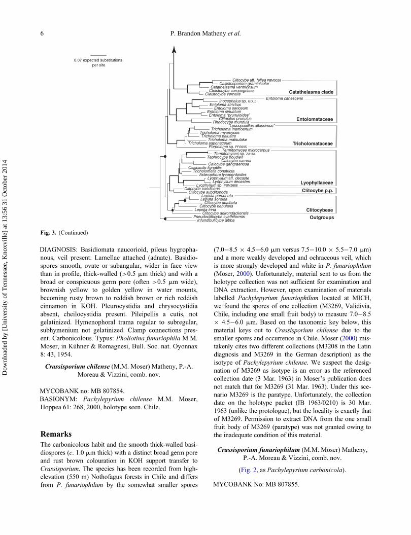
Fig. 3. Phylogeny of the Agaricoid clade based on a Maximum Likelihood and Bayesian Inference analysis of a supermatrix of fournuclear gene regions (5.8S rRNA, LSU-rRNA, SSU-rRNA and rpb2 conserved domains 5�7). Thickened branches indicate ML boot-strap support >70% and Bayesian posterior probability >0.95. Nodes that receive Bayesian posterior probabilities >0.95 but with<70% ML bootstrap are indicated by small black-filled circles. Clade nomenclature follows Matheny et al. (2006). Grey shaded taxonlabels indicate placement of species of Pachylepyrium or collections mislabelled Pachylepyrium.
Two new genera of dark-spored Agaricales 5
Dow
nloa
ded
by [
Uni
vers
ity o
f T
enne
ssee
, Kno
xvill
e] a
t 13:
56 3
1 O
ctob
er 2
014
DIAGNOSIS: Basidiomata naucorioid, pileus hygropha-
nous, veil present. Lamellae attached (adnate). Basidio-
spores smooth, ovate or subangular, wider in face view
than in profile, thick-walled (>0.5 mm thick) and with a
broad or conspicuous germ pore (often >0.5 mm wide),
brownish yellow to golden yellow in water mounts,
becoming rusty brown to reddish brown or rich reddish
cinnamon in KOH. Pleurocystidia and chrysocystidia
absent, cheilocystidia present. Pileipellis a cutis, not
gelatinized. Hymenophoral trama regular to subregular,
subhymenium not gelatinized. Clamp connections pres-
ent. Carbonicolous. Typus: Pholiotina funariophila M.M.
Moser, in K€uhner & Romagnesi, Bull. Soc. nat. Oyonnax
8: 43, 1954.
Crassisporium chilense (M.M. Moser) Matheny, P.-A.
Moreau & Vizzini, comb. nov.
MYCOBANK no: MB 807854.
BASIONYM: Pachylepyrium chilense M.M. Moser,
Hoppea 61: 268, 2000, holotype seen. Chile.
Remarks
The carbonicolous habit and the smooth thick-walled basi-
diospores (c. 1.0 mm thick) with a distinct broad germ pore
and rust brown colouration in KOH support transfer to
Crassisporium. The species has been recorded from high-
elevation (550 m) Nothofagus forests in Chile and differs
from P. funariophilum by the somewhat smaller spores
(7.0�8.5 £ 4.5�6.0 mm versus 7.5�10.0 £ 5.5�7.0 mm)
and a more weakly developed and ochraceous veil, which
is more strongly developed and white in P. funariophilum
(Moser, 2000). Unfortunately, material sent to us from the
holotype collection was not sufficient for examination and
DNA extraction. However, upon examination of materials
labelled Pachylepyrium funariophilum located at MICH,
we found the spores of one collection (M3269, Validivia,
Chile, including one small fruit body) to measure 7.0�8.5
£ 4.5�6.0 mm. Based on the taxonomic key below, this
material keys out to Crassisporium chilense due to the
smaller spores and occurrence in Chile. Moser (2000) mis-
takenly cites two different collections (M3208 in the Latin
diagnosis and M3269 in the German description) as the
isotype of Pachylepyrium chilense. We suspect the desig-
nation of M3269 as isotype is an error as the referenced
collection date (3 Mar. 1963) in Moser’s publication does
not match that for M3269 (31 Mar. 1963). Under this sce-
nario M3269 is the paratype. Unfortunately, the collection
date on the holotype packet (IB 1963/0210) is 30 Mar.
1963 (unlike the protologue), but the locality is exactly that
of M3269. Permission to extract DNA from the one small
fruit body of M3269 (paratype) was not granted owing to
the inadequate condition of this material.
Crassisporium funariophilum (M.M. Moser) Matheny,
P.-A. Moreau & Vizzini, comb. nov.
(Fig. 2, as Pachylepyrium carbonicola).
MYCOBANK No: MB 807855.
0.07 expected substitutions per site
Lyophyllum aff. decaste
Infundibulicybe gibba
Calocybe gangraenosa
Lyophyllum sp. PBM2688
Clitopilus prunulusRhodocybe mundula
Entoloma strictius
Clitocybe aff. fellea PBM3028
Lyophyllum decastes
Lepista personata
Callistosporium graminicolor
Entoloma “prunuloides”
Catathelasma ventricosum
Clitocybe adirondackensis
Inocephalus sp. GD_b
Cleistocybe carneogrisea
Pseudoclitocybe cyathiformis
Termitomyces microcarpus
Entoloma sinuatum
Clitocybe dealbata
Tricholoma inamoenum
Lepista irina
Cleistocybe vernalis
“Leucopaxillus albissimus”
Tricholoma matsutake
Ossicaulis lignatilis
Entoloma sericeum
Asterophora lycoperdoides
Porpoloma sp. PR3995
Tricholoma myomyces
Tricholoma saponaceum
Calocybe carnea
Clitocybe candicans
Termitomyces sp. ZA164
Entoloma canescens
Tricholomella constricta
Clitocybe nebularis
Tricholoma palustre
Tephrocybe boudieri
Clitocybe subditopoda
Lepista sordida
Catathelasma clade
Entolomataceae
Tricholomataceae
Lyophyllaceae
Clitocybe p.p.
Clitocybeae
Outgroups
Fig. 3. (Continued)
6 P. Brandon Matheny et al.
Dow
nloa
ded
by [
Uni
vers
ity o
f T
enne
ssee
, Kno
xvill
e] a
t 13:
56 3
1 O
ctob
er 2
014

BASIONYM: Pholiotina funariophila M.M. Moser, in
K€uhner & Romagnesi, Bull. Soc. nat. Oyonnax 8: 43,
1954, holotype seen. Austria.
� Pachylepyrium funariophilum (M.M. Moser) Singer,
in Singer & Moser, Mycopath. Mycol. Appl. 26(2-3): 171,
1965.
D Kuehneromyces carbonicola A.H. Sm., Beihefte zur
Sydowia 1: 53, 1957. Holotype seen. Idaho.
� Pachylepyrium carbonicola (A.H. Sm.) Singer,
Sydowia 11: 321, 1958 [1957].
� Pholiota subangularis A.H. Sm. & Hesler, The North
American Species of Pholiota: 44, 1968.
Remarks
Crassisporium funariophilum is geographically wide-
spread occurring in Europe, northern Africa and west-
ern North America (where it has been referred to as
Pachylepyrium carbonicola and Pholiota subangularis)
and may be expected elsewhere. Singer & Moser
(1965) and Singer (1969) also report it from Argentina,
but this material has not been revised in light of
description of P. chilense (see above). Moser (2000)
describes collections of P. carbonicola with a white
fugacious veil and similar ecology to P. funariophilum,
but with somewhat larger spores (8.2�12.1 £6.5�8.3 mm) than for P. funariophilum (7.6�10.0 £5.3�7.1 mm). However, pairwise comparison of ITS
sequences from the type collections of C. funariophi-
lum and P. carbonicola differ at only two nucleotide
positions (excluding three polymorphic sites among the
five sequences considered) strongly suggesting the two
species are conspecific. As such, P. funariophilum has
nomenclatural priority.
Crassisporium squarrulosum (Singer) Matheny, P.-A.
Moreau & Vizzini, comb. nov.
MYCOBANK no: MB 807856.
BASIONYM: Pachylepyrium squarrulosum Singer, Beih.
Nova Hedwigia 29: 281, 1969, holotype not seen. Chile.
Remarks
We have not studied material of C. sqarrulosum, but the
thick spore wall with a truncate germ pore and intense
‘ferruginous’ colouration in KOH described by Singer
(1969) are consistent with placement in Crassisporium
rather than with the type of Pachylepyrium in the Tubaria-
ceae or with Romagnesiella. The species is associated
with burnt debris and occurs at high elevations (1000 m)
in Chile. The type (M 6550) is reportedly at SGO. The
species differs most readily from C. chilense by the
flocculose-squarrose pileus surface and longer spores
(12.0�14.0 £ 6.5�8.0 mm).
Pholiota nubicola (Singer) Matheny & P.-A. Moreau,
comb. nov.
MYCOBANK No: MB 807857.
BASIONYM: Pachylepyrium nubicola Singer in Dennis,
Kew Bull. 15(1): 139, 1961, holotype seen. Venezuela.
Remarks
The ITS sequence produced from the holotype strongly
suggests that Pachylepyrium nubicola is a species of Pho-
liota. Consistent with this placement are the caespitose
and lignicolous habit, paler (yellowish) pigmented basi-
diospores with a thinner wall than in Crassisporium,
strongly gelatinized pileipellis composed of coarsely
incrusted yellowish hyphae, and gelatinized subhymenial
trama. Add to this the slightly phaseoliform spores with a
distinct germ pore (0.8�1.0 mm wide) and the squamulose
stipe covering, it is not surprising P. nubicola would be
closely related to Ph. gummosa (Lasch: Fr.) Singer as
described by Holec (2001).
In contrast to the protologue, our examination of the
type revealed a gelatinized pileipellis and cylindric to
subphaseoliform non-dextrinoid basidiospores, these
with a distinct germ pore. The spores measure 7.5�8.8
£ 4.5�4.8 mm, which is consistent with the proto-
logue. The basidia measure 17�28 £ 7�8 mm with
yellowish contents when mature. The lamellar edge
was observed to be sterile and yellow but without
reviving elements. The presence or absence of chryso-
cystidia could not be confirmed, but given the high
sequence similarity to ITS sequences labelled Ph. gum-
mosa and Ph. terrestris, we predict chrysocystidia will
be found in this species.
A taxonomic key to species of Crassisporium
1(a) Pileus surface flocculose-squarrose, spores 12�14 £6.5�8 mm. . . .................................C. squarrulosum Singer
1(b) Pileus surface glabrous or with marginal fibrils,
spores 7�11.5 £ 5.5�7 mm. . .. . .. . .. . .. . . ..................... .22(a) Spores mostly 8�11.5 £ 5.5�7 mm, in north temper-
ate forests of Europe, North Africa and western North
America (also reported from southern South America, but
this is likely C. chilense); veil well developed, white-
. . .. . .. . .. . .. . .. . . ..C. funariophilum (M.M. Moser) Singer
2(b) Spores mostly 7�9 £ 4.5�6 mm, in Nothofagus
forests in southern South America; veil weakly developed,
ochraceous. . .. . .. . .. . .. . .. . .. . . ...C. chilenseM.M. Moser
Two new genera of dark-spored Agaricales 7
Dow
nloa
ded
by [
Uni
vers
ity o
f T
enne
ssee
, Kno
xvill
e] a
t 13:
56 3
1 O
ctob
er 2
014

Romagnesiella Contu, P.-A. Moreau, Vizzini & A. de
Haan, gen. nov.
MYCOBANK No.: MB 519559.
TYPE SPECIES:Galerina clavus Romagn., 1944 [1942].
ETYMOLOGY: named in honour of Henri Romagnesi,
French mycologist (1912�1997) (gender: feminine).
DIAGNOSIS: Basidiomata naucorioid, lamellae distant,
adnate to subdecurrent; pileus dry, not hygrophanus; stipe
smooth, without a partial veil. Basidiospores smooth,
more or less ovate, not subangular, yellow in water
mounts, reddish ochre in KOH, not dextrinoid, germ pore
absent; necrobasidia numerous; cheilocystidia present,
edges of lamellae smooth and (sub)sterile, pleurocystidia
present but dispersed and infrequent, pileipellis filamen-
tous, hymenophoral trama regular, clamp connections fre-
quent. On unburnt soil or sand among mosses and grasses.
Typus: Galerina clavus Romagn., Bull. Trimest. Soc.
Mycol. France 58(4): 149 (1944 [1942]).
Romagnesiella clavus (Romagn.) Contu, P.-A. Moreau,
Vizzini & A. de Haan, comb. nov.
(Figs 1, 4�8).
MYCOBANK No.: MB 519560.
BASIONYM: Galerina clavus Romagn., Bull. Trimest.
Soc. Mycol. France 58(4): 149, 1944 [1942], lectotype
designated here (Fig. 14, p. 145, Romagnesi (1944)
[1942], MBT177567); epitype designated here (P.-A.
Moreau 06090110, LIP, MBT177568). Switzerland.
� Naucoria clavus (Romagn.) K€uhner & Romagn., Fl.
Anal. Champ. Sup.: 239 (1953, comb. inval., Art. 33.4).
MISAPPLICATIONS: Tubaria minima J.E. Lange sensu
Moreau in Matheny et al. (2007a: 571); Galerina embolus
Figs. 4�8. Anatomical features of Romagnesiella clavus (PAM06090110, epitype). (Fig. 4) Spores. (Fig. 5) Basidia and subhymenium.(Fig. 6) Cheilocystidia. (Fig. 7) Pleurocystidium. (Fig. 8) Pileipellis. Scale bars D 10 mm.
8 P. Brandon Matheny et al.
Dow
nloa
ded
by [
Uni
vers
ity o
f T
enne
ssee
, Kno
xvill
e] a
t 13:
56 3
1 O
ctob
er 2
014

(Fr.) Sacc. sensu Orton (1960: 239), sensu de Haan &
Walleyn (2009: 64).
BIBLIOGRAPHY: Romagnesi (1942: 144, protologue);
K€uhner & Romagnesi (1953: 239; description); Smith &
Singer (1964: 336); de Haan & Walleyn (2009: 64, 66:
description, picture); North African collections: Haus-
knecht & Zuccherelli (1993: 47), Moreau (2009: 199).
Description
Pileus 5�9 (12) mm diam, hemispheric-umbonate then §depressed around umbo, margin early inrolled becoming
shortly crenulate when old, even, not striate, densely fur-
furaceous-micaceous, grey-brown with somewhat pur-
plish tones when fresh, paler at margin, quickly fading
from margin to uniformly fleshy-ochre, without any trace
of veil. Lamellae adnexed-ventricose at first, becoming
shortly uncinate in age, distant with 14�16 L reaching
stipe, interspersed by 1�2 series of lamellulae, dull rusty
ochre even when young; edges smooth but (sub)sterile,
pale yellow. Stipe 15�25 £ 1 mm, flexuose, slightly
attenuate at base and inflated at apex, pruinose-floccose
just below lamellae, fibrillose below then glabrous against
a uniform dirty brown ground colour, slightly purplish
when young; no perceptible trace of veil (primordia not
observed). Context dark brown when fresh, pale ochre
when dry. Odour and taste fungoid, not remarkable.
Basidiospores (5.6) 6.2�6.7�7.3 (8.5) £ (3.6)
3.9�4.2�4.4 (5.0) mm, Q D 1.51�1.62�1.73 (n D 48),
ovate to obovate but longer spores more fusiform, smooth,
germ pore absent; bright yellow in water, amber yellow in
Melzer’s, warm reddish ochre in KOH, wall thickened up
to 0.3 (0.5) mm; content with a large central droplet, often
elongate. Basidia four-spored, 28�36 £ 7�9 mm, broadly
clavate, with long sterigmata, content often microguttulate;
necrobasidia abundant, with reddish-brown content. Chei-
locystidia 22�45 £ 5.5�7 mm, cylindrical-flexuose with
slightly thickened yellowish wall, mixed with fascicles of
terminal hyphae issued from trama with pear-shaped to
subglobose terminal cells, 9�14 mm wide, lamella edge
fertile to locally substerile. Pleurocystidia 38�42 £7.5�13 mm, cylindrical to subutriform, not very distinct
but not rare. Hymenophoral trama regular, with strongly
encrusted hyphae, 3�5 mm wide. Pileipellis a superficial
layer of short cells, these lobate, digitate, puzzle-like, fusi-
form or pyriform, 12�16 mm wide, more or less erected to
nearly hymeniform towards margin, pale in KOH, smooth,
issued from hyphae of subpellis; subpellis filamentous,
coarsely encrusted, thick-walled, deep yellow to reddish
brown in KOH, continuous with pileus context. Stipitipellis
a cutis with sparse to fasciculate (at apex) caulocystidia
measuring 16�25 £ 5�12 mm, cylindrical to clavate-pyri-
form, very rare below apex; superficial hyphae slender,
2�3.5 mm wide. Clamp connections frequent.
Habitat and distribution: Often on calcareous, min-
eral-rich, sandy or alluvial substrates in pioneer or dis-
turbed habitats including fixed coastal dunes and banks of
trails or paths among mosses and grasses. Less frequent in
secondarized dunes, scattered and never abundant. Europe
(Belgium, France, Switzerland) and reported from Italy,
the Netherlands, and North Africa. Fruiting Sept.�Nov.
Material studied: BELGIUM. Antwerpen: Antwerpen-
Linkeroever, Het Rot, 4 Sep 2004; 10 specimens, among
grass and mosses (Tortula ruralis and Ceratodon purpur-
eus) on sandy, calcareous soil, herb. A. de Haan n� 04101;Antwerpen-Linkeroever, Blokkersdijk, 9 Sep 2004, 2
specimens, among mosses on sandy, calcareous soil, herb.
A. de Haan n� 04113. Namur: Oignies-en-Thi�erache,l’Estache, 23 Sep 1999, 1 specimen on wet calcareous
soil, herb. A. de Haan n� 99100. Oost-Vlaanderen: Zwij-
naarde, Rijvisschepark, 15 Oct 1989, about ten carpo-
phores, in bare spot in mossy lawn, on sandy slightly
loamy soil, leg. P. Van der Veken, herb. A. de Haan n�
89017; same location, leg. P. Van der Veken, 4 speci-
mens, 29 Oct 2003, herb. A. de Haan n� 03088. West-
Vlaanderen: De Panne, Calmeijnbos, 3 Nov 1997, 4 speci-
mens, on humus-rich, calcareous soil, herb. A. de Haan n�
97088; Oostduinkerke, Doornpanne, 1 Nov 2001, 2 speci-
mens, among moss and lichens, on calcareous dune sand,
herb. A. de Haan n� 01080 (as ‘Galerina embolus’).
FRANCE. Pas-De-Calais: Equihen-Plage, dunes d’Ecault,
7 November 2004, five specimens in Phleo-Tortuletum
with Calamagrostis epigeos, calcareous fixed dune, leg.
A. Brabant & P.-A. Moreau, 7 Nov 2004, herb. P.-A. Mor-
eau n� 04110710 (LIP); same location, along a sandy path
amongst Calamagrostis epigeos, fixed calcareous dunes
with Hippophae rhamnoides, leg. C. Hannoire & P.-A.
Moreau, 31 Oct 2008, herb. P.-A. Moreau n� 08103102
(LIP). Seine: Paris, bois de Vincennes, 1 Oct 1932, herb.
R. K€uhner (G, as ‘Tubaria oligophylla’, ined.). ITALY:
Sardinia, prov. Olbia, Golfo Aranci, Golfo di Marinella,
in troops on sandy soil in a coastal grassland, leg. M.
Contu, 15 Dec 2007, herb. M. Contu (C15122007, TENN
063957). SWITZERLAND. Gr€aubunden: Rothenbrunnen,edge of path, riparian Alnus incana forest, on black allu-
vial humus, 1 Sep 2006, leg. B. Senn-Irlet & P.-A. Mor-
eau, herb. P.-A. Moreau n� 06090110 (epitype LIP),
TENN 063587, TENN 063976.
Remarks
Our interpretation of Galerina clavus is based on the
detailed protologue of Romagnesi (1944 [1942]), which
matches collections from Belgium and Switzerland.
Unfortunately, no original material of Galerina clavus
exists. The herbarium packet corresponding to one of the
two collections cited by Romagnesi (1942: 145) Yerres,
bois de Cercay, 18 Jun 1942, kept in herb. H. Romagnesi
Two new genera of dark-spored Agaricales 9
Dow
nloa
ded
by [
Uni
vers
ity o
f T
enne
ssee
, Kno
xvill
e] a
t 13:
56 3
1 O
ctob
er 2
014

(PC) was empty. The other cannot be located. Because a
figure that depicts G. clavus exists in the protologue, this
must serve as the lectotype. Original drawings of G.
clavusmade by Romagnesi also exist at PC. Thus, we des-
ignate PAM06090110 (LIP) as an epitype.
The species features some morphological and micro-
scopic variation. The epitype (sequenced here), showed
purplish-grey tones on the stipe as well as the pileus, but
the protologue only mentions this colour on the stipe. Bel-
gian collections described by de Haan & Walleyn (2009)
describe a more convex pileus, broadly adnate instead of
subdecurrent lamellae, a ‘weakly farinaceous’ taste, and
habitat in dry mineral spots in urban grasslands. However,
all collections show gradual variation in the pileus shape,
colour and lamellae attachment.
Coastal collections are probably better considered as var-
iants of R. clavus. Despite some variation in spore dimen-
sions we could not find any support for specific or
infraspecific distinctions. Detailed spore measurements
illustrate continuity in these variation patterns but apparent
differences (Appendix 2, see supplemental material online).
North African and Sardinian collections of Galerina
clavus, as described by Hausknecht & Zuccherelli (1993)
and Malencon (Moreau, 2009), differ from continental
collections by somewhat larger basidiospores [(7.0)
7.6�8.2�9.0 (10) £ (4.0) 4.9�5.2�5.5 (5.7) mm, Q D1.48�1.59�1.70], slightly larger cystidia, and a filamen-
tous pileipellis with a more or less continuous suprapellis
of slender cylindrical hyphae with sparse slightly upraised
terminal elements. It is possible that Mediterranean col-
lections may represent distinct populations. One collec-
tion from Sardinia was sequenced (C15122007, leg. M.
Contu), in which the ITS1 region reveals nine site differ-
ences with R. clavus PAM6090110, four of which, how-
ever, are polymorphic in C15122007. Galerina clavus has
also been reported from the Netherlands (www.
verspreidingsatlas.nl/046620).
Romagnesiella clavus is probably often confused with
other naucorioid species frequent in the same environment,
such as Galerina graminea (Velen.) K€uhner, Psilocybe
pratensis P.D. Orton, or Tubaria spp. The distant lamellae
and absence of a veil on the stipe are good distinctive field
characters. However, we discuss below three additional
species with which R. clavus could be confused.
Tubaria umbonata S. Lundell in Lundell & Nannfeldt
(1953: 23).
(Figs 9�13).
MYCOBANK No. 307168.
ISOTYPE: SWEDEN. Upland: Uppsala, Slottsbaken,
NW part below Gunillaklaken, 50 m from Stockholmsv€a-gen, 6 August 1944, leg. S. Lundell, ex Fungi exsiccati
Suecici 2041 (PC, about 20 well-preserved specimens).
Description
Exsiccata small to minute (2�6 mm), very slender,
entirely dark brown, without visible veil, with arcuate and
distant lamellae. Basidiospores (6.2) 6.5�7.2�8.0 (9.0) £(3.0) 3.2�3.7�4.2 mm, Q D 1.70�1.96�2.24 (n D 23;
see also Appendix 2, see supplemental material online),
pale yellow, slightly thick-walled (<0.5 mm thick), ochra-
ceous yellow in KOH, not collapsing, narrowly ovo-ellip-
soidal to ellipsoidal, with slightly guttulate content, not
dextrinoid. Basidia 22�34 £ 6.5�7.5 mm, 4-spored
(occasionally 2-spored), clavate more or less capitate,
often strangulate under apex before maturity, hyaline;
subhymenium 12�15 mm thick, filamentous-ramose, with
hyphae 2�2.5 mm wide. Cheilocytidia 16�30 £6�11.5 mm, often clustered�ampullaceous, clavate, ellip-
soidal, utriform, cylindrical, with a thin and smooth wall,
intermixed with some fertile basidia; lamella edge almost
sterile. Pleurocystidia if present, not studied. Hymenopho-
ral trama regular, yellowish, made of slender hyphae
2�6 mm wide with thick encrusted wall. Pileipellis with a
discontinuous suprapellis made ofC/¡ erected wide ellip-
soidal to cylindrical catenulate elements, 18�30 £6�13 mm, pale, slightly thick-walled, not or only locally
encrusted; subpellis made of slender hyphae 3.5�8 mmwide, distinctly encrusted by granular pigment remaining
yellow in KOH. Stipitipellis with sparse traces of filamen-
tous veil towards apex, composed of slender hyphae
3�5 mm wide; wall yellowish, up to 2 mm thick and
encrusted, terminal cells cylindrical with some vesicular
cells up to 20 mm wide; superficial hyphae slender, these
3�5 mm wide, with yellow walls up to 0.5 mm thick,
intermixed with large cylindrical hyphae 9�16 mm wide,
locally encrusted by gold-yellow pigment (KOH), espe-
cially at septa, and some sparse pale gleoplerous hyphae.
Clamp connections present at septa.
Remarks
Tubaria umbonata has not been revised nor documented
since its publication in Fungi Exsiccati Suecici (Lundell
& Nannfeldt 1953). We studied the isotype at PC (Fungi
Exsiccati Suecici fasc. 41�42). Based on our morphologi-
cal analysis, we conclude that T. umbonata represents a
genuine species of Tubaria (W.G. Sm.) Gillet, but with
rather narrow spores. DNA extraction of the PC material
of T. umbonata yielded no PCR amplicons. Illustrations
of anatomical features (Fig. 5) and their description
(above) from the isotype are presented.
Two additional species could be confused with R.
clavus. The name Tubaria minima J.E. Lange (Lange,
1940) was misapplied by Moreau (in Matheny et al.,
2007a) to collections of R. clavus. Although Bon (1992)
maintains T. minima as an autonomous species, Romag-
nesi (1942) considers it to be a synonym of T. minutalis
10 P. Brandon Matheny et al.
Dow
nloa
ded
by [
Uni
vers
ity o
f T
enne
ssee
, Kno
xvill
e] a
t 13:
56 3
1 O
ctob
er 2
014

Romagn. (Romagnesi, 1937), a position followed by mod-
ern authors. This species (sensu Lange (1940), non Mor-
eau) differs from G. clavus by its hygrophanous piles and
smaller spores (5.2�6.0 £ 3.2�3.8 mm), features that
reinforce its conspecificity with T. minutalis.
Tubaria embolus (Fr.) Sacc. is rather frequently cited
in the literature but has been interpreted several different
ways. Lange (1938: 655, pl. 127B) illustrates as
‘Tubaria embola’ a species with broadly adnexed lamel-
lae and yellow tones especially in the context (conform-
ing to Fries’ protologue, 1836�1838: 206), which seems
to represent Agrocybe pusiola (Fr.) R. Heim. Bon (1992)
cites the species in the genus Galerina, but examination
of his materials (LIP) showed that his concept was
unclear: coll. 741120 is Galerina uncialis (Britzelm.)
K€uhner, and coll. 70624 (as ‘Galerina cf. embolus’) is a
species with pleurocystidia close to Galerina
vittaeformis (Fr.) K€uhner. Orton (1960: 176) mentions
five reports of T. embolus (as ‘Galerina embolus’) from
sand dunes, with comparable microscopical characters
(but with notably long spores, 9�11 £ 4.5�6 mm, com-
patible with our coastal collections of R. clavus), but
pleurocystidia and necrobasidia are not mentioned.
Moreover, yellow tones are described towards the pileus
margin when dry, incompatible with any species known
to us. In addition de Haan & Walleyn (2009) describe
without illustrations a collection of G. embolus (reported
here as R. clavus) and also found in fixed dunes in Bel-
gium. Considering the ambiguities of the protologue
(Fries, 1836�1838), and the diversity of interpretations
proposed by various authors, we reject the name here.
Additional morphological and molecular study is
required to unravel the taxonomic relationships of these
variously interpreted collections to Romagnesiella.
Figs. 9�13. Anatomical features of Tubaria umbonata (Fungi Exsicatti Suecici fasc. 41�42, isotype). (Fig. 9) Spores. (Fig. 10) Basidiaand subhymenium. (Fig. 11) Cheilocystidia. (Fig. 12) pileipellis. (Fig. 13) Stipitipellis. Scale bars D 10 mm.
Two new genera of dark-spored Agaricales 11
Dow
nloa
ded
by [
Uni
vers
ity o
f T
enne
ssee
, Kno
xvill
e] a
t 13:
56 3
1 O
ctob
er 2
014

Discussion
Polyphyly of Pachylepyrium and recognition
of Crassisporium and Romagnesiella as new
genera
Our results strongly support the polyphyletic status of
Pachylepyrium. The type species of the genus, P. fulvi-
dula, lacks several of the features attributed to the residual
species. Originally described in Phaeomarasmius, P. ful-
vidula fruits on non-burnt woody debris and lacks the
broad germ pore observed in other species. Horak (1968)
reports seldom seeing any germ pore at all in the type of
P. fulvidula. Our examination of the isotype at MICH con-
firms this observation (a germ pore was not observed).
Thus, it is not surprising to see phylogenetic placement of
P. fulvidula apart from the residual Pachylepyrium spe-
cies. Pachylepyrium fulvidula resides in the Tubariaceae
(Fig. 3) where it is closely related to other species of
Flammulaster, Phaeomyces, Phaeomarasmius and Tuba-
ria (W.G. Sm.) Gillet, all of these also lacking a broad
germ pore (Horak, 2005).
We place three residual species of Pachylepyrium in the
new genus Crassisporium united by a combination of
basidiospore features (thick-walled spores with a broad
germ pore and rusty to reddish brown colouration in
KOH), anatomical features (non-gelatinous cutis, absence
of pleurocystidia and chrysocystidia, absence of a gelati-
nous subhymenial layer) and ecology (carbonicolous
habit). However, before our phylogenetic analysis based
on molecular data, we did not suspect that Galerina clavus
would be related to Crassisporium more so than to any
other group of Hymenogastraceae or Strophariaceae. In
order to point out differences between typical carbonico-
lous species with thick-walled pored spores (Crassispo-
rium species) and non-carbonicolous species with thinner-
walled (<0.5 mm) spores such as G. clavus, we have pro-
posed a new genus Romagnesiella to accommodate the
latter. No extra-European or North African species are
unequivocally attributable to Romagnesiella at present
without the addition of detailed morphological and molec-
ular study. Galerina fuegiana Singer from Patagonia
(Smith & Singer, 1964) is a possible candidate.
The inclusion of sequences of Crassisporium and
Romagnesiella in a multigene phylogenetic analysis of
the Agaricoid clade shows these two taxa form a well-sup-
ported group (Fig. 3) sister to the Strophariaceae s.lat.
consortium (Gulden et al., 2005), including the families
Hymenogastraceae and Strophariaceae s. str. of Matheny
et al. (2006). Inclusion of Crassisporium and Romagne-
siella in Strophariaceae s. str. would render the family
paraphyletic in this analysis. Consideration of a more
broadly conceived Hymenogastraceae, subsuming the
Strophariaceae, could be made since the name Hymeno-
gastraceae Vitt. 1831 pre-dates that of the Strophariaceae
Singer & A.H. Sm. 1946. However, additional taxon and
gene sampling are needed to resolve the relationship
between these two families.
An alternative scenario to consider is inclusion of the
three species of Crassisporium into one genus with R.
clavus, thereby describing only a single genus as new.
Samples of each group form a clade with strong support, a
synapomorphy of which are the basidiospores that deepen
various shades of reddish brown in KOH. However, we
favour separate genera for the two lineages for several
reasons: (1) species of Crassisporium are carbonicolous,
whereas those of Romagnesiella are non-carbonicolous;
(2) the lamellae are adnate to subdecurrent in Romagne-
siella but never subdecurrent in Crassisporium; (3) a veil
is absent in Romagnesiella but present in Crassisporium;
(4) the basidiospores of Crassisporium feature walls
>0.5 mm thick, a wide germ pore typically 1.0�1.5 mmthick, and are subangular in face view. These features
may be correlated with the fire ecology in that heat may
be required to induce germination (Claridge, Trappe, &
Hansen, 2009). Basidiospores of Romagnesiella have
thinner walls (<0.5 mm thick), no germ pore and are not
subangular; (5) pleurocystidia are present in Romagne-
siella but absent in Crassisporium; and (6) the relative
branch length differences between the two lineages corre-
spond to branch length differences between other genera
of Strophariaceae and Hymenogastraceae. Therefore, we
prefer to recognize the two lineages as separate genera.
The monophyly of the Agaricoid clade is recovered here
for the first time with high bootstrap and significant Bayes-
ian posterior probability. Most species in the Agaricoid
clade tend to have pigmented and thick-walled spores, per-
haps indicative of adaptations to novel environments (e.g.
dung, burnt sites) (Garnica et al., 2007). Many species of
the Agaricoid clade also feature multiple nuclei per spore
and an open pore type of hilum (Matheny et al., 2006).
AcknowledgementsThe authors are grateful to staff at herbaria G (P. Clerc),
IB (R. Kuhner), LIP (R. Courtecuisse and C. L�ecuru),MICH (R. Rabeler, P. Rogers), MPU (V. Bourgade, L.
Gomel and M.-J. Mauruc), PC (B. Buyck), PERTH (N.
Bougher), and the USDA Forest Service, Luqillo, Puerto
Rico (D. J. Lodge) for loan of collections. Egon Horak
(Innsbruck, Austria) is acknowledged for his study on Sar-
dinian collections sent by M. Contu. R�egis Courtecuisse
(Lille, France) is also acknowledged for his valuable sug-
gestions and bibliographic expertise. Aaron Wolfen-
barger, Emily Giles, Whitaker Hoskins, Sarah Sprague,
and Christine Braaten provided laboratory assistance at
the University of Tennessee. Three anonymous reviewers
and the Associate Editor, Karen Hansen, provided critical
feedback that helped improve this paper. Research was
conducted at the University of Tennessee, Universit�e LilleNord de France, and Universit�a di Torino.
12 P. Brandon Matheny et al.
Dow
nloa
ded
by [
Uni
vers
ity o
f T
enne
ssee
, Kno
xvill
e] a
t 13:
56 3
1 O
ctob
er 2
014

FundingThis work was supported by the U.S. National Science
Foundation under Grant DEB-0949517.
Supplemental dataSupplemental data for this article can be accessed here.
ReferencesAime, M. C., Vilgalys, R. & Miller, O. K. (2005). The Crepidota-
ceae (Basidiomycota, Agaricales). Phylogeny and taxonomyof the genera and revision of the family based on molecularevidence. American Journal of Botany, 92, 74�82.
Altekar, G., Dwarkadas, S., Huelsenbeck, J. P. & Ronquist, F.(2004). Parallel Metropolis-coupled Markov chain MonteCarlo for Bayesian phylogenetic inference. Bioinformatics,20, 407�415.
Ammirati, J. F., Parker, A. D. & Matheny, P.B. (2007). Cleisto-cybe, a new genus of Agaricales. Mycoscience, 48,282�289.
Baroni, T. J. & Matheny, P. B. (2011). A re-evaluation ofgasteroid and cyphelloid species of Entolomataceae fromeastern North America. Harvard Papers in Botany, 16,293�310.
Binder, M., Larsson, K.-H., Matheny, P. B. & Hibbett, D. S.(2010). Amylocorticiales ord. nov. and Jaapiales ord. nov.:early-diverging clades of Agaricomycetidae dominated bycorticioid forms.Mycologia, 102, 865�880.
Bon, M. (1992). Cl�e monographique des esp�eces gal�ero-naucorio€ıdes. Documents Mycologiques, 21, 1�89.
Claridge, A. W., Trappe, J. M. & Hansen, K. (2009). Do fungihave a role as soil stabilizers and remediators after forestfire? Forest Ecology and Management, 257, 1063�1069.
de Haan, A. & Walleyn, R. (2009). Studies in Galerina. Galeri-nae Flandriae (3). Fungi non Delineati, 46, 1�84.
Dennis, R. W. G. (1961). Fungi venezuelani: IV. Agaricales.Kew Bulletin, 15, 67�156.
Fannech�ere, G. (2005). Statistiques et notation des dimensionsdes spores. Bulletin Trimestriel de la Soci�et�e Mycologiquede France, 121, 255�292.
Fannech�ere, G. (2009). Mycom�etre 2.02. Available online, 2.VII.2009. Retrieved from http://mycolim.free.fr/DOC_SML/mycm202/Charg_Mycm202.htm, accessed 9 October 2014.
Fries, E. M. (1836�1838 [1838]). Epicrisis systematicis mycolo-gici, synopsis Hymenomycetum, I,. Uppsala.
Gardes, M. & Bruns, T. D. (1993). ITS primers with enhancedspecificity for basidiomycetes �applications to the identifi-cation of mycorrhizae and rusts. Molecular Ecology, 2,113�118.
Garnica, S., Weiss, M., Walther, G. & Oberwinkler, F. (2007).Reconstructing the evolution of agarics from nuclear genesequences and basidiospore ultrastructure. MycologicalResearch, 111, 1019�1029.
Gouy, M., Guindon, S. & Gascuel, O. (2010). SeaView version4: a multiplatform graphical user interface for sequencealignment and phylogenetic tree building. Molecular Biol-ogy and Evolution, 27, 221�224.
Gulden, G. (2012). Galerina Earle. In H. Knudsen & J. Vester-holt (Eds.), Funga Nordica. Agaricoid, Boletoid, Clavarioid,Cyphelloid and Gasteroid Genera, (pp. 886�903). Copen-hagen: Norsvamp.
Gulden, G., Stensrud, ;., Shalchian-Tabrizi, K. & Kauserud, H.2005. Galerina Earle: a polyphyletic genus in the consor-tium of dark-spored agarics.Mycologia, 97, 823�837.
Hausknecht, A. & Zuccherelli, A. (1993). Ritrovamenti interes-santi dal Ravennate. 1a parte. Alcune Agaricales a sporebrune o pi�u scure. Bollettino del Gruppo Micologico G Bre-sadola, 36, 35�61.
Holec, J. (2001). The genus Pholiota in central and westernEurope. Libri Botanici, 20, 1�220.
Horak, E. (1968). Synopsis generum Agaricalium (Die Gattung-stypen der Agaricales). Beitr€age zur Kryptogamenflora derSchweiz, 13, 1�741.
Horak, E. (2005). R€ohrlinge und Bl€atterpilze in Europa � unterder Mitarbeit von Anton Hausknecht (Bolbitiaceae) und P.A. Moreau (Alnicola),. Heidelberg: Elsevier SpektrumAkademischer.
Kirk, P., Cannon, P. F., Minter, D. W. & Stalpers, J. A. (2008).The dictionary of fungi, (10th ed.). Wallingford: CABInternational.
Kornerup, A. & Wanscher, J.H. (1967). Methuen handbook ofcolour, (2nd ed.). London: Methuen & Co.
K€uhner, R. & Romagnesi, H. (1953). Flore anaytique des cham-pignons sup�erieurs (Agarics, Bolets, Chanterelles),. Paris:Masson.
Lange, J.E. (1938). Studies in the Agarics of Denmark. Part XII.Hebeloma, Naucoria, Tubaria, Galera, Bolbitius, Pluteolus,Crepidotus, Pseudopaxillus, Paxillus. Dansk Botanisk Arkiv,9, 1�104.
Lange, J.E. (1940). Flora Agaricina Danica V, supplement(I�IX). Copenhagen: The Danish Botanical Society.
Lundell, S. & Nannfeldt, J. A. (1953). Fungi Exsiccati Suecici,41�42, 2001�2100.
Maddison, D. R. & Maddison, W. P. (2005). MacClade 4,, ver-sion 4.08 for OS X. Sunderland, MA: Sinauer Associates.
Matheny, P.B. (2005). Improving phylogenetic inference ofmushrooms with RPB1 and RPB2 nucleotide sequences(Inocybe; Agaricales). Molecular Phylogenetics and Evolu-tion, 35, 1�20.
Matheny, P. B. & Griffith, G. W. (2010). Mycoparasitism betweenSquamanita paradoxa and Cystoderma amianthinum (Cysto-dermateae, Agaricales). Mycoscience, 51, 456�461.
Matheny, P. B., Aime, M. C., Bougher, N. L., Buyck, B., Desjar-din D. E., . . . Horak, E. (2009). Out of the Palaeotropics?Historical biogeography and diversification of the cosmopol-itan ectomycorrhizal mushroom family Inocybaceae. Jour-nal of Biogeography, 36, 577�592.
Matheny, P. B., Austin, E. A., Birkebak, J. M. & Wolfenbarger,A. D. (2010). Craterellus fallax, a Black Trumpet mushroomfrom eastern North America with a broad host range.Mycor-rhiza, 20, 569�575.
Matheny, P. B., Curtis, J. M., Hofstetter, V., Aime, M. C., Mon-calvo, J.-M., . . . Ge, Z.-W. (2006). Major clades of Agari-cales: a multilocus phylogenetic overview. Mycologia, 98,982�995.
Matheny, P. B., Vellinga, E. C., Bougher, N., Ceska, O., Moreau,P.-A., Neves, M. A. & Ammirati, J. (2007a). Taxonomy ofdisplaced species of Tubaria.Mycologia, 99, 569�585.
Matheny, P. B., Wang, Z., Binder, M., Curtis, J. M., Lim, Y.-W.,. . . Nilsson, H. R. (2007b). Contributions of rpb2 and tef1 tothe phylogeny of mushrooms and allies (Basidiomycota,Fungi). Molecular Phylogenetics and Evolution, 43,430�451.
McMullan-Fisher, S. J. M., May, T. W., Robinson, R. M., Bell,T. L., Lebel, T., . . . Catcheside, P. (2011). Fungi and fire inAustralian ecosystems: a review of current knowledge,
Two new genera of dark-spored Agaricales 13
Dow
nloa
ded
by [
Uni
vers
ity o
f T
enne
ssee
, Kno
xvill
e] a
t 13:
56 3
1 O
ctob
er 2
014

management implications and future directions. AustralianJournal of Botany, 59, 70�90.
Moncalvo, J. M., Vilgalys, R., Redhead, S. A., Johnson, J. E.,James, T. Y., . . . Aime, M. C. (2002). One hundred and sev-enteen clades of euagarics. Molecular Phylogenetics andEvolution, 23, 357�400.
Moreau, P.-A. (2009). R�evision des Naucorioideae, Geophileaeet Cortinarieae naucorio€ıdes. In J.-C. Maire, P.-A. Moreau& G. Robich (Eds.), Compl�ements �a la Flore des Cham-pignons sup�erieurs du Maroc de G. Malencon & R. Bertault,(pp. 161�204). Nice: Confederatio Europaea MycologiaMediterraneensis.
Moser, M. (1954). Une pholiotine nouvelle int�eressante: Pholio-tina funariophila n. sp. avec quelques remarques �ecologiques.Bulletin de la Soci�et�e des Naturalistes d’Oyonnax, 8, 41�54.
Moser, M. (1978). Fungorum Rariorum Icones Coloratae VII.Vaduz: Springer Verlag.
Moser, M. (1983). Keys to Agarics and Boleti,, 4th ed. London:Roger Phillips.
Moser, M. (2000). Beobachtungen zur Gattung PachylepyriumSing. Hoppea, 61, 267�274.
O’Donnell, K. L., Cigelnik, E. & Benny, G.L. (1998). Phyloge-netic relationships among the Harpellales and Kickxellales.Mycologia, 90, 624�639.
Orton, P. D. (1960). New check list of British agarics and Boleti.Part III. Notes on genera and species in the list. Transactionsof the British Mycological Society, 43, 159�439.
Petersen, G., Knudsen, H. & Seberg, O. (2010). Alignment,clade robustness and fungal phylogenetics � Crepidotaceaeand sister families revisited. Cladistics, 26, 62�71.
Rambaut, A. (2009). FigTree: tree figure drawing tool,. Version1.2.3. Institute of Evolutionary Biology: University of Edin-burgh, Edinburgh. Retrieved from http://tree.bio.ed.ac.uk/,accessed 9 October 2014.
Rees, B. J., Midgley, D. J., Marchant, A., Perkins, A. & Orlo-vich, D. A. (2013). Morphological and molecular data forAustralian Hebeloma species do not support the generic sta-tus of Anamika.Mycologia, 105, 1043�1058.
Romagnesi, H. (1937). Florule mycologique des bois de laGrange et de l’�Etoile (Seine-et-Oise). Basidiomyc�etes.Revue de Mycologie, 2, 85�95.
Romagnesi, H. (1942). Description de quelques esp�ecesd’Agarics ochrospor�es. Bulletin Trimestriel de la Soci�et�eMycologique de France, 58, 121�149.
Ronquist, F. & Huelsenbeck, J. P. (2003). MrBayes 3: Bayesianphylogenetic inference under mixed models. Bioinformatics,19: 1572�1574.
Singer, R. (1957). New genera of fungi X. Pachylepyrium.Sydowia, 11, 320�322.
Singer, R. (1969). Mycoflora australis. Beihefte zur NovaHedwigia, 29, 1�405.
Singer, R. (1986). The Agaricales in modern taxonomy,, 4th ed.Koenigstein: Koeltz Scientific Books.
Singer, R. & Moser, M. (1965). Forest mycology and forest com-munities in South America. 1. The early fall aspect of themycoflora of the Cordillera Pelada (Chile). Mycopathologiaet Mycologia Applicata, 26, 129�191.
Smith, A. H. (1951). The North American species of Naemato-loma.Mycologia, 43, 467�521.
Smith, A. H. (1957). Additional new or unusual North Americanagarics. Beihefte zur Sydowia, 1, 46�61.
Smith, A. H. & Hesler, L. R. (1968). The North Americanspecies of Pholiota. New York, NY: Hafner PublishingCompany.
Smith, A. H. & Singer, R. (1964). A monograph of the genusGalerina. New York, NY: Hafner Publishing Company.
Stamatakis, A. (2006). RAxML-VI-HPC: maximum likelihood-based phylogenetic analyses with thousands of taxa andmixed models. Bioinformatics, 22, 2688�2690.
Thiers, B. (continuously updated). Index herbariorum: a globaldirectory of public herbaria and associated staff,. New YorkBotanical Garden’s Virtual Herbarium. Retrieved fromhttp://sweetgum.nybg.org/ih/, accessed 9 October 2014.
Veerkamp, M. (1998). Strong decline of carbonicolous fungi inthe Netherlands. De Levende Natuur, 99, 62�66.
Vilgalys, R. & Hester, M. (1990). Rapid genetic identificationand mapping of enzymatically amplified ribosomal DNAfrom several Cryptococcus species. Journal of Bacteriology,172, 4238�4246.
Walther, G., Garnica, S. & Weiß, M. (2005). The systematic rel-evance of conidiogenesis modes in the gilled Agaricales.Mycological Research, 109, 525�544.
Watling, R. & Gregory, N.M. (1993). British fungus flora.Agarics and Boleti 7. Cortinariaceae p.p,. Edinburgh: RoyalBotanical Garden.
White, T. J., Bruns, T., Lee, S. & Taylor, J. (1990). Amplifica-tion and directi sequencing of fungal ribosomal RNA genesfor phylogenies. In M. A. Innis, D. H. Gelfand, J. J. Sninsky& T. J. White (Eds.), PCR protocols: a guide to methodsand applications, (pp. 315�322). San Diego, CA: AcademicPress.
Wiens, J. J. (2006). Missing data and the design of phylogeneticanalyses. Journal of Biomedical Informatics, 39, 34�42.
Wiens, J. J. & Moen, D. S. (2008). Missing data and the accuracyof Bayesian phylogenetics. Journal of Systematics and Evo-lution, 46, 307�314.
Wiens, J. J. & Tu, J. (2012). Highly incomplete taxa can rescuephylogenetic analyses from the negative impacts of limitedtaxon sampling. Public Library of Science ONE, 7, e42925.
Associate Editor: Karen Hansen
14 P. Brandon Matheny et al.
Dow
nloa
ded
by [
Uni
vers
ity o
f T
enne
ssee
, Kno
xvill
e] a
t 13:
56 3
1 O
ctob
er 2
014