Craniopharyngiomas of the Third Ventricle: Trans-Lamina ...
9
SURGICAL APPROACH Craniopharyngiomas of the Third Ventricle: Trans-Lamina Terminalis Approach Giulio Maira, M.D., Carmelo Anile, M.D., Cesare Colosimo, M.D., Daniel Cabezas, M.D. Institute of Neurosurgery (GM, CA, DC), Universita ` Cattolica, Rome, and Institute of Radiology (CC), Universita ` degli Studi di Chieti, Chieti, Italy OBJECTIVE: Craniopharyngiomas usually grow on the cisternal surface of the hypothalamic region; these tumors can also grow from the infundibulum or tuber cinereum on the floor of the third ventricle, developing exclusively into the third ventricle. The aim of the present work was to establish the usefulness of the pterional trans-lamina terminalis approach for the removal of these tumors. METHODS: Eight patients who were surgically treated for craniopharyngiomas located exclusively within the third ventricle were considered. The initial symptoms were acute hydrocephalus in two cases, psychological disturbances in two, amenorrhea in two, headaches in one, and hypopituitarism in one. The diagnoses were established, in all cases except one, with magnetic resonance imaging. In all cases, the tumor completely filled the third ventricle. RESULTS: Total removal of the lesion was achieved in seven cases. One patient underwent partial removal. In the immediate postoperative period, no major complications were observed. Five patients required replacement hormonal therapy. All patients returned to a normal life. Many months after surgery, two patients exhibited psychological disturbances and died, the first because of voluntary withdrawal of replacement therapy (12 mo after surgery) and the second because of a severe imbalance in body fluids and electrolytes, with a subsequent hyperosmolar coma (27 mo after surgery). Only one patient who underwent initial total removal experienced a small recurrence of the lesion (30 mo after surgery); after 3 years, the lesion exhibited unchanged size. CONCLUSION: In our experience, the trans-lamina terminalis approach is a valid choice for the removal of purely intraventricular craniopharyngiomas. These tumors can be removed without significant sequelae related to the surgical approach. The proximity to the hypothalamus requires accurate neuroendocrine and electrolyte control in the postoperative period, in some cases even years after surgery. (Neurosurgery 47:857–865, 2000) Key words: Craniopharyngiomas, Lamina terminalis, Pterional approach, Third ventricle S uprasellar craniopharyngiomas (CRFs) frequently grow from remnants of Rathke’s pouch, located on the cister- nal surface of the hypothalamic region. They can second- arily involve the anterior third ventricle, with displacement or distraction of the floor (20, 35). However, these tumors can also grow from the infundibulum or tuber cinereum in the floor of the third ventricle, exclusively developing into the third ven- tricle (2, 16, 34, 40). Purely intraventricular CRFs are rare. To our knowledge, only 45 cases among adults and 27 cases among children have been reported (Tables 1 and 2). Tumors with “mainly intraventricular extension” (20) or “extending into the third ventricle” (8) were excluded from this analysis. Purely intraventricular CRFs represented 11% of all CRFs in a personal series (27), Sipos and Vajda (36) observed an inci- dence of 0.7%, and Villani et al. (43) described CRFs as 5.9% of all intraventricular tumors. These tumors present peculiar surgical and clinical problems because of their deep localiza- tion and their relationship with hypothalamic structures. Many surgical approaches have been used for the removal of these tumors (Table 3). Since 1978, we have operated on purely intraventricular CRFs via a pterional approach with opening of the lamina ter- minalis (LT), planning for radical surgical removal whenever possible. We report on our series of eight purely intraventricular CRFs that were surgically treated via this approach. PATIENTS AND METHODS Patients Eight patients who were surgically treated for CRFs located exclusively within the third ventricle were considered (Table 4). 857 Neurosurgery, Vol. 47, No. 4, October 2000
Transcript of Craniopharyngiomas of the Third Ventricle: Trans-Lamina ...
SURGICAL APPROACH
Craniopharyngiomas of the Third Ventricle: Trans-LaminaTerminalis Approach
Giulio Maira, M.D., Carmelo Anile, M.D.,Cesare Colosimo, M.D., Daniel Cabezas, M.D.Institute of Neurosurgery (GM, CA, DC), Universita Cattolica, Rome, andInstitute of Radiology (CC), Universita degli Studi di Chieti, Chieti, Italy
OBJECTIVE: Craniopharyngiomas usually grow on the cisternal surface of the hypothalamic region; these tumors canalso grow from the infundibulum or tuber cinereum on the floor of the third ventricle, developing exclusively intothe third ventricle. The aim of the present work was to establish the usefulness of the pterional trans-laminaterminalis approach for the removal of these tumors.
METHODS: Eight patients who were surgically treated for craniopharyngiomas located exclusively within the thirdventricle were considered. The initial symptoms were acute hydrocephalus in two cases, psychological disturbancesin two, amenorrhea in two, headaches in one, and hypopituitarism in one. The diagnoses were established, in all casesexcept one, with magnetic resonance imaging. In all cases, the tumor completely filled the third ventricle.
RESULTS: Total removal of the lesion was achieved in seven cases. One patient underwent partial removal. In theimmediate postoperative period, no major complications were observed. Five patients required replacementhormonal therapy. All patients returned to a normal life. Many months after surgery, two patients exhibitedpsychological disturbances and died, the first because of voluntary withdrawal of replacement therapy (12 moafter surgery) and the second because of a severe imbalance in body fluids and electrolytes, with a subsequenthyperosmolar coma (27 mo after surgery). Only one patient who underwent initial total removal experienced asmall recurrence of the lesion (30 mo after surgery); after 3 years, the lesion exhibited unchanged size.
CONCLUSION: In our experience, the trans-lamina terminalis approach is a valid choice for the removal of purelyintraventricular craniopharyngiomas. These tumors can be removed without significant sequelae related to thesurgical approach. The proximity to the hypothalamus requires accurate neuroendocrine and electrolyte controlin the postoperative period, in some cases even years after surgery. (Neurosurgery 47:857–865, 2000)
Key words: Craniopharyngiomas, Lamina terminalis, Pterional approach, Third ventricle
Suprasellar craniopharyngiomas (CRFs) frequently growfrom remnants of Rathke’s pouch, located on the cister-nal surface of the hypothalamic region. They can second-
arily involve the anterior third ventricle, with displacement ordistraction of the floor (20, 35). However, these tumors canalso grow from the infundibulum or tuber cinereum in the floorof the third ventricle, exclusively developing into the third ven-tricle (2, 16, 34, 40). Purely intraventricular CRFs are rare. To ourknowledge, only 45 cases among adults and 27 cases amongchildren have been reported (Tables 1 and 2). Tumors with“mainly intraventricular extension” (20) or “extending into thethird ventricle” (8) were excluded from this analysis.
Purely intraventricular CRFs represented 11% of all CRFs ina personal series (27), Sipos and Vajda (36) observed an inci-dence of 0.7%, and Villani et al. (43) described CRFs as 5.9% ofall intraventricular tumors. These tumors present peculiar
surgical and clinical problems because of their deep localiza-tion and their relationship with hypothalamic structures.Many surgical approaches have been used for the removal ofthese tumors (Table 3).
Since 1978, we have operated on purely intraventricularCRFs via a pterional approach with opening of the lamina ter-minalis (LT), planning for radical surgical removal wheneverpossible. We report on our series of eight purely intraventricularCRFs that were surgically treated via this approach.
PATIENTS AND METHODS
Patients
Eight patients who were surgically treated for CRFs locatedexclusively within the third ventricle were considered (Table 4).
857Neurosurgery, Vol. 47, No. 4, October 2000
They represent 11% of a surgical series of 72 CRFs that wereprimarily surgically treated by us between 1976 and 1997. Fe-male patients were predominant (62.5%), and patient agesranged from 17 to 51 years (mean, 34.5 yr). Follow-up periodsvaried from 4 to 21 years (mean, 12 yr). The initial symptomswere acute hydrocephalus in two cases, psychological distur-bances in two cases, amenorrhea in two cases, headaches in onecase, and hypopituitarism in one case.
Hormonal and imaging evaluations
Complete preoperative hormonal evaluations were per-formed for all patients. Four patients (50%) exhibited panhy-popituitarism, two (25%) exhibited hyperprolactinemia, one
(12.5%) exhibited an isolated impairment of gonadotropicfunctions, and one exhibited no hormonal deficits (12.5%).
The tumor diagnoses were established, in all cases exceptCase 1, by using magnetic resonance imaging. Magnetic res-onance imaging indicated mass lesions that were entirelyconfined to the third ventricle and were homogeneously en-hancing after gadolinium diethylenetriamine penta-aceticacid injection, without evidence of calcifications or cystic re-gions (Figs. 1–4). Magnetic resonance imaging was repeatedeach year during the follow-up period.
Surgical approach
In all cases, we used the pterional approach, which allowsaccess to virtually all parts of the anterior cisterns. Aftercareful inspection of the supra- and parasellar cisterns, re-moval of the tumor was performed by opening the LT be-tween the optic tracts and behind the chiasm.
Clinical outcomes
The surgical outcome was considered to be good if thepatient (even if requiring hormonal replacement therapy) re-sumed a normal life, being able to work or attend schoolwithout mnemonic, psychological, or hypothalamic disorders.The outcome was considered to be fair if the patient exhibitedmild neurological or psychological problems after resuming anormal life. It was considered to be poor if the patient expe-rienced severe neurological problems or compromised con-sciousness. Immediate postoperative and long-term hormonalevaluations were performed.
External radiotherapy
No patient received adjunctive external radiotherapy.
RESULTS
Surgical observations and results
For all patients, after opening of the LT, we observed asolid, compact, noncalcified mass, without oily or necroticcysts. The tumor was attached to the anterior infundibularpart of the wall of the third ventricle, whereas a clear dissec-tion plane was present between the tumor and the posteriorpart of the ventricular wall. Total removal of the tumor waspossible for seven patients (Table 5; Figs. 1–4). In one case, partof the tumor, extending toward the interpeduncular cistern,was left in place. In six patients (Patients 1, 3, 4, 5, 6, and 7)(Tables 4 and 5), the anterior floor of the ventricle was intactafter removal of the tumor. In one patient who underwenttotal removal (Patient 8) (Fig. 4) and in the patient whounderwent partial removal (Patient 2), the anterior ventricularfloor was opened by the tumoral mass protruding into theinterpeduncular cistern. In these patients, the interpeduncularcistern and the upper part of the basilar artery could beobserved through the hole in the floor of the ventricle, corre-sponding to the tuber cinereum, after tumor removal.
TABLE 1. Review of the Literature on Craniopharyngiomasof the Third Ventricle in Adults
Series (Ref. No.) No. of Cases
Dobos et al., 1953 (11) 1Cashion and Young, 1962 (5) 1Cashion and Young, 1971 (6) 2Long and Chou, 1973 (26) 4Rush et al., 1975 (34) 1Nanba and Tsuboi, 1977 (30) 1Fitz et al., 1978 (12) 1King, 1979 (17) 3Asari et al., 1980 (2) 1Kubota et al., 1980 (21) 1Goldstein et al., 1983 (15) 1Matthews, 1983 (28) 1Bose et al., 1985 (3) 2Carmel, 1985 (4) 2Lanzieri et al., 1985 (24) 4Kunishio et al., 1986 (22) 1Kunishio et al., 1987 (23) 1Urasaki et al., 1988 (40) 1Linden et al., 1989 (25) 1Fukushima et al., 1990 (14) 1Iwasaki et al., 1992 (16) 2Fujitsu et al., 1994 (13) 2Maira et al., 1995 (27) 6Sipos and Vajda, 1997 (36) 1Villani et al., 1997 (43) 2Urbach et al., 1998 (41) 1
TABLE 2. Review of the Literature on Craniopharyngiomasof the Third Ventricle in Infants
Series (Ref. No.) No. of Cases
Van Den Bergh and Brucher, 1970 (42) 2Long and Chou, 1973 (26) 2King, 1979 (17) 1Ravindram et al., 1980 (32) 1Mori et al., 1980 (29) 4Takahashi et al., 1985 (39) 3Klein and Rath, 1989 (18) 3Choux et al., 1991 (7) 11
858 Maira et al.
Neurosurgery, Vol. 47, No. 4, October 2000

Clinical results
The postoperative period was uneventful and the clinicalresults were considered to be good for six of seven patients
who underwent total removal. All of those patients returnedto normal lives, with no mnemonic, psychological, or hypo-thalamic deficits. Patient 8 remained confused for 40 daysafter surgery. A severe imbalance of body fluids and electro-
TABLE 3. Surgical Approaches Used for the Removal of Purely Intraventricular Craniopharyngiomasa
Series (Ref. No.) No. of Cases Total Removal Outcome
Transventricular approachDobos et al., 1953 (11) 1 No Died 15 d laterRush et al., 1975 (34) 1 No NRNanba and Tsuboi, 1977 (30) 1 No Died 2 mo laterKubota et al., 1980 (21) 1 No Died after reoperationMatthews, 1983 (28) 1 Yes GoodLanzieri et al., 1985 (24) 2 No NRIwasaki et al., 1992 (16) 2 Yes Good
Transcallosal approachLong and Chou, 1973 (26) 4 Yes Good for 1Asari et al., 1980 (2) 1 No Died 4 mo laterLanzieri et al., 1985 (24) 3 Yes for 2 NRKunishio et al., 1987 (23) 1 No Died 5 mo laterSipos and Vajda, 1997 (36) 1 Yes GoodVillani et al., 1997 (43) 2 Yes Good
Trans-lamina terminalis approachKing, 1979 (17) 2 No Good for 1Goldstein et al., 1983 (15) 1 No GoodCarmel, 1985 (4) 2 NR NRUrasaki et al., 1988 (40) 1 Yes NRFukushima et al., 1990 (14) 1 Yes GoodFujitsu et al., 1994 (13) 2 Yes GoodMaira et al., 1995 (27) 6 Yes for 5 Good for 5
a NR, not reported.
TABLE 4. Preoperative Data
Patient No. Sex/Age (yr) Initial Symptoms Hormonal Findings Year of Surgery
1 F/33 Amenorrhea Increased prolactinemia 19782 F/45 Psychological disturbances Panhypopituitarism 19813 F/17 Amenorrhea Increased prolactinemia 19844 M/45 Headaches No deficits 19845 F/25 Hypopituitarism Panhypopituitarism 19896 M/28 Hydrocephalus Panhypopituitarism 19907 F/51 Hydrocephalus Gonadotropin dysfunction 19958 M/25 Psychological disturbances Panhypopituitarism 1996
FIGURE 1. Patient 3, preoperative (A) and postoperative (B ) mag-netic resonance imaging scans. The preoperative sagittal magneticresonance imaging scan demonstrated a mass lesion located exclu-sively within the third ventricle (A). Total removal of the lesionwas observed (B).
FIGURE 2. Patient 5, preoperative (A) and postoperative (B )sagittal magnetic resonance imaging scans. Total removal ofthe lesion was observed (B ). The mass was almost homoge-neously enhanced with gadolinium, and there was no evi-dence of calcification or cystic regions.
Trans-Lamina Terminalis Approach 859
Neurosurgery, Vol. 47, No. 4, October 2000

lytes was present and required a long stay in the intensivecare unit. The patient slowly improved until a normal life wasresumed, but he required complete hormonal replacementtherapy. Panhypopituitarism, coupled with diabetes insipidusand a defective thirst mechanism, resulted in periodic hyper-natremia and serum hyperosmolarity. Preexisting mild psy-chological disturbances remained in the postoperative period,and the immediate surgical result was classified as fair.
Twenty-seven months after surgery, the patient was hospital-ized in another institution because of the appearance of severehyperthermia and water overload, with an electrolyte imbal-ance, and then died, probably as a result of inappropriatetreatment of hyperosmolarity.
The patient who underwent partial removal returned to anormal life, with persistence of the psychological disturbancesand hypopituitarism that were present before surgery.Twelve months later, the patient died as a result of voluntarywithdrawal from the replacement therapy. Visual functionswere preserved for all patients.
Hormonal findings
The postoperative hormonal findings are reported in Table5. Five patients required replacement hormonal therapy. Nor-mal pituitary functions were restored for one patient (Patient6). One patient (Patient 7) with a preoperative gonadotropicdeficit resumed normal menses.
Recurrences
For one of the six surviving patients for whom total re-moval was achieved, we observed, 30 months after surgery,the appearance of localized hypothalamic enhancement,strongly indicating a small recurrence, which has not yet beensurgically treated and which exhibits the same lesion size after3 years of follow-up monitoring (Fig. 3).
Histopathological findings
Histopathological findings indicated a prevalence of theadamantinomatous type of CRFs (75%), compared with thepapillary type (25%).
DISCUSSION
Purely intraventricular CRFs differ from the more com-mon suprasellar infundibular CRFs with respect to clinicalfeatures, neuroradiological findings, and surgical approaches.These patients may exhibit signs of increased intracranialpressure, secondary to obstructive hydrocephalus, or evi-dence of hypothalamic or pituitary dysfunction. Visual fielddefects are rare (15) and were absent in our series. Magneticresonance imaging demonstrated homogeneous masses thatwere entirely confined to the third ventricle and homoge-neously enhanced with contrast material, without evidence ofcalcification or cystic lesions (Figs. 1–4).
CRFs of the third ventricle present a particular problembecause of the difficulty of reaching them and the risk ofproducing damage to the optic pathways and the hypothala-mus, which constitute the walls of the ventricular cavity.Various surgical approaches have been used for their removal(Table 3).
Searching for an approach that permitted us to reach thehypothalamus from a short distance and to remove the tumorwith reduced risks of neurological damage, since 1978 wehave chosen the pterional approach, with opening of the LT,for exposure of the tumor. The LT offers easy access to theinferior part of the third ventricle where the tumor is attached,
FIGURE 3. Patient 7, coronal and sagittal preoperative (A andB ) and postoperative (C–F ) magnetic resonance imaging scans.Total removal of the mass was achieved (C and D). Recurrenceafter 30 months of follow-up monitoring was observed (E andF , arrows). Hydrocephalus (resulting from occlusion of the fora-men of Monro) present in the preoperative period improvedafter surgery.
FIGURE 4. Patient 8, sagittal and coronal preoperative (Aand B ) and postoperative (C and D) magnetic resonanceimaging scans. Total removal of the lesion can be observed.The third ventricle is almost virtual and is completely openedin the interpeduncular cistern.
860 Maira et al.
Neurosurgery, Vol. 47, No. 4, October 2000

at the side of the tuber cinereum, and it is readily exposed bystandard subfrontal or pterional approaches (13, 19, 33).
The LT is a soft, thin, white-matter structure located in theinferior part (two-thirds) of the anterior ventricular wall, be-tween the optic tracts, proceeding from the anterior commis-sure to the posterior limit of the chiasm (Fig. 5). It is crossed bythe anterior cerebral arteries and by the anterior communicat-ing artery. The LT, which is often distended by the mass, mustbe incised anterior to the anterior communicating artery, topermit observation of the tumor and access to the third ven-tricle. It is important to distinguish the LT from the thinned-out medial border of the optic tract and from the posteriorlimit of the chiasm. The supraoptic nuclei and the columns ofthe fornix lie in the anterior wall of the hypothalamus justdorsal to the optic chiasm and just lateral to the LT. Theorganum vasculosum of the LT, which is implicated in bodyfluid homeostasis and reproduction, lies beneath the anteriorcommissure in the midline of the LT. The floor of the thirdventricle is formed by the tuber cinereum. Damage to thisregion can result from excessive retraction or damage to per-forating vessels originating from the anterior cerebral artery(31).
Both the pterional-transsylvian and subfrontal approachesprovide exposure of the LT, through which the anteroinferior
portion of the third ventricle can be accessed (19, 20, 31, 33).The trans-LT approach was first described by Stookey andScarff (37) in 1936. They used this approach for the treatmentof tumor-related hydrocephalus. Dandy (10) had earlier de-scribed an approach to the third ventricle in which the chiasmwas divided. That approach was used by Cushing (9) in onecase for evacuation of the contents of a pituitary adenoma,and Svien (38) mentioned its use for limited procedures, suchas cyst drainage.
The use of the trans-LT approach was advocated for theremoval of an intraventricular CRF by King (17) in 1979.Excluding our previously reported cases, nine patients whowere surgically treated using this approach have been re-ported in the literature to date (Table 3).
In the literature, total removal of purely intraventricularCRFs was reported in 21 of 45 cases (46%) described. In onlynine of these cases was a trans-LT approach used. The resultsof surgery were good in 77% of the cases (Table 3).
In our series, we always used a pterional approach withopening of the LT, aiming at total removal of the tumorbecause of an absence of adhesions to the ventricular walls.During surgery, the chiasm was usually observed to be forcedtoward the tuberculum sellae by the tumor, making it, ineffect, prefixed. The incision of the LT was in the most anteriorpart of the ventricle, immediately posterior to the chiasm, andextended from one optic tract to the other (Fig. 5). Afteropening of the LT, the tumor was identified and dissected bythe anterolateral neural structures, with care being taken topreserve the visual pathways and the hypothalamic struc-tures. Distinguishing between tumor and normal tissue wasnot always easy but was facilitated by the differences in color(more white-yellow for the optic pathways and hypothalamusand more gray for the tumor) and by the fact that the tumorswere not infiltrative. Internal decompression was afforded bysmooth dissection, traction, and piecemeal removal. An ultra-sonic aspirator could be successfully used. Accurate skillfuluse of angled pituitary curettes was important to remove themore posteriorly located tumor. Endoscopy could be veryuseful for observation of this part of the tumor. The tumoralmass was progressively reduced until it could be pulledthrough the small hole corresponding to the opening of theLT. Anatomic maintenance of the ventricular floor and wallsand of the infundibulum was a major objective. The area of
TABLE 5. Postoperative Data
PatientNo.
RemovalImmediate
ResultsHormonal Findings
Follow-upPeriod (mo)
Late Results Recurrence
1 Total Good Panhypopituitarism 252 Good No2 Partial Fair Panhypopituitarism 12 Death No3 Total Good Panhypopituitarism 180 Good No4 Total Good Normal 180 Good No5 Total Good Panhypopituitarism 120 Good No6 Total Good Normal 108 Good No7 Total Good Normal 55 Good Yesa
8 Total Fair Panhypopituitarism 27 Death Noa Thirty months after surgery, localized hypothalamic enhancement was observed; the lesion has not yet been surgically treated and is
unchanged after 3 years of follow-up, monitoring.
FIGURE 5. Anatomic features of structures surrounding theLT and a view into the third ventricle after its opening.
Trans-Lamina Terminalis Approach 861
Neurosurgery, Vol. 47, No. 4, October 2000
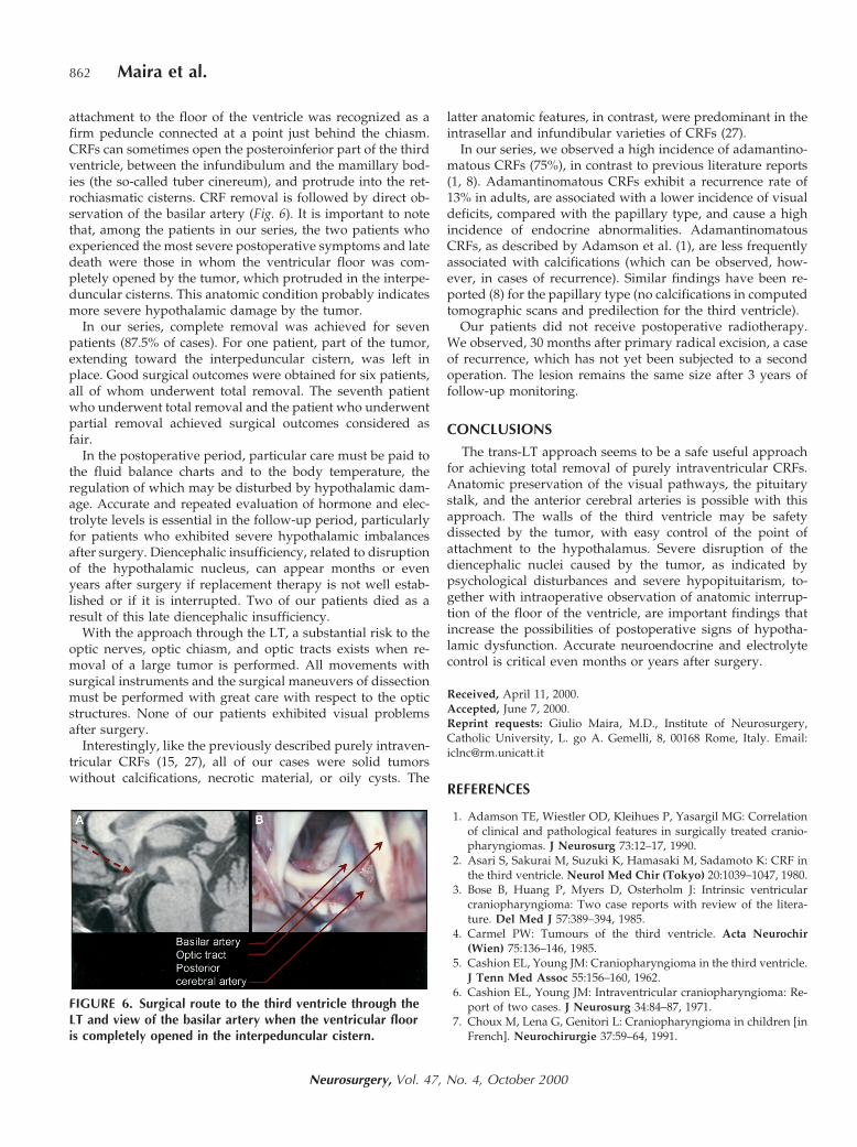
attachment to the floor of the ventricle was recognized as afirm peduncle connected at a point just behind the chiasm.CRFs can sometimes open the posteroinferior part of the thirdventricle, between the infundibulum and the mamillary bod-ies (the so-called tuber cinereum), and protrude into the ret-rochiasmatic cisterns. CRF removal is followed by direct ob-servation of the basilar artery (Fig. 6). It is important to notethat, among the patients in our series, the two patients whoexperienced the most severe postoperative symptoms and latedeath were those in whom the ventricular floor was com-pletely opened by the tumor, which protruded in the interpe-duncular cisterns. This anatomic condition probably indicatesmore severe hypothalamic damage by the tumor.
In our series, complete removal was achieved for sevenpatients (87.5% of cases). For one patient, part of the tumor,extending toward the interpeduncular cistern, was left inplace. Good surgical outcomes were obtained for six patients,all of whom underwent total removal. The seventh patientwho underwent total removal and the patient who underwentpartial removal achieved surgical outcomes considered asfair.
In the postoperative period, particular care must be paid tothe fluid balance charts and to the body temperature, theregulation of which may be disturbed by hypothalamic dam-age. Accurate and repeated evaluation of hormone and elec-trolyte levels is essential in the follow-up period, particularlyfor patients who exhibited severe hypothalamic imbalancesafter surgery. Diencephalic insufficiency, related to disruptionof the hypothalamic nucleus, can appear months or evenyears after surgery if replacement therapy is not well estab-lished or if it is interrupted. Two of our patients died as aresult of this late diencephalic insufficiency.
With the approach through the LT, a substantial risk to theoptic nerves, optic chiasm, and optic tracts exists when re-moval of a large tumor is performed. All movements withsurgical instruments and the surgical maneuvers of dissectionmust be performed with great care with respect to the opticstructures. None of our patients exhibited visual problemsafter surgery.
Interestingly, like the previously described purely intraven-tricular CRFs (15, 27), all of our cases were solid tumorswithout calcifications, necrotic material, or oily cysts. The
latter anatomic features, in contrast, were predominant in theintrasellar and infundibular varieties of CRFs (27).
In our series, we observed a high incidence of adamantino-matous CRFs (75%), in contrast to previous literature reports(1, 8). Adamantinomatous CRFs exhibit a recurrence rate of13% in adults, are associated with a lower incidence of visualdeficits, compared with the papillary type, and cause a highincidence of endocrine abnormalities. AdamantinomatousCRFs, as described by Adamson et al. (1), are less frequentlyassociated with calcifications (which can be observed, how-ever, in cases of recurrence). Similar findings have been re-ported (8) for the papillary type (no calcifications in computedtomographic scans and predilection for the third ventricle).
Our patients did not receive postoperative radiotherapy.We observed, 30 months after primary radical excision, a caseof recurrence, which has not yet been subjected to a secondoperation. The lesion remains the same size after 3 years offollow-up monitoring.
CONCLUSIONS
The trans-LT approach seems to be a safe useful approachfor achieving total removal of purely intraventricular CRFs.Anatomic preservation of the visual pathways, the pituitarystalk, and the anterior cerebral arteries is possible with thisapproach. The walls of the third ventricle may be safetydissected by the tumor, with easy control of the point ofattachment to the hypothalamus. Severe disruption of thediencephalic nuclei caused by the tumor, as indicated bypsychological disturbances and severe hypopituitarism, to-gether with intraoperative observation of anatomic interrup-tion of the floor of the ventricle, are important findings thatincrease the possibilities of postoperative signs of hypotha-lamic dysfunction. Accurate neuroendocrine and electrolytecontrol is critical even months or years after surgery.
Received, April 11, 2000.Accepted, June 7, 2000.Reprint requests: Giulio Maira, M.D., Institute of Neurosurgery,Catholic University, L. go A. Gemelli, 8, 00168 Rome, Italy. Email:[email protected]
REFERENCES
1. Adamson TE, Wiestler OD, Kleihues P, Yasargil MG: Correlationof clinical and pathological features in surgically treated cranio-pharyngiomas. J Neurosurg 73:12–17, 1990.
2. Asari S, Sakurai M, Suzuki K, Hamasaki M, Sadamoto K: CRF inthe third ventricle. Neurol Med Chir (Tokyo) 20:1039–1047, 1980.
3. Bose B, Huang P, Myers D, Osterholm J: Intrinsic ventricularcraniopharyngioma: Two case reports with review of the litera-ture. Del Med J 57:389–394, 1985.
4. Carmel PW: Tumours of the third ventricle. Acta Neurochir(Wien) 75:136–146, 1985.
5. Cashion EL, Young JM: Craniopharyngioma in the third ventricle.J Tenn Med Assoc 55:156–160, 1962.
6. Cashion EL, Young JM: Intraventricular craniopharyngioma: Re-port of two cases. J Neurosurg 34:84–87, 1971.
7. Choux M, Lena G, Genitori L: Craniopharyngioma in children [inFrench]. Neurochirurgie 37:59–64, 1991.
FIGURE 6. Surgical route to the third ventricle through theLT and view of the basilar artery when the ventricular flooris completely opened in the interpeduncular cistern.
862 Maira et al.
Neurosurgery, Vol. 47, No. 4, October 2000

8. Crotty TB, Scheithauer BW, Young WF, Davis DH, Shaw EG,Miller GM, Burger PC: Papillary craniopharyngioma: A clinico-pathological study of 48 cases. J Neurosurg 83:206–214, 1995.
9. Cushing HW: The craniopharyngiomas, in Bailey P (ed): Intracra-nial Tumors. Springfield, Charles C Thomas, 1933, pp 93–98.
10. Dandy WE: Diagnosis, localization and removal of tumors of thethird ventricle. Bull Johns Hopkins Hosp 33:188–194, 1922.
11. Dobos EI, Freed CG, Ashe SMP: An intrinsic tumor of the thirdventricle. J Neuropathol Exp Neurol 12:232–243, 1953.
12. Fitz CR, Wortzman G, Harwood-Nash DC, Holgare RC, Barry JF,Boldt DW: Computed tomography in craniopharyngiomas. Radi-ology 127:687–691, 1978.
13. Fujitsu K, Sekino T, Sakata K, Kawasaki T: Basal interfalcineapproach through a frontal sinusotomy with vein and nervepreservation: Technical note. J Neurosurg 80:575–579, 1994.
14. Fukushima T, Hirakawa K, Kimura M, Tomonaga M: Intraven-tricular craniopharyngioma: Its characteristics in magnetic reso-nance imaging and successful total removal. Surg Neurol 33:22–27, 1990.
15. Goldstein SJ, Wilson D, Young B, Guidry G: Craniopharyngiomaintrinsic to the third ventricle. Surg Neurol 20:249–253, 1983.
16. Iwasaki K, Kondo A, Takahashi JB, Yamanobe K: Intraventricularcraniopharyngioma: Report of two cases and review of the liter-ature. Surg Neurol 38:294–301, 1992.
17. King TT: Removal of intraventricular craniopharyngiomasthrough the lamina terminalis. Acta Neurochir (Wien) 45:277–286, 1979.
18. Klein HG, Rath SA: Removal of tumors in the third ventricleusing the lamina terminalis approach: Three cases of isolatedgrowth of craniopharyngiomas in the third ventricle. Childs NervSyst 5:144–147, 1989.
19. Konovalov AN: Technique and strategies of direct surgical man-agement of craniopharyngiomas, in Apuzzo MLJ (ed): Surgery ofthe Third Ventricle. Baltimore, Williams & Wilkins, 1987, pp542–553.
20. Konovalov AN, Gorelyshev SK: Surgical treatment of anteriorthird ventricle tumors. Acta Neurochir (Wien) 118:33–39, 1992.
21. Kubota T, Fujii H, Ikeda K, Ito H, Yamamoto S, Nakanishi I: Acase of intraventricular craniopharyngioma with subarachnoidhemorrhage [in Japanese]. No Shinkei Geka 8:495–501, 1980.
22. Kunishio K, Sunami M, Yamamoto Y, Asari S, Akagi T, Ohtsuki Y:An autopsy study on the origin of craniopharyngioma confined tothe third ventricle: Case report. Neurol Med Chir (Tokyo) 26:712–717, 1986.
23. Kunishio K, Yamamoto Y, Sunami M, Asari S, Akagi T, Ohtsuki Y:Craniopharyngioma in the third ventricle: Necropsy findings andhistogenesis. J Neurol Neurosurg Psychiatry 50:1053–1056, 1987.
24. Lanzieri CF, Sacher M, Som PM: CT changes in the septumpellucidum associated with intraventricular craniopharyngiomas.J Comput Assist Tomogr 9:507–510, 1985.
25. Linden CN, Martinez CR, Gonzalvo AA, Cahill DW: Intrinsicthird ventricle craniopharyngioma: CT and MR findings.J Comput Assist Tomogr 13:362–368, 1989.
26. Long DM, Chou SN: Transcallosal removal of craniopharyngio-mas within the third ventricle. J Neurosurg 39:563–567, 1973.
27. Maira G, Anile C, Rossi GF, Colosimo C: Surgical treatment ofcraniopharyngiomas: An evaluation of the transsphenoidal andpterional approaches. Neurosurgery 36:715–724, 1995.
28. Matthews FD: Intraventricular craniopharyngioma. AJNR Am JNeuroradiol 4:984–985, 1983.
29. Mori K, Handa H, Murata T, Takeuchi J, Miwa S, Osaka K: Resultsof treatment for craniopharyngiomas. Childs Brain 6:306–312,1980.
30. Nanba S, Tsuboi M: Craniopharyngioma in the third ventricle [inJapanese]. No To Shinkei 29:45–49, 1977.
31. Page RB: Diencephalic structures at risk in third ventricular sur-gery, in Apuzzo MLJ (ed): Surgery of the Third Ventricle. Baltimore,Williams & Wilkins, 1987, pp 553–556.
32. Ravindram M, Radhakrishnan VV, Rao VR: Communicating cys-tic craniopharyngioma. Surg Neurol 14:230–232, 1980.
33. Rhoton AL Jr, Yamamoto I: Operative approaches to the thirdventricle, in Wilkins RH, Rengachary SS (eds): Neurosurgery. NewYork, McGraw-Hill, 1996, ed 2, vol 1, pp 1435–1449.
34. Rush JL, Kusske JA, De Feo DR, Pribram HW: Intraventricularcraniopharyngioma. Neurology 25:1094–1096, 1975.
35. Samii M: Technical aspects of excision of giant basal tumors withthird ventricular involvement, in Apuzzo MLJ (ed): Surgery of theThird Ventricle. Baltimore, Williams & Wilkins, 1987, pp 684–697.
36. Sipos L, Vajda J: Craniopharyngioma of the third ventricle. ActaNeurochir (Wien) 139:92–93, 1997.
37. Stookey B, Scarff J: Occlusion of the aqueduct of Sylvius byneoplastic and non-neoplastic processes, with a rational surgicaltreatment for the relief of the resultant obstructive hydrocephalus.Bull Neurol Inst N Y 5:348–377, 1936.
38. Svien JH: Surgical experiences with craniopharyngiomas. J Neuro-surg 23:148–155, 1965.
39. Takahashi H, Nakazawa S, Shimura T: Evaluation of postopera-tive intratumoral injection of bleomycin for craniopharyngiomain children. J Neurosurg 62:120–127, 1985.
40. Urasaki E, Fukumura A, Ito Y, Itoyama Y, Yamada M, Ushio Y,Yokota A, Wada S: Craniopharyngioma in the third ventricle [inJapanese]. No To Shinkei 16:1399–1404, 1988.
41. Urbach H, Behrens E, Von Deimling A, Reul J: Solid craniophar-yngioma in the 3rd ventricle: Differential diagnostic aspects [inGerman]. Aktuelle Radiol 8:95–97, 1998.
42. Van Den Bergh R, Brucher JM: The transventricular approach incranio-pharyngioma of the 3rd ventricle: Neurosurgical and neu-ropathologic aspects [in French]. Neurochirurgie 16:51–65, 1970.
43. Villani R, Papagno C, Tomei D, Grimoldi N, Spagnoli D, Bello B:Transcallosal approach to tumors of the third ventricle: Surgicalresults and neuropsychological evaluation. J Neurosurg Sci 41:41–50, 1997.
COMMENTS
The majority of extra-axial cranial base tumors, includingacoustic neuromas, meningiomas, and pituitary adenomas,are extra-arachnoid lesions, in that they are covered by a layerof arachnoid membrane that separates them from the under-lying subarachnoid space and the important neurovascularstructures contained within it. Therefore, the separation ofthese tumors from the surrounding neurovascular structuresshould proceed along the tumor-arachnoid membrane inter-face and not between the arachnoid membrane and the neu-rovascular structures. Craniopharyngiomas (CRFs) differ inthis regard, in that they can be completely extra-arachnoid (as,for example, when they are completely intrasellar), they canbe partially extra-arachnoid and partially intra-arachnoid, orthey can be completely intra-arachnoid, and in fact partiallyintrapial, interdigitating with the neuropil of the hypotha-lamic floor. Finally, they can also be completely intraventric-ular. The explanation for such varied anatomic relationshipsbetween CRFs and the surrounding pia/arachnoid mem-branes and the ependyma of the third ventricle can be foundin the anatomic development of these tumors. Two important
Trans-Lamina Terminalis Approach 863
Neurosurgery, Vol. 47, No. 4, October 2000

embryological events play a role in this respect. First, as theanterior pituitary gland develops from the ventral portion ofRathke’s sac, it begins to rotate, with its inferior part turningcounterclockwise toward the hypothalamic floor (1). In theprocess, remnants of the columnar epithelial cells from Rath-ke’s duct that may have remained attached to Rathke’s sac arebrought into contact with the neuroectodermal layer of theventral aspect of the cerebral vesicle, the precursor of theinfundibulum and of the third ventricle floor. The secondimportant event is the development of the pia/arachnoidmembranes from the mesoderm intervening between the sto-modeum and the cerebral vesicle. Depending on the sequenceof these two events, CRFs can arise either extrapially, extra-arachnoidally, intrapially, or in fact intraventricularly. Theformer scenario occurs when the pial membrane develops inapproximately the fifth week of gestation, before the anteriorpituitary gland rotates, in which case columnar epithelial cellsthat may have remained attached to the anterior pituitarygland are excluded from the subpial space by the alreadyformed pia membrane. Depending on the degree of rotationof the anterior pituitary gland, these residual columnar epi-thelial cells may be brought into contact with the pia mem-brane high along the hypothalamic floor, low along the stalk,or even lower along the simultaneously developing posteriorlobe. Consequently, if a CRF arises from these cell remnants,it develops either intra- arachnoidally (but extrapially), par-tially intra-arachnoidally and partially extra-arachnoidally, orcompletely extra-arachnoidally. In contrast, when the devel-opment of the pia membrane lags in time, the rotation of theanterior pituitary gland can bring the residual columnar epi-thelial cells directly into contact with the neuroectoderm ofthe developing cerebral vesicle, where they can remain im-planted. If these cells then undergo squamous metaplasia anddevelop into a CRF, such a tumor remains intimately adherentto and interdigitated with the neuropil of the hypothalamicfloor. It is not difficult to imagine that the columnar epithelialcells can easily migrate through the signal neuroectodermallayer of the early cerebral vesicle and develop into an intra-ventricular CRF (2).
Clearly, the surgical approach for third-ventricle CRFsshould be chosen with consideration of the embryological andsurgical anatomic features of these tumors. An importantdistinction should be made between purely intraventricularCRFs and those that are retrochiasmatic and insinuate them-selves into the floor of the third ventricle, simulating anintraventricular presence. My own preference has alwaysbeen to approach purely intraventricular CRFs using thetransfrontal/transcallosal/transforaminal route and to ap-proach retrochiasmatic CRFs via a subfrontal approach inconjunction with a pterional (but with good access to themidline) craniotomy that includes an orbito-zygomatic-clinoidal cranial base dissection. I have rarely found it neces-sary to open the lamina terminalis (LT) to reach such retro-chiasmatic tumors, because it has always been possible to findan avenue between the ipsilateral optic nerve and track onone side and the carotid artery on the other, working under-neath the optic nerves and chiasm. I have occasionally re-
sorted to removal of the tuberculum sellae to gain betteraccess to the retrochiasmatic space, especially in patients witha prefixed chiasm. Opening the LT as a means of reaching intothe retrochiasmatic space, which anatomically requires open-ing of the floor of the third ventricle as well, has always mademe uncomfortable. Finally, to the best of my recollection, theLT in our patients was usually crowded by the anterior cere-bral arteries, the anterior communicating artery, and the per-forators emanating from these vessels. The contributions ofthe authors should certainly be considered by neurosurgeonsendeavoring to remove CRFs in this region. However, I thinkthat this method should not be the principle approach for theremoval of CRFs, because it may be associated with consid-erable risks, but should be considered an ancillary route to beused when all other extra-axial approaches have proveninadequate.
Ivan S. CiricEvanston, Illinois
The trans-LT approach is useful in the surgical treatment ofCRFs, as the results presented in this article testify. It can beused for tumors that are located largely in the third ventricle,as in this report, and it is also useful for CRFs when the opticnerves are short. In the case of short optic nerves, if thetuberculum sellae is removed and the LT is opened, then thetumor can be pushed from behind, via the third ventricle,under the chiasm and out between the optic nerves.
Another surgical option for CRFs located largely in thethird ventricle is to remove them from above, via an interfor-niceal approach. In this approach, a craniotomy is performedalong the sagittal sinus, the corpus callosum is divided, andthe septum pellucidum is followed to the interforniceal fis-sure. The fornices are separated, which provides a clear viewof the third ventricle. The tumor is removed piecemeal; at theend, the floor of the third ventricle is usually wide open andthe basilar bifurcation is in sight. Although this approachinitially seems to involve a greater distance, it is surprisinglyeasy to perform and is very well tolerated by patients, per-haps better than the trans-LT approach. The details of thisapproach have been published (1).
Russel H. Patterson, Jr.New York, New York
1. Apuzzo MLJ (ed): Surgery of the Third Ventricle. Baltimore, Wil-liams & Wilkins, 1998, ed 2.
The authors have documented the usefulness of thetrans-LT approach, and they point out that the approach canbe used with either a subfrontal or pterional craniotomy. It isusually possible to predict the tumors for which a trans-LTapproach will be helpful, on the basis of preoperative mag-netic resonance imaging scans showing the chiasm pushedforward and the tumor located within the third ventricle. Iprefer to approach the LT along the inferior surface of thefrontal lobe, either via the medial part of a pterional approachor, more frequently, via a small frontal craniotomy above the
864 Maira et al.
Neurosurgery, Vol. 47, No. 4, October 2000
supraorbital rim. The LT can be exposed by working along thesylvian fissure in the middle portion of the pterional ap-proach. However, the oblique angle at which the LT is viewedthrough the pterional approach makes it difficult to see pos-teriorly into the third ventricle, whereas the more medialsubfrontal route provides observation of the third ventriclebehind the LT even posterior to the aqueduct or to the basilarapex if the floor has been penetrated by the tumor. Care isrequired to preserve the perforating arteries, which arise fromthe anterior communicating artery and ascend on the LT,
because these supply the columns of the fornix and damagemay result in memory deficits. The LT is usually observedbelow the anterior communicating artery. All of the tumorswithin the third ventricle in this series were solid. We haveobserved several cystic CRFs within the third ventricle, whichwere removed via the trans-LT approach. The authors haveachieved admirable results with this difficult group of tumors.
Albert L. Rhoton, Jr.Gainesville, Florida
Trans-Lamina Terminalis Approach 865
Neurosurgery, Vol. 47, No. 4, October 2000