Cranial electrotherapy stimulation and transcranial pulsed current
8
Cranial electrotherapy stimulation and transcranial pulsed current stimulation: A computer based high-resolution modeling study Abhishek Datta a, b, ⁎, Jacek P. Dmochowski a , Berkan Guleyupoglu a , Marom Bikson a , Felipe Fregni b, c, ⁎⁎ a Neural Engineering Laboratory, Department of Biomedical Engineering, The City College of New York of CUNY, New York, NY 10031, USA b Laboratory of Neuromodulation, Spaulding Rehabilitation Hospital, Harvard Medical School, Boston, MA 02114, USA c Berenson-Allen Center for Noninvasive Brain Stimulation, Beth Israel Deaconess Medical Center, Harvard Medical School, Boston, MA 02215, USA abstract article info Article history: Accepted 24 September 2012 Available online 5 October 2012 Keywords: Cranial electrotherapy stimulation CES Brain stimulation Computer based modeling Brainstem The field of non-invasive brain stimulation has developed significantly over the last two decades. Though two techniques of noninvasive brain stimulation — transcranial direct current stimulation (tDCS) and transcranial magnetic stimulation (TMS) — are becoming established tools for research in neuroscience and for some clinical applications, related techniques that also show some promising clinical results have not been devel- oped at the same pace. One of these related techniques is cranial electrotherapy stimulation (CES), a class of transcranial pulsed current stimulation (tPCS). In order to understand further the mechanisms of CES, we aimed to model CES using a magnetic resonance imaging (MRI)-derived finite element head model including cortical and also subcortical structures. Cortical electric field (current density) peak intensities and distribu- tions were analyzed. We evaluated different electrode configurations of CES including in-ear and over-ear montages. Our results confirm that significant amounts of current pass the skull and reach cortical and subcortical structures. In addition, depending on the montage, induced currents at subcortical areas, such as midbrain, pons, thalamus and hypothalamus are of similar magnitude than that of cortical areas. Incre- mental variations of electrode position on the head surface also influence which cortical regions are modu- lated. The high-resolution modeling predictions suggest that details of electrode montage influence current flow through superficial and deep structures. Finally we present laptop based methods for tPCS dose design using dominant frequency and spherical models. These modeling predictions and tools are the first step to advance rational and optimized use of tPCS and CES. © 2012 Elsevier Inc. All rights reserved. Introduction The field of non-invasive brain stimulation has developed signifi- cantly during the last two decades. The use of neurophysiological, neuroimaging and computer-based modeling tools have contributed to this increased interest and development of this field. As a conse- quence, techniques that have been explored and used in the past are now being re-explored, with different, optimized parameters of stimulation. Transcranial direct current stimulation is one such exam- ple. The use of neurophysiological markers such as transcranial magnetic stimulation-induced cortical excitability and computer- based modeling has optimized parameters of stimulation such as electrode montage, intensity and duration of stimulation (Brunoni and Fregni, 2011; Datta et al., 2008, 2010; Miranda et al., 2006; Nitsche and Paulus, 2000; Wagner et al., 2006, 2007) One highly used method of noninvasive transcranial electrical stimulation — cranial electrotherapy stimulation (CES), with relatively broad clinical use, has not been fully explored. CES has had relatively broad clinical use following FDA clearance in 1978, and is historically a derivative of neuromodulation approaches dating to the early 20th century including cranial electrostimulation therapy (CET) and electrosleep (ES). CES devices use transcranial pulse current stimulation with dose parameters typically 50 μA to 5 mA intensity, around 100 Hz, typically applied over a session (around 30 min) using surface electrodes on the infra- or supra-auricular struc- tures (Zaghi et al., 2010). Although the CES technique has been used for several decades (Edelmuth et al., 2010) and has been reported to be effective for the treatment of insomnia, depression and anxiety (FDA label indications) in several clinical studies, the mechanisms of action remain unknown. Due to its effect mainly on vegetative symptoms of psychiatric disorders such as sleep, impaired attention and fatigue, it is purported that the application of CES through the maxillo-occipital junction causes current to reach the sub-cortical and brain stem structures. It has been shown that stimulation of these structures causes increased secretion of neurotransmitters, namely serotonin, NeuroImage 65 (2013) 280–287 ⁎ Correspondence to: A. Datta, T-463 Steinman Hall, Grove School of Engineering, The City College of CUNY, 160 Convent Ave, New York, NY 10031, USA. Fax: +1 212 6506727. ⁎⁎ Correspondence to: F. Fregni, Spaulding Rehabilitation Hospital, 125 Nashua Street, Boston, MA 02114, USA. Fax: +1 617 975 5322. E-mail addresses: [email protected] (A. Datta), [email protected] (F. Fregni). 1053-8119/$ – see front matter © 2012 Elsevier Inc. All rights reserved. http://dx.doi.org/10.1016/j.neuroimage.2012.09.062 Contents lists available at SciVerse ScienceDirect NeuroImage journal homepage: www.elsevier.com/locate/ynimg
Transcript of Cranial electrotherapy stimulation and transcranial pulsed current

NeuroImage 65 (2013) 280–287
Contents lists available at SciVerse ScienceDirect
NeuroImage
j ourna l homepage: www.e lsev ie r .com/ locate /yn img
Cranial electrotherapy stimulation and transcranial pulsed current stimulation:A computer based high-resolution modeling study
Abhishek Datta a,b,⁎, Jacek P. Dmochowski a, Berkan Guleyupoglu a, Marom Bikson a, Felipe Fregni b,c,⁎⁎a Neural Engineering Laboratory, Department of Biomedical Engineering, The City College of New York of CUNY, New York, NY 10031, USAb Laboratory of Neuromodulation, Spaulding Rehabilitation Hospital, Harvard Medical School, Boston, MA 02114, USAc Berenson-Allen Center for Noninvasive Brain Stimulation, Beth Israel Deaconess Medical Center, Harvard Medical School, Boston, MA 02215, USA
⁎ Correspondence to: A. Datta, T-463 Steinman Hall, GCity College of CUNY, 160 Convent Ave, New York, NY 100⁎⁎ Correspondence to: F. Fregni, Spaulding RehabilitatioBoston, MA 02114, USA. Fax: +1 617 975 5322.
E-mail addresses: [email protected] (A. Dat(F. Fregni).
1053-8119/$ – see front matter © 2012 Elsevier Inc. Allhttp://dx.doi.org/10.1016/j.neuroimage.2012.09.062
a b s t r a c t
a r t i c l e i n f oArticle history:Accepted 24 September 2012Available online 5 October 2012
Keywords:Cranial electrotherapy stimulationCESBrain stimulationComputer based modelingBrainstem
The field of non-invasive brain stimulation has developed significantly over the last two decades. Though twotechniques of noninvasive brain stimulation— transcranial direct current stimulation (tDCS) and transcranialmagnetic stimulation (TMS) — are becoming established tools for research in neuroscience and for someclinical applications, related techniques that also show some promising clinical results have not been devel-oped at the same pace. One of these related techniques is cranial electrotherapy stimulation (CES), a class oftranscranial pulsed current stimulation (tPCS). In order to understand further the mechanisms of CES, weaimed to model CES using a magnetic resonance imaging (MRI)-derived finite element head model includingcortical and also subcortical structures. Cortical electric field (current density) peak intensities and distribu-tions were analyzed. We evaluated different electrode configurations of CES including in-ear and over-earmontages. Our results confirm that significant amounts of current pass the skull and reach cortical andsubcortical structures. In addition, depending on the montage, induced currents at subcortical areas, suchas midbrain, pons, thalamus and hypothalamus are of similar magnitude than that of cortical areas. Incre-mental variations of electrode position on the head surface also influence which cortical regions are modu-lated. The high-resolution modeling predictions suggest that details of electrode montage influence currentflow through superficial and deep structures. Finally we present laptop based methods for tPCS dose designusing dominant frequency and spherical models. These modeling predictions and tools are the first step toadvance rational and optimized use of tPCS and CES.
© 2012 Elsevier Inc. All rights reserved.
Introduction
The field of non-invasive brain stimulation has developed signifi-cantly during the last two decades. The use of neurophysiological,neuroimaging and computer-based modeling tools have contributedto this increased interest and development of this field. As a conse-quence, techniques that have been explored and used in the pastare now being re-explored, with different, optimized parameters ofstimulation. Transcranial direct current stimulation is one such exam-ple. The use of neurophysiological markers such as transcranialmagnetic stimulation-induced cortical excitability and computer-based modeling has optimized parameters of stimulation such aselectrode montage, intensity and duration of stimulation (Brunoniand Fregni, 2011; Datta et al., 2008, 2010; Miranda et al., 2006;
rove School of Engineering, The31, USA. Fax: +1 212 6506727.n Hospital, 125 Nashua Street,
ta), [email protected]
rights reserved.
Nitsche and Paulus, 2000; Wagner et al., 2006, 2007) One highlyused method of noninvasive transcranial electrical stimulation —
cranial electrotherapy stimulation (CES), with relatively broad clinicaluse, has not been fully explored.
CES has had relatively broad clinical use following FDA clearance in1978, and is historically a derivative of neuromodulation approachesdating to the early 20th century including cranial electrostimulationtherapy (CET) and electrosleep (ES). CES devices use transcranialpulse current stimulation with dose parameters typically 50 μA to5 mA intensity, around 100 Hz, typically applied over a session (around30 min) using surface electrodes on the infra- or supra-auricular struc-tures (Zaghi et al., 2010). Although the CES technique has been used forseveral decades (Edelmuth et al., 2010) and has been reported to beeffective for the treatment of insomnia, depression and anxiety (FDAlabel indications) in several clinical studies, the mechanisms of actionremain unknown. Due to its effect mainly on vegetative symptoms ofpsychiatric disorders such as sleep, impaired attention and fatigue, itis purported that the application of CES through the maxillo-occipitaljunction causes current to reach the sub-cortical and brain stemstructures. It has been shown that stimulation of these structurescauses increased secretion of neurotransmitters, namely serotonin,

281A. Datta et al. / NeuroImage 65 (2013) 280–287
beta endorphin, and norepinephrine (Shealy, 1989); thus being poten-tially involved with the mechanisms underlying the behavioral effectsof CES (Schroeder and Barr, 2001).
In one of the few controlled studies where the physiologic mecha-nism of action of CES was investigated, electroencephalographic (EEG)changes were reported (Schroeder and Barr, 2001). CES led to changesin alpha and beta frequency ranges suggesting potential neuroplasticand cognitive effects of this technique. Interestingly, similar changesin alpha and beta bands were shown to be associated with a reductionin the emotional-cognitive aspects of pain in a study using transcranialdirect current stimulation, which is another type of non-invasive brainstimulation (Maeoka et al., 2012). Though these results are promising,additional studies must be done due to the lack of mechanistic studies,particularly in CES (Edelmuth et al., 2010). Table 1 includes a summaryof the most recent studies with CES therapy published in the past15 years. Moreover a recurring point of contention over the years hasbeen whether low current CES applied through the electrode sites(ear lobes, mastoid processes or the temporal areas) can even reachthe underlying cortex to influence neural activity. In fact very limitedeffort has been invested to quantify the spatial distribution of currentswithin the human brain using this technique.
Since it is technically difficult to directly assess current flow in struc-tures within the human head, simulations of current flow via computermodeling can be used to predict the intensity and spatial distribution ofcurrent flowduring transcranial stimulation. Concentric-spheremodelshave previously been used to calculate CES induced electric fields(Ferdjallah et al., 1996). In recent years, advances in modeling andimaging tools have allowed the development of models with increasedrealism and precision, resulting in high-resolution (1 mm3) MRIderived head models that capture gyri/sulci anatomical details (Dattaet al., 2009) as well as examine current density distributions throughsub-cortical target regions (Dasilva et al., 2012; Parazzini et al., 2012).
We adapted a previously developed high-resolution individual-ized model of tDCS (Datta et al., 2009) for simulating the effects ofCES. We modeled the conventional ear-clip electrode montage andcompared it with several novel montages (Brain Gear, Switzerland).We determined induced surface cortical electrical field (EF) topredict spatial focality. In addition, sub-cortical and brain-stemstructures implicated in the purported CES beneficial effects wereindividually analyzed.
Methods
In order to better understand which brain regions are modulatedduring cranial electrotherapy stimulation (CES), we carried out ahigh-resolution finite element (FE) model analysis. For comparison,we showed the effects of conventional therapy using ear-clip elec-trodes versus multiple novel montages like the in-ear, ear-hook andthe over-the-ear montages.
Table 1Summary of randomized CES trials.
Author Year Patient# Design Sham controlled
Rose 2009 44 Parallel Randomized Trial Yes
Schroeder 2001 12 Cross-over Trial Yes
Southworth 1999 52 Parallel Randomized Trial Yes
Scherder et al., 2006 21 Parallel Randomized Trial Yes
Abbreviations: CES: cranial electrical stimulation; TCES: transcutaneous cranial electrical st
MRI derived high-resolution model
The human head model was derived from a high spatial resolution(1 mm3) 3 T MRI of a male adult healthy subject with no neurologicalpathologies. Using a combination of tools from FMRIB SoftwareLibrary (FSL) and Simpleware, the head model was segmented intotissue compartments representing the scalp, skull, CSF, eye region,muscle, gray matter, white matter, and air respectively. In additionto analyzing current flow patterns through structures thought to beimplicated in the beneficial effects of CES, structures such as cingulatecortex, thalamus, insula, pituitary gland, pineal gland, hypothalamus,midbrain, pons, and medulla oblongata were also segmented. Thehead model was limited to the masks being directly derived fromthe MRI acquisition volume. An artificial neck and shoulder regionwas thus fused onto the existing segmented head. Stimulation elec-trodes of various sizes (as mentioned below) were imported as CADmodels and placed onto the existing segmented volume to modelthe different CES montages. The entire model (head and the elec-trodes) were meshed and exported to a commercial FE solver(COMSOL 3.5a) for final computation of current flow patterns.
Electrode montages
We modeled the following CES montages representing the con-ventional and the novel montages (see Fig. 1):
1) Conventional ear-clip montage (montage 1):The stimulation elec-trodes were placed mimicking conventional CES stimulation usingear-clip electrodes. The left ear-clip electrode was energized to anormal current density boundary condition corresponding to1 mA total injected current. The right ear-clip electrode wasapplied as the ground boundary condition. All other externalsurfaces were treated as insulated.
2) Novel in-ear electrode montage (montages 2 and 3):Stimulationelectrodes were placed resembling the in-ear headphone loca-tions. The left in-ear electrode was energized to a normal currentdensity boundary condition corresponding to 1 mA total injectedcurrent. The right in-ear electrode was applied as the groundboundary condition. All other external surfaces were treated asinsulated. In addition, the In-Ear electrode montage was alsosolved at 150 Hz (montage 3).
3) Novel ear-hook electrode montage (montage 4):Stimulation elec-trodes were placed resembling the ear-hook headphone locations.The left ear-hook electrode was energized to a normal currentdensity boundary condition corresponding to 1 mA total injectedcurrent. The right ear-hook electrode was applied as the groundboundary condition. All other external surfaces were treated asinsulated.
4) Novel over-the-ear electrode montage (4 contacts) (montage 5):Stimulation electrodes were placed resembling the over-the-ear
Blinded Clinical effects
Double blinded The CES group showed improvements in sleep disturbance,and depression though neither was statistically significant.
Double blinded .5 and 100 Hz CES elicited frequency distribution shifts.100 Hz CES produced greater overall change. These resultssuggest beneficial changes in mental state.
Not Stated CES significantly improved attention and concentration in anormal adult population.
Blinded No significant improvements on cognition and (affective)behavior were found between CES treatment and controlgroups.
imulation; CBF: cerebral blood flow.

Fig. 1. Cranial electrotherapy stimulation (CES)/transcranial pulsed current stimulation (tPCS) electrode montages modeling in the present study.
282 A. Datta et al. / NeuroImage 65 (2013) 280–287
headphone locations. The four left over-the-ear electrodes wereeach energized to a normal current density boundary conditioncorresponding to 0.25 mA current (leading to 1 mA total currentinjected across the head). The four right over-the-ear electrodeswere each applied as the ground boundary condition. All otherexternal surfaces were treated as insulated.
5) Novel over-the-ear electrode montage (2 contacts) (montage 6):Stimulation electrodes were placed resembling the over-the-earheadphone locations. The two left over-the-ear electrodes wereeach energized to a normal current density boundary conditioncorresponding to 0.5 mA current (leading to 1 mA total currentinjected across the head). The two right over-the-ear electrodeswere each applied as the ground boundary condition. All otherexternal surfaces were treated as insulated.
The Laplace equation was solved and induced cortical surface elec-tric field (EF) magnitude maps for the different electrode montageswere determined. The following isotropic electrical conductivities atDC in (S/m) were assigned: scalp (0.465); skull (0.01); CSF (1.65);gray matter (0.276); white matter (0.126); eye (0.4); muscle(0.334); hypothalamus (0.201); glands (0.5); air (1e−15); and elec-trode (5.8e7). The following isotropic electrical conductivities corre-sponding to 150 Hz in (S/m) were assigned: scalp (0.002); skull(0.02); CSF (1.65); gray matter (0.092); white matter (0.059); eye(0.4); muscle (0.282); hypothalamus (0.075); glands (0.522); air(1e−15); and electrode (5.8e7). The cingulate cortex, insula, andthe thalamus were assigned gray matter conductivity while themidbrain, pons, and the medulla oblongata were assigned whitematter conductivity (DaSilva et al., 2012; Gabriel et al., 1996).

Fig. 2. Cortical surface electric field magnitude plots for cortical, subcortical and brain-stem regions across all montages. All plots are plotted to their maximum peak.
283A. Datta et al. / NeuroImage 65 (2013) 280–287

Fig. 3. Cortical surface electric field magnitude plots for cortical, subcortical and brain-stem regions across all montages. The ear-hook montage led to the maximum peak inducedelectric field magnitude and all plots are scaled to this maximum peak (apart from montage 3 — 150 Hz simulation).
284 A. Datta et al. / NeuroImage 65 (2013) 280–287

Table 2Different classes of tPCS are summarized including temporal waveform (function), the associated magnitude spectrum (frequency content), and clinical references including dose using“CES”. The Fourier series were generated using the same parameters for T, τ, and A across all classes and the same parameters for h, D0, Ton, and Toff where applicable. Note that n is adiscrete function of 1/T (or Toff in the case of Class III). In Class III, the CES casewouldhaveD0 set to zerowhichwould lower thepeak at zero. In Class II, hr=(h+1)/h, in Class III, Tr=Ton/Toff and in all classes, P=A(τ/T). The references indicated are: 1Limoge et al. (1999), 2Brown (1975), 3Bystritsky et al. (2008), 4http://www.net1device.com/specs.htm, 5Liss StimulatorManual, Model No. SBL-502-B, 6Richthofen and Mellor (1979), 7Dimitrov and Ralev (2009), 8Liss Stimulator Manual, Model No.SBL-501-M.
Magnitude spectrum Notes
Class I(A) -
Monophasic pulse
-ES2
-CET6
-EA2
00
P
10/T 20/T 30/TFrequency
Class I(B) -
Monophasic pulse with
DC offset
-ES2,6
-EA2
-CET2,6
-TCET6,2
00
10/T 20/T 30/TFrequency
P+
D0
Class II(A) -
Biphasic pulse -CES7
00
10/T 20/T 30/TFrequency
.63
7A
Class II(B) -
Biphasic pulse with delay
-TCET
-CET
-NET4
-CES5
00
10/T 20/T 30/TFrequency
1.4
4P
Class II(C) -
Asymmetric biphasic pulse
00
10/T 20/T 30/TFrequency
.57
6(A
/h)
Class II(D) -
Asymmetric biphasic pulse
with delay
-CES3
-NET
00
10/T 20/T 30/TFrequency
.91
5P
hr
Class III -
Monophasic pulse train
-LC1
-TCES1
-CES8
Waveform
C
A
T tτ
CA
TD t
τ
CA
2τ tτ
CA
2τ tTτ
C
A
A/h
hτ+τ tτ
C
A
A/h
hτ+τ T tτ
D
CA
T tτ
00
10/T 20/T 30/TFrequency
Tr(
P+
D0)
285A. Datta et al. / NeuroImage 65 (2013) 280–287
Results
Figs. 2 and 3 summarize results using each electrode montage. InFig. 2, all panels are scaled to their individual peak electric field mag-nitude (peak/scale indicated), which highlights the electric field localmaximum in each case and the regional spatial distribution. In Fig. 3,each cortical and sub-cortical column is re-scaled to one value (peakof the ear-hook montage) to highlight the relative electric field in-tensities and distributions across montages (except for the 150 Hzcase).
The conventional ear-clip montage resulted in a 0.10 V/m peakinduced cortical electric field. Maximal currents were induced inthe temporal sides of the cortex and in the medulla oblongata (seeDiscussion) with diffuse activation in the midbrain, pons, thalamus,insula, and hypothalamus.
For the in-ear montage, a similar spatial profile of induced cur-rents was predicted; however the peak induced electric field in thecortex was higher (0.16 V/m). The in-ear montage thus led to higherinduced EF magnitudes in the midbrain, pons, hypothalamus, andthe insula.
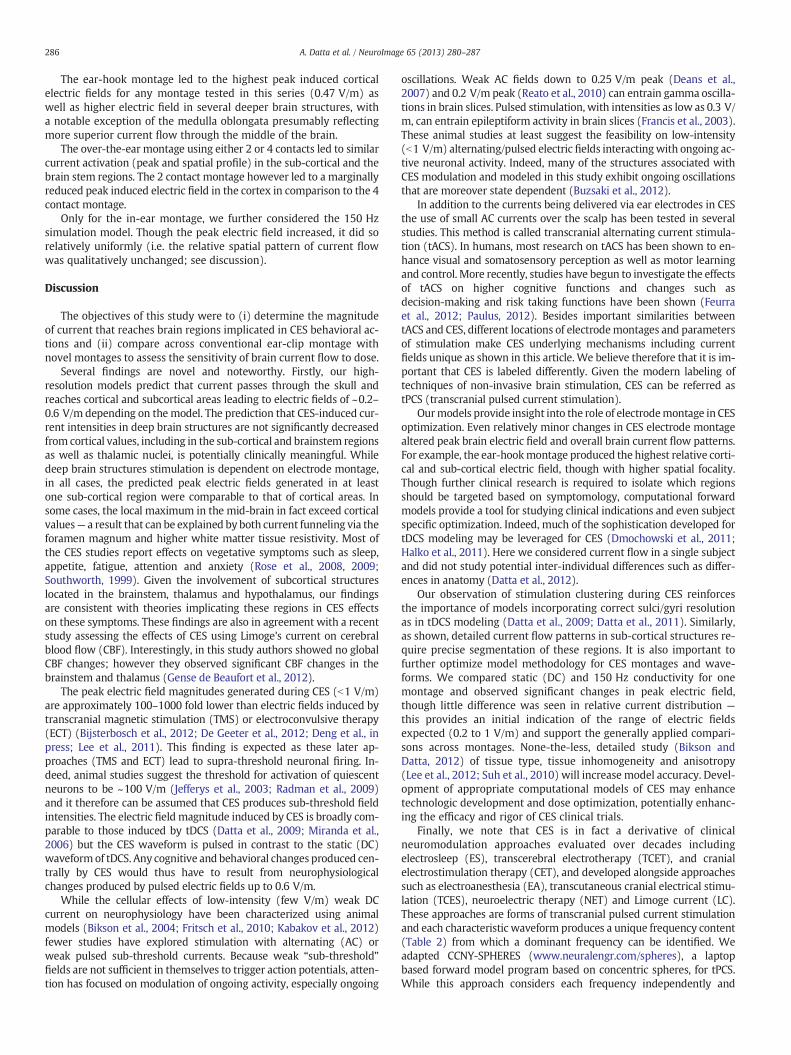
286 A. Datta et al. / NeuroImage 65 (2013) 280–287
The ear-hook montage led to the highest peak induced corticalelectric fields for any montage tested in this series (0.47 V/m) aswell as higher electric field in several deeper brain structures, witha notable exception of the medulla oblongata presumably reflectingmore superior current flow through the middle of the brain.
The over-the-ear montage using either 2 or 4 contacts led to similarcurrent activation (peak and spatial profile) in the sub-cortical and thebrain stem regions. The 2 contact montage however led to a marginallyreduced peak induced electric field in the cortex in comparison to the 4contact montage.
Only for the in-ear montage, we further considered the 150 Hzsimulation model. Though the peak electric field increased, it did sorelatively uniformly (i.e. the relative spatial pattern of current flowwas qualitatively unchanged; see discussion).
Discussion
The objectives of this study were to (i) determine the magnitudeof current that reaches brain regions implicated in CES behavioral ac-tions and (ii) compare across conventional ear-clip montage withnovel montages to assess the sensitivity of brain current flow to dose.
Several findings are novel and noteworthy. Firstly, our high-resolution models predict that current passes through the skull andreaches cortical and subcortical areas leading to electric fields of ~0.2–0.6 V/m depending on the model. The prediction that CES-induced cur-rent intensities in deep brain structures are not significantly decreasedfrom cortical values, including in the sub-cortical and brainstem regionsas well as thalamic nuclei, is potentially clinically meaningful. Whiledeep brain structures stimulation is dependent on electrode montage,in all cases, the predicted peak electric fields generated in at leastone sub-cortical region were comparable to that of cortical areas. Insome cases, the local maximum in the mid-brain in fact exceed corticalvalues— a result that can be explained by both current funneling via theforamen magnum and higher white matter tissue resistivity. Most ofthe CES studies report effects on vegetative symptoms such as sleep,appetite, fatigue, attention and anxiety (Rose et al., 2008, 2009;Southworth, 1999). Given the involvement of subcortical structureslocated in the brainstem, thalamus and hypothalamus, our findingsare consistent with theories implicating these regions in CES effectson these symptoms. These findings are also in agreement with a recentstudy assessing the effects of CES using Limoge's current on cerebralblood flow (CBF). Interestingly, in this study authors showed no globalCBF changes; however they observed significant CBF changes in thebrainstem and thalamus (Gense de Beaufort et al., 2012).
The peak electric field magnitudes generated during CES (b1 V/m)are approximately 100–1000 fold lower than electric fields induced bytranscranial magnetic stimulation (TMS) or electroconvulsive therapy(ECT) (Bijsterbosch et al., 2012; De Geeter et al., 2012; Deng et al., inpress; Lee et al., 2011). This finding is expected as these later ap-proaches (TMS and ECT) lead to supra-threshold neuronal firing. In-deed, animal studies suggest the threshold for activation of quiescentneurons to be ~100 V/m (Jefferys et al., 2003; Radman et al., 2009)and it therefore can be assumed that CES produces sub-threshold fieldintensities. The electric field magnitude induced by CES is broadly com-parable to those induced by tDCS (Datta et al., 2009; Miranda et al.,2006) but the CES waveform is pulsed in contrast to the static (DC)waveformof tDCS. Any cognitive and behavioral changes produced cen-trally by CES would thus have to result from neurophysiologicalchanges produced by pulsed electric fields up to 0.6 V/m.
While the cellular effects of low-intensity (few V/m) weak DCcurrent on neurophysiology have been characterized using animalmodels (Bikson et al., 2004; Fritsch et al., 2010; Kabakov et al., 2012)fewer studies have explored stimulation with alternating (AC) orweak pulsed sub-threshold currents. Because weak “sub-threshold”fields are not sufficient in themselves to trigger action potentials, atten-tion has focused on modulation of ongoing activity, especially ongoing
oscillations. Weak AC fields down to 0.25 V/m peak (Deans et al.,2007) and 0.2 V/m peak (Reato et al., 2010) can entrain gamma oscilla-tions in brain slices. Pulsed stimulation, with intensities as low as 0.3 V/m, can entrain epileptiform activity in brain slices (Francis et al., 2003).These animal studies at least suggest the feasibility on low-intensity(b1 V/m) alternating/pulsed electric fields interactingwith ongoing ac-tive neuronal activity. Indeed, many of the structures associated withCES modulation and modeled in this study exhibit ongoing oscillationsthat are moreover state dependent (Buzsaki et al., 2012).
In addition to the currents being delivered via ear electrodes in CESthe use of small AC currents over the scalp has been tested in severalstudies. This method is called transcranial alternating current stimula-tion (tACS). In humans, most research on tACS has been shown to en-hance visual and somatosensory perception as well as motor learningand control. More recently, studies have begun to investigate the effectsof tACS on higher cognitive functions and changes such asdecision-making and risk taking functions have been shown (Feurraet al., 2012; Paulus, 2012). Besides important similarities betweentACS and CES, different locations of electrodemontages and parametersof stimulation make CES underlying mechanisms including currentfields unique as shown in this article. We believe therefore that it is im-portant that CES is labeled differently. Given the modern labeling oftechniques of non-invasive brain stimulation, CES can be referred astPCS (transcranial pulsed current stimulation).
Ourmodels provide insight into the role of electrodemontage in CESoptimization. Even relatively minor changes in CES electrode montagealtered peak brain electric field and overall brain current flow patterns.For example, the ear-hookmontage produced the highest relative corti-cal and sub-cortical electric field, though with higher spatial focality.Though further clinical research is required to isolate which regionsshould be targeted based on symptomology, computational forwardmodels provide a tool for studying clinical indications and even subjectspecific optimization. Indeed, much of the sophistication developed fortDCS modeling may be leveraged for CES (Dmochowski et al., 2011;Halko et al., 2011). Here we considered current flow in a single subjectand did not study potential inter-individual differences such as differ-ences in anatomy (Datta et al., 2012).
Our observation of stimulation clustering during CES reinforcesthe importance of models incorporating correct sulci/gyri resolutionas in tDCS modeling (Datta et al., 2009; Datta et al., 2011). Similarly,as shown, detailed current flow patterns in sub-cortical structures re-quire precise segmentation of these regions. It is also important tofurther optimize model methodology for CES montages and wave-forms. We compared static (DC) and 150 Hz conductivity for onemontage and observed significant changes in peak electric field,though little difference was seen in relative current distribution —
this provides an initial indication of the range of electric fieldsexpected (0.2 to 1 V/m) and support the generally applied compari-sons across montages. None-the-less, detailed study (Bikson andDatta, 2012) of tissue type, tissue inhomogeneity and anisotropy(Lee et al., 2012; Suh et al., 2010) will increase model accuracy. Devel-opment of appropriate computational models of CES may enhancetechnologic development and dose optimization, potentially enhanc-ing the efficacy and rigor of CES clinical trials.
Finally, we note that CES is in fact a derivative of clinicalneuromodulation approaches evaluated over decades includingelectrosleep (ES), transcerebral electrotherapy (TCET), and cranialelectrostimulation therapy (CET), and developed alongside approachessuch as electroanesthesia (EA), transcutaneous cranial electrical stimu-lation (TCES), neuroelectric therapy (NET) and Limoge current (LC).These approaches are forms of transcranial pulsed current stimulationand each characteristic waveform produces a unique frequency content(Table 2) from which a dominant frequency can be identified. Weadapted CCNY-SPHERES (www.neuralengr.com/spheres), a laptopbased forward model program based on concentric spheres, for tPCS.While this approach considers each frequency independently and

287A. Datta et al. / NeuroImage 65 (2013) 280–287
concentric spheres evidently do not represent cortical folding or explicitdeep brain structures, CCNY-SPHERES provides an immediately accessi-ble tool for dose exploration using tPCS. High-resolution FEMmodels, asdeveloped in this study, are more resource intensive, and may thus beevaluated on those dose approaches considered promising based onCCNY-SPHERES pre-screening.
Acknowledgments
This work was partially funded by a gift from To Be First AG to theLaboratory of Neuromodulation, Spaulding Rehabilitation Hospital(M.B and A.D.). We are thankful to Noelle Chiavetta for her editorialrevision on this manuscript.
References
Bijsterbosch, J.D., Barker, A.T., et al., 2012. Where does transcranial magnetic stimula-tion (TMS) stimulate? Modelling of induced field maps for some common corticaland cerebellar targets. Med. Biol. Eng. Comput. 50 (7), 671–681.
Bikson, M., Datta, A., 2012. Guidelines for precise and accurate computational modelsof tDCS. Brain Stimul. 5 (3), 430–431.
Bikson, M., Inoue, M., et al., 2004. Effects of uniform extracellular DC electric fields onexcitability in rat hippocampal slices in vitro. J. Physiol. 557 (Pt. 1), 175–190.
Brunoni, A.R., Fregni, F., 2011. Clinical trial design in non-invasive brain stimulationpsychiatric research. Int. J. Methods Psychiatr. Res. 20 (2), e19–e30.
Buzsaki, G., Anastassiou, C.A., et al., 2012. The origin of extracellular fields and currents— EEG, ECoG, LFP and spikes. Nat. Rev. Neurosci. 13 (6), 407–420.
Dasilva, A.F., Mendonca, M.E., et al., 2012. tDCS-induced analgesia and electrical fieldsin pain-related neural networks in chronic migraine. Headache.
Datta, A., Bansal, V., et al., 2009. Gyri-precise head model of transcranial direct currentstimulation: improved spatial focality using a ring electrode versus conventionalrectangular pad. Brain Stimul. 2 (4), 201–207 (207 e201).
Datta, A., Bikson, M., et al., 2010. Transcranial direct current stimulation in patientswith skull defects and skull plates: high-resolution computational FEM study offactors altering cortical current flow. Neuroimage 52 (4), 1268–1278.
Datta, A., Elwassif, M., et al., 2008. Transcranial current stimulation focality using discand ring electrode configurations: FEM analysis. J. Neural Eng. 5 (2), 163–174.
Datta, A., Baker, J.M., et al., 2011. Individualized model predicts brain current flow dur-ing transcranial direct-current stimulation treatment in responsive stroke patient.Brain Stimul. 4 (3), 169–174.
Datta, A., Truong, D., et al., 2012. Inter-individual variation during transcranial directcurrent stimulation and normalization of dose using MRI-derived computationalmodels. Front. Psychiatry 3 (91). http://dx.doi.org/10.3389/fpsyt.2012.0009191.
De Geeter, N., Crevecoeur, G., et al., 2012. A DTI-based model for TMS using the inde-pendent impedance method with frequency-dependent tissue parameters. Phys.Med. Biol. 57 (8), 2169–2188.
Deans, J.K., Powell, A.D., et al., 2007. Sensitivity of coherent oscillations in rat hippo-campus to AC electric fields. J. Physiol. 583 (Pt. 2), 555–565.
Deng, Z.D., Lisanby, S.H., et al., in press. Electric field depth-focality tradeoff in transcra-nial magnetic stimulation: simulation comparison of 50 coil designs. Brain Stimul.http://dx.doi.org/10.1016/j.brs.2012.02.005.
Edelmuth, R.C., Nitsche, M.A., et al., 2010. Why do some promising brain-stimulationdevices fail the next steps of clinical development? Expert Rev. Med. Devices 7(1), 67–97.
Ferdjallah, M., Bostick Jr., F.X., et al., 1996. Potential and current density distributions ofcranial electrotherapy stimulation (CES) in a four-concentric-spheres model. IEEETrans. Biomed. Eng. 43 (9), 939–943.
Feurra, M., Galli, G., et al., 2012. Transcranial alternating current stimulation affectsdecision making. Front. Syst. Neurosci. 6 (39), 1–2.
Francis, J.T., Gluckman, B.J., et al., 2003. Sensitivity of neurons to weak electric fields.J. Neurosci. 23 (19), 7255–7261.
Fritsch, B., Reis, J., et al., 2010. Direct current stimulation promotes BDNF-dependentsynaptic plasticity: potential implications for motor learning. Neuron 66 (2), 198–204.
Gabriel, C., Gabriel, S., Corthout, E., 1996. The dielectric properties of biological tissues:I. Literature survey. Phys. Med. Biol. 41 (11), 2231–2249 (Nov, Review).
Gense de Beaufort, D., Sesay, M., et al., 2012. Cerebral blood flow modulation by trans-cutaneous cranial electrical stimulation with Limoge's current. J. Neuroradiol. 39(3), 167–175.
Halko, M.A., Datta, A., et al., 2011. Neuroplastic changes following rehabilitative trainingcorrelate with regional electrical field induced with tDCS. Neuroimage 57 (3),885–891.
Jefferys, J.G., Deans, J., et al., 2003. Effects of weak electric fields on the activity ofneurons and neuronal networks. Radiat. Prot. Dosimetry 106 (4), 321–323.
Kabakov, A.Y., Muller, P.A., et al., 2012. Contribution of axonal orientation to pathway-dependent modulation of excitatory transmission by direct current stimulation inisolated rat hippocampus. J. Neurophysiol. 107 (7), 1881–1889.
Lee, W.H., Deng, Z.D., et al., 2011. Influence of white matter conductivity anisotropy onelectric field strength induced by electroconvulsive therapy. Conf. Proc. IEEE Eng.Med. Biol. Soc. 2011, 5473–5476.
Lee, W.H., Deng, Z.D., Kim, T.S., Laine, A.F., Lisanby, S.H., Peterchev, A.V., 2012. Regionalelectric field induced by electroconvulsive therapy in a realistic finite element headmodel: influence of white matter anisotropic conductivity. Neuroimage 59 (3),2110–2123 (Feb 1, Epub 2011 Oct 18).
Miranda, P.C., Lomarev, M., et al., 2006. Modeling the current distribution duringtranscranial direct current stimulation. Clin. Neurophysiol. 117 (7), 1623–1629.
Maeoka, H., Matsu, A., Hiyamizu, M., Morioka, S., Ando, H., 2012. Influence of transcranialdirect current stimulation of the dorsolateral prefrontal cortex on pain relatedemotions: a study using electroencephalographic power spectrum analysis. Neurosci.Lett. 512 (1), 12–16.
Nitsche, M.A., Paulus, W., 2000. Excitability changes induced in the human motor cortexby weak transcranial direct current stimulation. J. Physiol. 527 (Pt. 3), 633–639.
Parazzini, M., Fiocchi, S., et al., 2012. Electric field and current density distribution in ananatomical head model during transcranial direct current stimulation for tinnitustreatment. Bioelectromagnetics 33 (6), 476–487.
Paulus, W., 2012. Transcranial electrical stimulation (tES–tDCS; tRNS, tACS) methods.Neuropsychol. Rehabil. 21 (5), 602–617.
Radman, T., Ramos, R.L., et al., 2009. Role of cortical cell type and morphology insubthreshold and suprathreshold uniform electric field stimulation in vitro. BrainStimul. 2 (4), 215–228 (228 e211–213).
Reato, D., Rahman, A., et al., 2010. Low-intensity electrical stimulation affects networkdynamics by modulating population rate and spike timing. J. Neurosci. 30 (45),15067–15079.
Rose, K.M., Taylor, A.G., et al., 2009. Effects of cranial electrical stimulation on sleepdisturbances, depressive symptoms, and caregiving appraisal in spousal caregiversof persons with Alzheimer's disease. Appl. Nurs. Res. 22 (2), 119–125.
Rose, K.M., Taylor, A.G., et al., 2008. Cranial electrical stimulation: potential use inreducing sleep and mood disturbances in persons with dementia and their familycaregivers. Fam. Community Health 31 (3), 240–246.
Scherder, E.J.A., van Tol, M.J., et al., 2006. High-frequency cranial electrostimulation(CES) in patients with probable Alzheimer's disease. Am. J. Phys. Med. Rehabil.Neurophysiol. 85 (7), 614–618.
Schroeder, M.J., Barr, R.E., 2001. Quantitative analysis of the electroencephalogram duringcranial electrotherapy stimulation. Clin. Neurophysiol. 112 (11), 2075–2083.
Shealy, Y.F., 1989. Synthesis and evaluation of some new retinoids for cancer chemo-prevention. Prev. Med. 18 (5), 624–645.
Southworth, S., 1999. A study of the effects of cranial electrical stimulation on attentionand concentration. Integr. Physiol. Behav. Sci. 34 (1), 43–53.
Suh, H.S., Lee,W.H., Cho, Y.S., Kim, J.H., Kim, T.S., 2010. Reduced spatial focality of electricalfield in tDCS with ring electrodes due to tissue anisotropy. Conf. Proc. IEEE Eng. Med.Biol. Soc. 2010, 2053–2056.
Wagner, T., Fregni, F., et al., 2006. Transcranial magnetic stimulation and stroke: acomputer-based human model study. Neuroimage 30 (3), 857–870.
Wagner, T., Fregni, F., et al., 2007. Transcranial direct current stimulation: a computer-based human model study. Neuroimage 35 (3), 1113–1124.
Zaghi, S., Acar, M., et al., 2010. Noninvasive brain stimulation with low-intensity electricalcurrents: putativemechanisms of action for direct and alternating current stimulation.Neuroscientist 16 (3), 285–307.
References for tables
Limoge, A., Robert, C., Stanley, T.H., 1999. Transcutaneous cranial electrical stimulation(TCES): A review 1998 Neuroscience and Biobehavioral Reviews 23, 529–538.
Brown, C.C., 1975. Electroanesthesia and electrosleep. Am. Psychol. 30, 402–410.Bystritsky, A., Kerwin, L., Feusner, J., 2008. A pilot study of cranial electrotherapy stim-
ulation for generalized anxiety disorder. J. Clin. Psychiatry. 69 (3), 412–417.von Richthofen, Carmen L., Mellora, Clive S., 1979. Cerebral electrotherapy:methodological
problems in assessing its therapeutic effectiveness. Psychol. Bull. 86, 1264–1271.Dimitrov, D. Tz, Ralev, N.D., 2009. Signals and Systems for Electrosleep. Electron. Elec-
tric. Eng. 5 (93), 95–98.Liss Body Stimulator, Bipolar Model No. SBL-502-B Manual.Liss Body Stimulator, Monopolar ModelNo.SBL-501-M Manual.