COVID19 INFECTION CONTROL ISSUES FOR LUNG FUNCTION.
16
1 COVID19 INFECTION CONTROL ISSUES FOR LUNG FUNCTION. Prepared by ARTP COVID19 Group The purpose of this document is to summarise the information, evidence and guidance for infection control relevant to lung function testing services in patients with suspected or confirmed COVID19. It is divided into 4 sections covering the following areas; 1. Information about the virus and the infection risks 2. PPE: masks & filters 3. Environmental considerations and cleaning procedures 4. Current and future considerations for lung function services 1. COVID19 COVID19 (known as SARS‐CoV‐2, 2019‐nCoV or novel coronavirus) emerged in December 2019 in Wuhan, China. This novel coronavirus initially caused a national outbreak of severe pneumonia in China, and rapidly spread around the world as a pandemic. The COVID19 virus has a range of diameters between 0.08 - 0.12 μm. It is currently understood that its transmission is highly likely to be by droplet and airborne routes. There is considerable controversy about the difference between an aerosol and a droplet. In general, aerosols are liquid or solid particles suspended in air. (Tellier 2009; Judson 2019). They can be visible, like fog, but are most often invisible, like dust or pollen. Aerosols can be produced by speaking, coughing, and sneezing which produce droplets that are sufficiently small to remain airborne. Whilst some activity produces lots of droplets, over time the total exposure is small. However, normal breathing produces few droplets but is prolonged for 24 hours, so the total droplet load is considerable. See Table 1 (Nicas, 2005) Table 1 Activity Number of droplets produced Small (1-2 μm) aerosols? Normal breathing (5 min) A few Some Single strong nasal exhalation Few to a few hundred Some Counting out loud (talking) Few dozen to a few hundred. Maybe a few thousand (Xie, 2007) Mostly Cough Few hundred to many thousand Mostly Sneeze Few hundred thousand to a few million Mostly
Transcript of COVID19 INFECTION CONTROL ISSUES FOR LUNG FUNCTION.

1
COVID19 INFECTION CONTROL ISSUES FOR LUNG FUNCTION.
Prepared by ARTP COVID19 Group The purpose of this document is to summarise the information, evidence and guidance for infection control relevant to lung function testing services in patients with suspected or confirmed COVID19. It is divided into 4 sections covering the following areas;
1. Information about the virus and the infection risks 2. PPE: masks & filters 3. Environmental considerations and cleaning procedures 4. Current and future considerations for lung function services
1. COVID19
COVID19 (known as SARS‐CoV‐2, 2019‐nCoV or novel coronavirus) emerged in December 2019 in Wuhan, China. This novel coronavirus initially caused a national outbreak of severe pneumonia in China, and rapidly spread around the world as a pandemic. The COVID19 virus has a range of diameters between 0.08 - 0.12 µm. It is currently understood that its transmission is highly likely to be by droplet and airborne routes. There is considerable controversy about the difference between an aerosol and a droplet. In general, aerosols are liquid or solid particles suspended in air. (Tellier 2009; Judson 2019). They can be visible, like fog, but are most often invisible, like dust or pollen. Aerosols can be produced by speaking, coughing, and sneezing which produce droplets that are sufficiently small to remain airborne. Whilst some activity produces lots of droplets, over time the total exposure is small. However, normal breathing produces few droplets but is prolonged for 24 hours, so the total droplet load is considerable. See Table 1 (Nicas, 2005)
Table 1
Activity Number of droplets produced Small (1-2 µm) aerosols?
Normal breathing (5 min) A few Some
Single strong nasal exhalation Few to a few hundred Some
Counting out loud (talking) Few dozen to a few hundred. Maybe a few thousand (Xie, 2007)
Mostly
Cough Few hundred to many thousand Mostly
Sneeze Few hundred thousand to a few million
Mostly

2
The droplet distribution is also influenced by a very large number of factors, including relative humidity, temperature, ventilation pattern and rate, initial velocity, shape of the human body, and droplet nuclei size and composition. (Xie 2007; Chen2010) Public Health England has published extensive guidance regarding infection control and COVID19 (April 2020) which is regularly updated. Some of the key points they have raised which has relevance for lung function operators include:
Droplet precautions
Used to prevent and control infection transmission over short distances via droplets (>5μm) from the respiratory tract of one individual directly onto a mucosal surface or conjunctivae of another individual.
Droplets penetrate the respiratory system to above the alveolar level. The maximum distance for cross transmission from droplets has not been definitively determined, although, a precautionary approach is recommended, and close contact has been defined as within 2 metres (approximately 6 feet) of a patient.
Note (i) Lung function staff often have to come into close contact with the patient.
Airborne precautions
Used to prevent and control infection transmission without necessarily having close contact via aerosols (≤5μm) from the respiratory tract of one individual directly onto a mucosal surface or conjunctivae of another individual.
Interrupting transmission of COVID19 requires both droplet and contact precautions. If an aerosol generating procedure (AGP) is being undertaken, then airborne precautions are required in addition to contact precautions.
Note (ii) Lung function testing is currently not deemed to be an aerosol generating procedure.
In addition to standard infection control precautions (SICPs), droplet precautions should be used for patients known to be or possibly infected with COVID19 in all healthcare settings.
COVID19 virus is expelled as droplets from the respiratory tract of an infected individual (for example during coughing and sneezing) directly onto a mucosal surface or conjunctiva of a susceptible individual(s) or environmental surface(s).
Droplets travel only short distances through the air; a distance of at least 2 metres has been used for deploying droplet precautions. However, this distance should be considered as the minimum rather than an absolute.
Note (iii) Lung function testing can often induce coughing and sometimes sneezing.
Lung function staff were initially advised by ARTP COVID19 Group to wear basic PPE consisting of; gloves, surgical mask & apron and the use of standard infection control precautions. Subsequently, with the increased viral loads in hospitals, and the

3
concern about (i) the close proximity to the patient, (ii) the likelihood of inducing coughing, and (iii) the lack of evidence that testing may produce AGPs, ARTP guidance suggested the addition of an FFP3 mask and visor when lung function testing all patients during the COVID19 pandemic until evidence to the contrary was available.
Relevant information about COVID19 helps lung function staff understand the rationale about infection control decisions and procedures. Firstly, consider the size of the COVID19 virus:
Table 2a: Covid19 Relative Size
Virus, Cell, Droplet Size (µm )
Rhinovirus 0.03
Polio virus 0.03
Influenza virus 0.10
COVID19 0.12
Rabies virus 0.15
T4 bacteriophage 0.20
Small pox virus 0.30
Staphylococcus 1.0
Lactobacillus 2.0
E Coli 2.0
Table 2b: For comparative purposes
Virus, Cell, Droplet Size (µm )
Droplet nuclei <0.5
Red blood cell 8.0
Respiratory droplets <5.0-10.0
Skin cell 30.0
Pollen 90.0
Human egg 130.0
IMPORTANT Note (iv): FFP1-FFP3 & N95-100 FILTER CAPACITY >0.3 µm diameter
2. Protective Equipment
There are 4 main types of filtration mechanisms:
Diffusion: occurs with Brownian Motion and does not follow the air flow lines.
Interception: the particle comes into contact with the fibre and adheres to the media and is caught.

4
Inertial impact: inertia causes the particle to separate from the air flow and collide with the fibre. The greater the movement, more the probability that the particle will collide with the filter fibre.
Electrostatic attraction: as it nears a fibre, an electrostatic force pulls the particle.
However, more than one of these processes can occur during filtration of air, depending on the filter material and design. Particles “pass through” filters because:
They are too small to be captured by the filter medium.
They avoid capture by taking an alternate flow passage - bypass leakage.
They are forced through a pore space by the pressure differential across the medium.
They are migrated through tortuous passages due to the effects of flow surges.
Table 3: Respirator standards and filtering capacity
Respirator standard
Filter capacity (removes x% of all particles that are 0.3 microns
in diameter or larger
FFP1 80%
FFP2 94%
N95 95%
FFP3 99.95%
N100 99.97%
HEPA air purifiers 99.97%
N95 face masks capture 95% of particles down to 0.3 microns. This means that 5% still get through the protection. HEPA air purifiers, by contrast, are 99.97% effective at 0.3 microns, and are much more efficient than face masks. Every filter has a range of particle sizes that are collected inefficiently. Above and below this range, particles will be collected with greater efficiency. For fibrous non-electret filters, this size is about 0.3 micrometers (µm); for electret filters, it ranges from 0.06 to 0.1 µm. The point of inefficiency is most important. As flow increases, particles in this range will be collected less efficiently. Most filter tests use worst-case conditions with high flow rates (80 to 90 L/min) and particle sizes in the least efficiency range. This guarantees that filter efficiency will be high at typical, lower flow rates for all particle sizes. Respirator filter certification tests use 84 L/min, well above the typical 10 to 30 L/min breathing rates. The N95 designation means the filter exhibits at least 95% efficiency in the least efficient particle size range.
5
Note (v) Spirometry testing regularly sees flow rates between 200-600 L/min. However, most of this is captured in the disposable B/V filters used universally now.
Infectious aerosols are inhalable Contrary to popular belief, the larger particles (5 to 15 micrometers [µm]) will not immediately drop to the ground but will remain airborne for several minutes. Smaller particles (less than 5 µm) will remain in the air for many minutes or even hours.
3. Environmental Considerations and Cleaning Procedures
Cleaning the room once the patient has been discharged or left the room (PHE, April 2020)
Clearance of aerosols is dependent on the ventilation and air change within the room. Once an end to dispersion can be defined (such as the patient leaving the room), a single air change is estimated to remove 63% of airborne contaminants and similarly with each subsequent air change. After 5 air changes, less than 1% of the original airborne contamination is thought to remain.
In an isolation room with 10 to 12 air changes per hour (ACH) a minimum of 30 minutes will reduce contamination to less than 1%. In a side room with 6 ACH, one hour would be a pragmatic time, allowing for aerosols settling out as well as being removed by ventilation.
Cleaning COVID19 contaminated parts/devices related to lung function testing services.
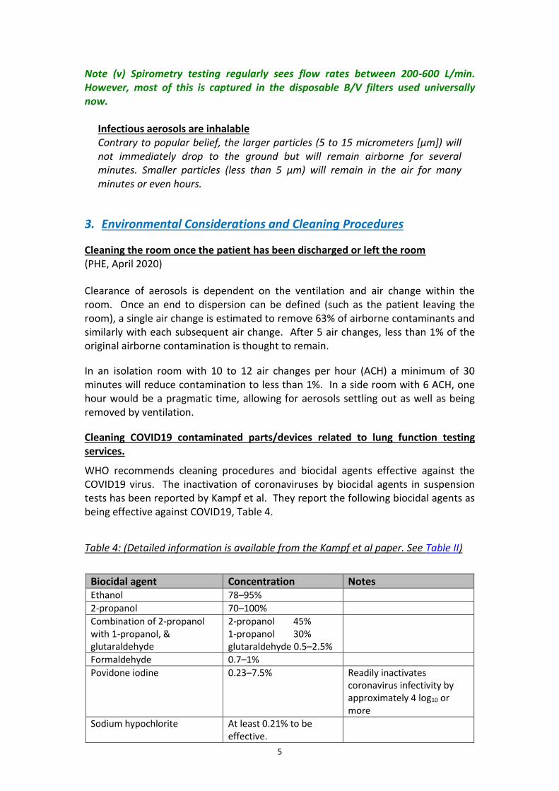
WHO recommends cleaning procedures and biocidal agents effective against the COVID19 virus. The inactivation of coronaviruses by biocidal agents in suspension tests has been reported by Kampf et al. They report the following biocidal agents as being effective against COVID19, Table 4.
Table 4: (Detailed information is available from the Kampf et al paper. See Table II)
Biocidal agent Concentration Notes Ethanol 78–95%
2-propanol 70–100%
Combination of 2-propanol with 1-propanol, & glutaraldehyde
2-propanol 45% 1-propanol 30% glutaraldehyde 0.5–2.5%
Formaldehyde 0.7–1%
Povidone iodine 0.23–7.5% Readily inactivates coronavirus infectivity by approximately 4 log10 or more
Sodium hypochlorite At least 0.21% to be effective.

6
Hydrogen peroxide 0.5% incubation time of 1 min.
Benzalkonium chloride 0.2% - 0.5% tested Uncertain results - reject
Chlorhexidine digluconate 0.02% Ineffective
A solution of 1:100 of 5% sodium hypochlorite results in a final concentration of 0.05%. Data with coronaviruses suggests that a concentration of 0.1% is effective in 1 minute. For the disinfection of small surfaces ethanol (62–71%; carrier tests) revealed a similar efficacy against coronavirus. A concentration of 70% ethanol is also recommended by the WHO for disinfecting small surfaces. Practically this translates to the following guidance for the lung function department:
a. Detergent/soap and water Soap and water are your first line of defence to remove the virus from surfaces. Soap interferes with the fats in the virus shell and lift the virus from surfaces and this is then rinsed off by water. (Of course, you also need to wash your hands when you come in from the shops and wash your food as normal.)
b. Bleach
The active ingredient in bleach, sodium hypochlorite, is very effective (Kampf et al, 2020) at killing the virus. There is a need to leave the bleach to work for 10-15 minutes, then give the surface a wipe with a clean cloth. The bleach works by destroying the ribonucleic acid (RNA) of the virus – which gives the blueprint for making more virus particles when a subject becomes infected.
c. Alcohol based cleaning fluids
Surgical spirit is mostly made up of ethanol which has been shown to kill coronaviruses in as little as 30 seconds. Like bleach, the alcohol destroys the protein and RNA that the virus is made up of.
4. Post COVID19 Lung Function
Since COVID19 is a novel virus, the only similar experience that healthcare systems have had with this is based around the SARS cases in 2010. However, it appears that the lung function changes from COVID19 may be more severe than SARS, so the likelihood of follow up lung function seems greater. Dr James Gill, Locum GP & Honorary Clinical Lecturer, Warwick Medical School Looking at the SARS cases recovery: “27.8% of patients demonstrated persistent changes on chest X-rays 12 months after their recovery. Whilst post-infection lung function was within normal range for those patients, they also demonstrated reduced exercise tolerances ” ( H u i , 2 0 1 0 ) . That finding of normal lung function testing post S.A.R.S. recovery is important, as it has not been possible to draw a convincing connection between pulmonary function and post infection reduced exercise capacity (Su, 2007).
7
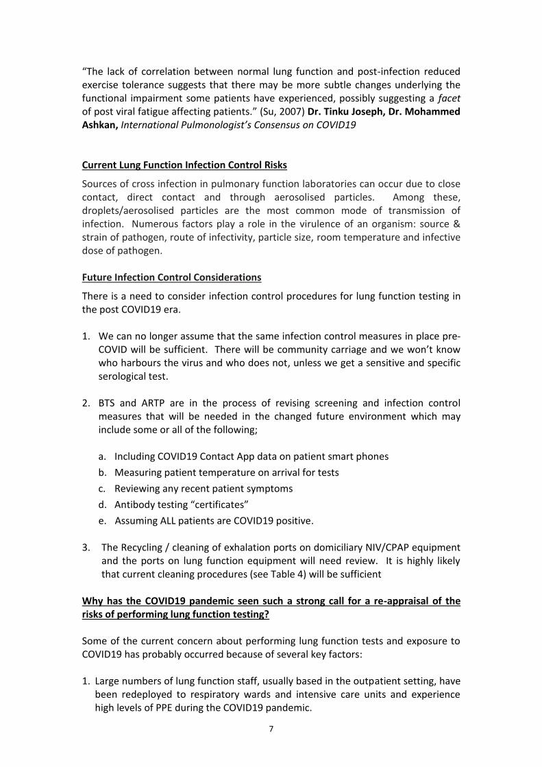
“The lack of correlation between normal lung function and post-infection reduced exercise tolerance suggests that there may be more subtle changes underlying the functional impairment some patients have experienced, possibly suggesting a facet of post viral fatigue affecting patients.” (Su, 2007) Dr. Tinku Joseph, Dr. Mohammed Ashkan, International Pulmonologist’s Consensus on COVID19 Current Lung Function Infection Control Risks
Sources of cross infection in pulmonary function laboratories can occur due to close contact, direct contact and through aerosolised particles. Among these, droplets/aerosolised particles are the most common mode of transmission of infection. Numerous factors play a role in the virulence of an organism: source & strain of pathogen, route of infectivity, particle size, room temperature and infective dose of pathogen. Future Infection Control Considerations
There is a need to consider infection control procedures for lung function testing in the post COVID19 era. 1. We can no longer assume that the same infection control measures in place pre-
COVID will be sufficient. There will be community carriage and we won’t know who harbours the virus and who does not, unless we get a sensitive and specific serological test.
2. BTS and ARTP are in the process of revising screening and infection control
measures that will be needed in the changed future environment which may include some or all of the following;
a. Including COVID19 Contact App data on patient smart phones
b. Measuring patient temperature on arrival for tests
c. Reviewing any recent patient symptoms
d. Antibody testing “certificates”
e. Assuming ALL patients are COVID19 positive. 3. The Recycling / cleaning of exhalation ports on domiciliary NIV/CPAP equipment
and the ports on lung function equipment will need review. It is highly likely that current cleaning procedures (see Table 4) will be sufficient
Why has the COVID19 pandemic seen such a strong call for a re-appraisal of the risks of performing lung function testing? Some of the current concern about performing lung function tests and exposure to COVID19 has probably occurred because of several key factors: 1. Large numbers of lung function staff, usually based in the outpatient setting, have
been redeployed to respiratory wards and intensive care units and experience high levels of PPE during the COVID19 pandemic.

8
2. Respiratory physiologists are not used to the uncertainty that is accepted in emergency medicine research, which by its nature has to be rapid, often uncontrolled and uses small samples.
3. As a result of 1 and 2 above, the perception of COVID19 with death, dying and infection spread is altered in respiratory physiologists.
4. Previously lung function staff have taken usual SICPs for testing patients with
common infections such as influenza and tuberculosis where the risk of serious infection is reduced by staff vaccination or medication (e.g. triple therapy) respectively. Never have staff had to knowingly face an infection that can lead to unprecedented numbers of healthcare workers being admitted to intensive care for life-threatening conditions.
5. Respiratory physiologists may not be good at distinguishing between possible and
likely, as they are used to large data series with complex study designs. Sharing of professional expertise and some joint research should help alleviate some of these concerns in due course.
PHE Infection Control Guidance (27th April 2020) The most recent guidance for infection control and COVID19 states that in the following locations;
(a) Working in a higher risk acute care area with possible or confirmed case(s) where higher risk acute areas include: ICU/HDUs; ED resuscitation areas; wards with non-invasive ventilation; operating theatres; endoscopy units for upper Respiratory, ENT or upper GI endoscopy; and other clinical areas where AGPs are regularly performed.
Filtering face piece respirator
(b) Working in an inpatient, maternity, radiology area with possible or confirmed case(s) – direct patient care (within 2 metres)
A fluid-resistant (Type IIR) surgical mask
The following should always be used in both these locations;
Disposable fluid-resistant coverall/gown
Disposable Plastic Apron
Disposable Gloves
Eye/face protection The key issue for Lung Function testing is whether the maximal respiratory manoeuvres exhaling forcibly from total lung capacity (TLC) to residual volume (RV) generates aerosols either during the test or if the patient starts coughing after the test. There has been no published research evidence on the actual risks of generating aerosols in these tests and possible spread of COVID19 to the operators.
9
For reference, the ATS/ERS 2005 guidance for infection control is attached (Appendix 1). This may be used together with this ARTP document to help clarify concerns lung function staff may have about PPE and testing on patients with proven or actual COVID19 infection to Infection Control teams. In absence of any irrefutable evidence that lung function testing is or isn’t an aerosol generating procedure, ARTP COVID19 Group cannot make a definitive recommendation either way, but suggest that Lung Function departments discuss this issue with their local Infection Prevention Control teams.
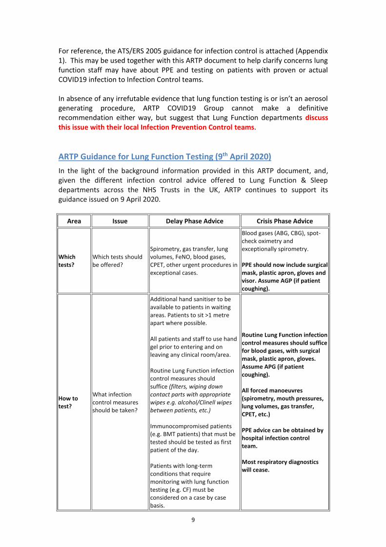
ARTP Guidance for Lung Function Testing (9th April 2020)
In the light of the background information provided in this ARTP document, and, given the different infection control advice offered to Lung Function & Sleep departments across the NHS Trusts in the UK, ARTP continues to support its guidance issued on 9 April 2020.
Area Issue Delay Phase Advice Crisis Phase Advice
Which tests?
Which tests should be offered?
Spirometry, gas transfer, lung volumes, FeNO, blood gases, CPET, other urgent procedures in exceptional cases.
Blood gases (ABG, CBG), spot-check oximetry and exceptionally spirometry. PPE should now include surgical mask, plastic apron, gloves and visor. Assume AGP (if patient coughing).
How to test?
What infection control measures should be taken?
Additional hand sanitiser to be available to patients in waiting areas. Patients to sit >1 metre apart where possible. All patients and staff to use hand gel prior to entering and on leaving any clinical room/area. Routine Lung Function infection control measures should suffice (filters, wiping down contact parts with appropriate wipes e.g. alcohol/Clinell wipes between patients, etc.) Immunocompromised patients (e.g. BMT patients) that must be tested should be tested as first patient of the day. Patients with long-term conditions that require monitoring with lung function testing (e.g. CF) must be considered on a case by case basis.
Routine Lung Function infection control measures should suffice for blood gases, with surgical mask, plastic apron, gloves. Assume APG (if patient coughing). All forced manoeuvres (spirometry, mouth pressures, lung volumes, gas transfer, CPET, etc.) PPE advice can be obtained by hospital infection control team. Most respiratory diagnostics will cease.

10
APPENDIX 1: ATS/ERS Guidelines (2005) This is copied from the ATS/ERS lung function standards (without permission) but is widely available (Brusasco, 2005) HYGIENE AND INFECTION CONTROL The goal of infection control is to prevent the transmission of infection to patients/subjects and staff during pulmonary function testing. The number of documented cases of infection transmission is very small (Refs), but the potential is real (see Level of infection risk section). This set of recommendations focuses on equipment used to measure spirometry, diffusing capacity and lung volumes. Organisms may also be transmitted via pulse oximeter probes and nebulisers used to administer bronchodilators (Botman, 1987; Dautzenburg, 2001. Although infection risks increase with exposure to blood, this document does not deal with the risks of arterial blood gases. Pulmonary laboratories performing blood gas analysis should follow the same infection-control procedures used by their clinical laboratory. Infection can be transmitted by direct contact or by indirect means, which is discussed as follows: Transmission by direct contact There is potential for transmission of upper respiratory diseases, enteric infections and blood-borne infections through direct contact. Although hepatitis and HIV contagion are unlikely via saliva, transmission becomes a possibility with open sores on the oral mucosa or bleeding gums. The most likely surfaces for contact are mouthpieces and the immediate proximal surfaces of valves or tubing. Transmission by indirect contact There is potential for transmission of tuberculosis (TB), various viral infections, opportunistic infections and nosocomial pneumonia through aerosol droplets. The most likely surfaces for possible contamination by this route are mouthpieces, proximal valves and tubing. Prevention
Transmission to operators
Prevention of infection transmission to operators exposed to contaminated spirometer surfaces can be accomplished through proper hand washing and use of barrier devices, such as suitable gloves. To avoid operator exposure and cross-contamination, hands should be washed immediately after direct handling of mouthpieces, tubing, breathing valves or interior spirometer surfaces. Gloves should be worn when handling potentially contaminated equipment if the operator has any open cuts or sores on his/her hands. Hands should always be washed between patients. Indications and techniques for hand washing during pulmonary function testing have previously been reviewed (Denison, 1989).
Cross-contamination
To avoid cross-contamination, reusable mouthpieces, breathing tubes, valves and manifolds should be disinfected or sterilised regularly. Mouthpieces, nose clips
11
and any other equipment that comes into direct contact with mucosal surfaces should be disinfected, sterilised or, if disposable, discarded after each use. The optimal frequency for disinfection or sterilisation of tubing, valves or manifolds has not been established. However, any equipment surface showing visible condensation from expired air should be disinfected or sterilised before reuse. Since the use of cold sterilising agents is not without risk, laboratory staff should take care to follow the manufacturer's recommendations concerning proper handling of these products. Some respiratory equipment may be damaged by some methods of sterilisation. For example, heat sterilisation or cold sterilisation chemicals could damage some flow sensors, tubing or seals. Manufacturers should explicitly describe acceptable methods of cleaning and disinfecting their equipment, including recommended chemicals and concentrations, as well as safety precautions for the operators. Manufacturers' recommendations should be followed; however, a hospital infection control department’s requirements will probably supersede both manufacturers' recommendations and those in this document. If hospital infection control recommendations have the potential to harm instruments, compromises may have to be negotiated. Volume-based spirometers
Volume-based spirometers used with a closed-circuit technique should be flushed between subjects with room air at least five times over the entire volume range of the spirometer to enhance clearance of droplet nuclei. The breathing tube and mouthpiece should be decontaminated or changed between patients. When the open-circuit technique is used and the patient/subject only exhales into the spirometer AND the room, only the portion of the circuit through which rebreathing occurs must be decontaminated between patients. For example, when a pneumotachometer system is used, either avoid having the patient inspire through the device, or decontaminate or replace the resistive element and tubing between subjects. Alternatively, a disposable sensor may be used. Disposable sensors, when appropriately used, avoid the need for decontamination of sensors and mouthpieces (see Disposable in-line filters section). When an open-circuit technique (either volume or flow spirometers) is used without inspiration from the measuring system, only the mouthpiece would need to be changed or decontaminated between subjects. However, it is difficult, if not impossible, to assure that patients do not inhale through the device. A low-resistance one-way valve may be used to prevent inhalation, and, if used, must be demonstrated not to alter the spirometric measurements. Not having patients inspire through the device may make it difficult to assess test quality because of the absence of an inspiratory tracing. Hence, this technique should be used with caution. Disassembling, cleaning and/or sensor replacement will usually require recalibration of the spirometer.

12
Tuberculosis
In settings where TB or other diseases that are spread by droplet nuclei are likely to be encountered, proper attention to environmental engineering controls, such as ventilation, air filtration or ultraviolet decontamination of air, should be used to prevent disease transmission. Haemoptysis and oral lesions
Special precautions should be taken when testing patients with haemoptysis, open sores on the oral mucosa or bleeding gums. Tubing and breathing valves should be decontaminated before reuse, and internal spirometer surfaces should be decontaminated with accepted disinfectants for blood-transmissible agents. Other known transmissible infectious diseases
Extra precautions should be taken for patients with known transmissible infectious diseases. Possible precautions include the following:
1) Reserving equipment for the sole purpose of testing infected patients
2) Testing such patients at the end of the day to allow time for spirometer disassembly and disinfection
and
3) Testing patients in their own rooms with adequate ventilation and appropriate protection for the operator.
Disposable in-line filters
These may be an effective and less expensive method of preventing equipment contamination. The influence of commercially available in-line filters on forced expiratory measures, such as forced vital capacity (FVC) and forced expiratory volume in one second (FEV1) has not been well characterised. A low-impedance barrier device was found not to have a significant effect on FVC and FEV1 (Johns, 1995), whereas a barrier filter has been shown to cause small but significant reductions in FEV1 (-44 mL) and peak expiratory flow (PEF; -0.47 L·s−1), but did not appear to affect DL,CO, alveolar volume or TLC (Fuso, 1995). Although significant differences between measurements with and without filters have been demonstrated for FVC, FEV1, airway resistance and specific airway conductance (sGaw) (Side, 1999), these differences were unrelated to the average values of the measurements (except for sGaw), and the limits of agreement were within the range of intra-individual short-term repeatability for almost all of the function indices. Thus, the effect of a filter with optimal characteristics is not considered to be clinically significant, and no appreciable classification error was found in diagnostic tests. If in-line filters are used, the measuring system should meet the minimum recommendations for accuracy, precision (reproducibility), flow resistance and back pressure with the filter installed. Airflow resistance must be measured with in-line filters in place if that is how patients are tested. Manufacturers of in-line filters should provide evidence that their filter does not alter standard lung
13
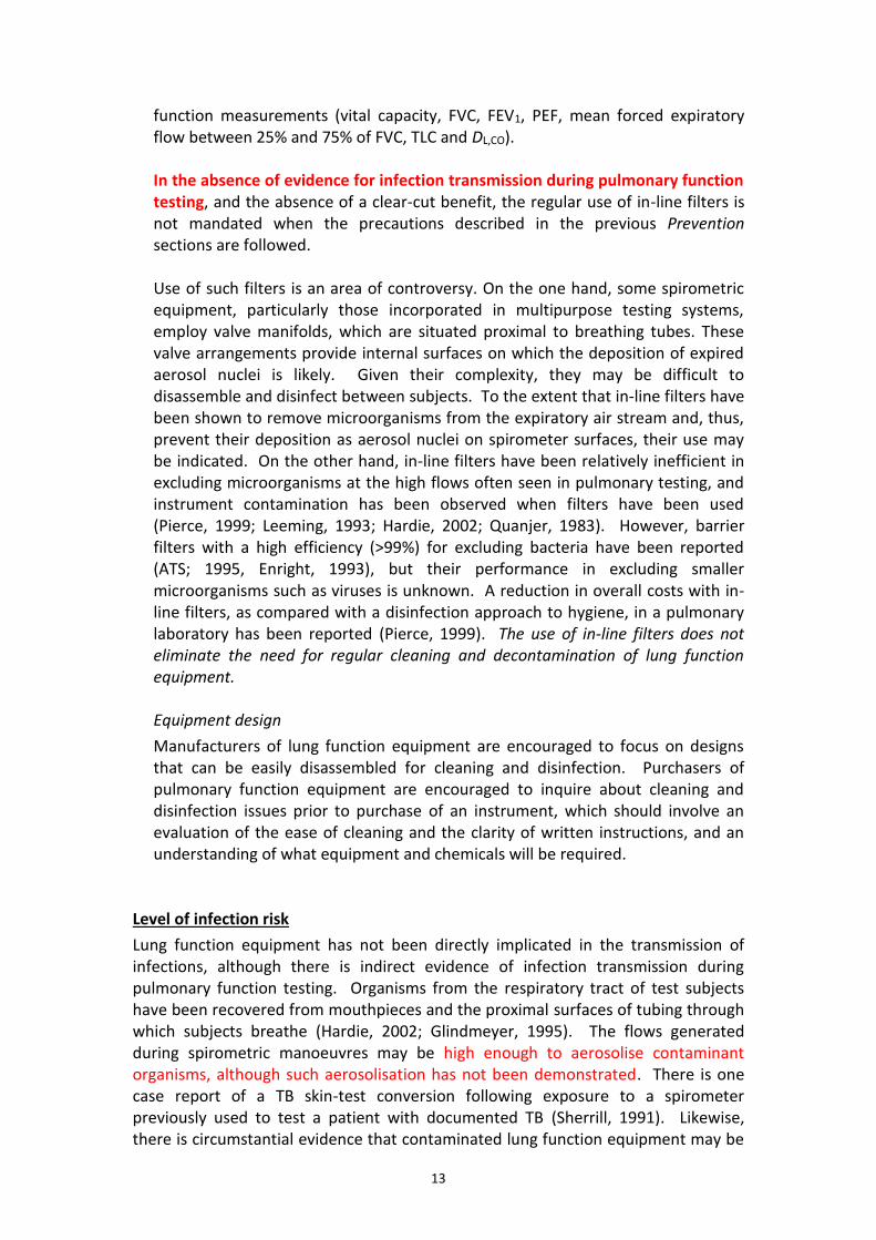
function measurements (vital capacity, FVC, FEV1, PEF, mean forced expiratory flow between 25% and 75% of FVC, TLC and DL,CO). In the absence of evidence for infection transmission during pulmonary function testing, and the absence of a clear-cut benefit, the regular use of in-line filters is not mandated when the precautions described in the previous Prevention sections are followed. Use of such filters is an area of controversy. On the one hand, some spirometric equipment, particularly those incorporated in multipurpose testing systems, employ valve manifolds, which are situated proximal to breathing tubes. These valve arrangements provide internal surfaces on which the deposition of expired aerosol nuclei is likely. Given their complexity, they may be difficult to disassemble and disinfect between subjects. To the extent that in-line filters have been shown to remove microorganisms from the expiratory air stream and, thus, prevent their deposition as aerosol nuclei on spirometer surfaces, their use may be indicated. On the other hand, in-line filters have been relatively inefficient in excluding microorganisms at the high flows often seen in pulmonary testing, and instrument contamination has been observed when filters have been used
(Pierce, 1999; Leeming, 1993; Hardie, 2002; Quanjer, 1983). However, barrier filters with a high efficiency (>99%) for excluding bacteria have been reported (ATS; 1995, Enright, 1993), but their performance in excluding smaller microorganisms such as viruses is unknown. A reduction in overall costs with in-line filters, as compared with a disinfection approach to hygiene, in a pulmonary laboratory has been reported (Pierce, 1999). The use of in-line filters does not eliminate the need for regular cleaning and decontamination of lung function equipment. Equipment design
Manufacturers of lung function equipment are encouraged to focus on designs that can be easily disassembled for cleaning and disinfection. Purchasers of pulmonary function equipment are encouraged to inquire about cleaning and disinfection issues prior to purchase of an instrument, which should involve an evaluation of the ease of cleaning and the clarity of written instructions, and an understanding of what equipment and chemicals will be required.
Level of infection risk
Lung function equipment has not been directly implicated in the transmission of infections, although there is indirect evidence of infection transmission during pulmonary function testing. Organisms from the respiratory tract of test subjects have been recovered from mouthpieces and the proximal surfaces of tubing through which subjects breathe (Hardie, 2002; Glindmeyer, 1995). The flows generated during spirometric manoeuvres may be high enough to aerosolise contaminant organisms, although such aerosolisation has not been demonstrated. There is one case report of a TB skin-test conversion following exposure to a spirometer previously used to test a patient with documented TB (Sherrill, 1991). Likewise, there is circumstantial evidence that contaminated lung function equipment may be

14
implicated in increasing the prevalence of Burkholderia cepacia infections among cystic fibrosis patients at one centre (Burrow, 1986). There is evidence that pneumotachometer-based systems are less susceptible to bacterial contamination than water-sealed spirometers (Ware, 1990). In addition, it is well documented that community water supplies can be contaminated with Mycobacteria spp. and Pseudomonas aeruginosa organisms (Sherrill, 1992; Pistelli, 2000; Wang, 1993). Thus, there is a potential for both patients/subjects and healthcare workers to deposit microorganisms onto spirometer surfaces (including mouthpieces, nose clips, tubing and any internal or external machine surface), which could subsequently come into direct or indirect contact with other patients or healthcare workers. This does not pose an appreciable threat to patients/subjects/workers with competent immune systems. It has been argued that immunocompromised patients may require only a relatively small infective dose of either opportunistic organisms or common pathogens for infection to occur. However, there is no direct evidence that routine pulmonary function testing poses an increased risk of infection to immunocompromised patients. Concerns for the protection of immunocompromised patients, along with increased public and provider awareness of hospital infection-control issues since the 1990s, has led many laboratory directors to routinely use in-line filters to reassure patients and laboratory personnel that their protection has been considered.
15
References American Thoracic Society. Standardization of Spirometry: 1994 update. Am J Respir Crit Care Med 1995; 152: 1107–1136. Botman MJ, de Krieger RA. Contamination of small volume medication nebulizers and its association with oropharyngeal colonization. J Hosp Infect 1987;10:204–208.
Brusasco, V. Crapo, R. Viegi. G. ATS/ERS TASK FORCE: STANDARDISATION OF LUNGFUNCTION TESTING: General considerations for lung function testing. Eur Respir J 2005; 26: 153–161 Burrows B, Lebowitz MD, Camilli AE, Knudson RJ. Longitudinal changes in forced expiratory volume in one second in adults. Methodologic considerations and findings in healthy nonsmokers. Am Rev Respir Dis 1986; 133: 974–980. Chen C, Zhao B. Some questions on dispersion of human exhaled droplets in ventilation room: answers from numerical investigation. Indoor Air. 2010;20(2):95–111. Dautzenberg B. Prevention of nosocomial infection during nebulization and spirometry. Rev Pneumol Clin 2001;57:91–98.
Denison DM, Cramer DS, Hanson PJV. Lung function testing and AIDS. Respir Med 1989;83:133–138.
Enright PL, Kronmal RA, Higgins M, Schenker M, Haponik EF. Spirometry reference values for women and men 65 to 85 years of age. Cardiovascular health study. Am Rev Respir Dis 1993; 147: 125–133.
Fuso L, Accardo D, Bevignani G, Ferrante E, Della Corte A, Pistelli R. Effects of a filter at the mouth on pulmonary function tests. Eur Respir J 1995;8:314–317.
Glindmeyer HW, Lefante JJ, McColloster C, Jones RN, Weill H. Blue-collar normative spirometric values for Caucasian and African-American men and women aged 18 to 65. Am J Respir Crit Care Med 1995; 151: 412–422. Hardie JA, Buist AS, Vollmer WM, Ellingsen I, Bakke PS, Morkve O. Risk of over-diagnosis of COPD in asymptomatic elderly never-smokers. Eur Respir J 2002; 20:1117–1122. Hui DSC. Chan PKS. Severe acute respiratory syndrome and coronavirus. Infect Dis Clin North Am 2010 Sep; 24(3): 619-638 Johns DP, Ingram C, Booth H, Williams TJ, Walters EH. Effect of a microaerosol barrier filter on the measurement of lung function. Chest 1995;107:1045–1048.
Judson SD, Munster VJ. Nosocomial Transmission of Emerging Viruses via Aerosol-Generating Medical Procedures. Viruses. 2019;11(10):940. Kampf, G. Todt, D. Pfaender, S. & Steinmann E. Persistence of coronaviruses on inanimate surfaces and their inactivation with biocidal agents. J Hosp Infect| 2020; 104 (3), 246-251, March 01, 2020 Leeming JP, Kendrick AH, Pryce-Roberts D, Smith D, Smith EC. Use of filters for the control of cross-infection during pulmonary function testing. J Hosp Infect 1993;23:245–246 Nicas M, Nazaroff WW, Hubbard A. Toward understanding the risk of secondary airborne infection: emission of respirable pathogens. J Occup Environ Hyg. 2005;2(3):143–154. Pierce RJ. Infection control in the respiratory laboratory: risk, costs, expediency. Aust N Z J Med 1999;29:3–4.
Pistelli F, Bottai M, Viegi G, et al. Smooth reference equations for slow vital capacity and flow-volume curve indexes. Am J Respir Crit Care Med 2000; 161: 899–905. Quanjer PH. Standardized Lung Function Testing. Bull Eur Physiopathol 1983; 19: Suppl. 5, 22–27. Sherrill DL, Lebowitz MD, Knudson RJ, Burrows B. Continuous longitudinal regression equations for pulmonary function measures. Eur Respir J 1992; 5:452–462. Sherrill DL, Lebowitz MD, Knudson RJ, Burrows B. Methodology for generating continuous prediction equations for pulmonary function measures. Comput Biomed Res 1991; 24: 249–260.

16
Side EA, Harrington G, Thien F, Walters EH, Johns DP. A cost-analysis of two approaches to infection control in a lung function laboratory. Aust N Z J Med 1999;29:9–14.
Su, MC. Hsieh, YT. Wang, YH. Lin, AS. Chung, YH. Lin, MC. Exercise capacity and pulmonary function in hospital workers recovered from severe acute respiratory syndrome. Respiration 2007; 74:511-516
Tabalan OC, Williams WW, Martone WJ. Infection control in pulmonary function laboratories. Infect Control 1985;6:442–444.
Tellier R. Aerosol transmission of influenza A virus: a review of new studies. J R Soc Interface. 2009;6 (Suppl 6): S783–S790. Wang X, Dockery DW, Wypij D, Fay ME, Ferris BG Jr. Pulmonary function between 6 and 18 years of age. Pediatr Pulmonol 1993; 15: 75–88. Ware JH, Dockery DW, Louis TA, Xu XP, Ferris BG Jr, Speizer FE. Longitudinal and cross-sectional estimates of pulmonary function decline in never-smoking adults. Am J Epidemiol 1990; 132: 685–700.
Xie X, Li Y, Chwang AT, Ho PL, Seto WH. How far droplets can move in indoor environments–revisiting the Wells evaporation-falling curve. Indoor Air. 2007;17(3):211–225.