COVID-19 Guidelines: Critical Care
21
COVID-19 Guidelines: Critical Care 2020. 3. 2. The Korean Society of Critical Care Medicine The Korean Academy of Tuberculosis and Respiratory diseases The Korean Society of Infectious Diseases The Korean Society for Antimicrobial Therapy Translation and Editing by Africa Future Foundation And Korean Global Health Forum in the United Kingdom
Transcript of COVID-19 Guidelines: Critical Care

COVID-19 Guidelines: Critical Care
2020. 3. 2.
The Korean Society of Critical Care Medicine
The Korean Academy of Tuberculosis and Respiratory diseases
The Korean Society of Infectious Diseases
The Korean Society for Antimicrobial Therapy
Translation and Editing by Africa Future Foundation And Korean Global Health Forum in the United Kingdom
1
1. Screening and Triage
1) COVID-19 can be complicated by mild respiratory tract infection and progress to severe
pneumonia, acute respiratory distress syndrome (ARDS), sepsis and septic shock.
* Refer to [Appendix 1] Clinical manifestations associated with COVID-19
2) Early identification of patients with severe symptoms allows for optimized treatments.
○ For high-risk patients with oxygen saturation (SpO2) below 90% on room air or
considered to apply supplemental oxygen, a further care plan should be developed
with a critical care team.
○ The early warning score (EWS) determined by vital signs can be used for the early
recognition of critically ill patients, even if patients hospitalized with COVID-19 present
mild symptoms, not requiring oxygen therapy.
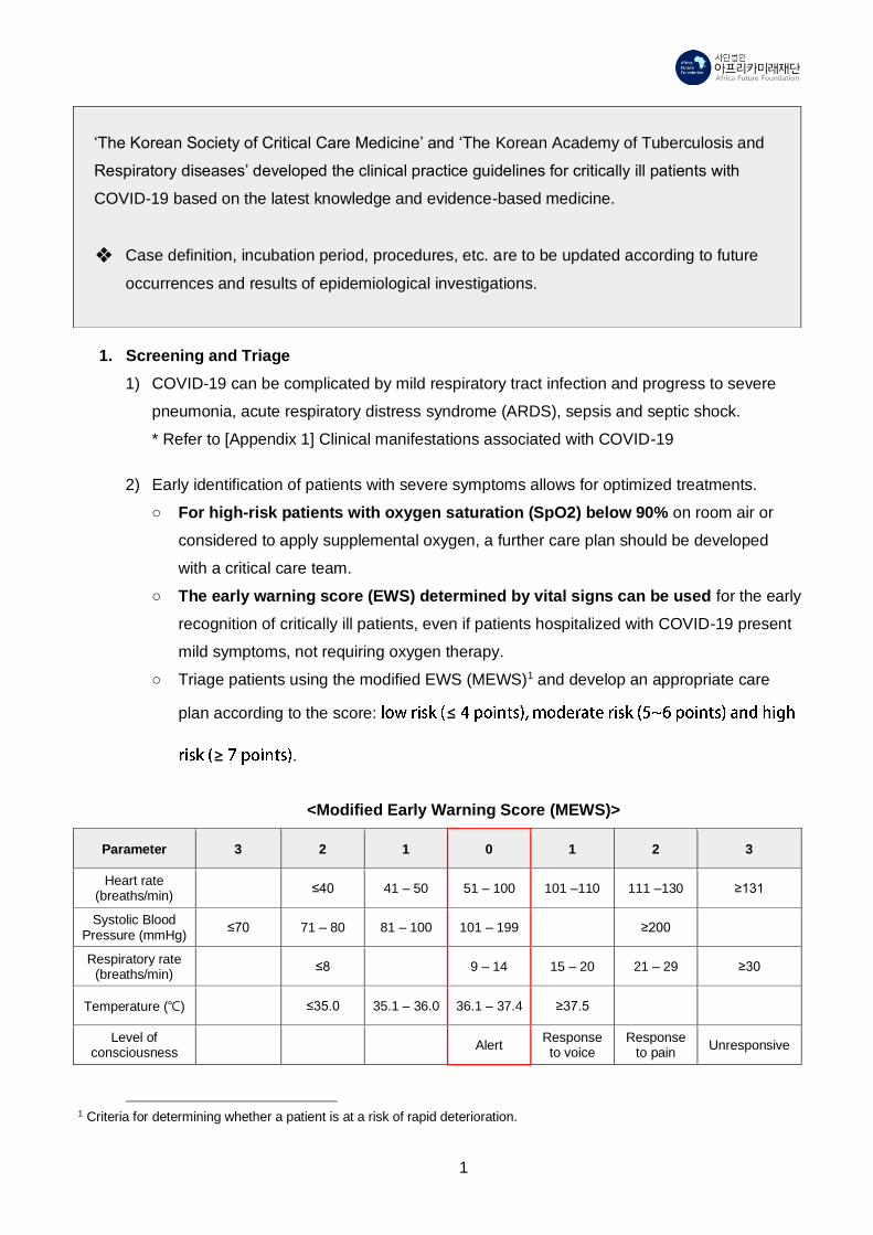
○ Triage patients using the modified EWS (MEWS)1 and develop an appropriate care
plan according to the score: ≤
≥ .
<Modified Early Warning Score (MEWS)>
Parameter 3 2 1 0 1 2 3
Heart rate (breaths/min)
≤40 41 – 50 51 – 100 101 –110 111 –130 ≥131
Systolic Blood Pressure (mmHg)
≤70 71 – 80 81 – 100 101 – 199 ≥200
Respiratory rate (breaths/min)
≤8 9 – 14 15 – 20 21 – 29 ≥30
Temperature (℃) ≤35.0 35.1 – 36.0 36.1 – 37.4 ≥37.5
Level of consciousness
Alert Response to voice
Response to pain
Unresponsive
1 Criteria for determining whether a patient is at a risk of rapid deterioration.
‘The Korean Society of Critical Care Medicine’ and ‘The Korean Academy of Tuberculosis and
Respiratory diseases’ developed the clinical practice guidelines for critically ill patients with
COVID-19 based on the latest knowledge and evidence-based medicine.
❖ Case definition, incubation period, procedures, etc. are to be updated according to future
occurrences and results of epidemiological investigations.
2
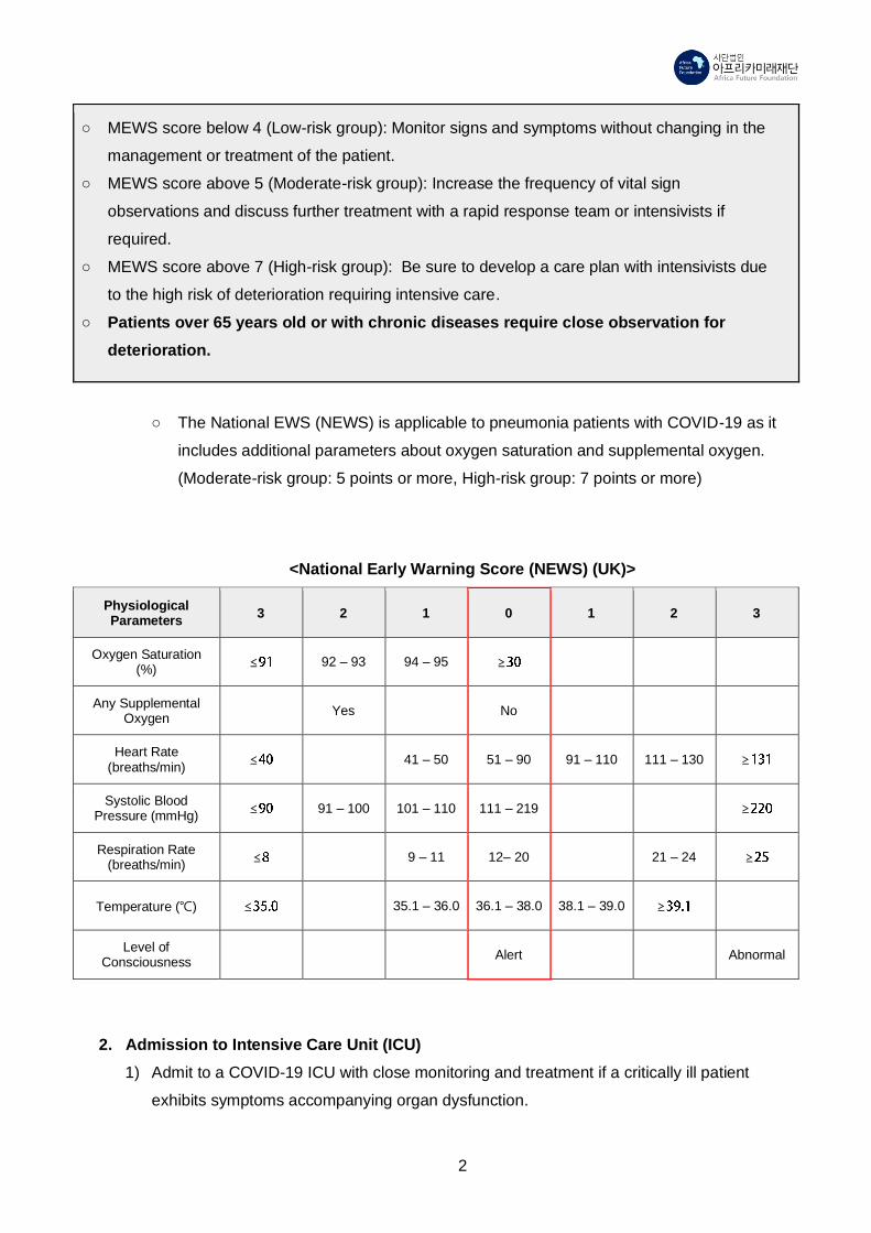
○ MEWS score below 4 (Low-risk group): Monitor signs and symptoms without changing in the
management or treatment of the patient.
○ MEWS score above 5 (Moderate-risk group): Increase the frequency of vital sign
observations and discuss further treatment with a rapid response team or intensivists if
required.
○ MEWS score above 7 (High-risk group): Be sure to develop a care plan with intensivists due
to the high risk of deterioration requiring intensive care.
○ Patients over 65 years old or with chronic diseases require close observation for
deterioration.
○ The National EWS (NEWS) is applicable to pneumonia patients with COVID-19 as it
includes additional parameters about oxygen saturation and supplemental oxygen.
(Moderate-risk group: 5 points or more, High-risk group: 7 points or more)
<National Early Warning Score (NEWS) (UK)>
Physiological Parameters
3 2 1 0 1 2 3
Oxygen Saturation (%)
≤ 92 – 93 94 – 95 ≥
Any Supplemental Oxygen
Yes No
Heart Rate (breaths/min)
≤ 41 – 50 51 – 90 91 – 110 111 – 130 ≥
Systolic Blood Pressure (mmHg)
≤ 91 – 100 101 – 110 111 – 219 ≥
Respiration Rate (breaths/min)
≤ 9 – 11 12– 20 21 – 24 ≥
Temperature (℃) ≤ 35.1 – 36.0 36.1 – 38.0 38.1 – 39.0 ≥
Level of Consciousness
Alert Abnormal
2. Admission to Intensive Care Unit (ICU)
1) Admit to a COVID-19 ICU with close monitoring and treatment if a critically ill patient
exhibits symptoms accompanying organ dysfunction.

3
○ Severe pneumonia
– Respiratory rate (RR) over 30 breaths/min
– Severe dyspnea
– SpO2 less than 90 % on room air
○ Acute Respiratory Distress Syndrome (ARDS)
○ Sepsis or septic shock
2) General disease progression of critically ill patients with COVID-19
○ Approximately 15 ~ 20% of total patients with COVID 19 are critically ill.
○ Approximately 5% of total patients with COVID-19 admit to an intensive care unit.
– The majority of critically ill patients (>90%) develop hypoxemic respiratory distress:
70% out of them require mechanical ventilation.
– Shock occurs in approximately 30% of them.
– Acute kidney injury occurs in approximately 10~30% of them.
❖ The above analysis is only based on cases in Wuhan, China2. Generalization of the
result to other countries may be limited.
Week 1 Week 2
Ward
(HD 4)
Ward
(HD 5)
Ward
(HD 6)
Ward
(HD 7)
ICU
(HD 8)
ICU
(HD 9)
ICU
(HD 10)
ICU
(HD 11)
Risk of respiratory
deterioration such as
hypoxemia and dyspnea
Risk of
respiratory
failure
Risk of ARDS Risk of multiple
organ failure
Symptomatic
treatment
Symptomatic
treatment
Oxygen
therapy HFNC HFNC/MV MV MV (ECMO)
* ARDS, acute respiratory distress syndrome; HFNC, high-flow nasal cannula; MV, mechanical ventilation; ECMO,
extracorporeal membrane oxygenation; HD, Hospital day
2) ICU admission decision
○ The need, as well as the expected benefits and risks from an ICU admission, should be
discussed with patients and their caregivers (or legal representatives).
○ Planned and unplanned admission should be differentiated. It is recommended to
prepare a structured advance healthcare directive prior to admission regardless of the
2 Yang X, Yu Y, Xu J, Shu H, Xia J, Liu H, et al. Clinical course and outcomes of critically ill patients with SARS-CoV-2
pneumonia in Wuhan, China: a single-centered, retrospective, observational study. Lancet Respir Med. 2020.

4
admission decision.
○ For resource utilization, if the hospital is unable to provide appropriate critical care due
to the shortage of health workers and medical equipment, patients should be
transferred to other available hospitals through consultation with the Transfer Support
Team at the National Medical Center (☎ 1800-3323), after informing patients and
caregivers. Supplemental personnel and resources can be requested if the health
status of a patient is not likely to endure the transfer.
○ The intensivist prioritizes patients for ICU admission on the basis of objective criteria
using a severity-of-disease classification system or performing triage.
3) Management of nursing staff
○ The recommended nurse-to-patient ratio is 2:1 per duty; types of work shifts can be
adjusted according to the context of the hospital.
○ It is required that the second nurse assists the supplement and administration of
procedure outside the room, and separate personnel monitors the donning and doffing
of PPE.
3. Treatment of Critically Ill Patients
* Refer to [Appendix 2] Treatment recommendation for critical cases with COVID-19.
1) Treatment of hypoxemic respiratory failure and acute respiratory distress syndrome
○ ECMO treatment should be conducted only in healthcare facilities that have a
multidisciplinary team specializing in ECMO and have experienced more than twenty
cases per year.
○ A multidisciplinary approach with objective indicators (e.g. Respiratory ECMO Survival
Prediction score; RESP score) is recommended for the final decision as ECMO is a
resource-intensive intervention.
Parameter Score Parameter Score
Age (year) Neuromuscular blockade agents before
ECMO 1
18 – 49 0 Nitric oxide before ECMO -1
50 – 59 -2 Bicarbonate infusion before ECMO -2
≥ 60 -3 Cardiac arrest before ECMO -2
Immunocompromised status* -2 PaCO2, mmHg
Mechanical ventilation < 75 0

5
prior to initiation of ECMO ≥ 75 -1
< 48 hours 3 Peak inspiratory pressure, cmH2O
48 hours – 7 days 1 < 42 0
> 7 days 0 ≥ 42 1
Acute Respiratory diagnosis group (select only one)
Total score -22 to 15
Viral pneumonia 3
Bacterial pneumonia 3
Asthma 11 Hospital Survival by Risk Class
Trauma/ burn 3 Total RESP score
Risk Class
Aspiration pneumonitis 5 ≥ 6 Ⅰ
Other acute respiratory diagnosis 1 3 to 5 Ⅱ
Non-respiratory and chronic respiratory diagnosis
0 -1 to 2 Ⅲ
Central nervous system dysfunction† -7 -5 to -2 Ⅳ
Acute associated (non-pulmonary) infection ‡
-3 ≤ -6 Ⅴ
An online calculator is available at www.respscore.com
* “immunocompromised” is defined as hematological malignancies, solid tumor, solid organ transplantation, human
immunodeficiency virus (HIV), and cirrhosis.
† “Central nervous system dysfunction” diagnosis combined neurotrauma, stroke, encephalopathy, cerebral
embolism, and seizure and epileptic syndrome.
‡ “Acute associated (non-pulmonary) infection” is defined as another bacterial, viral, parasitic, or fungal infection that
did not involve the lung.
❖ ECMO can be applied to patients with RESP score above -1, and it is not recommended for
patients with poor prognosis, RESP score below -6.
4. Infection Control in ICUs
Healthcare workers should take care of patients in an airborne infection isolation room. In
particular, all healthcare workers in the critical care team should carry out the following
guidelines regarding procedures in ICUs.
1) Personal Protective Equipment (PPE) of healthcare workers
○ Healthcare workers wear basic PPE (N95 Masks, goggles or face shields, disposable
waterproof long-sleeved gowns, gloves) for patient examination and treatment
○ When performing procedures likely to produce more concentrated aerosols (e.g.
intubation, bronchoscopy, cardiopulmonary resuscitation), healthcare workers should
wear a full-body protective suit and level D PPE (N95 Masks or PAPR, goggles or face

6
shields, a full-body protective suit, gloves, and a hat or hood).
○ The doctor responsible for the procedure and the assisting nurse are recommended to
wear a powered air-purifying respirator (PAPR) if available.
* Refer to “[Appendix 3] COVID-19 Practical Guidance for Healthcare Facilities (PPE)”
in this document.
2) Closed suctioning system must be applied for intubated patients and remain closed except
in case of an emergency such as acute airway obstruction.
3) Inhalation therapy (nebulizer) is prohibited except for patients with underlying medical
conditions such as chronic obstructive pulmonary disease (COPD) and asthma. Health
workers responsible for the therapy should wear a full-body protective suit in accordance
with the guideline for concentrated aerosol-generating procedures (level D PPE).
4) Level D PPE is not compulsory for inhalation therapies using a nebulizer directly attached
to the mechanical ventilation circuit, as there is no risk of exposure to aerosols.
5) Non-invasive mechanical ventilation is not recommended. Tracheostomy should be
performed only in patients who have clinically improved and received more than two
negatives for COVID-19. Also, the final decision for performing the procedure is left to the
medical team.
6) During the procedure, maintain the frequency of supply and exhaust ventilation to at least
6 to 12 times per hour (to the maximum frequency if possible).
7) During aerosol-generating procedures, follow the guidelines regarding infection control to
manage aerosols. The isolation room should be cleaned, disinfected, and evacuated for a
while, as the risk of exposure to aerosol is high when maximal ventilation is operated.
(Follow the guidelines for ventilation and disinfection.)
* Refer to “COVID-19 Guidelines: Hospital-level Healthcare Facilities (Edition 6).”
(KCDC,2020.2)
8) A full-body protective suit (level D PPE) is recommended for the personnel who need to
enter the isolation room.

7
Appendix 1 Clinical manifestations associated with COVID-19
The table below is excerpted from “Clinical management of severe acute respiratory infection (SARI) when COVID-19
disease is suspected: Interim guidance” published by WHO (January 2020).
Category Clinical signs and symptoms
Mild illness
• Patients with uncomplicated upper respiratory tract viral infection, may have
non-specific symptoms such as fever, cough, sore throat, nasal congestion,
malaise, headache, or muscle pain.
• The elderly and immunosuppressed may present with atypical symptoms.
These patients have no signs of dehydration, sepsis, or dyspnea.
Mild pneumonia • Patient with pneumonia but no signs of severe pneumonia
Severe
pneumonia
•
≤
Acute
respiratory
distress
syndrome
(ARDS)
• Onset: within 1 week of a known clinical insult or new or worsening
respiratory symptoms
• Chest imaging (radiograph, CT scan, or lung ultrasound): bilateral opacities,
not fully explained by volume overload, lobar or lung collapse, or nodules.
• Origin of pulmonary infiltrates: respiratory failure not fully explained by
cardiac failure or fluid overload. Need objective assessment (e.g.
echocardiography) to exclude hydrostatic cause of infiltrates/edema if no
risk factor present.
• Oxygenation impairment in adults:
· Mild ARDS: 200 mmHg ≤ 300 mmHg
(with PEEP or ≥ or non-ventilated)
· Moderate ARDS: 100 mmHg ≤200 mmHg
( ≥
· Severe ≤
≥
· ≤

8
Sepsis
• Life-threatening organ dysfunction caused by a dysregulated host response to
suspected or proven infection
• Signs of organ dysfunction include: altered mental status, difficult or fast
breathing, low oxygen saturation, reduced urine output, fast heart rate, weak
pulse, cold extremities or low blood pressure, skin mottling, or laboratory
evidence of coagulopathy, thrombocytopenia, acidosis, high lactate or
hyperbilirubinemia.
Septic Shock
• P
≥

9
Appendix 2 Treatment recommendation for critical cases with COVID-19
1. Management of Hypoxemic respiratory failure and acute respiratory distress syndrome
(ARDS)
○ Hypoxemic respiratory failure: Patients may continue to have increased work of breathing
or hypoxemia even when oxygen is delivered via a face mask with reservoir bag (flow rates
of 10–15 L/min, which is typically the minimum flow required to maintain bag inflation; FiO2
0.60–0.95)
○ Hypoxemic respiratory failure in ARDS commonly results from intrapulmonary ventilation-
perfusion mismatch or shunt and usually requires mechanical ventilation.
○ When a patient initiates mechanical ventilation, a critical care team led by intensivists must
be exclusively responsible for intensive care of critically ill patients and prepare a 24-hour-
staffed system in the intensive care unit.
○ Pneumonia patients who are suspected with COVID-19 should immediately receive
empirical antibiotics concerning the chance of general pneumonia. Empirical antibiotic
therapy should be modified based on microbiologic and clinical results.
○ High-flow nasal cannula (HFNC) or non-invasive ventilation (NIV) should only be used in
selected patients.
2) High-flow nasal cannula (HFNC)
○ HFNC can deliver 60L/min of gas flow and FiO2 100%. Patients with hypercapnia
(exacerbation of obstructive lung disease, cardiogenic pulmonary edema), hemodynamic
instability, multiorgan failure, or abnormal mental status should generally not receive HFNC,
although HFNC reduces the need for intubation compared with standard oxygen therapy.
Patients receiving HFNC should be in a monitored setting and cared for by experienced
personnel capable of endotracheal intubation.
○ As the unnecessary application of HFNC may delay intubation when to discontinue HFNC
should be decided based on the objective predictor (e.g. ROX index3).
○ If ROX index is measured less than 4.88 at 2 hours, 6 hours and 12 hours after HFNC
initiation, HFNC fails to ventilate so that it should be changed to mechanical ventilation
3) Non-invasive ventilation (NIV)
○ NIV guidelines make no recommendation on use in hypoxemic respiratory failure or
3 ROX index: the ratio of oxygen saturation as measured by pulse oximetry (SpO2)/FiO2 (%) to respiratory rate (RR):
[(SpO2, %)/FiO2, %) x100]/RR

10
pandemic viral illness
○ There is a risk of delayed intubation, large tidal volumes, and injurious transpulmonary
pressures.
○ Patients with hemodynamic instability, multiorgan failure, or abnormal mental status should
generally not receive NIV. Patients receiving NIV should be in a monitored setting and
cared for by experienced personnel capable of endotracheal intubation.
4) Conventional mechanical ventilation
○ Endotracheal intubation should be performed by experienced healthcare workers wearing
PPE for airborne precautions. Patients with ARDS, especially young children or those who
are obese or pregnant, may desaturate quickly during intubation. Pre-oxygenate with 100%
FiO2 for 5 minutes, and intubate rapidly after an airway assessment that identifies no signs
of difficult intubation.
○ Tidal volume should maintain below 6 ml/kg predicted body weight when applying a
ventilator. Tidal volume up to 8 mL/kg predicted body weight is allowed if undesirable side-
effects occur (e.g. dyssynchrony, pH < 7.15).
○ It is recommended to keep plateau pressure below 30 cmH₂O in the volume target mode
and driving pressure below 15cmH₂O without PEEP in the pressure target mode. The use
of deep sedation may be required to control patient-ventilator synchrony and achieve tidal
volume targets.
○ In addition to the lung-protective ventilation (LPV) above, the following interventions can be
applied.
– In patients with severe ARDS, prone position over 12 hours is recommended.
– Use a conservative fluid management strategy for ARDS patients without tissue
hypoperfusion. This is a strong recommendation; the main effect is to shorten the
duration of ventilation.
– In patients with moderate or severe ARDS, higher positive end expiratory pressure
(PEEP) instead of lower PEEP is suggested. PEEP titration requires consideration of
benefits (reducing atelectrauma and improving alveolar recruitment) vs risks (end-
inspiratory overdistension leading to lung injury and higher pulmonary vascular
resistance)
– Recruitment maneuvers (RMs) can be used to improve hypoxemia, and the benefits
and risks of RMs are similar to those of the increased PEEP.
– Monitoring of patients to identify those who respond to the initial application of higher
PEEP or a different RM protocol, and stopping these interventions in non-responders,
is suggested.

11
– In patients with moderate-severe ARDS (PaO2/FiO2 < 150), neuromuscular blockade
by continuous infusion should not be routinely used. The ACURASYS trial4 reported
that continuous infusion of neuromuscular blockade in severe ARDS was associated
with a reduction in mortality. According to the ROSE trial5 with a larger sample, high
PEEP and the use of neuromuscular blockade did not improve mortality compared to
the light sedation protocol without neuromuscular blockade. However, continuous
neuromuscular blockade may still be considered in patients with ARDS in certain
situations: ventilator dyssynchrony despite sedation, such that tidal volume limitation
cannot be reliably achieved; or refractory hypoxemia or hypercapnia.
– Extracorporeal membrane oxygenation (ECMO) is suggested for severe hypoxemia
which is unresponsive to the lung protective ventilation (LPV). A multicenter RCT of
ECMO (EOLIA trial6) found no statistically significant difference in the 60-day mortality
between veno-venous ECMO and standard medical management (including prone
positioning and neuromuscular blockade). However, ECMO was associated with a
reduced risk of the composite outcome of mortality and crossover to ECMO, and a post
hoc Bayesian analysis of this RCT showed that ECMO is very likely to reduce mortality
across a range of prior assumptions. In patients with MERS-CoV infection, ECMO vs
conventional treatment was associated with reduced mortality in a cohort study. (refer
to the RESP Score)
– Nitrogen oxides (NO) gas inhalation, use of systemic steroids, or high-frequency
ventilation are not recommended due to the lack of evidence for decreasing mortality.
– If mechanical ventilation is predicted to last more than a week, aggressive rehabilitation
should be provided to critically ill patients for early recovery and weaning of mechanical
ventilation. However, aggressive rehabilitation should start after testing negative more
than twice, and designated physiatrists and physical therapists must be arranged.
2. Early hemodynamic resuscitation in sepsis
1) Fluid therapy
○ In hemodynamic resuscitation of sepsis, give at least 30 mL/kg isotonic crystalloids in the
first 3 hours. Do not use hypotonic crystalloids, starches or gelatins for resuscitation.
○ Perfusion targets include urine output (> 0.5 mL/kg/hr), and improvement of skin mottling
and capillary refill, level of consciousness, and lactate. Consider dynamic indices of volume
4 L, Forel JM, Gacouin A, Penot-Ragon C, Perrin G, Loundou A, et al. Neuromuscular blockers in early acute respiratory
distress syndrome. N Engl J Med. 2010. 5 Moss M, Huang DT, Brower RG, Ferguson ND, Ginde AA, Gong MN, et al. Early neuromuscular blockade in the acute
respiratory distress syndrome. N Engl J Med. 2019. 6 Combes A, Hajage D, Capellier G, Demoule A, Lavoué S, Guervilly C, et al. Extracorporeal membrane oxygenation for
severe acute respiratory distress syndrome. N Engl J Med. 2018.

12
responsiveness to guide volume administration. These indices include passive leg raises,
stroke volume measurements after fluid challenges, or variations in systolic pressure, pulse
pressure or inferior vena cava size.
○ Fluid resuscitation may lead to volume overload, including respiratory failure. If there is no
response to fluid loading or signs of volume overload appear (e.g. jugular venous
distension, crackles on lung auscultation or pulmonary edema), then reduce or discontinue
fluid administration.
○ Colloids such as starch are associated with an increased risk of death and acute kidney
injury compared to crystalloids. The effects of gelatins are less clear, but they are more
expensive than crystalloids. Hypotonic solutions are less effective at increasing
intravascular volume. Surviving Sepsis also suggests albumin for resuscitation when
patients require substantial amounts of crystalloids, but this conditional recommendation is
based on low-quality evidence
2) Septic shock
○ Recognize septic shock when vasopressors are needed to maintain mean arterial pressure
(MAP) ≥ 65 mmHg AND lactate is ≥ 2 mmol/L, in the absence of hypovolemia.
○ In the absence of a lactate measurement, use blood pressure (i.e. MAP) and clinical signs
of perfusion to define shock.
○ Standard care includes early recognition of shock and the following treatments within 1 hour
of recognition: antimicrobial therapy, and initiation of fluid bolus and vasopressors for
hypotension
○ Other details follow the treatment guidelines for septic shock
3) Vasopressor
○ Administer vasopressors when shock persists during or after fluid resuscitation. The initial
blood pressure target is mean arterial blood pressure (MAP) ≥ 65 mmHg.
○ Vasopressors (i.e. norepinephrine, epinephrine, vasopressin, and dopamine) are most
safely given through a central venous catheter at a strictly controlled rate, but it is also
possible to safely administer them via peripheral vein and intraosseous needle.
○ Norepinephrine is considered first-line in adult patients; epinephrine or vasopressin can be
added to achieve the MAP target. Because of the risk of tachyarrhythmia, reserve
dopamine for selected patients with low risk of tachyarrhythmia or those with bradycardia.
○ If central venous catheters are not available, vasopressors can be given through a
peripheral IV, but use a large vein and closely monitor for signs of extravasation and local
tissue necrosis. If extravasation occurs, stop infusion.
○ Monitor blood pressure frequently and titrate the vasopressor to the minimum dose

13
necessary to maintain perfusion and prevent side-effects. If signs of poor perfusion and
cardiac dysfunction persist despite achieving MAP target with fluids and vasopressors,
consider an inotropic agent such as dobutamine.
4) Corticosteroids
○ Corticosteroids should be avoided unless indicated for another reason (e.g. COPD or septic
shock specified by 'Surviving Sepsis7') due to the potential downside of prolonged shedding
of coronavirus, as has been observed in patients with MERS-CoV.
○ Intravenous Injection of 200 mg hydrocortisone per day can be administered for septic
shock not reactive to appropriate fluid treatment and vasopressor. The continuous
administration is preferred to the repeated bolus injections.
3. Prevention of complications
Implement the following interventions to prevent complications associated with critical illness.
These interventions are based on ‘Surviving Sepsis’ or other guidelines and are generally limited
to feasible recommendations based on high-quality evidence.
7 Rhodes A, Evans LE, Alhazzani W, Levy MM, Antonelli M, Ferrer R et al. Surviving Sepsis Campaign: International
Guidelines for Management of Sepsis and Septic Shock: 2016. Intensive Care Med. 2017;43(3):304-77. Epub 2017/01/20.
doi: 10.1007/s00134-017-4683-6. PubMed PMID: 28101605.

14
<Prevention of complications>
Anticipated
outcome Interventions
Reduce days of
invasive mechanical
ventilation
• Use weaning protocols that include daily assessment for readiness to breathe
spontaneously.
• Minimize continuous or intermittent sedation, targeting specific titration endpoints
(light sedation unless contraindicated) or with daily interruption of continuous
sedative infusions.
Reduce incidence of
ventilator- associated
pneumonia
• Keep patient in semi-recumbent position (head of bed elevation 30–45º)
• Use a closed suctioning system; periodically drain and discard condensate in
tubing
• Use a new ventilator circuit for each patient; once a patient is ventilated, change
circuit if it is soiled or damaged, but not routinely.
• Change heat moisture exchanger when it malfunctions, when soiled, or every 5–
7 days
Reduce incidence of
venous
thromboembolism
• Use pharmacological prophylaxis (low molecular-weight heparin [preferred if
available] or heparin 5000 units subcutaneously twice daily) in adolescents and
adults without contraindications. For those with contraindications, use
mechanical prophylaxis (intermittent pneumatic compression devices).
Reduce incidence of
catheter-related
bloodstream infection
• Use a checklist with completion verified by a real-time observer as a reminder of
each step needed for sterile insertion and as a daily reminder to remove catheter
if no longer needed.
Reduce incidence of
pressure ulcers • Turn patient every 2 hours
Reduce incidence of
stress ulcers and
gastrointestinal
bleeding.
• Give early enteral nutrition (within 24–48 hours of admission)
• Administer histamine-2 receptor blockers or proton-pump inhibitors in patients
with risk factors for GI bleeding. Risk factors for gastrointestinal bleeding include
mechanical ventilation for ≥ 48 hours, coagulopathy, renal replacement therapy,
liver disease, multiple comorbidities, and higher organ failure score.
Reduce incidence of
ICU-related
weakness.
• Actively mobilize the patient early in the course of illness when safe to do so
4. Pharmacological treatment of COVID-19
Currently, there is no randomized controlled trial recommending anti-COVID-19 treatment for
suspected or confirmed patients. We provisionally follow the ‘(Korean) Expert Recommendations
for Pharmacological Treatment of COVID-19.’
15
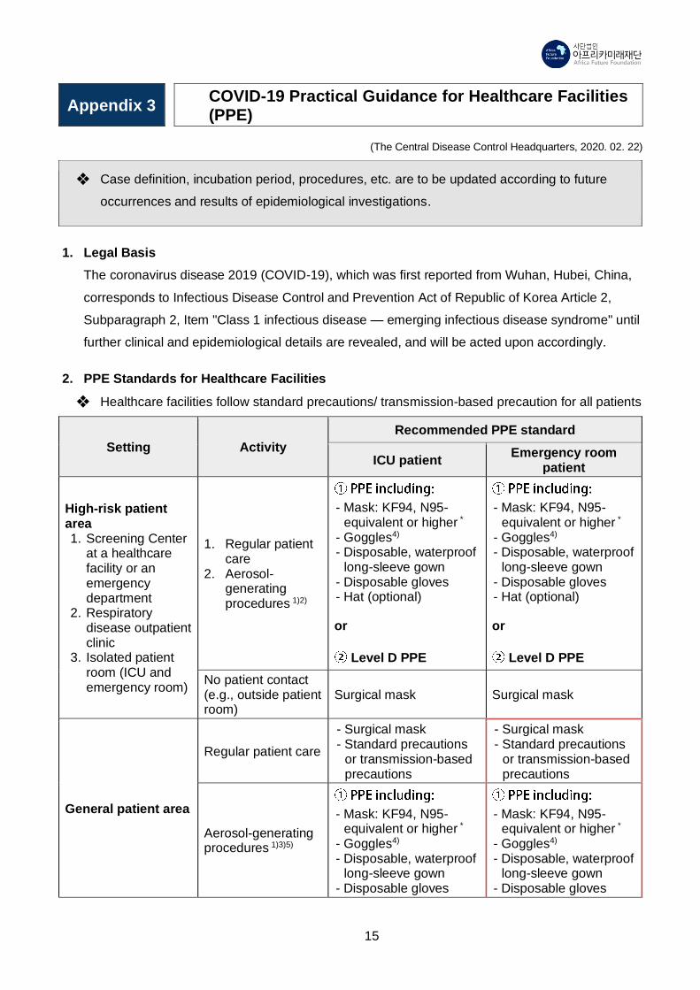
Appendix 3 COVID-19 Practical Guidance for Healthcare Facilities (PPE)
(The Central Disease Control Headquarters, 2020. 02. 22)
1. Legal Basis
The coronavirus disease 2019 (COVID-19), which was first reported from Wuhan, Hubei, China,
corresponds to Infectious Disease Control and Prevention Act of Republic of Korea Article 2,
Subparagraph 2, Item "Class 1 infectious disease — emerging infectious disease syndrome" until
further clinical and epidemiological details are revealed, and will be acted upon accordingly.
2. PPE Standards for Healthcare Facilities
❖ Healthcare facilities follow standard precautions/ transmission-based precaution for all patients
Setting Activity
Recommended PPE standard
ICU patient Emergency room
patient
High-risk patient area 1. Screening Center
at a healthcare facility or an emergency department
2. Respiratory disease outpatient clinic
3. Isolated patient room (ICU and emergency room)
1. Regular patient care
2. Aerosol- generating procedures 1)2)
- Mask: KF94, N95-equivalent or higher *
- Goggles4) - Disposable, waterproof
long-sleeve gown - Disposable gloves - Hat (optional)
or
Level D PPE
- Mask: KF94, N95-equivalent or higher *
- Goggles4) - Disposable, waterproof
long-sleeve gown - Disposable gloves - Hat (optional)
or
Level D PPE
No patient contact (e.g., outside patient room)
Surgical mask Surgical mask
General patient area
Regular patient care
- Surgical mask - Standard precautions
or transmission-based precautions
- Surgical mask - Standard precautions
or transmission-based precautions
Aerosol-generating procedures 1)3)5)
- Mask: KF94, N95-equivalent or higher *
- Goggles4) - Disposable, waterproof
long-sleeve gown - Disposable gloves
- Mask: KF94, N95-equivalent or higher *
- Goggles4) - Disposable, waterproof
long-sleeve gown - Disposable gloves
❖ Case definition, incubation period, procedures, etc. are to be updated according to future
occurrences and results of epidemiological investigations.

16
- Hat (optional)
or
Level D PPE
- Hat (optional)
or
Level D PPE
No patient contact Surgical mask Surgical mask
Area without patients No patient contact Surgical mask Surgical mask
* Including PAPR (Powered Air Purifying Respirator)
1) Aerosol-generating procedures
○ Aerosol-generating procedures which increase the transmission risk of respiratory
infectious disease are endotracheal intubation, cardiopulmonary resuscitation,
bronchoscopy, airway open suction (including tracheostomy care), autopsy and non-
invasive positive airway pressure (BiPAP and CPAP).
○ Acknowledging the controversy, limited evidence shows that procedures generating aerosol
include high-frequency oscillatory ventilation, nebulizer therapy, or induced sputum
examination.
○ Nasopharyngeal aspiration (NPA) and high-flow oxygen have a risk of infectious droplet
dispersion, so that, in high-risk patients’ area, it should be performed following the condition
for aerosol-generating procedures.
○ Other procedures should be evaluated by the personnel who is in charge of the nosocomial
infection control.
2) In high-risk patients' area, patients should be placed at an airborne infection isolation room
before performing aerosol-generating procedures.
3) In general patients' area, patients should be placed at a room with good air ventilation before
performing aerosol-generating procedures (e.g. at least 6 ventilation cycles per hour or use
portable HEPA filter (e.g. IQ Air)).
4) Eye should be protected by face shield or goggles.
5) Patients should be checked for considerations on an operation bed after preoperative
examination and sedation, and healthcare workers should follow the standard precautions/
transmission-based precaution when intubating for a planned operation.

17
Appendix 4 COVID-19 Guidelines: Hospital-level Healthcare Facilities (Edition 6)
Appendix 4 excerpted from “COVID-19 Guidelines: Hospital-level Healthcare Facilities (Edition 6)” published by The
Central Disease Control Headquarters
1. Donning and Doffing PPE
1) How to don (put on) PPE
○ Prepare all equipment according to the PPE recommendations per healthcare setting and
put on equipment in proper sequence and method.
2) How to doff (take off) PPE
○ Remove PPE at a place safe from pathogens (e.g. changing room outside isolation room)
and be careful not to contaminate body parts and surroundings.
○ Take caution not to contaminate surroundings while removing PPE, and do so in the proper
sequence and method; immediately discard them as healthcare waste.
Category
Sequence for N95 or equivalent
respiratory protection equipment and
coveralls
Sequence for PAPR and coveralls
Donning
order
1 Hand hygiene Hand hygiene
2 (Inner) Gloves (Inner) Gloves
3 Lower part of full-body protective suit Full-body protective suit
4 Shoe covers (or boots) Shoe covers (or boots)
5 N95 equivalent respiratory protection
equipment
Powered Air Purifying Respirator
(PAPR)8
6 Goggles (or face shield) Hood
7 Upper part of full-body protective suit and
tighten hood Connect PAPR and hood
8 (Outer) Gloves (Outer) Gloves
(Remove PPE outside of infectious areas such as isolation rooms)
Doffing9
order
1 (Outer) Gloves (Outer) Gloves
2 Glove disinfection Glove disinfection
8 Follow manufacturer instructions for putting on and taking off PAPR and tubing since it can be different for each product. 9 The inner glove can be contaminated while taking off the PPE. Therefore, it is advisable to sanitize the gloved hand after
removing each element of the PPE.

18
3 Full body protective suit Powered Air Purifying Respirator
(PAPR)
4 Shoe covers (or boots) Hood
5 Glove disinfection Full body protective suit
6 Goggles (or face shield) Shoe covers (or boots)
7 N95 equivalent respiratory protection
equipment (Inner) Gloves
8 (Inner) Gloves Hand hygiene
9 Hand hygiene -
2. COVID-19 PPE recommendation by situation
Situation
PPE
Respiratory protection Body protection Eye
protection
Surgical mask
KF94, N95-
equivalent
Electronic respirator
Disposable gloves10
Disposable waterproof
long-sleeved gown
Coveralls including
shoe covers
Goggle/ face shield
POE screening (epidemiological investigation)
● ● ● ●
Screening center: administrative staff
● ● ●
Screening center: clinical staff ● ● ● ●
Transport (ambulance driver)11 ● ●
Transport (quarantine officer, PHC personnel, EMT, etc.)
● ● ● ●
Ambulance disinfection ● ● ● ●
Suspected patient care: entering room, evaluation, nursing
● ● ● ●
Aerosol-inducing procedures12 ● ● ● ●
10 Double glove while examining, treating, nursing, testing, or cleaning around suspected or confirmed patients to mitigate
the risk of exposure from glove perforation. 11 If driving an ambulance without a barrier separating the driver seat from the patient compartment, wear a full-body suit,
shoe cover, KF94, N95-equivalent respiratory protection equipment, and gloves and wear goggles/face shield if necessary. 12 Aerosol-inducing procedures refer to endotracheal intubation, CPR, bronchoscopy, tracheostomy care, autopsy,
continuous positive airway pressure (CPAP) therapy, nebulizer therapy and other procedures for expectoration.
19
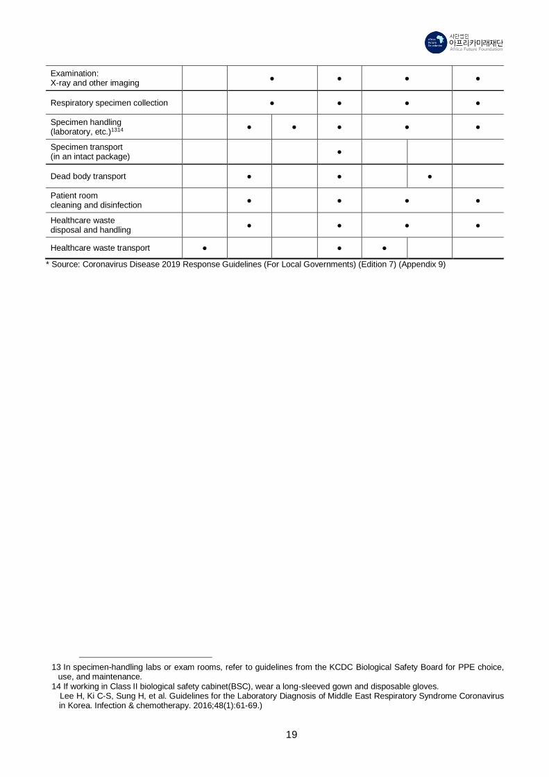
Examination: X-ray and other imaging
● ● ● ●
Respiratory specimen collection ● ● ● ●
Specimen handling (laboratory, etc.)1314
● ● ● ● ●
Specimen transport (in an intact package)
●
Dead body transport ● ● ●
Patient room cleaning and disinfection
● ● ● ●
Healthcare waste disposal and handling
● ● ● ●
Healthcare waste transport ● ● ●
* Source: Coronavirus Disease 2019 Response Guidelines (For Local Governments) (Edition 7) (Appendix 9)
13 In specimen-handling labs or exam rooms, refer to guidelines from the KCDC Biological Safety Board for PPE choice,
use, and maintenance. 14 If working in Class II biological safety cabinet(BSC), wear a long-sleeved gown and disposable gloves.
Lee H, Ki C-S, Sung H, et al. Guidelines for the Laboratory Diagnosis of Middle East Respiratory Syndrome Coronavirus in Korea. Infection & chemotherapy. 2016;48(1):61-69.)

20
Disclaimer
The English version is an unofficial translation of the original in Korean for information
purposes only. In case of a discrepancy, the Korean original will prevail.
The original document was developed by the Korea Centers for Disease Control and
Prevention and has been translated from Korean to English by the Africa Future
Foundation (AFF) in cooperation with the Korean Global Health Forum in the UK
(KGHF).
To maintain consistent terminology in related guidelines, this document used the
following source as reference for the glossary and overlapping contents:
“Coronavirus Disease 2019 Response Guidelines for Local Governments,” KCDC,
translated by the COVID Translate Project (www.covidtranslate.org), accessed 24 April
2020.
Reproduction of any content of this document is prohibited other than reproductions for
individual, non-commercial, and informational use. This limited permission to recopy
does not allow you to modify or incorporate any portion of the contents in any work or
publication, regardless of the medium.
Africa Future Foundation (AFF) [email protected] / https://africaff.org/
Korean Global Health Forum (KGHF) [email protected]