Coverings of the CNS 1) Bone – Cranium, Vertebrae 2) Meninges – Three connective tissue...
27
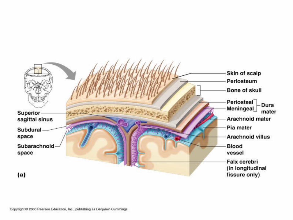
Coverings of the CNS • 1) Bone – Cranium, Vertebrae • 2) Meninges – Three connective tissue membranes covering the brain and spinal cord • a) Dura Mater – outermost , composed of tough fibrous connective tissue. Vascular. Attached to cranium but not to the vertebrae. Epidural Space exists between the vertebra and Dura Mater. Composed of fat. • b) Arachnoid Mater – middle layer. Thin, web-like layer • c) Pia Mater – innermost layer. Very thin, vascular. Clings to the surface of the brain. Aids in nourishing underlying brain cells
-
Upload
solomon-lewis -
Category
Documents
-
view
216 -
download
0
Transcript of Coverings of the CNS 1) Bone – Cranium, Vertebrae 2) Meninges – Three connective tissue...

Coverings of the CNS• 1) Bone – Cranium, Vertebrae• 2) Meninges – Three connective tissue membranes
covering the brain and spinal cord• a) Dura Mater – outermost , composed of tough
fibrous connective tissue. Vascular. Attached to cranium but not to the vertebrae. Epidural Space exists between the vertebra and Dura Mater. Composed of fat.
• b) Arachnoid Mater – middle layer. Thin, web-like layer
• c) Pia Mater – innermost layer. Very thin, vascular. Clings to the surface of the brain. Aids in nourishing underlying brain cells

• Subarachnoid Space – exists between the Arachnoid and Pia Mater. Filled with cerebrospinal fluid.

The Brain• Composed of 100 Billion Neurons – Divided
into 4 major regions
• 1) Cerebrum – largest region. Surface has elevated ridges called Gyri, separated by shallow grooves called Sulci and less numerous but deeper grooves called Fissures.
• Sulci and fissures divide the cerebrum into lobes named for the cranial bones above them.
• Divided into hemispheres by the Longitudinal Cerebral Fissure.
• Hemispheres connected by a bridge of nerve fibers called the Corpus Callosum

• Impulses cross over to the other side of the body in the brainstem. Impulses from the right side of the brain control muscles on the left side of the body.
• Hemisphere Dominance – both hemispheres participate in basic functions. In most people one side acts as a dominate hemisphere for other functions. 90% of people are left hemisphere dominant

• Within cerebral hemispheres and brainstem are interconnected cavities called Ventricles
• The ventricles are continuous with the central canal of the spinal cord and are filled with Cerebrospinal Fluid
• Cerebrospinal fluid (CSF) completely surrounds the brain and functions to:
• 1) support and protect
• 2) Maintain ion concentration of the CNS
• 3) Remove wastes


Functions of the Cerebrum
• 1) Interpret sensory impulses
• 2) Initiate voluntary muscle movement
• 3) Store Information
• 4) Reasoning
• 5) Personality, Intelligence

• 2) Diencephalon – Sits atop the brainstem and consists of 3 regions
• Thalamus – relay station for sensory impulses
• Hypothalamus – Plays a role in the regulation of body temperature, water balance and metabolism. Also thirst, appetite, pain and pleasure centers are in the hypothalamus.
• Epithalamus – tissues lining the epithalamus form cerebrospinal fluid

• 3) Brain Stem – Bundle of nerve tissue that connects the cerebrum to the spinal cord. It also has many areas of gray matter that controls vital activities. 3 Main Regions
• a) Midbrain – Center for auditory and visual reflexes
• b) Pons – Relay impulses from the medulla to the cerebrum
• c) Medulla Oblongata – Regulates blood pressure, heart rate, breathing and certain reflexes

• 4) Cerebellum – Acts as a control center in the coordination of skeletal muscle movements
Cranial Nerves• 12 pairs of nerves arise from the underside
of the brain that mostly serve the head and neck
• Numbered in order, front to back• Most are mixed nerves, but three are
sensory only• Olfactory – Sense of smell• Optic – Sense of Vision• Vestibulocochlear – Hearing and balance• Vagus – Sensations and movements of
Visceral organs.


Spinal Cord
• Extends from the foramen magnum to the disk between the 1st and 2nd Lumbar vertebrae
• It is surrounded and protected by the meninges
• The meninges extends below the end of the cord and provide for safe sampling of CSF below L3 (spinal tap)
• Spinal cord gives rise to 31 pairs of spinal nerves that exit the vertebrae and serve the body close by.

• Nerves exiting below the end of the cord travel through the vertebral canal and form the Cauda Equina (horse’s tail)
• Cross section of the cord reveals a core of gray matter surrounded by white matter. Pattern of gray matter resembles a butterfly
• Neurons of the gray matter are interneurons. Neurons in the white matter are nerve tracts
• Central canal filled with Cerebrospinal Fluid
• Divided into right and left halves much like the brain

Spinal Cord Functions
• 1) Reflex Center (gray matter)
• 2) Conduct impulses to and from the brain (white matter)

Peripheral Nervous System
• Nerves that branch out of the CNS to different parts of the body
• 2 Divisions – Somatic and Autonomic• Somatic Nerves – Nerves that lead to the
skin and skeletal muscles involved in conscious activities
• Autonomic Nerves – Nerves that lead to the visceral organs involved in unconcious activities

Autonomic Nervous System
• Further subdivided into the Sympathetic and Parasympathetic divisions
• Impulses from one set of nerves activate an organ. Impulses from the other nerves inhibit the organ
• Sympathetic division is concerned with preparing the body for energy expending, stressful or emergency situations

• 31 pairs of spinal nerves originate from the spinal cord
• All are mixed nerves. Not named but numbered
• 8 pairs of Cervical nerves
• 12 pairs of Thoracic nerves
• 5 pairs of Lumbar nerves
• 5 pairs of Sacral nerves
• 1 pair of Coccygeal nerves


• Each spinal nerve emerges from the spinal cord as two short branches or “roots”
• The dorsal root is composed of sensory fibers and the ventral root is composed of motor fibers
• The dorsal and ventral root unite to form a spinal nerve which passes outward from the vertebral canal through the intervertebral foramen
• After emerging from the vertebral canal, main portions of the spinal nerves combine to form complex networks called plexuses

Plexuses
• 1) Cervical Plexus – C1-C4 – muscles of the neck, diaphragm
• 2) Brachial Plexus – C5-T1 – arm, forearm, hand
• 3) Lumbosacral Plexus – T12-S5 – Lower abdomen, legs