Correlation Between Microstructure and Ageing of … · Correlation Between Microstructure and...
15
HAL Id: jpa-00249634 https://hal.archives-ouvertes.fr/jpa-00249634 Submitted on 1 Jan 1997 HAL is a multi-disciplinary open access archive for the deposit and dissemination of sci- entific research documents, whether they are pub- lished or not. The documents may come from teaching and research institutions in France or abroad, or from public or private research centers. L’archive ouverte pluridisciplinaire HAL, est destinée au dépôt et à la diffusion de documents scientifiques de niveau recherche, publiés ou non, émanant des établissements d’enseignement et de recherche français ou étrangers, des laboratoires publics ou privés. Correlation Between Microstructure and Ageing of Iron Manganite Thermistors T. Battault, R. Legros, M. Brieu, J. Coudere, L. Bernard, A. Rousset To cite this version: T. Battault, R. Legros, M. Brieu, J. Coudere, L. Bernard, et al.. Correlation Between Microstructure and Ageing of Iron Manganite Thermistors. Journal de Physique III, EDP Sciences, 1997, 7 (5), pp.979-992. <10.1051/jp3:1997169>. <jpa-00249634>
-
Upload
vuongthuan -
Category
Documents
-
view
220 -
download
0
Transcript of Correlation Between Microstructure and Ageing of … · Correlation Between Microstructure and...

HAL Id: jpa-00249634https://hal.archives-ouvertes.fr/jpa-00249634
Submitted on 1 Jan 1997
HAL is a multi-disciplinary open accessarchive for the deposit and dissemination of sci-entific research documents, whether they are pub-lished or not. The documents may come fromteaching and research institutions in France orabroad, or from public or private research centers.
L’archive ouverte pluridisciplinaire HAL, estdestinée au dépôt et à la diffusion de documentsscientifiques de niveau recherche, publiés ou non,émanant des établissements d’enseignement et derecherche français ou étrangers, des laboratoirespublics ou privés.
Correlation Between Microstructure and Ageing of IronManganite Thermistors
T. Battault, R. Legros, M. Brieu, J. Coudere, L. Bernard, A. Rousset
To cite this version:T. Battault, R. Legros, M. Brieu, J. Coudere, L. Bernard, et al.. Correlation Between Microstructureand Ageing of Iron Manganite Thermistors. Journal de Physique III, EDP Sciences, 1997, 7 (5),pp.979-992. <10.1051/jp3:1997169>. <jpa-00249634>
J. Phys III llrance 7 (1997) 979-992 MAY1997, PAGE 979
Correlation Between Microstructure and Ageing of IronManganite Thermistors
T. Battault (~), R. Legros (~), M. Brieu (~), J-J- Couderc (~), L. Bernard (~) and
A. Rousset (~>*)
(~) Laboratoire de Chimie des Mat4riaux Inorganiques (**), Universitd Paul Sabatier,
118 route de Narbonne, 31062 Toulouse Cedex, France.
(~) Laboratoire de Physique des Sohdes (***), INSA, Complexe Scientifique de Rangueii,31077 Toulouse Cedex, France
(Received 26 June 1996, revised 21 November 1996, accepted 31 January 1997)
PACS.81.40.Rs Electrical and magnetic properties (related to treatment conditions)
PACS.61.16.Bg Transmission, reflection and scanning electron microscopy(including EBIC)
PACS.61.72 Mm Grain and twin boundaries
Abstract. Negative Temperature Coefficient (NTC) thermistors made of spinel structure
transition metal manganites usually display ageing phenomena under thermal stress. Their re-
sistance drift depends on their composition, crystal structure (cubic ortetragonal) and heat
treatments We have previously shownm
iron manganite thermistors, Mn3-~Fe~04 (with0 < ~ < 1.51), that the ageing is due to the migration of Fe~+ and Mn~+ ions between tetrahedral
and octahedral sites of the spmel structure. Iron manganites were investigated by Transmission
Electron Microscopy ITEM) in order to relate microstructure to electrical stability For iron
manganites with iron content z < 0.78, two dimensional defects result in a domain microstruc-
ture (microtwins). As ~ increases and exceeds 0.78, the domain structure gradually vanishes and
transforms into a tweed microstructure lx=
1.05) and, for z > 1.30, no bidimensional defects
are observed. Thus it is suggested that the microstructural disturbance plays an important role
in the kinetics of the ion migration during the ageing of the studied ceramics.
R4sum4. Les thermistances h Coefficient de Temp4rature N4gatif (CTN) 4Iabordes h partirde manganites de m4taux de transition h structure spinelle pr4sentent, sous contrainte ther-
mique, Ie phdnomAne de vieilhssement. La dative de leur r4sistance ddpend de la composition
chimique, de la structure cristallographique (cubiqueou
quadratique) et des traitements ther-
miques Pr4c4demment, nous avons montr4, pour Ies thermistances h base de manganites de fer
de composition Mn3-~Fe~04 (avec 0 < z < 1,51), que le vieillissement est dfi h une migra-tion des ions Fe~+ et Mn~+ entre Ies sites t4traddriques et octaddriques de la structure spinelle.
Une dtude des manganites de fer a 4t4 r4alis4e par Microscopie #Iectronique h Transmission
(MET) afin de relier la microstructure h la stabilitd dlectrique. Pour les manganites de fer ayant
une teneur en ferz < 0,78, la microstructure en forme de domaines (micromaclages) rdsulte
de la prdsence de deux types de ddfauts bidimensionnels Pour des teneurs supdrieures, jusqu'h
0,78, cette microstructure disparait graduellement et se transforme en une microstructure tweed
(* Author for correspondence (e-mail roussetfliris.ups-tlse fr)(**) CNRS ESA 5070
(*** CNRS ERS 111
© Les #ditions de Physique 1997
980 JOURNAL DE PHYSIQUE III N°5
(~=
1, 05) et, pour ~ > 1, 30, aucun d4faut bidimensionnel n'est observ4 Ces observations nous
ont conduits h sugg4rer que ces diR4rences dans la microstructure mfluencent grandement la
cindtique de migration des ions durant Ie vieillissement des cdramiques dtudiAes.
1. Introduction
Transition-metal manganites are technologically important for use in thermally sensitive resis-
tors [1-5]. They have gained extensive use as temperature sensors over a number of years, and
are widely used for temperature measurement in air conditioners, refrigerators, medical, and
other fields.
The electrical transport phenomena of these materials are frequently interpreted in terms
of phonon-assisted jump of carriers among localized states, the so-called hopping conductivity
11, 6]. Unfortunately, Negative Temperature Coefficient (NTC) thermistors made of spinet
structure transition metal manganites usually display ageing phenomena under thermal stress:
their resistance R increases with time. Their resistance drift AR/R depends on their chemical
composition, their crystal structure (cubicor tetragonal), and the heat treatments applied ii, 8].
In a previous paper [9] we presented the results of Transmission Electron Microscopy (TEM)characterization of Ni and Ni-Co manganites. These experiments generated new information
concerning the microstructure and phase composition of these materials after slow cooling(6 ° C /h) or quenching. The improvement in electrical stability might be related to the existence
of a "tweed" structure, i,e. fine-scaled bidimensionai lattice defects parallel to (l10).Several processes have been proposed to explain the origin of ageing, namely ion oxidation,
and ionic and/or electronic migration [8,10]. The one most commonly retained is migrationof cations during various heat treatments. Since ageing is believed to be correlated to atomic
diRusion in the spinel lattice, the intergranular defects could act as barriers against ion mobility'thus explaining the better thermal stability of quenched ceramic.
In spite of its high resistivity, iron manganites Mn3-~Fe~04 have NTC thermistor charac-
teristics ii Ii. Using M6ssbauer results, we have previously shown that the origin of the ageingin these thermistors is due to the migration of Fe~+ and Mn~+ ions between tetrahedral (A)and octahedral (B) sites of the spinel structure and inverse migration of Mn~+ ions [12].
The aim of this paper is to extend TEM characterization to a series of iron manganitesMn3-~Fex04 with iron content 0 I z < 1.51, to determine the role of defects on ion migration
during ageing.
2. Experimental
2.I. PREPARATION OF CERAMIC SAMPLES. Thermal decomposition of coprecipitated for-
mate precursors is a direct method for the preparation of pure, homogeneous iron manganitespinel powders ill], and consequently improves measurement reproducibility. The obtained
oxide powders were mixed with an organic binder pressed into disks at a pressure of 400 MPa.
The green disks were fired at l180 °C in air and sintered 4 hours, then quenched in air. In
this latter treatment, the samples were simply taken out of the furnace (mean cooling rate
300 °C min~~).A batch of iron manganites were prepared with formula Mn3-~Fe~04 and with 0 < z < 1.51.
The specifications of all samples used in this study are listed in Table 1.
N°5 MICROSTRUCTURE AND AGEING OF IRON MANGANITES 981
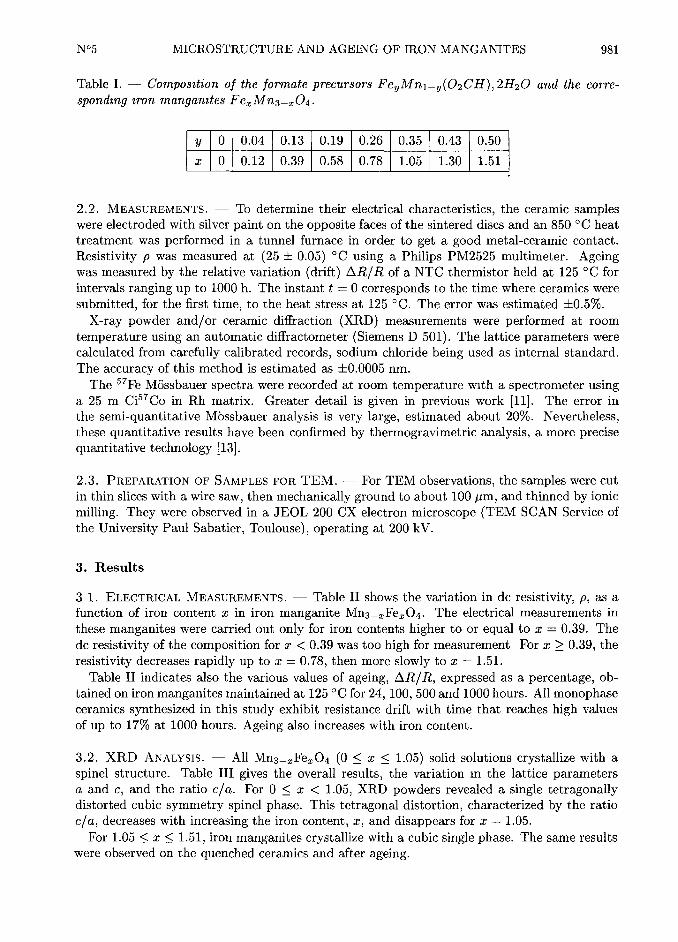
Table I. Composition of the formate precursors Fe~mni-y(02CH), 2H20 and the corre-
sponding iron mangamtes Fe~Mn3-~04.
y 0 0.04 0.13 0.19 0.26 0.35 0.43 0.50
x 0 0.12 0.39 0.58 0.78 1.05 1.30 1.51
2.2. MEASUREMENTS. To determine their electrical characteristics, the ceramic samples
were electroded with silver paint on the opposite faces of the sintered discs and an 850 °C heat
treatment was performed in a tunnel furnace in order to get a good metal-ceramic contact.
Resistivity p was measured at (25 + 0.05) °C using a Philips PM2525 multimeter. Ageing
was measured by the relative variation (drift) AR/R of a NTC thermistor held at 125 °C for
intervals ranging up to 1000 h. The instant t=
0 corresponds to the time where ceramics were
submitted, for the first time, to the heat stress at 125 °C. The error was estimated +0.5$l.
X-ray powder and/or ceramic diffraction (XRD) measurements were performed at room
temperature using an automatic diffractometer (Siemens D 501). The lattice parameters were
calculated from carefully calibrated records, sodium chloride being used as internal standard.
The accuracy of this method is estimated as +0.0005 nm.
The ~~Fe M6ssbauer spectra were recorded at room temperature with a spectrometer using
a 25 mCi~~co in Rh matrix. Greater detail is given in previous work ill]. The error in
the semi-quantitative Mbssbauer analysis is very large, estimated about 20$l. Nevertheless,these quantitative results have been confirmed by thermogravimetric analysis, a more precisequantitative technology [13].
2.3. PREPARATION OF SAMPLES FOR TEM. For TEM observations, the samples were cut
in thin slices with a wire saw, then mechanically ground to about 100 ~Jm, and thinned by ionic
milling. They were observed in a JEOL 200 CX electron microscope (TEM SCAN Service of
the University Paul Sabatier, Toulouse), operating at 200 kV.
3. Results
3 1. ELECTRICAL MEASUREMENTS. Table II shows the variation in dc resistivity, p, as a
function of iron content ~ in iron manganite Mn3-~Fe~04. The electrical measurements in
these manganites were carried out only for iron contents higher to or equal to ~ =0.39. The
dc resistivity of the composition for ~ < 0.39 was too high for measurement For x > 0.39, the
resistivity decreases rapidly up to x =0.78, then more slowly to x =
1.51.
Table II indicates also the various values of ageing, AR/R, expressed as a percentage, ob-
tained on iron manganites maintained at 125 ° C for 24, 100, 500 and 1000 hours. All monophaseceramics synthesized in this study exhibit resistance drift with time that reaches high values
of up to 17~ at 1000 hours. Ageing also increases with iron content.
3.2. XRD ANALYSIS. All Mn3-~Fe~04 (0 < ~ < l.05) solid solutions crystallize with a
spinet structure. Table III gives the overall results, the variation mthe lattice parameters
a and c, and the ratio cla. For 0 < ~ < l.05, XRD powders revealed a single tetragonallydistorted cubic symmetry spinet phase. This tetragonal distortion, characterized by the ratio
cla, decreases with increasing the iron content, ~, and disappears for ~ =1.05.
For 1.05 < ~ < l.51, iron manganites crystallize with a cubic single phase. The same results
were observed on the quenched ceramics and after ageing.

982 JOURNAL DE PHYSIQUE III N°5
Table II. Electrical characteristics of iron mangamtes: resistimty p and ageing AR/R at
125 °C.
Iron content, z 0.39 0.58 0.78 1.05 1 30 1.51
p (flcm) 1.43 x10~ 9.88 x
10~ 2.44 x10~ 2 02 x 10~ 1.80 x
10~ 1.60 x10~
24 h 3.3 3.2 3.1 2.2 9 1 11.0
AR/R 100 h 5A 5.8 4 8 6.4 11 6 11.9
(~) 500 h 7.2 6.4 10 9 13.4 13 8 13.3
1000 h 10.0 10.4 14 0 15.0 15 4 17.0
Table III. Lattice parameters and cla ratio as afknction of iron content, ~, for the iron
manganite powders.
Iron content, ~ 0 0,12 0.39 0.58 0.78 1.05 1.30 1.51
a(nm) 0.8141 0.8192 0.8225 0.8259 0.8274 0.8392 0.8486 0.8501
c(nm) 0.9456 0.9400 0.9300 0.9181 0.9019
cla 1.16 1.14 1.13 1.11 1.09 1 1 1
3.3. M6sSBAUER SPECTROSCOPY. M6ssbauer spectroscopy was carried out on two iron
manganites with two different crystallographic structures before (t=
0), after 1000 hours ageing(t
=1000 h): tetragonal, for
~ =0.58, and cubic, for ~ =
l.05. All the M0ssbauer spectraof these four iron manganites have the same shape. They all exhibit two doublets indicatingthe presence of two non-equivalent sites of the Fe~+ ions in octahedral and tetrahedral sites
of the spinet structure. From quantitative analysis [14] we computed the percentage of Fe~+
ions in each site. Thus knowing the total number of iron ions in the iron manganite (here 0.58
and 1.05), we calculated the number of Fe~+ ions in both sites. All the results are reported in
Table IV. Considering the large error of about 20$l in the semi-quantitative M6ssbauer results,the value of Fe~+ ions in tetrahedral sites 0.29 and 0.23 for
x =0.58 and 1.05 respectively
must be assumed to be the same. After calculating the cationic distributions, we shall consider
the mean values.
3.4. TEM RESULTS. The observations were performed on quenched ceramics. Electron
diffraction confirms the XRD results concerning the crystal structure of the samples, i-e- they
are tetragonal (space group I411amd) for x < 1.05 and cubic spinel (space group Fd3m) for
Table IV. Number of Fe~+ions m each site of the spinet structure for the iron manganites
Feo 58Mn2 4204 and Fei o5Mni 9504 at t=
0 and t=
1000 hours.
Octahedral sites Tetrahedral sites
Samples I II I II mean value
t=
0 0.29 0.82 0.29 0.23 0.26
t=
1000 h 0.52 0.96 0.06 0.09 0.08
Iron content of the samples=
0.58 and II=
1.05.

N°5 MICROSTRUCTURE AND AGEING OF IRON MANGANITES 983
o
0.44 Am
Fig. 1. Bright field electron micrograph Domain microstructure in Mn2 6iFeo 3g04 Laths inter-
nally twinned. f~.~
~~~A
' ','~ ~
~jjx(~l':
k'; .'
~
0.65 Am
Fig 2 Bright field electron micrograph. Domain microstructure in Mn2 42Feo 5804 Laths inter-
naIly twinned.
x > 1.05. Moreover, TEM results show that, as long as the samples have a tetragonal crystalline structure, a domain microstructure is observed.
. ~ =0.39 (Fig, I): TEM reveals laths of about 0.5-1 ~Jm wide; each of them is internally
twinned, with the twins being about 5-20 nm wide. The domain walls are parallel to the
(101) planes of the cubic cell.
. x =0.58 (Fig. 2): A domain structure is as well observed. The lath width ranges from 0.4
to 0.8 ~Jm, and the internal twins are stiff present and well marked (10-20 nm wide).
. x =0.78 (Fig. 3): The lath width clearly decreases (50-100 nm), and the internal twins
inside each lath are hardly visible (about 5 nmwide). So, as soon as the tetragonality of the
sample decreases, the lath width also decreases and the internal twinning (lamellae) tends to
vanish. All the twin walls are parallel to (101).
. x =1.05 (Fig. 4): For this
xvalue, a special microstructure is observed, the sample
being relatively heterogeneous. Fine striations are observed parallel to (l10) space at a range

984 JOURNAL DE PHYSIQUE III N°5
~m
Fig. 3. Bright field electron micrograph. Domain microstructure in Mn2 22Feo 7s04 Two sets of
nearly perpendicular domains. Note the wall interactions at crossing (arrow).
jioij
jioij
0.25
Fig. 4. Bright field electron micrograph. "Tweed" microstructure in Mni g5Fei o504.
from a few nanometers to 40 nm. In those areas where the striations are more dense, this
microstructure is very similar to the "tweed structure" already reported by the authors in Ni
and Co manganites [9].
. x > 1.05 (Figs. 6 and 7) Grains free of two-dimensional defects are now observed.
Note that for x =1.30, dislocation pile-ups are frequently observed forming low-angle bound-
aries (Fig. 6). These dislocations have the well-known Burgers vector of the spinel structure
b=
(l10) [16], generally dissociated into two colinear partials b=
(l10). The dissociation2 4
width is about 5-6 nm.

N°5 MICROSTRUCTURE AND AGEING OF IRON MANGANITES 985
m One s&,d prose
m GJ eJX Two s~>d proses
eJA Three &~<d prose%
~ Q
x m
Cu~c s@net
~ ~
~~'
C
~f
m
f~ m m ~ w
m Q ~~
1 X
x "
e j b
Wh
~W
a-Mn~O~
O O< 02 03 04 OS OS OS <O
R=Mn/tfe+&tnl
~.,5 ,2
19,6
~3
X
la) o-Fe203 (structure corundum) (e) a-Fe203 + a-Mn203
16) a-Mn203 (C-rare-earth oxide structure) (f) a-Fe203 + cubic spinel(c) cubic spinel (g) cubic spinel + tetragonal spinel(d) tetragonal spinel (h) o-Mn203 + tetragonal spinel
(I) a-Mn203 + cubic spinel
Fig 5. Equilibrium diagram of the system Fe304-Mn304 (15j.
1.12
Fig. 6. Bright field electron micrograph. Low angle boundary in Mni mFe13004. No two-
dimensional defects are observed

986 JOURNAL DE PHYSIQUE III N°5
Fig. 7. Bright field electron micrograph. These grains (Mni 4gFei 5104)are free of two-dimensional
defects.
Table V. Iron manganite ceramics before ageing (t=
0): cationic distrtbutions computedfrom Mbssbauer data.
Iron content, x Cationic distributions
~.~~ ~~~~S~~~~2[~~~~7~~~~1~~~2j°~
~'~~ ~~~~1~~~~9[~~~~9~~~~2~~~~9j°~
~'~~ ~~~~1~~~~9[~~~~9~~~~2~ll~~9j°~
~'~~ ~ll~~7~~~~3[~~~~2~ll~~5~ll~~3j°~
l.30
1.51
4. Discussion
4.I. ELECTRICAL PROPERTIES. The cationic distributions given by Mn)+~Fe(+[Fe(+Mn(+~Mn(+]O(~ with x = y + z
ill]can be inferred by correlation of the results obtained
by XRD, M0ssbauer spectroscopy and electrical measurements. Table V recalls the cationic
distributions of iron manganite for 0.39 < x < 1.51 before ageing (t=
0). Moreover, the
origin of the ageing observed on iron manganite thermistors has been identified in a previouswork [12]. This is related to the migration of Fe~+ ions in octahedral sites. The number of
Fe~+ ions in tetrahedral sites is nearly constant (see Tab. V),so we can assume that, as z
increases, the number of Fe~+ ions, which migrate toward octahedral sites, remains constant.
Thus the number of Fe3+ ions remaining in A sites after 1000 hours ageing is constant and
equal to 0.08, as indicated by the M0ssbauer results for two values ofx
(0.58 and 1.05) in
Table IV. On light of this assumption, Table VI reports the cationic distributions obtained for
different iron contents in iron manganites after 1000 hours ageing (tiooo h).According to the cationic distributions proposed for iron manganites before and after ageing
(Tabs. V and VI), both Mn~+ and Mn~+are present in B sites so that the conditions are
correct for electron hopping from Mn2+ and Mn~+ [Iii. The conductivity of the material is
determined by the number of ions capable of either donating or accepting electrons in this
N°5 MICROSTRUCTURE AND AGEING OF IRON MANGANITES 987
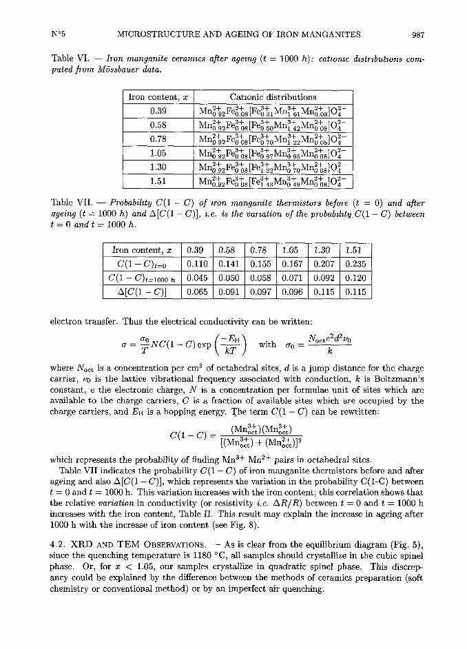
Table VI. Iron manganite ceramics ajter ageing (t=
1000 h): catiomc distributions com-
puted from Mbssbauer data.
Iron content, xCationic distributions
~'~~ [~~~~l~ll~~1~ll~~Sj°~
~'~~ 05[~~~~0~ll~~2~ll~~Sj°~
0.78 ~~]O(~
i.05
~'~~ 08j°~
~'~~ 08[~~~~3~ll~~9~ll~~8j°~
Table VII. Probability C(I C) of iron manganite thermistors before (t=
0) and ajterageing (t
=1000 h) and A[C(I C)],
i.e. is the variation of the probability, C(I C) between
t=
0 and t=
1000 h.
Iron content, x 0.39 0.58 0.78 1.05 1.30 1.51
C(I C)t=o 0.l10 0.141 0.155 0.167 0.207 0.235
C(I C)i=locoh
0.045 0.050 0.058 0.071 0.092 0.120
A[C(I-C)] 0.065 0.091 0.097 0.096 0.lls 0.lls
electron transfer. Thus the electrical conductivity can be written:
where Noct is a concentration per cm3 of octahedral sites, d is a jump distance for the chargecarrier, vo is the lattice vibrational frequency associated with conduction, k is Boltzmann's
constant, e the electronic charge, N is a concentration per formulae unit of sites which are
available to the charge carriers, C is a fraction of available sites which are occupied by the
charge carriers, and EH is a hopping energy. The term C(I C) can be rewritten:
~j~ ~~~
lmlll$)lMIllS)llmnls) + lmnls)l~
which represents the probability of finding Mn~+ Mn~+ pairs in octahedral sites.
Table VII indicates the probability Gil C) of iron manganite thermistors before and after
ageing and also A [Cl I C)], which represents the variation in the probability Cl I-C) between
t=
0 and t=
1000 h. This variation increases with the iron content; this correlation shows that
the relative variation in conductivity (or resistivity I.e. AR/R) between t=
0 and t=
1000 h
increases with the iron content, Table II. This result may explain the increase in ageing after
1000 h with the increase of iron content (see Fig. 8).
4.2. XRD AND TEM OBSERVATIONS. As is clear from the equilibrium diagram (Fig. 5),since the quenching temperature is l180 °C, all samples should crystallize in the cubic spinelphase. Or, for x < 1.05, our samples crystallize in quadratic spinel phase. This discrep-
ancy could be explained by the difference between the methods of ceramics preparation (softchemistry or conventional method)
or by an imperfect air quenching.
988 JOURNAL DE PHYSIQUE III N°5
18
16--~_
14"
'~ ~ii
~~,f_ -0 58
W-078
( 8 -105
6~~~~"~~°
isi
4
o
0 200 400 600 800 1000
Time (hours)
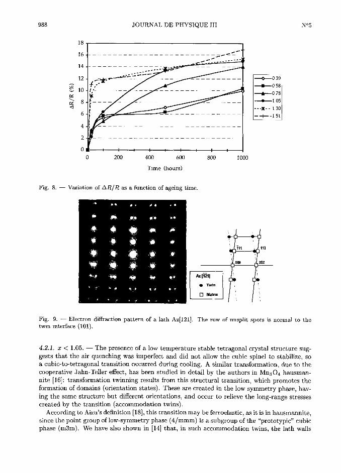
Fig. 8. Variation of AR/Ras a function of ageing time.
1". TWn
n Mawx
Fig. 9. Electron diRraction pattern of a lath Az[121j. The row of unsplit spots is normal to the
twin interface (101).
4.2.1. x < 1.05. The presence of a low temperature stable tetragonal crystal structure sug-gests that the air quenching was imperfect and did not allow the cubic spinel to stabilize, so
a cubic-to-tetragonal transition occurred during cooling. A similar transformation, due to the
cooperative Jahn-Tefler effect, has been studied in detail by the authors in Mn304 hausman-
nite [16]: transformation twinning results from this structural transition, which promotes the
formation of domains (orientation states). These are created in the low symmetry phase, hav-
ing the same structure but different orientations, and occur to relieve the long-range stresses
created by the transition (accommodation twins).According to Aizu's definition [18], this transition may be ferroelastic, as it is in hausmannite,
since the point group of low-symmetry phase (4 /mmm) is a subgroup of the "prototypic" cubic
phase (m3m). We have also shown in [14] that, in such accommodation twins, the lath walls
N°5 MICROSTRUCTURE AND AGEING OF IRON MANGANITES 989
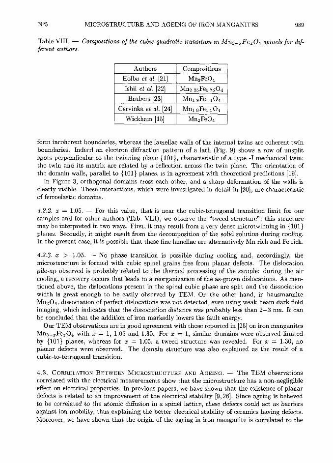
Table VIII. Compositions of the cubic-quadratic transition m Mn3-~Fe~04 spinets for dif-ferent authors.
Authors Compositions
Holba et al. [21] Mn2Fe04
Ishii et al. [22] Mn2 25Feo 7504
Brabers [23] Mni 9Fei i04
Cervinka et al. [24] Mni 9Fei i04
Wickham [15] Mn2Fe04
form incoherent boundaries, whereas the lamellae walls of the internal twins are coherent twin
boundaries. Indeed an electron diffraction pattern of a lath (Fig. 9) shows a row of unsplitspots perpendicular to the twinning plane (101), characteristic of a type -I mechanical twin:
the twin and its matrix are related by a reflection across the twin plane. The orientation of
the domain walls, parallel to (101) planes, is in agreement with theoretical predictions [19].In Figure 3, orthogonal domains cross each other, and a sharp deformation of the walls is
clearly visible. These interactions, which were investigated in detail in [20], are characteristic
of ferroelastic domains.
4.2.2, x =1.05. For this value, that is near the cubic-tetragonal transition limit for our
samples and for other authors (Tab. VIII),we observe the "tweed structure" this structure
may be interpreted in two ways. First, it may result from a very dense microtwinning in (101)planes. Secondly, it might result from the decomposition of the solid solution during cooling.In the present case, it is possible that these fine lamellae are alternatively Mn rich and Fe rich.
4.2.3. x > 1.05. No phase transition is possible during cooling and, accordingly, the
microstructure is formed with cubic spinel grains free from planar defects. The dislocation
pile-up observed is probably related to the thermal processing of the sample: during the air
cooling, a recovery occurs that leads to a reorganization of the as-grown dislocations. As men-
tioned above, the dislocations present in the spinel cubic phase are split and the dissociation
width is great enough to be easily observed by TEM. On the other hand, in hausmannite
Mn304, dissociation of perfect dislocations was not detected, even using weak-beam dark field
imaging, which indicates that the dissociation distance was probably less than 2-3 nm. It can
be concluded that the addition of iron markedly lowers the fault energy.Our TEM observations are in good agreement with those reported in [25] on iron manganites
Mn3-~Fe~04 with x =I, 1.05 and 1.30. For
~ =l, similar domains were observed limited
by (101) planes, whereas for~ =
l.05, a tweed structure was revealed. For ~ =l.30, no
planar defects were observed. The domain structure was also explained as the result of a
cubic-to-tetragonal transition.
4.3. CORRELATION BETWEEN MicRosTRucTuRE AND AGEING. The TEM observations
correlated with the electrical measurements show that the microstructure has a non-negligibleeffect on electrical properties. In previous papers, we have shown that the existence of planardefects is related to an improvement of the electrical stability [9, 26]. Since ageing is believed
to be correlated to the atomic diffusion in a spinel lattice, these defects could act as barriers
against ion mobility, thus explaining the better electrical stability of ceramics having defects.
Moreover, we have shown that the origin of the ageing in iron manganite is correlated to the

990 JOURNAL DE PHYSIQUE III N°5
1000 h
Time (h)24 h
0 39 0.58 0 78 1.05 1.30 51
Iron content, x '
Mic~ostn~cbwe : Bidimensional defects No bidi~nensional defects
Fig 10. Variation of ageing at 24 and 1000 hours as a function of iron content and microstructure.
migration of Fe3+ and Mn~+ ions between the A and B sublattices of spinel structure. Does
the microstructure of solid-solution Mn3-~Fe~04 have any effect on electrical properties?
From Figure 10, it is clear that the ageing after 1000 hours is correlated with the microstruc-
ture. For ~ =0.39 and
~ =0.58, TEM reveals large laths and internal twins, and the ageing
is about 10~. For ~ =0.78, the lath width clearly decreases and ageing (t
=1000 h) increases
to 14%. For x =1.05, a value close to the limit between the tetragonal and cubic phases,
the domain structure disappears and TEM reveals a tweed microstructure, as the ageing in-
creases about 15$l. Lastly, when grains free of two-dimensional defects are observed, the ageingreaches about 17$l. Unambiguously, the presence of planar defects hampers ion migration and
consequently prevents the increase of AR/R. The phenomenon is enhanced at 24 hours of
ageing. From Figure 10, it is clear that, as soon as the sample has the tetragonal structure
with a more or less pronounced domain structure, the variation of resistance between t=
0
and t=
24 hours is very low, AR/R is about 3$l (high density of planar defects); AR/Rincreases abruptly for x =
1.30 (AR /R=
9, l~) and reaches about 11$l with no defects. So
the presence of defects does play a role in the kinetics of ion migration: it slows down the ion
migration at the beginning of the ageing.
5. Conclusion
Previously, we have shown that the origin of ageing of iron manganite thermistors is due to
the migration of Fe3+ and Mn~+ ions between tetrahedral and octahedral sites of the spinel
structure. In this paper, the electrical measurements show that the ageing depends on the
composition, as it increases with the iron content. TEM observations reveal two-dimensional
defects for z < 1.05 and no planar defects are detected for x > 1.05.
The correlation between the microstructure and the ageing indicates that the presence of
these defects hampers ion migration. Consequently:
I) the ageing kinetic is slowed down by the existence of defects;
it) defects stabilize the electrical properties.

N°5 MICROSTRUCTURE AND AGEING OF IRON MANGANITES 991
References
iii Macklen E-D-, Thermistors (Electrochem. Publ. Ayr. Scotland, 1979).
[2] Jabry E., Boissier G., Rousset A., Carnet R. and Lagrange A., Preparation of semi-
conducting NTC Thermistors by Chemical Method., J. Phys. Colloq. France 47 (1986)Cl-843-847.
[3] Feltz A., T6pfer J. and Schiirmeister F., Conductivity data and preparation routes for
NiMn204 thermistor ceramics, J. Eur. Ceram. Soc. 9 (1992) 187-191.
[4] Macklen E.D., Electrical conductivity and cation distribution in nickel manganite, J. Phys.Chem. Solids 47 (1986) 1073-1079.
[5] Metz R., Caffin J-P-, Legros R. and Rousset A., The preparation, characterization and
electrical properties of copper manganites Cu~Mn3-z04, (0 < z < I), J. Mater. So. 24
(1989) 83-87.
[6] Vervey E-J-M-, Haaij P-W-, Romeijn F-C- and Van Oosternout C-W-, Controlled-valencysemiconductors, Philips Res. Rep. 5 (1950) 173-187.
iii Caflin J-P-, Thesis, University Paul Sabatier (Toulouse, France, 1986).
[8] Metz R., Thesis, University Paul Sabatier (Toulouse, France, 1991).
[9] Brieu M., Couderc J-J-, Rousset A. and Legros R., TEM Characterization of nickel and
nickel-cobalt manganite ceramics, J. Eur. Ceram. Soc. 11 (1993) iii-iii-
[l0] Fritsch S., Thesis, University Paul Sabatier (Toulouse, France, 1995).ii Ii Battault T., Legros R. and Rousset A., Structural and electrical properties of iron mangan-
ite spinels in relation with cationic distribution., J. Eur. Ceram. Soc. 15 (1995) l141-l147.
[12] Battault T., Thesis, University Paul Sabatier (Toulouse, France, 1995).[13] Gillot B., Laarj M., Kacim S., Battault T., Legros R. and Rousset A., Cationic distribution
and oxidation kinetics of divalent manganese ions in iron manganite spinels Mn3-~Fe~04
(0 < x < 1.50), Solid State Ionics 83 (1996) 215-263.
[14] Singh V-K-, Khatri N-K- and Lokanathan S., M6ssbauer study of Co~Mn3-~-yFe~04 and
Ni~Mn3-~-yFey04 systems, Ind. J. Pure 8 Appl. Phys. 20 (1982) 83-89.
[15] Wickham D-G-, The Chemical Composition of Spinels in the System Fe304-Mn304, J.
Inorg. Nucl. Chem. 31 (1969) 313-320.
[16] Couderc J-J-, Fritsch S., Brieu M., Vanderschaeve G., Fagot M. and Rousset A., A Trans-
mission Electron Microscopy Study of Lattice Defects in Mn304 Hausmannite, Phdos.
Mag. B 70 (1994) 1077-1094.
iii] Dorris S-E- and Mason T-O-, Electrical properties and cations valencies mMn304, J.
Amertcan Ceramic Society 71 (1988) 379-385.
[18] Aizu K., Possible Species of Ferromagnetic, Ferroelectric and Ferroelastic Crystals, Phys.Rev. B 2 (1970) 754-772.
[19] Sapriel J., Domain-wall Orientations in Ferroelastics, Phys. Rev. 812 (1975) 5128-5140.
[20] Snoeck E., Casanove M-J-, Baules P. and Roucaud C., Ferroelastic behaviour of the
(Ln)Ba2Cu306+~ Orthorhombic Phase, Ferroelectrics 97 (1989) 181-186.
[21] Holba P., Khilla M-A- and Krupicka S., On the Miscibility Gap of Spinels Mn~Fe3-~04+~,J. Phys. Chem. Solids 34 (1973) 387-395.
[22] Ishii M., Nakahira M. and Yamanaka T., Infrared absorption spectra and cation distribu-
tion in (Mn,Fe)304, Solid State Comm~nications 11 (1972) 209-212.

992 JOURNAL DE PHYSIQUE III N°5
[23] Brabers V-A-M-, Cation Migration, Cation Valencies and the Cubic-Tetragonal Transition
in Mn~Fe3-~04, J. Phys. Chem. Solids 32 (1971) 2181-2191.
[24] Cervinka L., Hosemann R, and Vogel W., Paracrystalline lattice distorsions and mi-
crodomains in manganese ferrites near the cubic to tetragonal transition, Acta Cryst.A 26 (1970) 277-289.
[25] Van Landuyt J., De Ridder R., Brabers V-A M. and Amelinckx S, Jahn-Teller Domains
in Mn~Fe3-~04 as observed by Electron Microscopy, Mat. Res. Bull. 7 (1972) 327-338.
[26] Rousset A., Lagrange A., Brieu M., Couderc J-J- et Legros R., Influence de la microstruc-
ture sur la stabilitA Alectrique des thermistances CTN, J. Phys. III France 3 (1993)833-845.