Correlation between Macrophage Activation …iai.asm.org/content/57/4/1311.full.pdf · INFECTION...
7
INFECTION AND IMMUNITY, Apr. 1989, p. 1311-1317 Vol. 57, No. 4 0019-9567/89/041311-07$02.00/0 Copyright © 1989, American Society for Microbiology Correlation between Macrophage Activation and Bactericidal Function and Mycobacterium leprae Antigen Presentation in Macrophages of Leprosy Patients and Normal Individuals SMITA D. DEEAI, TANNAZ J. BIRDI, AND NOSHIR H. ANTIA* The Foundation for Medical Research, 84-A, R. G. Thadani Marg, Worli, Bombay 400 018, India Received 22 September 1988/Accepted 10 January 1989 The killing of Mycobacterium leprae by resting and gamma interferon (IFN--y)-activated macrophages in normal subjects and leprosy patients was assessed. Resting macrophages from normal individuals demon- strated the ability to kill M. Ieprae. For macrophages from tuberculoid patients, killing of M. leprae was only achieved in the presence of IFN--y, suggesting that initial T-cell activation occurs prior to the killing of M. kprae. In contrast, though activation with IFN--y rendered the lepromatous macrophage microbicidal, it fiailed to induce lymphocyte proliferation, suggesting a defect at either the antigen-presenting cell or the lymphocyte level or both. The concept that T-cell anergy is primarily due to lack of lymphokine generation was ruled out by our results, since responsiveness was restored in only a small proportion of lepromatous patients after exogenous lymphokine addition. In conclusions this study demonstrated that killing and antigen presentation are two independent events. It appears that the ability of the macrophages per se to kill M. leprae may be of greater importance than lymphocyte-mediated activation for protection against M. leprae infection. Cellular immunity in chronic infections is determined primarily by the extent to which macrophages are activated. Some microbes are killed soon after phagocytosis, while others bypass the conventional microbicidal mechanisms and multiply in the permissive environment provided by macrophages (30). In the case of lepromatous leprosy, macrophages have been highlighted as important suppressor cells in the chain of events occurring after the host-parasite interaction (8, 25, 26). Our earlier studies have shown a defective regulation of M. leprae phagocytosis (19) initiating a series of distur- bances in the metabolism of macrophages (5, 7). This prob- ably aids the survival of the parasite and its replication within the susceptible host cell. Various studies indicate gamma interferon (IFN-y) as the major macrophage-activating factor found in antigen- and mitogen-stimulated lymphocyte culture supernatants which augment the microbicidal capacity of phagocytes (30). In contrast to tuberculoid patients and normal individuals, lepromatous leprosy patients have been reported to lack circulating T lymphocytes capable of responding to M. leprae by proliferation (12) or by the release of IFN--y (20), resulting in inadequate macrophage activation to eliminate the mycobacteria. This study aims at assessing whether in vitro activation with IFN--y can restore the ability of defective macrophages (i) to kill M. leprae through oxidative mechanisms and (ii) to process and present M. leprae antigens to T lymphocytes. Due to the inability to directly measure M. leprae viability in vitro, workers have resorted to the measurement of killing by indirect methods (5, 10, 17, 23, 28, 29, 32). In the present study, we have assessed M. leprae viability by using the following two methods: (i) growth in the mouse footpad (28) and (ii) the down-regulation of the macrophage Fc receptor (4, 5). Antigen processing and presentation have been assessed by human leukocyte antigen DR (HLA-DR) expression, * Corresponding author. antigen-specific monocyte-lymphocyte physical interaction, and lymphoproliferation. MATERIALS AND METHODS Subjects. Leprosy patients were classified according to the Ridley and Jopling classification (24). The choice of lepro- matous patients was restricted to long-term-treated leproma- tous patients who were bacteriologically negative (BI-ve), i.e., who did not demonstrate acid-fast bacilli in smears from multiple sites. All tuberculoid patients were treated for durations varying from 1 to 6 years. Occupational contacts who had various degrees of exposure to leprosy patients were used as normal controls. Antigens. (i) M. leprae was obtained from infected arma- dillo tissue stored at -80°C. The bacteria were harvested by homogenizing the armadillo tissue. The bacterial pellet ob- tained after differential centrifugation to minimize tissue contamination was suspended in saline, stored at 4°C, and used within a week. (ii) Purified protein derivative. Purified protein derivative (PPD) was obtained from the Statens Serum Institute, Copenhagen, Denmark, and was used at an optimal concen- tration of 4 pugIml. Monocyte-macrophage cultures. Mononuclear cells were isolated from heparinized peripheral blood on a Lymphoprep gradient (Nyegaard, Oslo, Norway) and depleted of lympho- cytes by adhering them to glass for 24 h. Monocytes were further maintained for 7 days by changing the culture me- dium every 48 h to obtain differentiated macrophage cul- tures. The cells were cultured in minimal essential medium (MEM) containing 30% human AB serum at a concentration of 3 x 10⁶ cells per ml either in petri dishes (Falcon, 55 mm; Becton Dickinson Labware, Oxnard, Calif.) in 3 ml of medium or on glass cover slips in Leighton tubes in 0.7 ml of medium. Determination of M. leprae viability. (i) Infection of mono- cyte-macrophage monolayers. The cultures were infected with 5 x 10⁶ M. leprae cells per ml for 24 h before the uningested M. leprae were washed off. 1311 on July 15, 2018 by guest http://iai.asm.org/ Downloaded from
Transcript of Correlation between Macrophage Activation …iai.asm.org/content/57/4/1311.full.pdf · INFECTION...

INFECTION AND IMMUNITY, Apr. 1989, p. 1311-1317 Vol. 57, No. 40019-9567/89/041311-07$02.00/0Copyright © 1989, American Society for Microbiology
Correlation between Macrophage Activation and BactericidalFunction and Mycobacterium leprae Antigen Presentation inMacrophages of Leprosy Patients and Normal Individuals
SMITA D. DEEAI, TANNAZ J. BIRDI, AND NOSHIR H. ANTIA*The Foundation for Medical Research, 84-A, R. G. Thadani Marg, Worli, Bombay 400 018, India
Received 22 September 1988/Accepted 10 January 1989
The killing of Mycobacterium leprae by resting and gamma interferon (IFN--y)-activated macrophages innormal subjects and leprosy patients was assessed. Resting macrophages from normal individuals demon-strated the ability to kill M. Ieprae. For macrophages from tuberculoid patients, killing of M. leprae was onlyachieved in the presence of IFN--y, suggesting that initial T-cell activation occurs prior to the killing of M.kprae. In contrast, though activation with IFN--y rendered the lepromatous macrophage microbicidal, it fiailedto induce lymphocyte proliferation, suggesting a defect at either the antigen-presenting cell or the lymphocytelevel or both. The concept that T-cell anergy is primarily due to lack of lymphokine generation was ruled outby our results, since responsiveness was restored in only a small proportion of lepromatous patients afterexogenous lymphokine addition. In conclusions this study demonstrated that killing and antigen presentationare two independent events. It appears that the ability of the macrophages per se to kill M. leprae may be ofgreater importance than lymphocyte-mediated activation for protection against M. leprae infection.
Cellular immunity in chronic infections is determinedprimarily by the extent to which macrophages are activated.Some microbes are killed soon after phagocytosis, whileothers bypass the conventional microbicidal mechanismsand multiply in the permissive environment provided bymacrophages (30).
In the case of lepromatous leprosy, macrophages havebeen highlighted as important suppressor cells in the chain ofevents occurring after the host-parasite interaction (8, 25,26). Our earlier studies have shown a defective regulation ofM. leprae phagocytosis (19) initiating a series of distur-bances in the metabolism of macrophages (5, 7). This prob-ably aids the survival of the parasite and its replicationwithin the susceptible host cell.
Various studies indicate gamma interferon (IFN-y) as themajor macrophage-activating factor found in antigen- andmitogen-stimulated lymphocyte culture supernatants whichaugment the microbicidal capacity of phagocytes (30). Incontrast to tuberculoid patients and normal individuals,lepromatous leprosy patients have been reported to lackcirculating T lymphocytes capable of responding to M.leprae by proliferation (12) or by the release of IFN--y (20),resulting in inadequate macrophage activation to eliminatethe mycobacteria.
This study aims at assessing whether in vitro activationwith IFN--y can restore the ability of defective macrophages(i) to kill M. leprae through oxidative mechanisms and (ii) toprocess and present M. leprae antigens to T lymphocytes.Due to the inability to directly measure M. leprae viability
in vitro, workers have resorted to the measurement of killingby indirect methods (5, 10, 17, 23, 28, 29, 32). In the presentstudy, we have assessed M. leprae viability by using thefollowing two methods: (i) growth in the mouse footpad (28)and (ii) the down-regulation of the macrophage Fc receptor(4, 5).Antigen processing and presentation have been assessed
by human leukocyte antigen DR (HLA-DR) expression,
* Corresponding author.
antigen-specific monocyte-lymphocyte physical interaction,and lymphoproliferation.
MATERIALS AND METHODS
Subjects. Leprosy patients were classified according to theRidley and Jopling classification (24). The choice of lepro-matous patients was restricted to long-term-treated leproma-tous patients who were bacteriologically negative (BI-ve),i.e., who did not demonstrate acid-fast bacilli in smears frommultiple sites. All tuberculoid patients were treated fordurations varying from 1 to 6 years. Occupational contactswho had various degrees of exposure to leprosy patientswere used as normal controls.
Antigens. (i) M. leprae was obtained from infected arma-dillo tissue stored at -80°C. The bacteria were harvested byhomogenizing the armadillo tissue. The bacterial pellet ob-tained after differential centrifugation to minimize tissuecontamination was suspended in saline, stored at 4°C, andused within a week.
(ii) Purified protein derivative. Purified protein derivative(PPD) was obtained from the Statens Serum Institute,Copenhagen, Denmark, and was used at an optimal concen-tration of 4 pugIml.Monocyte-macrophage cultures. Mononuclear cells were
isolated from heparinized peripheral blood on a Lymphoprepgradient (Nyegaard, Oslo, Norway) and depleted of lympho-cytes by adhering them to glass for 24 h. Monocytes werefurther maintained for 7 days by changing the culture me-dium every 48 h to obtain differentiated macrophage cul-tures. The cells were cultured in minimal essential medium(MEM) containing 30% human AB serum at a concentrationof 3 x 10⁶ cells per ml either in petri dishes (Falcon, 55 mm;Becton Dickinson Labware, Oxnard, Calif.) in 3 ml ofmedium or on glass cover slips in Leighton tubes in 0.7 ml ofmedium.
Determination of M. leprae viability. (i) Infection of mono-cyte-macrophage monolayers. The cultures were infectedwith 5 x 10⁶ M. leprae cells per ml for 24 h before theuningested M. leprae were washed off.
1311
on July 15, 2018 by guesthttp://iai.asm
.org/D
ownloaded from
1312 DESAI ET AL.
(ii) Activation with IFN--y. IFN--y was added at an optimalconcentration of 10 U/ml either simultaneously with thebacilli or 24 h postinfection.
(iii) Treatment with superoxide dismutase (SOD) fromcanine blood. SOD (Sigma Chemical Co., St. Louis, Mo.)was added at a concentration of 10 p1g/ml simultaneouslywith IFN--y and M. leprae.
(iv) Viability assays. Viability of M. leprae was assessed bydetermination of the footpad growth curve (28) and by the Fcreceptor assay (4, 5).Footpad growth curve. Macrophage cultures maintained in
petri dishes were scraped off with a rubber policeman,suspended in saline, and exposed to 10 cycles of freeze-thawing. The released bacilli were suspended in a knownvolume of saline and counted by the method of Shepard andMcRae (28). A total of 10 mice were inoculated with 104organisms per footpad. The footpads were harvested at 6, 7,8, 10, and 12 months postinoculation. A sample of the M.leprae suspension derived from the armadillo tissue and usedfor infecting the macrophage cultures in the assay systemwas used as a positive control.
Fc receptor assay. This assay has been used as a measureof viability since our earlier studies showed that erythrocyterosetting (EA rosetting) levels are reduced in bacteriologi-cally negative lepromatous patients only in the presence ofviable M. leprae and not after the addition of autoclaved orrifampin-treated M. leprae (5). Macrophage cultures weremaintained on glass cover slips for 72 h after infection beforeEA rosetting was carried out. This duration of incubationwas essential to distinguish between the direct action ofIFN--y on the macrophages per se (since IFN-y is known totransiently increase the Fc receptor levels in macrophages)and its effect on the intracellular M. leprae. A similarprotocol has been used earlier to distinguish between bacte-riostatic and bactericidal drugs (2, 4).Sheep erythrocytes (SRBCs) in a 2% suspension in MEM
were sensitized with an equal volume of goat anti-SRBCantibody (1:900 dilution; Wellcome Research Laboratories,Beckenham, England). A suspension of 1% sensitizedSRBCs was overlaid on the macrophage monolayer andallowed to rosette for 30 min at 37°C in an atmosphere of 5%C02. Nonrosetted SRBCs were removed by washing, andthe monolayers were fixed in 2.5% glutaraldehyde andstained by the Ziehl-Neelsen acid-fast staining method. Atotal of 200 cells were counted, and the percentage of cellswith three or more SRBCs attached was determined.
Antigen-specific monocyte-lymphocyte physical interaction.Mononuclear cells from peripheral blood were isolated on aLymphoprep gradient. The cells so obtained consisted of 80to 90% lymphocytes and 10 to 20% monocytes. They weresuspended in MEM containing 20% inactivated human ABserum in a concentration of 4 x 10⁶ cells per ml anddistributed into Leighton tubes containing cover slips. M.Ieprae was added (3 x 10⁶ per tube) in the presence orabsence of activating agents (IFN-y [10 U/ml] and recombi-nant interleukin-2 [20 U/ml]), and the cells were incubated at37°C for 18 h in an atmosphere of 5% CO2.The nonrosetted lymphocytes were then washed off, and
the cells were fixed in 2.5% glutaraldehyde and' stained bythe Ziehl-Neelsen acid-fast staining method. The percentageof monocytes with two or more lymphocytes adhering tothem was determined and expressed as the'percent interac-tion. (IFN-y and recombinant interleukin-2 were generousgifts from, respectively, M. Harboe, I.G.R.I., Oslo, Nor-way, and Francis Singaglia, Hoffman-La Roche, Basel,Switzerland.)
HLA-DR antigen expression on macrophages. Macrophagecultures derived from peripheral blood of bacteriologicallynegative patients and normal individuals were incubated inthe presence of media containing IFN--y or M. leprae (orboth) for 24 h at concentrations similar to those used for theFc assay. Before being stained, the cultures were washedand incubated with freshly collected human serum for 1 h.The cover slips were washed with saline, flooded withanti-HLA-DR (Biotin conjugate, 1: 103 dilution; BectonDickinson and Co., Paramus, N.J.), and incubated in a moistchamber for 30 min. Unbound antibody was removed bywashing the cultures three times with phosphate-bufferedsaline. The cover slips were fixed in cold absolute alcohol for30 min, washed, and then flooded with anti-immunoglobulinG (anti-IgG) (Avidin-rhodamine conjugate, 1:103 dilution;Becton Dickinson) and incubated in a moist chamber for 30min. The cover slips were washed, mounted in phosphate-buffered saline-glycerol, and examined under a fluorescentmicroscope under epi-illumination. The percentage of cellsstained positive in IFN--y-treated cultures in the presence orabsence of M. leprae was compared with that in controlcultures from the same individual.Lymphocyte proliferation assay. Mononuclear cells were
separated on a Lymphoprep gradient, and the cell count wasadjusted to 106 cells per ml. Cells were suspended in MEMsupplemented with 20% human AB serum and antibiotics. A100-,u portion of this cell suspension was added to each wellof a flat-bottomed 96-well microdilution plate.
Viable M. leprae at a concentration of 3 x 10⁶ bacilli perml was added in the presence or absence of concanavalin A(ConA)-derived lymphokines from mononuclear cells. Cul-tures were maintained in a 5% CO2 atmosphere at 37°C for 5days. Eighteen hours before termination 0.5 ,uCi of tritiatedthymidine was added (specific activity, 26,000 mCi/mmol) toeach well. Cultures were harvested onto glass microfiberfilters. Sample disks were dried and counted in a liquidscintillation counter (Kontron). Results were expressed interms of stimulation index, which was calculated as the ratioof the mean counts per minute of cultures stimulated simul-taneously with M. Ieprae and lymphokines to the meancounts per minute of the cultures stimulated with lympho-kines alone.
Preparation of ConA supernatants. ConA supernatantswere prepared by incubating 3 x 10⁶ mononuclear cells fromnormal individuals in '2 ml of MEM containing 25% humanAB serum with 20 ,ug of ConA (Sigma) per ml for 2 days at37°C in a 5% CO2 atmosphere. At the end of the incubationperiod, the cells were sedimented at 2,000 rpm (IECCENTRA-7R) for 10 min at 4°C. Supernatants were re-moved, filtered through a 0.2-pum filter (Millipore Corp.,Bedford, Mass.) and stored at -90°C until further use. ConAsupernatant controls were prepared in the same way asdescribed above except that addition of ConA was made atthe end of the 2-day incubation period: ConA supernatantsand supernatants from control cultures were used at anoptimal concentration of 50 p,l per well.
Modified lymphoproliferative assay. Monocytes were en-riched on a Nycodenz-monocyte gradient (Nyegaard) fromperipheral blood mononuclear cells. Monocytes were added(104 per well) to a flat-bottomed 96-well microdilution plate.T lymphocytes were primarily obtained on a Lymphoprepgradient and further enriched on a nylon-wool column. TheT lymphocytes were used at a concentration of 105 per 100p.l. Viable M. leprae at a concentration of 3 x 10⁶/ml or 10 Uof IFN--y per ml (or both) was added for 24 h before theaddition of T cells. Cultures were maintained for 5 days and
INFECT. IMMUN.
on July 15, 2018 by guesthttp://iai.asm
.org/D
ownloaded from

M. LEPRAE KILLING AND ANTIGEN PRESENTATION 1313
.1
ILEPROMATOUS ITUBERCULOID I NORMAL
10
-W00
co 10L.11111 lii II1L c ,
1~ ~~--A
.1~~~~~~~~~J-3tr
zI-
Co0
1 2 3 4 1 2 3 4 1 2 3
FIG. 1. M. leprae viability within resting and activated macro-phages from normal individuals and from tuberculoid and leproma-tous leprosy patients as assessed by the mouse footpad technique.Each set of bars represents. results obtained from a single individualand depicts growth of bacilli obtained at 12 months postinoculation.Resting macrophages from normal individuals were able to kill M.leprae. However, macrophages from leprosy patients were able tokill M. leprae only after activation with IFN-y. AFB, Acid-fastbacilli. Lanes: 1, original M. Ieprae from armadillo tissue used forinfecting macrophages; 2, M. leprae harvested from infected mac-rophage cultures; 3, M. Ieprae harvested from infected macrophagesstimulated simultaneously with IFN--y; 4, M. Ieprae harvested frommacrophages activated with IFN--y 24 h after M. Ieprae infection.
processed routinely as described above. Results were ex-pressed as counts per ininute plus or minus the standatderror of the mean.
Statistical analysis. All cultures were set up and read atleast in duplicate and occasionally in triplicate. The statisti-cal significance of the difference between each test and therespective control in the assays was determined by Student'st test.
RESULTS
M. leprae viability within resting or activated human mac-rophages from normal individuals and leprosy patients. (i)Mouse footpad assay. The intracellular bacilli were liberatedfrom the infected macrophages by 10 cycles of freeze-thawing. To determine whether this method of extractionaffected the viability ofM. leprae per se, a suspension of thebacilli was subjected to freeze-thawing and compared withan untreated suspension. No loss of viability of the bacilliwas noted, as comparable growth was observed with bothpreparations (data not shown).M. leprae derived from 7-day-old resting macrophage
cultures from four normal individuals did not show anygrowth in the mouse footpad (Fig. 1). This indicated thatmacrophages of normal individuals had the ability to kill M.leprae. In contrast, M. leprae derived from macrophages
4
40z
20 24
0
ML M.L+IFN M.L(o/n) M.L M.L+IFN+IFN
FIG. 2. Effect of IFN--y activation on the decrease in EA roset-ting induced by viable M. leprae in monocyte (B) and macrophage(A) cultures. Reduction in EA rosetting induced by viable M. leprae(M.L) was abrogated by simultaneous addition of IFN-,y (M.L +IFN) (P < 0.001, Student's t test). However, it was fourid to be lessefficacious when administered 24 h after M. Ieprae infection [M.L(o/n) + IFN]. Each line represents results obtained from a singlelepromatous patient. The percent decrease in EA rosetting inlepromatous macrophages was calculated as follows: [(control val-ues - experimental values)/(control values)] x 100.
from three tuberculoid and four lepromatous cases displayedthe characteristic growth pattern in the mouse footpad,indicating the absence of detectable antimycobacterial activ-ity. However, activation with IFN--y rendered the macro-phages microbicidal in both groups of patients, providedthey were added along with the M. leprae. When IFN--y wasadministered 24 h after infection with M. leprae, its efficacyappeared to be reduced because growth could be observed intwo of four lepromatous cases.
(ii) Fc receptor assay. Our earlier studies have shown thatEA rosetting levels are reduced in bacteriologically negativelepromatous patients only in the presence of viable M.Ieprae and not after the addition of autoclaved or rifampin-treated M. leprae (5). Figure 2A summarizes data fromexperiments with macrophages from 12 lepromatous leprosypatients. The percent decrease in EA rosetting caused by invitro infection of M. leprae was completely negated bysimultaneous addition of IFN--y (P < 0.001, Student's t test).However, in five of the eight lepromatous cases tested nosignificant difference was seen if activation with IFN--y wasattempted 24 h after M. Ieprae infection.No significant difference was noted in the killing ability of
monocytes and differentiated macrophage cultures (Fig. 2B).The discrepancy noted between the two viability assays
was not significant. The Fc receptor assay, besides being ashorter in vitro assay, is more sensitive since it is anindicator of metabolic inactivation and also has a built-inamplification via diffusible factors as a result of infectionwith viable bacilli (4).M. leprae viability within lepromatous macrophages in the
presence of SOD. Earlier studies from our laboratory havedetermined that, in contrast to macrophages from normalsubjects, macrophages from leprosy patients are incapable
-LHL
VOL. 57, 1989
l
.-
on July 15, 2018 by guesthttp://iai.asm
.org/D
ownloaded from
1314 DESAI ET AL.
o80-Z
w
0
cc 60-
4
w
z 40
w
4
w
cc 20-
w
0
M.L M.L+IFN ML+IFN M.L M.L+IFN M.L+IFN
+SOD +SOD
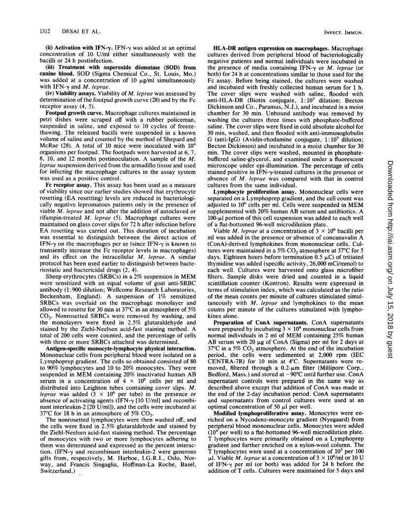
FIG. 3. The effect of SOD on EA rosetting of macrophages andmonocytes induced by viable M. leprae (M.L) in the presence ofIFN-y. Superoxide was involved in the killing of M. leprae bymacrophages and monocytes of the two patients denoted by + andO (P < 0.001, Student's t test). Each symbol represents resultsobtained from a single lepromatous patient. Results are expressed asthe percent decrease in EA rosetting from uninfected cultures.M.L+IFN, M. leprae and IFN-y added simultaneously;M.L+IFN+SOD, M. leprae, IFN--y, and SOD added simulta-neously.
of producing superoxide anions in response to viable M.leprae (18). To investigate whether the killing of M. leprae inIFN-y-activated lepromatous macrophages was due to theproduction of superoxide anions, monocyte-macrophagecultures were treated simultaneously with M. Ieprae andIFN--y in the presence of SOD.
In the present study, the decrease in EA rosetting was notabrogated in the presence of SOD in two of the cases
studied. This suggests that superoxide anions were notinvolved in the killing of M. leprae in these cases (Fig. 3).The effect of IFN--y on HLA-DR antigen expression on
macrophages from normal subjects and leprosy patients. Ourearlier study (6) had established that M. leprae infection ofmacrophage cultures from bacteriologically negative lepro-matous patients resulted in a significant decrease of HLA-DR-positive cells. In the present study, when IFN--y wasadded simultaneously with M. leprae, a significant increasein the percentage of macrophages expressing HLA-DR an-tigen, which was comparable to that seen with activationwith IFN-y alone, was observed (P < 0.005, Student's t test)(Table 1). However, addition of IFN-y 24 h postinfectionalso enhanced HLA-DR expression almost threefold overthat seen with infected cells alone (P < 0.001).
In tuberculoid patients and normal individuals, the ex-pected increase of HLA-DR-positive cells was observedafter stimulation with IFN--y and M. leprae (Table 1).
Monocyte-lymphocyte physical interaction in normal sub-jects and leprosy patients in response to M. leprae or activatingstimuli or both. Monocyte-lymphocyte interaction consti-tutes the first step in the development of a specific immuneresponse. Such a physical interaction is known to requirefunctionally intact macrophages, while the role of the lym-phocyte is considered passive because binding is not de-creased by metabolic poisoning of lymphocytes (16, 31).Besides macrophages, such clusters are also known to occurbetween dendritic cells and lymphocytes (14).
In our earlier study (6), we had reported a positiveantigen-dependent interaction in tuberculoid patients andnormal individuals, while in lepromatous patients the inter-action remained below the base line (<10%). The base-linevalue of interaction was established in normal individuals inthe absence of any antigen. M. leprae viability was notrequired for the antigen-dependent interaction. Thus, in thepresent set of experiments, the inability of ConA-derivedlymphokines and recombinant interleukin-2 to increase the
TABLE 1. Effect of IFN--y on HLA-DR antigen expression on macrophages from normal subjects and leprosy patients
Patient type % HLA-DR-positive cells with:and no. Control M. leprae IFN-y IFN-y + M. leprae" M. leprae + IFN-yb
Normal1 20 20 52 752 18 20 48 383 17 18 32 41Mean ± SEM 18 ± 1 19 ± 1 44 ± 6 51 ± 12P' NSd <0.01 <0.025
Tuberculoid1 25 35 70 722 19 22 50 463 15 26 31 34Mean ± SEM 20 ± 3 28 + 4 50 ± 11 50 ± 11P NS <0.05 <0.05
Lepromatous1 26 10 58 53 302 20 9 72 53 223 16 7 35 55 25Mean SEM 21 ± 3 9 1 55 ± 11 54 + 1 25 ± 3P <0.025 <0.005 <0.005 NSea M. Ieprae and IFN-y added simultaneously.b IFN-y added 24 h after M. leprae infection.' P is calculated relative to control.d NS, Not significant.e Not significant compared with control but significantly higher (P < 0.001) than infection with M. leprae alone.
INFECT. IMMUN.
on July 15, 2018 by guesthttp://iai.asm
.org/D
ownloaded from
M. LEPRAE KILLING AND ANTIGEN PRESENTATION 1315
degree of interaction in response to M. leprae in lepromatouscases (data not shown) is not surprising, even though killingof M. leprae was demonstrated.Modulation by IFN--y of the antigen-presenting capacity of
lepromatous macrophages to autologous T lymphocytes. Tocheck whether decreased IFN--y production could be di-rectly linked to aberrant macrophage function in leproma-tous patients, a coculture system of IFN--y-activated macro-phages and autologous T cells was used. It was essential toseparate the cells and treat the macrophages independentlywith IFN--y because it is known to have an antimitotic effecton lymphocytes. (See section on the lymphoproliferativeassay in Materials and Methods.)No response was noted with M. leprae when 105 resting,
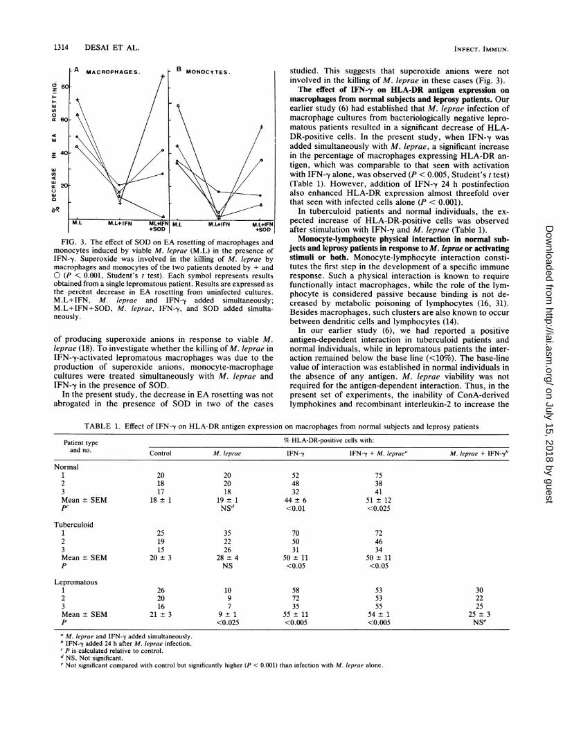
autologous, nylon-wool-separated T lymphocytes wereadded to macrophages stimulated with M. leprae and IFN-yfor 24 h (Fig. 4A). Significant T-cell proliferation was ob-served by using an identical protocol with PPD as depicted inthe two representative cases (Fig. 4B) (P < 0.001, Student'st test).M. leprae responsiveness in lepromatous leprosy patients
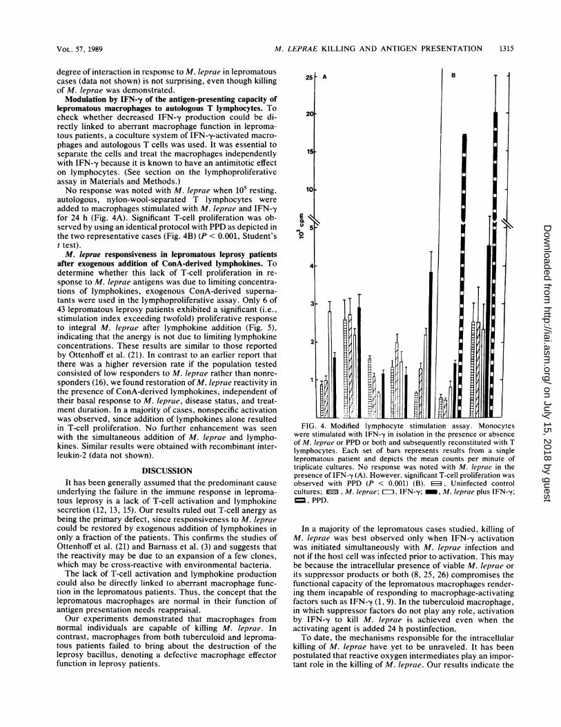
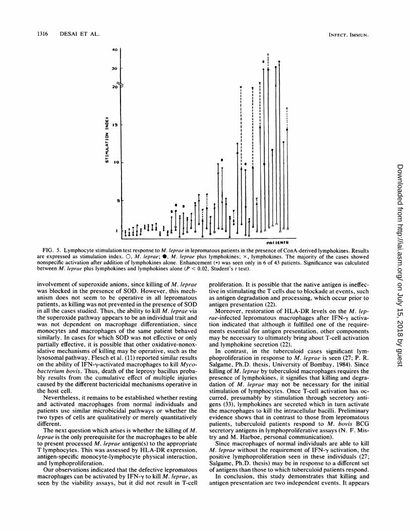
after exogenous addition of ConA-derived lymphokines. Todetermine whether this lack of T-cell proliferation in re-sponse to M. leprae antigens was due to limiting concentra-tions of lymphokines, exogenous ConA-derived superna-tants were used in the lymphoproliferative assay. Only 6 of43 lepromatous leprosy patients exhibited a significant (i.e.,stimulation index exceeding twofold) proliferative responseto integral M. leprae after lymphokine addition (Fig. 5),indicating that the anergy is not due to limiting lymphokineconcentrations. These results are similar to those reportedby Ottenhoff et al. (21). In contrast to an earlier report thatthere was a higher reversion rate if the population testedconsisted of low responders to M. Ieprae rather than nonre-sponders (16), we found restoration of M. leprae reactivity inthe presence of ConA-derived lymphokines, independent oftheir basal response to M. Ieprae, disease status, and treat-ment duration. In a majority of cases, nonspecific activationwas observed, since addition of lymphokines alone resultedin T-cell proliferation. No further enhancement was seenwith the simultaneous addition of M. leprae and lympho-kines. Similar results were obtained with recombinant inter-leukin-2 (data not shown).
DISCUSSIONIt has been generally assumed that the predominant cause
underlying the failure in the immune response in leproma-tous leprosy is a lack of T-cell activation and lymphokinesecretion (12, 13, 15). Our results ruled out T-cell anergy asbeing the primary defect, since responsiveness to M. lepraecould be restored by exogenous addition of lymphokines inonly a fraction of the patients. This confirms the studies ofOttenhoff et al. (21) and Barnass et al. (3) and suggests thatthe reactivity may be due to an expansion of a few clones,which may be cross-reactive with environmental bacteria.The lack of T-cell activation and lymphokine production
could also be directly linked to aberrant macrophage func-tion in the lepromatous patients. Thus, the concept that thelepromatous macrophages are normal in their function ofantigen presentation needs reappraisal.Our experiments demonstrated that macrophages from
normal individuals are capable of killing M. leprae. Incontrast, macrophages from both tuberculoid and leproma-tous patients failed to bring about the destruction of theleprosy bacillus, denoting a defective macrophage effectorfunction in leprosy patients.
10
4-
3
2-
tAll1llll~~IHnt VildlFIG. 4. Modified lymphocyte stimulation assay. Monocytes
were stimulated with IFN--y in isolation in the presence or absenceof M. leprae or PPD or both and subsequently reconstituted with Tlymphocytes. Each set of bars represents results from a singlelepromatous patient and depicts the mean counts per minute oftriplicate cultures. No response was noted with M. leprae in thepresence of IFN-y (A). However, significant T-cell proliferation wasobserved with PPD (P < 0.001) (B). ED, Uninfected controlcultures; , M. leprae; =II IFN-y; _, M. Ieprae plus IFN--y;=, PPD.
In a majority of the lepromatous cases studied, killing ofM. leprae was best observed only when IFN--y activationwas initiated simultaneously with M. leprae infection andnot if the host cell was infected prior to activation. This maybe because the intracellular presence of viable M. leprae orits suppressor products or both (8, 25, 26) compromises thefunctional capacity of the lepromatous macrophages render-ing them incapable of responding to macrophage-activatingfactors such as IFN-y (1, 9). In the tuberculoid macrophage,in which suppressor factors do not play any role, activationby IFN--y to kill M. leprae is achieved even when theactivating agent is added 24 h postinfection.To date, the mechanisms responsible for the intracellular
killing of M. leprae have yet to be unraveled. It has beenpostulated that reactive oxygen intermediates play an impor-tant role in the killing of M. leprae. Our results indicate the
VOL. 57, 1989
on July 15, 2018 by guesthttp://iai.asm
.org/D
ownloaded from
1316 DESAI ET AL.
40
30
20
Laz 15
z0
-I
U) 101
I~~~~~~~~~~~~~~~~~~ I III
PAT IENTO
FIG. 5. Lymphocyte stimulation test response to M. leprae in lepromatous patients in the presence of ConA-derived lymphokines. Resultsare expressed as stimulation index. 0, M. Ieprae; 0, M. Ieprae plus lymphokines; x, lymphokines. The majority of the cases showednonspecific activation after addition of lymphokines alone. Enhancement (*) was seen only in 6 of 43 patients. Significance was calculatedbetween M. Ieprae plus lymphokines and lymphokines alone (P < 0.02, Student's t test).
involvement of superoxide anions, since killing of M. lepraewas blocked in the presence of SOD. However, this mech-anism does not seem to be operative in all lepromatouspatients, as killing was not prevented in the presence of SODin all the cases studied. Thus, the ability to kill M. leprae viathe superoxide pathway appears to be an individual trait andwas not dependent on macrophage differentiation, sincemonocytes and macrophages of the same patient behavedsimilarly. In cases for which SOD was not effective or onlypartially effective, it is possible that other oxidative-nonox-idative mechanisms of killing may be operative, such as thelysosomal pathway. Flesch et al. (11) reported similar resultson the ability of IFN--y-activated macrophages to kill Myco-bacterium bovis. Thus, death of the leprosy bacillus proba-bly results from the cumulative effect of multiple injuriescaused by the different bactericidal mechanisms operative inthe host cell.
Nevertheless, it remains to be established whether restingand activated macrophages from normal individuals andpatients use similar microbicidal pathways or whether thetwo types of cells are qualitatively or merely quantitativelydifferent.The next question which arises is whether the killing of M.
Ieprae is the only prerequisite for the macrophages to be ableto present processed M. leprae antigen(s) to the appropriateT lymphocytes. This was assessed by HLA-DR expression,antigen-specific monocyte-lymphocyte physical interaction,and lymphoproliferation.Our observations indicated that the defective lepromatous
macrophages can be activated by IFN--y to kill M. leprae, asseen by the viability assays, but it did not result in T-cell
proliferation. It is possible that the native antigen is ineffec-tive in stimulating the T cells due to blockade at events, suchas antigen degradation and processing, which occur prior toantigen presentation (22).Moreover, restoration of HLA-DR levels on the M. lep-
rae-infected lepromatous macrophages after IFN--y activa-tion indicated that although it fulfilled one of the require-ments essential for antigen presentation, other componentsmay be necessary to ultimately bring about T-cell activationand lymphokine secretion (22).
In contrast, in the tuberculoid cases significant lym-phoproliferation in response to M. Ieprae is seen (27; P. R.Salgame, Ph.D. thesis, University of Bombay, 1984). Sincekilling of M. leprae by tuberculoid macrophages requires thepresence of lymphokines, it signifies that killing and degra-dation of M. leprae may not be necessary for the initialstimulation of lymphocytes. Once T-cell activation has oc-curred, presumably by stimulation through secretory anti-gens (33), lymphokines are secreted which in turn activatethe macrophages to kill the intracellular bacilli. Preliminaryevidence shows that in contrast to those from lepromatouspatients, tuberculoid patients respond to M. bovis BCGsecretory antigens in lymphoproliferative assays (N. F. Mis-try and M. Harboe, personal communication).
Since macrophages of normal individuals are able to killM. leprae without the requirement of IFN-y activation, thepositive lymphoproliferation seen in these individuals (27;Salgame, Ph.D. thesis) may be in response to a different setof antigens than those to which tuberculoid patients respond.
In conclusion, this study demonstrates that killing andantigen presentation are two independent events. It appears
I
INFECT. IMMUN.
5 t
on July 15, 2018 by guesthttp://iai.asm
.org/D
ownloaded from

M. LEPRAE KILLING AND ANTIGEN PRESENTATION 1317
that the ability of the macrophage per se to kill M. Iepraemay be of greater importance than lymphocyte-mediatedactivation for protection against M. leprae infection.
ACKNOWLEDGMENTS
We thank E. Storrs, Florida Institute of Technology, Melbourne,Fla., and LEPRA for the armadillo tissue. We are grateful to DeepaVaradkar and the animal house staff for assistance in the mousefootpad studies. We are also grateful to the staff of AcworthHospital, Wadala, Bombay, for the supply of patient material.
LITERATURE CITED1. Agarwal, S., N. Vemuri, and P. R. Mahadevan. 1986. Macro-
phage membrane alterations in leprosy as determined by changein sialic acid level. J. Clin. Lab. Immunol. 19:119-122.
2. Ambrose, E. J., N. H. Antia, T. J. Birdi, P. R. Mahadevan, L.Mester, N. F. Mistry, R. Mukherjee, and V. P. Shetty. 1985. Theaction of deoxyfructose serotonin on intracellular bacilli and onhost response in leprosy. Lepr. Rev. 55:221-227.
3. Barnass, S., J. Mace, J. Steel, P. Torres, and B. Gervasoni, R.Ravioli, J. A. de las Terencio, G. A. W. Rook, and M. F. R.Waters. 1986. Prevalence and specificity of the enhancing effectof 3 types of IL-2 on T cell responsiveness in 97 lepromatousleprosy patients of mixed ethnic origin. Clin. Exp. Immunol.64:41-49.
4. Birdi, T. J., and N. H. Antia. 1984. An in vitro method ofscreening for drug sensitivity. IRCS Med. Sci. 12:256-257.
5. Birdi, T. J., N. F. Mistry, P. R. Mahadevan, and N. H. Antia.1983. Alterations in the membrane of macrophages from leprosypatients. Infect. Immun. 41:121-127.
6. Birdi, T. J., N. F. Mistry, P. R. Mahadevan, and N. H. Antia.1984. Antigen specific macrophage-lymphocyte interaction inlepromatous leprosy. J. Clin. Lab. Immunol. 13:189-194.
7. Birdi, T. J., P. R. Salgame, and N. H. Antia. 1979. The role ofmacrophages in leprosy as studied by protein synthesis ofmacrophages from resistant and susceptible hosts. A mouse andhuman study. Lepr. India 51:23-42.
8. Birdi, T. J., P. R. Salgame, P. R. Mahadevan, and N. H. Antia.1984. An indomethacin sensitive suppressor factor released bymacrophages of leprosy patients. J. Biosci. 6:125-134.
9. Churchill, W. H., and C. Wong. 1980. Mediator-induced mac-rophage activation, as shown by enhanced cytotoxicity fortumor, requires macrophage surface fucose and sialic acid. Cell.Immunol. 55:490-498.
10. Dhople, A. M., and K. J. Green. 1985. Adenosine triphosphateand (3H)-thymidine as indicators of metabolic status and viabil-ity of Mycobacterium leprae. IRCS Med. Sci. 13:779-780.
11. Flesch, I. E. A., and S. H. E. Kaufmann. 1988. Attempts tocharacterize the mechanisms involved in mycobacterial growthinhibition by gamma-interferon-activated bone marrow macro-phages. Infect. Immun. 56:1464-1469.
12. Godal, T., B. Mykestad, D. R. Sameul, and B. Myrvang. 1971.Characterization of the cellular immune defect in lepromatousleprosy: a specific lack of circulating Mycobacterium lepraereactive lymphocytes. Clin. Exp. Immunol. 9:821-831.
13. Haregewoin, A., J. Longley, G. Bjune, A. S. Mustafa, and T.Godal. 1985. The role of interleukin-2 (IL-2) in the specificunresponsiveness of lepromatous leprosy to Mycobacteriumleprae: studies in vitro and in vivo. Immunol. Lett. 11:249-252.
14. Inaba, K., and R. M. Steinman. 1986. Accessory cell-T lympho-cyte interactions. Antigen-dependent and independent cluster-ing. J. Exp. Med. 163:247-261.
15. Kaplan, G., and Z. A. Cohn. 1985. Cellular immunity in lepro-
matous and tuberculoid leprosy. Immunol. Lett. 11:205-209.16. Lipsky, P. E., and A. S. Rosenthal. 1975. Macrophage-lympho-
cyte interaction. II. Antigen-mediated physical interaction be-tween immune guinea pig lymph node lymphocytes and synge-neic macrophages. J. Exp. Med. 141:138-154.
17. Mahadevan, P. R., R. Jagannathan, A. Bhagria, S. Vejare, andS. Agarwal. 1986. Host-pathogen interaction-new in vitro drugtest systems against Mycobacterium leprae their possibilitiesand limitations. Lepr. Rev. 57(Suppl. 3):182-200.
18. Marolia, J., and P. R. Mahadevan. 1987. Superoxide productionfrom macrophages of leprosy patients after stimulation withMycobacterium leprae. J. Biosci. 12:273-279.
19. Mistry, N. F., T. J. Birdi, and N. H. Antia. 1986. M. lepraephagocytosis and its association with membrane changes inmacrophages from leprosy patients. Parasite Immunol. 8:129-138.
20. Nogueria, N., G. Kaplan, E. Levy, E. N. Sarno, P. Kushner, A.Granelli-Piperno, L. Vieira, C. V. Gould, W. Levis, R. Steinman,Y. K. Yip, and Z. A. Cohn. 1983. Defective interferon produc-tion in leprosy. Reversal with antigen and interleukin 2. J. Exp.Med. 158:2165-2170.
21. Ottenhoff, T. H. M., D. G. Elferink, and R. R. P. deVries. 1984.Unresponsiveness to M. leprae in lepromatous leprosy in vitro:reversible or not? Int. J. Lepr. 52:419-424.
22. Pierce, S. K., and E. Margoliasch. 1988. Antigen processing: aninterim report. Trends Biochem. Sci. 13:27-29.
23. Prasad, H. K., and I. Nath. 1981. Incorporation of 3H-thymidinein Mycobacterium leprae within differentiated human macro-phages. J. Med. Microbiol. 14:279-293.
24. Ridley, D. S., and W. H. Jopling. 1966. Classification of leprosyaccording to immunity, a five group system. Int. J. Lepr.34:255-273.
25. Salgame, P. R., P. R. Mahadevan, and N. H. Antia. 1983.Mechanisms of immunosuppression in leprosy: presence ofsuppressor factor(s) from macrophages of lepromatous patients.Infect. Immun. 40:1119-1126.
26. Satish, M., L. K. Bhutani, A. K. Sharma, and I. Nath. 1983.Monocyte-derived soluble suppressor factor(s) in patients withlepromatous leprosy. Infect. Immun. 42:890-899.
27. Shankar, P., D. Wallach, and M. A. Bach. 1985. Interleukein-2induced T-cell response to M. leprae in lepromatous leprosy:reversion of a suppressor mechanism or expansion of a small M.leprae reactive T-cell pool. Int. J. Lepr. 53:649-652.
28. Shepard, C. C., and D. H. McRae. 1965. M. leprae in mice:minimal infectious dose, relationship between staining qualityand infectivity, and effect of cortisone. J. Bacteriol. 89:365-372.
29. Sibley, D. L., and J. L. Krahenbuhl. 1987. Mycobacteriumleprae-burdened macrophages are refractory to activation bygamma interferon. Infect. Immun. 55:446-450.
30. Skamene, E., and P. Gros. 1983. Role of macrophages inresistance against infectious diseases. Clin. Immunol. Allergy3:539-560.
31. Thomas, J., J. Schroer, K. Yokomuro, J. Blake, and A.Rosenthal. 1980. Macrophage lymphocyte interaction and ge-netic control of immune responsiveness, p. 165-179. In M.Escobar and H. Friedman (ed.), Macrophages and lympho-cytes, part B. Plenum Publishing Corp., New York.
32. Vejare, S., and P. R. Mahadevan. 1987. Importance of deter-mining viability of Mycobacterium leprae inside macrophages-an in vitro method using uracil. J. Biosci. 11:455-463.
33. Wiker, H. G., M. Harboe, S. Nagai, M. E. Patarroyo, C.Ramirez, and N. Cruz. 1986. MPB 59, a widely cross reactingprotein of Mycobacterium bovis. Int. Arch. Allergy Appl.Immunol. 81:307-314.
VOL. 57, 1989
on July 15, 2018 by guesthttp://iai.asm
.org/D
ownloaded from