Correlation Between Functional Myocardial Perfusion ... · imaging, angiography, or autopsy. This...
3
Copyright © 2019 Asian Society of Cardiovascular Imaging 27 INTRODUCTION Myocardial bridging (MB) is a congenital anomaly charac- terized by an intramyocardial route of a coronary artery. e coronary arteries tunnel into the myocardium rather than rest- ing on top of it [1]. MB can occur in almost all epicardial arter- ies; however, the incidence is highest in the leſt anterior descend- ing artery (LAD) [1]. MB patients are oſten asymptomatic, and diagnosis is oſten made from incidental findings of diagnostic imaging, angiography, or autopsy. is congenital anomaly has a good prognosis, with a reported 5-year survival greater than 97% [2]. However, prognosis can vary from benign to fatal. In symptomatic patients, MB can present with a variety of clinical manifestations ranging from myocardial ischemia, myocardial infarction, cardiac arrhythmia, to sudden cardiac death [2]. CASE REPORT A 56-year-old Indian woman with a history of diabetes mel- litus presented with atypical chest pain. Her electrocardiogram (ECG), chest radiograph, cardiac enzymes and other blood pa- rameters were unremarkable. Using the one-day, stress-rest gat- ed Tc-99m tetrofosmin protocol, myocardial perfusion imaging (MPI) was performed for cardiac assessment. Pharmacological stress was applied using dipyridamole infused over 4 minutes at 140 µg/kg/min. ere were no symptoms or ECG changes to suggest dipyridamole-induced myocardial ischemia. e myo- cardial perfusion single photon emission computed tomogra- phy (SPECT) images showed mild reversible reduced tracer uptake in the apical anterior segment of the leſt ventricle (Fig. 1). Other myocardial segments showed no demonstrable per- fusion defect. e leſt ventricular cavity was not dilated. Subsequently, the patient underwent cardiac CT, which showed a 2-cm-long superficial MB located at the mid segment of the LAD (Figs. 2 and 3). e lumen appeared patent without stenosis. e leſt circumflex and right coronary arteries were otherwise normal. The coronary calcium score (Agatston cc is is an Open Access article distributed under the terms of the Creative Commons Attribution Non-Commercial License (https://creativecommons.org/licenses/by- nc/4.0) which permits unrestricted non-commercial use, distribution, and reproduc- tion in any medium, provided the original work is properly cited. CVIA Correlation Between Functional Myocardial Perfusion Imaging and Anatomical Cardiac CT in a Case of Myocardial Bridging Mohd Fazrin Mohd Rohani 1,2 , Ahmad Khairuddin Mohamed Yusof 3 , Hazlin Hashim 2 1 Hospital Universiti Sains Malaysia, Kubang Kerian, Malaysia 2 Advanced Medical and Dental Institute, Universiti Sains Malaysia, Bertam, Malaysia 3 Institut Jantung Negara, Kuala Lumpur, Malaysia Received: October 23, 2018 Revised: November 20, 2018 Accepted: November 28, 2018 Corresponding author Mohd Fazrin Mohd Rohani, MD Hospital Universiti Sains Malaysia, Jabatan Perubatan Nuklear, Hospital Universiti Sains Malaysia, Jalan Raja Perempuan Zainab Ii, 16150, Kubang Kerian, Malaysia Tel: 60-97673486 Fax: 60-97676691 E-mail: [email protected] Myocardial perfusion imaging (MPI) with single photon emission computed tomography (SPECT) is generally used to evaluate myocardial ischemia in patients with known or sus- pected coronary artery disease. Cardiac CT, on the other hand, enables non-invasive visualiza- tion of the coronary vessels. Myocardial bridging (MB) is a congenital anomaly characterized by an intramyocardial route of a coronary artery. It is commonly considered benign and rarely requires further evaluation or intervention. However, its clinical complications can be hazard- ous. We report a patient whose SPECT MPI study demonstrated a mild reversible perfusion defect at the apical anterior segment of the left ventricle that was later attributed to MB vi- sualized on cardiac CT. Key words Myocardial perfusion imaging · Myocardial bridging · Multidetector computed tomography · SPECT. pISSN 2508-707X / eISSN 2508-7088 CVIA 2019;3(1):27-29 https://doi.org/10.22468/cvia.2018.00241 CASE REPORT
Transcript of Correlation Between Functional Myocardial Perfusion ... · imaging, angiography, or autopsy. This...
Copyright © 2019 Asian Society of Cardiovascular Imaging 27
INTRODUCTION
Myocardial bridging (MB) is a congenital anomaly charac-terized by an intramyocardial route of a coronary artery. The coronary arteries tunnel into the myocardium rather than rest-ing on top of it [1]. MB can occur in almost all epicardial arter-ies; however, the incidence is highest in the left anterior descend-ing artery (LAD) [1]. MB patients are often asymptomatic, and diagnosis is often made from incidental findings of diagnostic imaging, angiography, or autopsy. This congenital anomaly has a good prognosis, with a reported 5-year survival greater than 97% [2]. However, prognosis can vary from benign to fatal. In symptomatic patients, MB can present with a variety of clinical manifestations ranging from myocardial ischemia, myocardial infarction, cardiac arrhythmia, to sudden cardiac death [2].
CASE REPORT
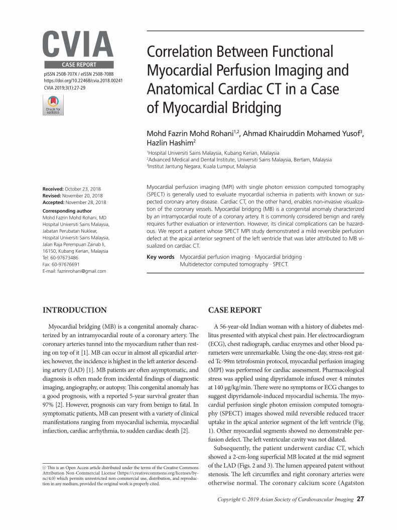
A 56-year-old Indian woman with a history of diabetes mel-litus presented with atypical chest pain. Her electrocardiogram (ECG), chest radiograph, cardiac enzymes and other blood pa-rameters were unremarkable. Using the one-day, stress-rest gat-ed Tc-99m tetrofosmin protocol, myocardial perfusion imaging (MPI) was performed for cardiac assessment. Pharmacological stress was applied using dipyridamole infused over 4 minutes at 140 µg/kg/min. There were no symptoms or ECG changes to suggest dipyridamole-induced myocardial ischemia. The myo-cardial perfusion single photon emission computed tomogra-phy (SPECT) images showed mild reversible reduced tracer uptake in the apical anterior segment of the left ventricle (Fig. 1). Other myocardial segments showed no demonstrable per-fusion defect. The left ventricular cavity was not dilated.
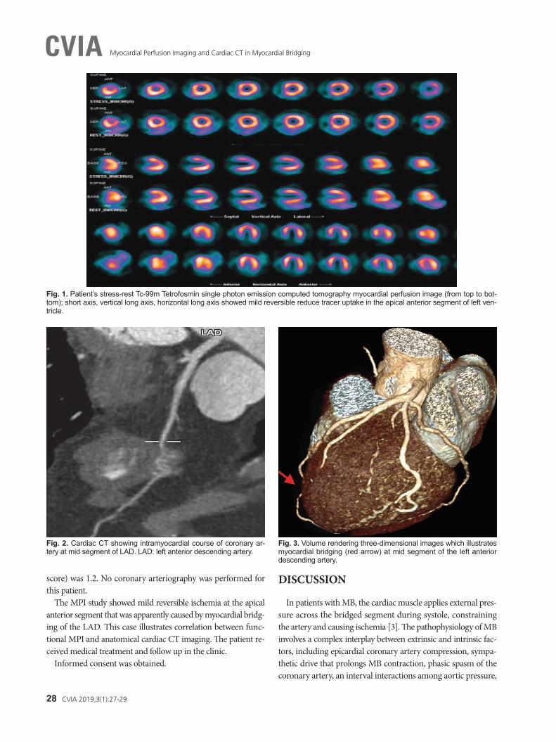
Subsequently, the patient underwent cardiac CT, which showed a 2-cm-long superficial MB located at the mid segment of the LAD (Figs. 2 and 3). The lumen appeared patent without stenosis. The left circumflex and right coronary arteries were otherwise normal. The coronary calcium score (Agatston
cc This is an Open Access article distributed under the terms of the Creative Commons Attribution Non-Commercial License (https://creativecommons.org/licenses/by-nc/4.0) which permits unrestricted non-commercial use, distribution, and reproduc-tion in any medium, provided the original work is properly cited.
CVIA Correlation Between Functional Myocardial Perfusion Imaging and Anatomical Cardiac CT in a Case of Myocardial BridgingMohd Fazrin Mohd Rohani1,2, Ahmad Khairuddin Mohamed Yusof3, Hazlin Hashim2
1Hospital Universiti Sains Malaysia, Kubang Kerian, Malaysia 2Advanced Medical and Dental Institute, Universiti Sains Malaysia, Bertam, Malaysia 3Institut Jantung Negara, Kuala Lumpur, Malaysia
Received: October 23, 2018Revised: November 20, 2018Accepted: November 28, 2018
Corresponding authorMohd Fazrin Mohd Rohani, MDHospital Universiti Sains Malaysia, Jabatan Perubatan Nuklear, Hospital Universiti Sains Malaysia, Jalan Raja Perempuan Zainab Ii, 16150, Kubang Kerian, MalaysiaTel: 60-97673486Fax: 60-97676691E-mail: [email protected]
Myocardial perfusion imaging (MPI) with single photon emission computed tomography (SPECT) is generally used to evaluate myocardial ischemia in patients with known or sus-pected coronary artery disease. Cardiac CT, on the other hand, enables non-invasive visualiza-tion of the coronary vessels. Myocardial bridging (MB) is a congenital anomaly characterized by an intramyocardial route of a coronary artery. It is commonly considered benign and rarely requires further evaluation or intervention. However, its clinical complications can be hazard-ous. We report a patient whose SPECT MPI study demonstrated a mild reversible perfusion defect at the apical anterior segment of the left ventricle that was later attributed to MB vi-sualized on cardiac CT.
Key words Myocardial perfusion imaging · Myocardial bridging · Multidetector computed tomography · SPECT.
pISSN 2508-707X / eISSN 2508-7088
CVIA 2019;3(1):27-29https://doi.org/10.22468/cvia.2018.00241
CASE REPORT
28 CVIA 2019;3(1):27-29
Myocardial Perfusion Imaging and Cardiac CT in Myocardial BridgingCVIA
DISCUSSION
In patients with MB, the cardiac muscle applies external pres-sure across the bridged segment during systole, constraining the artery and causing ischemia [3]. The pathophysiology of MB involves a complex interplay between extrinsic and intrinsic fac-tors, including epicardial coronary artery compression, sympa-thetic drive that prolongs MB contraction, phasic spasm of the coronary artery, an interval interactions among aortic pressure,
score) was 1.2. No coronary arteriography was performed for this patient.
The MPI study showed mild reversible ischemia at the apical anterior segment that was apparently caused by myocardial bridg-ing of the LAD. This case illustrates correlation between func-tional MPI and anatomical cardiac CT imaging. The patient re-ceived medical treatment and follow up in the clinic.
Informed consent was obtained.
Fig. 1. Patient’s stress-rest Tc-99m Tetrofosmin single photon emission computed tomography myocardial perfusion image (from top to bot-tom); short axis, vertical long axis, horizontal long axis showed mild reversible reduce tracer uptake in the apical anterior segment of left ven-tricle.
Fig. 2. Cardiac CT showing intramyocardial course of coronary ar-tery at mid segment of LAD. LAD: left anterior descending artery.
Fig. 3. Volume rendering three-dimensional images which illustrates myocardial bridging (red arrow) at mid segment of the left anterior descending artery.

www.e-cvia.org 29
Mohd Fazrin Mohd Rohani, et al CVIAdiastolic flow, heart rate, and transmural perfusion gradient [1,3]. Additionally, impaired diastolic flow can cause depres-surization of the septal branches within the MB, resulting in an intrabridge ischemia, also known as the “branch steal” phenom-enon [1,3].
Presently, there are no standardized diagnostic criteria or a gold standard for evaluation of MB [1]. The presence of MB is often noted on anatomical imaging, such as CT scan, which al-lows characterization of its location, length, and depth; arterial constriction and assessment of atherosclerosis [1,4]. However, the functional significance of MB is often determined by SPECT or positron emission tomography (PET) MPI. Based on the study by Gawor et al. [5] of 42 patients without coronary artery disease, the occurrence of myocardial perfusion defect in bridging patients is linked to the degree of coronary artery nar-rowing during systole, and patients usually experienced no symptoms if the coronary artery constriction was less than 50% and were symptomatic if constriction was greater than 70% [5].
Hybrid cardiac imaging with SPECT/CT [6] and PET/CT [4] allows concurrent evaluation of the coronary vascular anat-omy and myocardial blood flow. In a study by Monroy-Gonza-lez et al. [4] involving 131 patients who underwent both 13N-am-monia PET and cardiac CT, there was no significant correlation between quantitative perfusion analysis and the anatomical fea-tures of MB, such as its length and depth, displayed on CT [4]. Furthermore, myocardial perfusion reserve (MPR), a marker of microvascular dysfunction quantified by PET, was found to be low in patients with MB of the LAD [4]. MPR measured by PET can be used as a tool to identify MB patients who are at in-creased risk of cardiac event [4].
In practice, a combination of both functional and anatomi-cal imaging can guide the clinician in treatment planning and monitoring of MB [6]. In a case report by Lim et al. [7], a 43- year-old man with established MB refractory to medical treat-ment underwent a combination of cardiac CT with MPI. The study demonstrated MB in the mid LAD and reversible perfu-sion defect in the anteroseptal wall [7]. The MB was surgically corrected with supraarterial myotomy, and subsequent imaging showed successful unroofing of the MB segment with a normal result on myocardial perfusion study [7].
Treatment is often unnecessary in the asymptomatic MB pa-tient. For symptomatic patients, medical treatment with beta
blockers causes reduction in compression by the muscular band, slows the heart rate, and prolongs the diastolic period [1]. Invasive therapy such as percutaneous coronary intervention and coronary artery bypass graft, are usually reserved for se-verely symptomatic refractory cases and cases that cannot be controlled with oral medications [1,2]. To date, there are no ap-propriate guidelines on suitable medical treatment or revascu-larization techniques for MB. Additionally, it is uncertain if in-tervention in bridging patients with mild or moderate ischemia achieves better outcomes. Thus, there is uncertainty in optimal patient management.
In conclusion, although uncommon, MB should be included in the differential diagnosis for chest pain. A combination of an-atomical and functional imaging in patients with MB provides valuable hemodynamic information on the bridging segment, which guides the clinician’s determination of a treatment plan and aids in monitoring of therapy response.
Conflicts of InterestThe authors declare that they have no conflict of interest.
REFERENCES
1. Tarantini G, Migliore F, Cademartiri F, Fraccaro C, Iliceto S. Left anterior descending artery myocardial bridging: a clinical approach. J Am Coll Cardiol 2016;68:2887-2899.
2. Ko SM, Choi JS, Nam CW, Hur SH. Incidence and clinical significance of myocardial bridging with ECG-gated 16-row MDCT coronary angiog-raphy. Int J Cardiovasc Imaging 2008;24:445-452.
3. Gould KL, Johnson NP. Myocardial bridges: lessons in clinical coronary pathophysiology. JACC Cardiovasc Imaging 2015;8:705-709.
4. Monroy-Gonzalez AG, Alexanderson-Rosas E, Prakken NHJ, Juarez-Orozco LE, Walls-Laguarda L, Berrios-Barcenas EA, et al. Myocardial bridging of the left anterior descending coronary artery is associated with reduced myocardial perfusion reserve: a 13N-ammonia PET study. Int J Cardiovasc Imaging 2018 Sep 28 [Epub]. https://doi.org/10.1007/s10554-018-1460-8.
5. Gawor R, Kuśmierek J, Płachcińska A, Bieńkiewicz M, Drożdż J, Pi-otrowski G, et al. Myocardial perfusion GSPECT imaging in patients with myocardial bridging. J Nucl Cardiol 2011;18:1059-1065.
6. Singh D, Vadi SK, Parmar M, Sood A, Mehrotra S, Mittal BR. Role of 99mTc-sestamibi gated SPECT/CT myocardial perfusion imaging in the manage-ment of patients with myocardial bridging and its correlation with coro-nary angiography. J Cardiol Cases 2017;15:136-140.
7. Lim JW, Lee H, Her K, Park HW, Shin KE. Myocardial CT perfusion im-aging for pre- and postoperative evaluation of myocardial ischemia in a patient with myocardial bridging: a case report. Medicine (Baltimore) 2017;96:e8277.