Knee Osteoarthritis. The Valgus and Varus tests Knee range of motion Effusion Crepitus.

University of North DakotaUND Scholarly Commons
Physical Therapy Scholarly Projects Department of Physical Therapy
1993
Correlation between Forefoot Varus and PassiveKnee ExtensionScott B. NiceUniversity of North Dakota
Follow this and additional works at: https://commons.und.edu/pt-grad
Part of the Physical Therapy Commons
This Scholarly Project is brought to you for free and open access by the Department of Physical Therapy at UND Scholarly Commons. It has beenaccepted for inclusion in Physical Therapy Scholarly Projects by an authorized administrator of UND Scholarly Commons. For more information,please contact [email protected].
Recommended CitationNice, Scott B., "Correlation between Forefoot Varus and Passive Knee Extension" (1993). Physical Therapy Scholarly Projects. 331.https://commons.und.edu/pt-grad/331
CORRELATION BETWEEN FOREFOOT VARUS AND PASSIVE KNEE EXTENSION
by
Scott B. Nice Bachelor of Science Physical Therapy
University of North Dakota, 1987
An Independent Study
Submitted to the Graduate Faculty of the
Department of Physical Therapy
School of Medicine
University of North Dakota
in partial fulfillment of the requirements
for the degree of
Master of Physical Therapy
Grand Forks, North Dakota May 1993

This Indepedent Study, sumitted by Scott B. Nice in partial fulfillment of the requirements for the Degree of Master of Phyiscal Therapy from the University of North Dakota, has been read by the Chairperson of Physical Therapy under whom the work has been done and is hereby approved.
Physical Therapy)
ii
Title
Department
Degree
PERMISSION
Correlation Between Forefoot Varus and Passive Knee Extension
Physical Therapy
Master of Physical Therapy
In presenting this independent study in partial fulfillment of the requirements for a graduate degree from the University of North Dakota, I agree that the library of the University shall make it freely available for inspection. I further agree that permission for extensive copying for scholarly purposes may be granted by the professor who supervised my independent study or, in his absence, by the Chairperson of the department or the Dean of the Graduate School. It is understood that any copying or publication or other use of this independent study of part thereof for financial gain shall not be allowed without my written permission. It is also understood that due recognition shall be given to me and to the University of North Dakota in any scholarly use which may be made of any material in my independent study.
Signature
Date
iii
TABLE OF CONTENTS
LIST OF TABLES
ACKNOWLEDGEMENTS
ABSTRACT
CHAPTER
I.
II.
III.
IV.
V.
APPENDIX
A.
B.
REFERENCES
Introduction
Experimental Hypotheses
Literature review
Materials and Methods
Results
Discussion
Screening Questionnaire
Consent Form
iv
v
vi
vii
1
1
2
6
8
11
13
14
15
LIST OF TABLES
Table Page
1. Total Forefoot Measurements .................... 8
2. Total Passive Knee Extension Measurements ...... 9
\.1
ACKNOWLEDGEMENTS
The editor expresses sincere appreciation to Bud
Wessman, who has provided an unmeasurable amount of time,
effort and input to bring this project to conclusion.
vi
ABSTRACT
The purpose of this study was to examine if a
relationship exists between the amount of passive knee
extension measured along with the degree of forefoot varus.
The intent was to demonstrate that a positive relationship
does exist.
Twenty single limbs were tested and all met the
criteria set. Forefoot measurements were taken in the prone
position, the plane of the lesser metatarsal bones was
measured in relation to the bisection of the posterior
aspect of the heel. Passive knee extension was recorded by
measuring the distance of the lift of the calcaneus from the
table, with the thigh stabilized and traction employed
through the great toe.
Direct positive results were generated when correlated
mean values of knee and foot groups were compared. When
classified as knee values with forefoot varus types compared
to those with a forefoot valgus, highly significant results
were generated.
It was concluded that the great toe traction technique
may provide examiners with a valuable screening tool to
predict certain generalities of their patients. Upon which
physical therapy measures can be employed.
vii
I. INTRODUCTION
The purpose of this study was to investigate the
relationship between the degree of forefoot varus and the
amount of passive knee extension present in any given
individual. The topic originated as result the of four
years of biomechanical lower extremity evaluation and gait
assessment in the physical therapy clinical environment. As
subsequent evaluations were performed, and more and more
patients assessed, certain generalities became evident. To
determine if such generalities do truly exist, a non-bias
method to test the hypothesis had been designed. If true,
the answer to the research question will be that a positive
correlation does exist between the degree of forefoot varus
and the degree of passive knee extension. This study may
provide a simple screening procedure for those who are
involved in gait analysis, to develop insight and guidance
towards diagnosing the patient's problem, and in designing a
proper treatment regime.
1
II. LITERATURE REVIEW
The foot performs many essential dynamic functions
during gait that permits the body to progress forward in a
normal walking pattern.~ Rapid passive pronation of the
foot occuring immediately after heel contact, with
resultant supination in response to the ground reaction
forces imposed, permits the foot to convert from a mobile
adaptor to a rigid lever, allowing for normal propulsion at
toe off.~,2 This driving force is dependent on many
factors, most important being the delicate balance of the
subtalar joint motions of pronation and supination as they
occur normally during their appropriate phases of gait.
Subtalar joint neutral is that position at which the
subtalar joint will function optimally, allowing maximum
pronation and supination to occur. 2 ,3
The subtalar joint has been described as a torque
convertor that has direct functional correlation with the
midtarsal joint, as well as the other joints of the entire
lower extremity. When held in a subtalar neutral position,
the midtarsal joint loses its ability to dorsiflex, evert,
and abduct. The midtarsal joint is then considered to be
10cked.,,2,3,4 This is accomplished in a open kinetic
position by applying a dorsiflectory force to the fourth and
2
3
fifth metatarsal segments. This force is only taken until
resistance is met. 2 ,3
Pronation can be defined as a tri-plane motion
involving elements of plantarflexion, eversion, and
adduction.~,2,3,4 Excessive pronation has been described as
a common compensatory motion of gait. Pronation that
continues beyond midstance prevents the conversion of the
foot into a rigid lever for propulsion, and imposes
functional limitations upon the joint structures of the
lower limb as the foot prepares to leave the ground.~ The
resultant subtalar joint pronation and its compulsory
triplane motion of calcaneal eversion and talar adduction
with plantarflexion, continues in response to the flexion
moment at the knee. Obligatory internal rotation of the
tibia also occurs, placing stress on the knee joint.~,2,3
In order to maintain talocrural joint congruen~y during the
initial loading phase of gait prolonged, excessive subtalar
joint pronation necessitates additional internal rotation
and inclination of the tibia and flexion of the knee. Such
forces occuring during midstance through terminal stance,
result in delayed or reduced extension of the tibiofemoral
joint. Torsional stress is created as the lower extremity
attempts to extend while the tibia is prolonged in medial
inclination.~ Coplin 5 studied this phenomenon, and divided
her subjects into two groups, a pronating group and a normal
group. She tested all of her subjects for their total
4
available passive transverse rotatory motion at the knee at
90, 15 and five degrees of knee flexion. Coplin's5 results
manifested that tibial rotation was significantly greater in
the pronatory group when compared to normals at five degrees
of knee flexion. Although her studies were performed in a
non-weightbearing position, it appears that as the knee
moves towards terminal extension, an increase in transverse
rotation at the knee is noted.
The anterior cruciate ligament is a key element in this
study because of its relationship with knee extension. The
anterior cruciate is the ligament that has been noted to
prevent hyperextension (recurvatum) of the knee and anterior
movement of the tibia on the femur.G",s The anterior
cruciate also becomes taut in knee extension, and checks
movement of the lateral condyle. G" Combining the above two
statements, the anterior cruciate ligament has a very
important function with respect to terminal rotation and
locking of the knee.'
The other key element is an appreciation for the amount
of forefoot varus present. It should be noted that in
conjunction with forefoot varus, it is common to also assess
the position of the subtalar joint, but in this study, only
the forefoot position will be addressed. When present, even
a small degree of forefoot varus may influence total lower
extremity joint motion in a weight bearing position. 2, 3,4
5
Most commonly, these compensatory motions are demonstrated
by the lower extremity assuming a position of some degree of
pronation. 2 ,3,4

III. MATERIALS AND METHODS ..
The materials utilized in this study were basic and
simple; a goniometer, metric ruler, plinth, and thigh
stabilization belt. Twenty subjects were tested, none with
any past orthopedic, neurological, or congenital abnormality
that would influence the result of the test. Upon screening
each subject, a single extremity from each was examined for
the study. The first step of the procedure was to assess
the forefoot in a subtalar joint neutral position with the
subject in a prone position.
Up~m establishing the subtalar joint neutral position,
assessm~pt of forefoot position was accomplished. Beginning
by bisecting the calcaneus along its posterior aspect, all
of the soft tissue structures, including the fat pad of the
heel, were negated. Upon bisection, stabilization of the
subtalar joint in its neutral position was achieved by
applying a loading force to the fourth and fifth metatarsal
heads to lock the forefoot. The angle at which the plane of
the lesser metatarsal bones were inverted or everted in
relationship to the bisection of the posterior aspect of the
heel, was measured and recorded.
The thigh must be stabilized to adequately assess
passive knee extension. With the subject positioned supine
6
7
on a plinth, a mobilization belt was placed just cephalad to
the superior pole of the patella, and fastened around the
thigh and the plinth to ensure proper stabilization.
The technique of great toe traction was employed to
elicit the amount of passive knee extension present.
Passive knee extension was then measured by having the
examiner apply traction to the great toe and lift the
calcaneus off of the table until a joint end feel is
evident. The assessment was made by measuring, in
millimeters, the height of the inferior part of the
calcaneus from the plinth. "A zero reading would
indicate that no heel lift was demonstrated, and that no
passive knee extension existed.
The results of each individual test were compiled and
analyzed statistically using a two-tailed analysis of
variance.
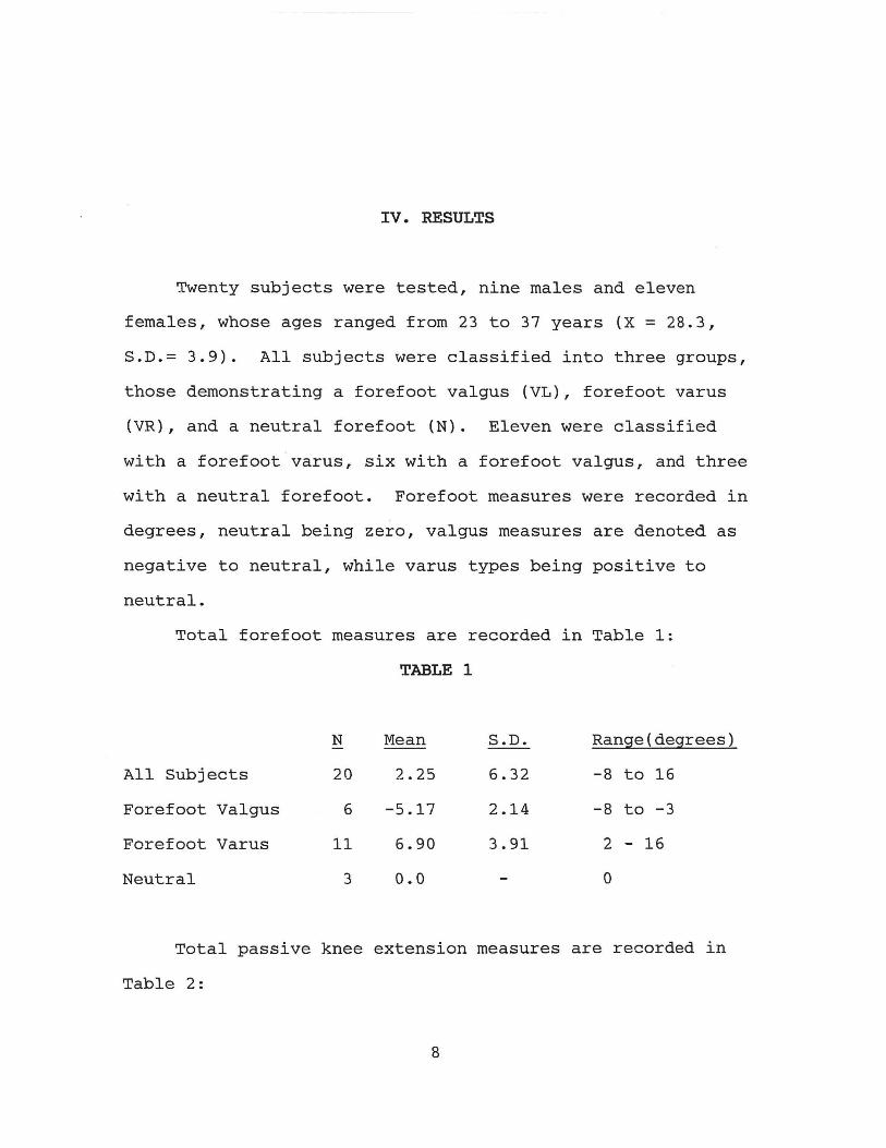
IV. RESULTS
Twenty subjects were tested, nine males and eleven
females, whose ages ranged from 23 to 37 years (X = 28.3,
S.D.= 3.9). All subjects were classified into three groups,
those demonstrating a forefoot valgus (VL), forefoot varus
(VR) , and a neutral forefoot (N). Eleven were classified
with a forefoot varus, six with a forefoot valgus, and three
with a neutral forefoot. Forefoot measures were recorded in
degrees, neutral being zero, valgus measures are denoted as
negative to neutral, while varus types being positive to
neutral.
Total forefoot measures are recorded in Table 1:
All Subjects
Forefoot Valgus
Forefoot Varus
Neutral
N
20
6
11
3
TABLE 1
Mean
2.25
-5.17
6.90
0.0
S.D.
6.32
2.14
3.91
Range(degrees)
-8 to 16
-8 to -3
2 - 16
o
Total passive knee extension measures are recorded in
Table 2:
8
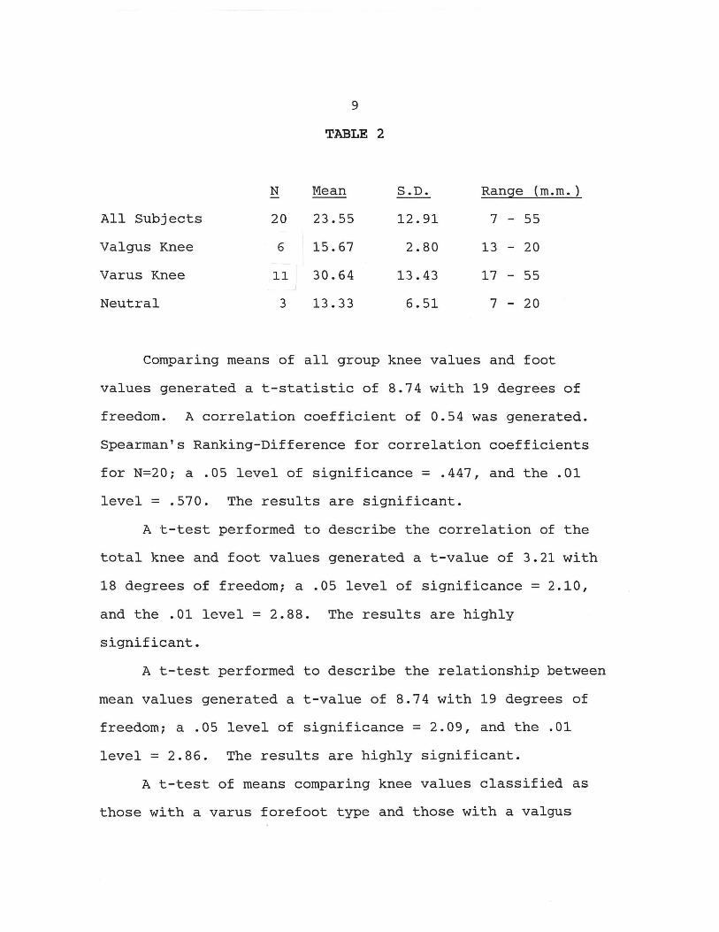
All Subjects
Valgus Knee
Varus Knee
Neutral
N
20
6
11
3
9
TABLE 2
Mean
23.55
15.67
30.64
13.33
S.D.
12.91
2.80
13.43
6.51
Range (m.m.)
7 - 55
13 - 20
17 - 55
7 - 20
Comparing means of all group knee values and foot
values generated a t-statistic of 8.74 with 19 degrees of
freedom. A correlation coefficient of 0.54 was generated.
Spearman's Ranking-Difference for correlation coefficients
for N=20; a .05 level of significance = .447, and the .01
level = .570. The results are significant.
A t-test performed to describe the correlation of the
total knee and foot values generated a t-value of 3.21 with
18 degrees of freedom; a .05 level of significance = 2.10,
and the .01 level = 2.88. The results are highly
significant.
A t-test performed to describe the relationship between
mean values generated a t-value of 8.74 with 19 degrees of
freedom; a .05 level of significance = 2.09, and the .01
level = 2.86. The results are highly significant.
A t-test of means comparing knee values classified as
those with a varus forefoot type and those with a valgus
10
forefoot type described a t-value of 3.45 with 10 degrees of
freedom; a .05 level of significance = 2.23, and the .01
level = 3.17. The results are highly significant.
V. DISCUSSION
The results of this study indicated a significant
relationship between the degree of forefoot varus measured
and the amount of passive knee extension present. There
appears to be a direct relationship between the two groups.
Therefore the null hypothesis can be rejected. A
significant difference was also demonstrated between the
average means of the passive knee extension measurements
when the measurements are classified as knee passive
extension measures with an identified forefoot varus
posture, and knee passive extension measurements with an
identified forefoot valgus posture. The average mean of the
forefoot varus' groups knee measurements was almost two
times that of those of the forefoot valgus knee
measurements.
Coplins felt that she was not able to prove the theory
that in a closed-chain weightbearing position, pronators
have greater tendency to show a increase in passive knee
rotation. She did find that however in a non-weightbearing
position, increased passive knee internal rotation did occur
at five degrees of knee flexion. s This study indicated that
terminal passive knee extension was greater on the average
11
12
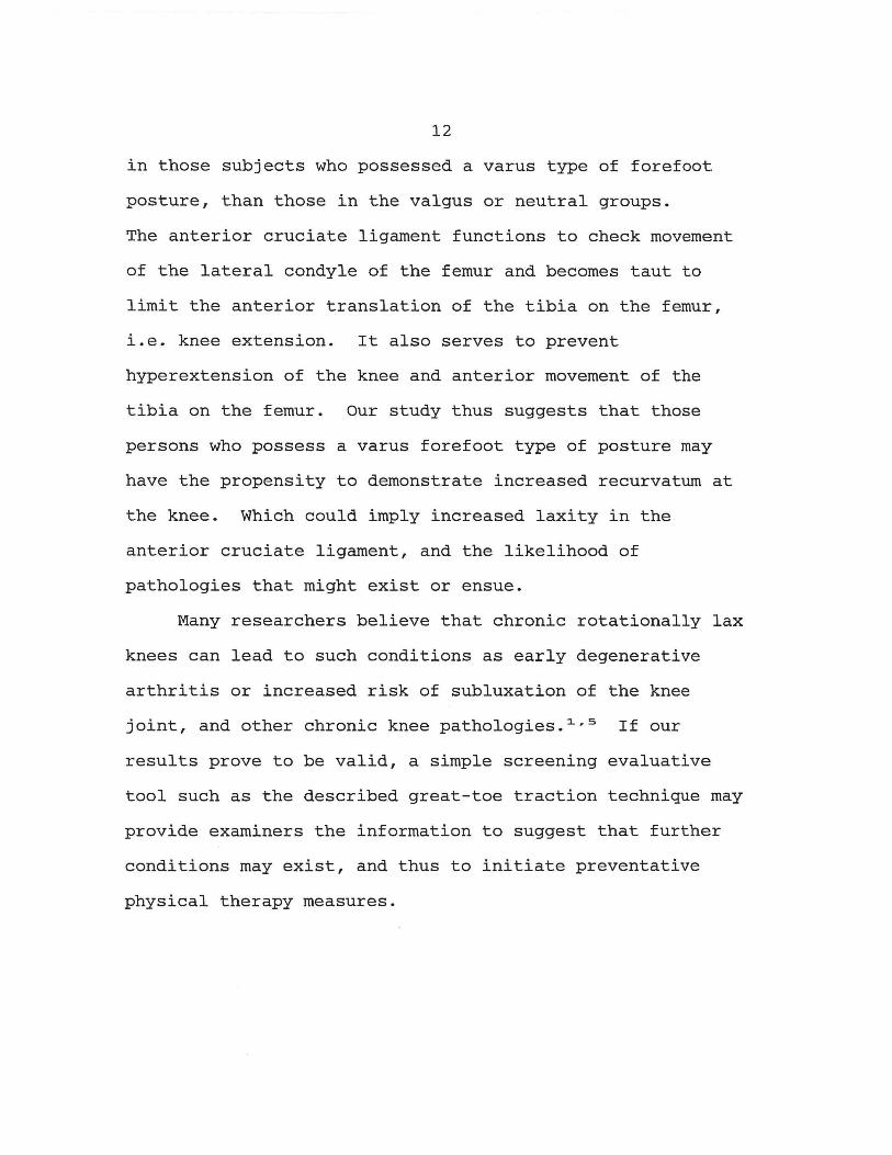
in those subjects who possessed a varus type of forefoot
posture, than those in the valgus or neutral groups.
The anterior cruciate ligament functions to check movement
of the lateral condyle of the femur and becomes taut to
limit the anterior translation of the tibia on the femur,
i.e. knee extension. It also serves to prevent
hyperextension of the knee and anterior movement of the
tibia on the femur. Our study thus suggests that those
persons who possess a varus forefoot type of posture may
have the propensity to demonstrate increased recurvatum at
the knee. Which could imply increased laxity in the
anterior cruciate ligament, and the likelihood of
pathologies that might exist or ensue.
Many researchers believe that chronic rotationally lax
knees can lead to such conditions as early degenerative
arthritis or increased risk of subluxation of the knee
joint, and other chronic knee pathologies.~,5 If our
results prove to be valid, a simple screening evaluative
tool such as the described great-toe traction technique may
provide examiners the information to suggest that further
conditions may exist, and thus to initiate preventative
physical therapy measures.
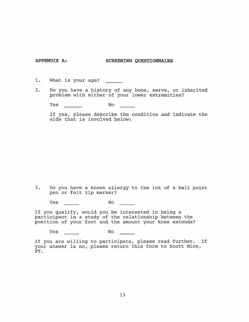
APPENDIX A: SCREENING QUESTIONNAIRE
1. What is your age?
2. Do you have a history of any bone, nerve, or inherited problem with either of your lower extremities?
Yes No
If yes, please describe the condition and indicate the side that is involved below:
3. Do you have a known allergy to the ink of a ball point pen or felt tip marker?
Yes No
If you qualify, would you be interested in being a participant in a study of the relationship between the position of your foot and the amount your knee extends?
Yes No
If you are willing to participate, please read further. If your answer is no, please return this form to Scott Nice, PT.
13
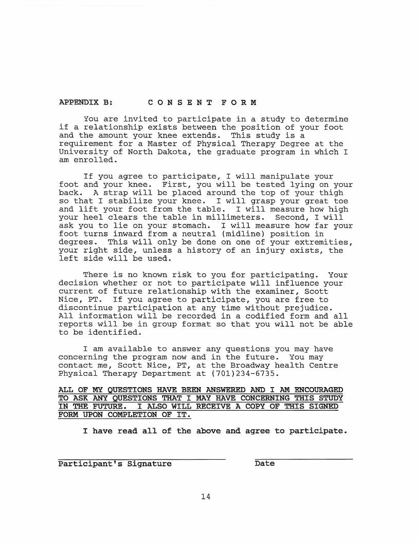
APPENDIX B: CONSENT FORM
You are invited to participate in a study to determine if a relationship exists between the position of your foot and the amount your knee extends. This study is a requirement for a Master of Physical Therapy Degree at the University of North Dakota, the graduate program in which I am enrolled.
If you agree to participate, I will manipulate your foot and your knee. First, you will be tested lying on your back. A strap will be placed around the top of your thigh so that I stabilize your knee. I will grasp your great toe and lift your foot from the table. I will measure how high your heel clears the table in millimeters. Second, I will ask you to lie on your stomach. I will measure how far your foot turns inward from a neutral (midline) position in degrees. This will only be done on one of your extremities, your right side, unless a history of an injury exists, the left side will be used.
There is no known risk to you for participating. Your decision whether or not to participate will influence your current of future relationship with the examiner, Scott Nice, PT. If you agree to participate, you are free to discontinue participation at any time without prejudice. All information will be recorded in a codified form and all reports will be in group format so that you will not be able to be identified.
I am available to answer any questions you may have concerning the program now and in the future. You may contact me, Scott Nice, PT, at the Broadway health Centre Physical Therapy Department at (701)234-6735.
ALL OF MY QUESTIONS HAVE BEEN ANSWERED .AND I AM ENCOURAGED TO ASK ANY QUESTIONS THAT I MAY HAVE CONCERNING THIS STUDY IN THE FUTURE. I ALSO WILL RECEIVE A COPY OF THIS SIGNED FORM UPON COMPLETION OF IT.
I have read all of the above and agree to participate.
Participant's Signature Date
14
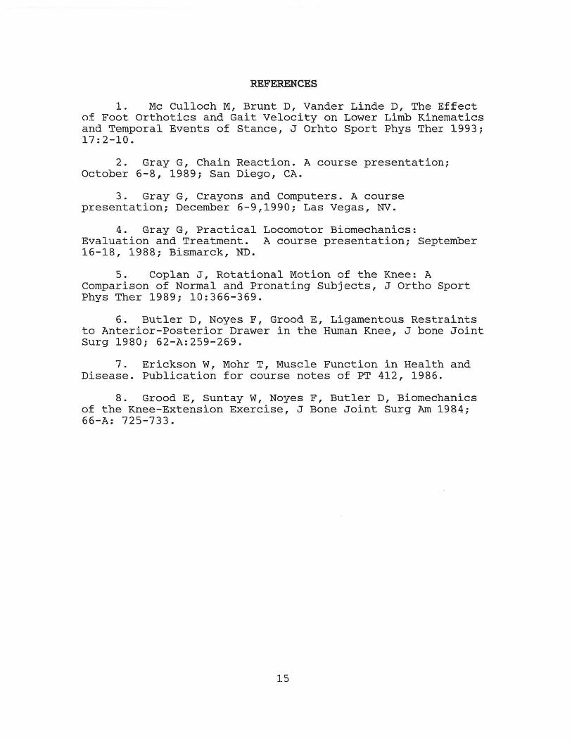
REFERENCES
1. Mc Culloch M, Brunt D, Vander Linde D, The Effect of Foot Orthotics and Gait Velocity on Lower Limb Kinematics and Temporal Events of Stance, J Orhto Sport Phys Ther 1993; 17:2-10.
2. Gray G, Chain Reaction. A course presentation; October 6-8, 1989; San Diego, CA.
3. Gray G, Crayons and Computers. A course presentation; December 6-9,1990; Las Vegas, NV.
4. Gray G, Practical Locomotor Biomechanics: Evaluation and Treatment. A course presentation; September 16-18, 1988; Bismarck, ND.
5. Coplan J, Rotational Motion of the Knee: A Comparison of Normal and Pronating Subjects, J Ortho Sport Phys Ther 1989; 10:366-369.
6. Butler D, Noyes F, Grood E, Ligamentous Restraints to Anterior-Posterior Drawer in the Human Knee, J bone Joint Surg 1980; 62-A:259-269.
7. Erickson W, Mohr T, Muscle Function in Health and Disease. Publication for course notes of PT 412, 1986.
8. Grood E, Suntay W, Noyes F, Butler D, Biomechanics of the Knee-Extension Exercise, J Bone Joint Surg Am 1984; 66-A: 725-733.
15