Coronary Syndromes Clinical Aspects - Specchia · ACUTE CORONARY SYNDROMES – Acute Myocardial...
78
Coronary Syndromes Clinical Aspect G.Specchia
Transcript of Coronary Syndromes Clinical Aspects - Specchia · ACUTE CORONARY SYNDROMES – Acute Myocardial...
Coronary Syndromes
Clinical Aspect
G.Specchia
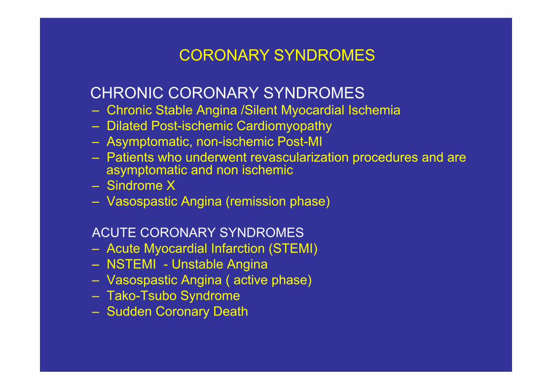
CORONARY SYNDROMES
CHRONIC CORONARY SYNDROMES – Chronic Stable Angina /Silent Myocardial Ischemia – Dilated Post-ischemic Cardiomyopathy – Asymptomatic, non-ischemic Post-MI – Patients who underwent revascularization procedures and are
asymptomatic and non ischemic – Sindrome X – Vasospastic Angina (remission phase)
ACUTE CORONARY SYNDROMES – Acute Myocardial Infarction (STEMI) – NSTEMI - Unstable Angina – Vasospastic Angina ( active phase) – Tako-Tsubo Syndrome – Sudden Coronary Death
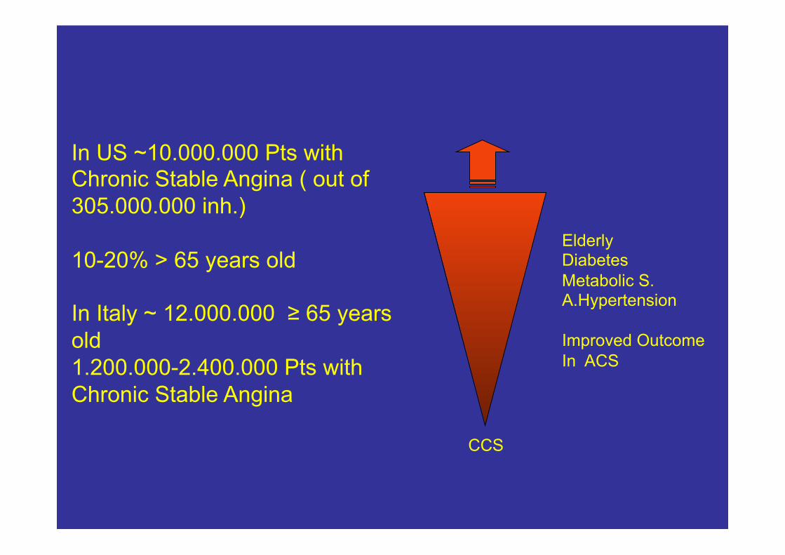
CCS
Elderly Diabetes Metabolic S. A.Hypertension Improved Outcome In ACS
In US ~10.000.000 Pts with Chronic Stable Angina ( out of 305.000.000 inh.) 10-20% > 65 years old In Italy ~ 12.000.000 ≥ 65 years old 1.200.000-2.400.000 Pts with Chronic Stable Angina
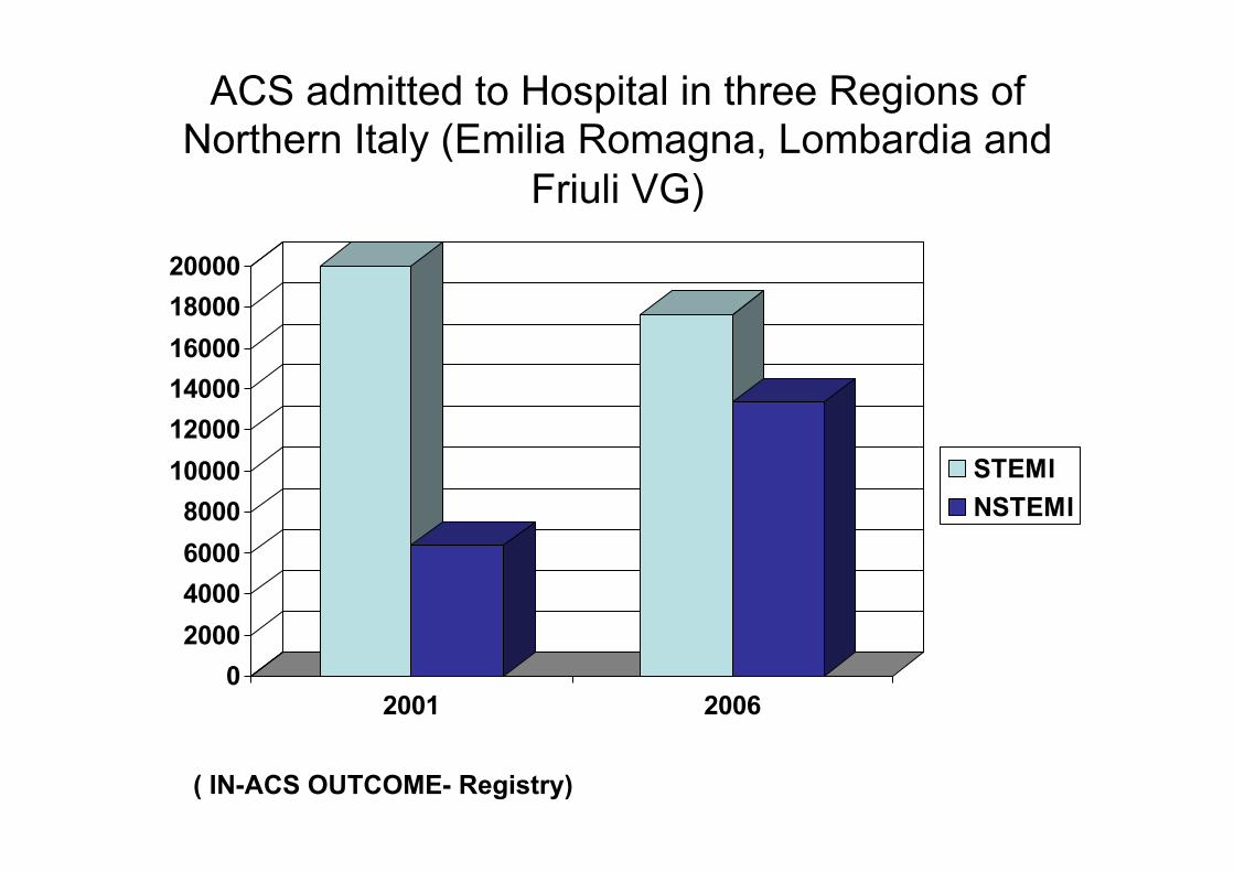
ACS admitted to Hospital in three Regions of Northern Italy (Emilia Romagna, Lombardia and
Friuli VG)
02000400060008000100001200014000160001800020000
2001 2006
STEMINSTEMI
( IN-ACS OUTCOME- Registry)
The Most Important clinical difference between Acute and
Chronic Coronary Syndromes is in the outcome
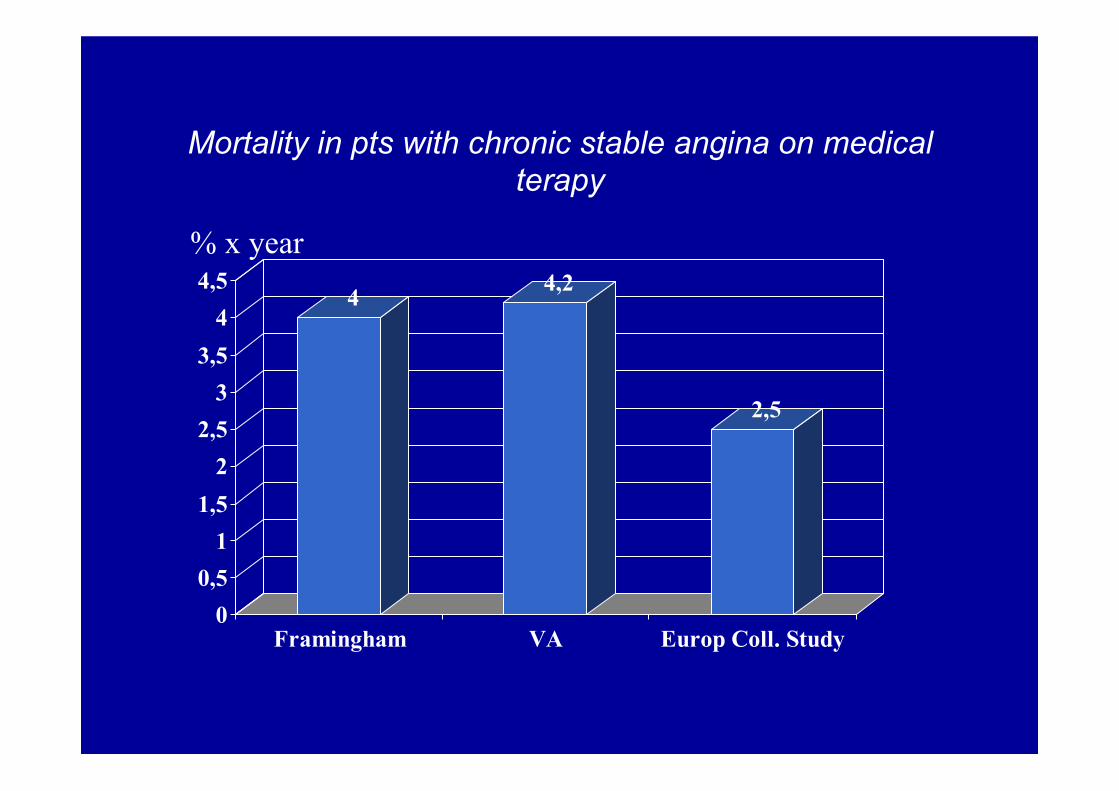
Mortality in pts with chronic stable angina on medical terapy
4 4,2
2,5
00,5
11,5
22,5
33,5
44,5
Framingham VA Europ Coll. Study
% x year

Cardiac Event x Year pts with Chronic Stable Angina on Medical Therapy
0
0,5
1
1,5
2
2,5
3
3,5
4
death or fatal MI
Italian OD1 1982COURAGE Trial 2007
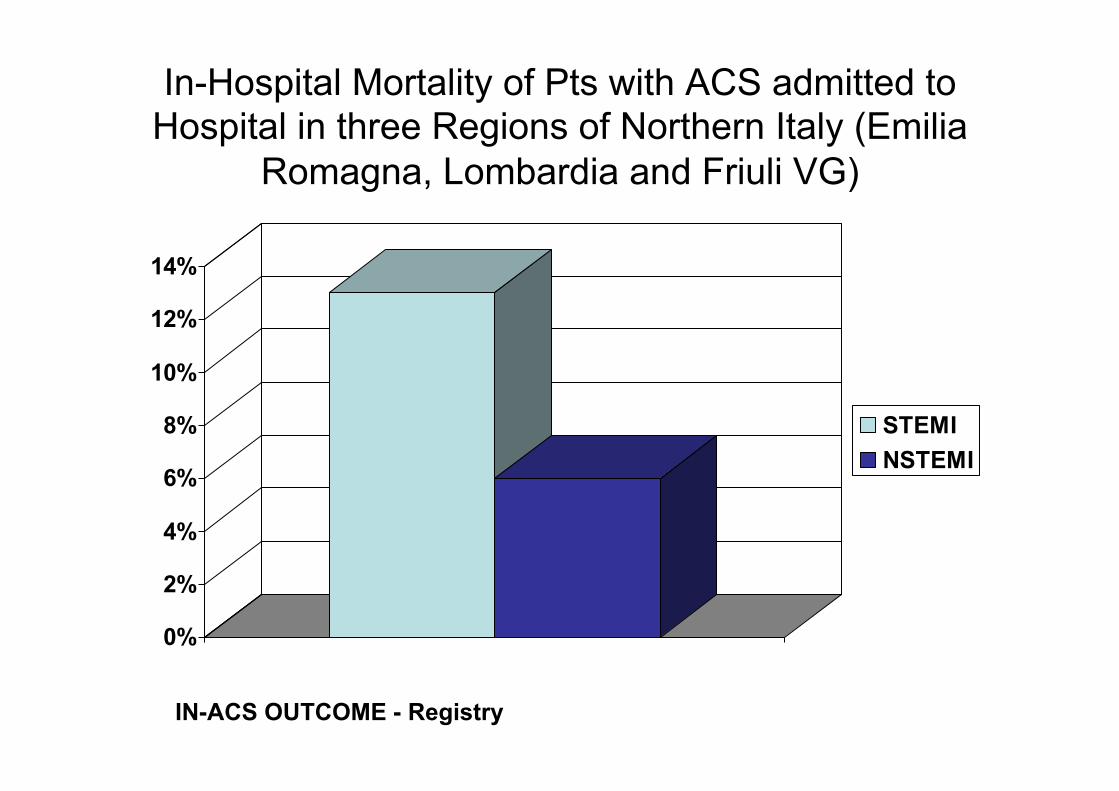
In-Hospital Mortality of Pts with ACS admitted to Hospital in three Regions of Northern Italy (Emilia
Romagna, Lombardia and Friuli VG)
0%
2%
4%
6%
8%
10%
12%
14%
STEMINSTEMI
IN-ACS OUTCOME - Registry
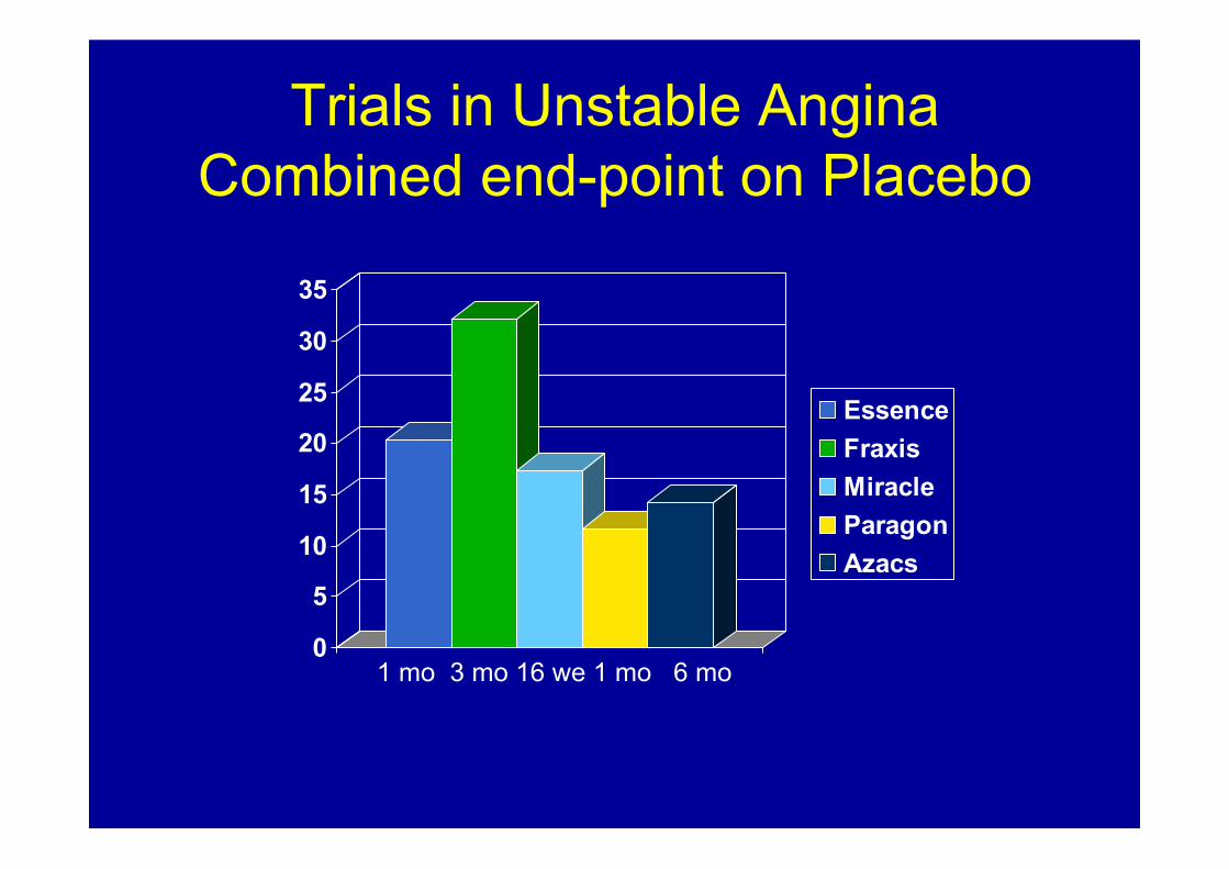
Trials in Unstable Angina Combined end-point on Placebo
0
5
10
15
20
25
30
35
EssenceFraxisMiracleParagonAzacs
1 mo 3 mo 16 we 1 mo 6 mo
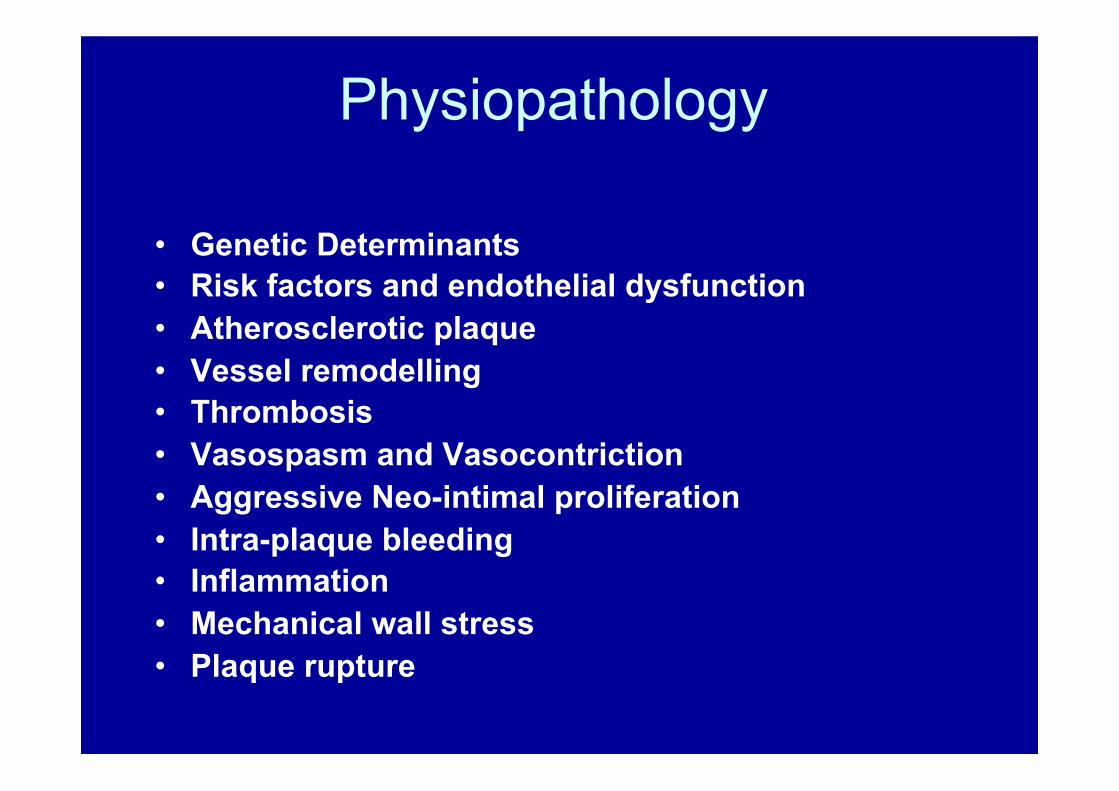
Physiopathology
• Genetic Determinants • Risk factors and endothelial dysfunction • Atherosclerotic plaque • Vessel remodelling • Thrombosis • Vasospasm and Vasocontriction • Aggressive Neo-intimal proliferation • Intra-plaque bleeding • Inflammation • Mechanical wall stress • Plaque rupture


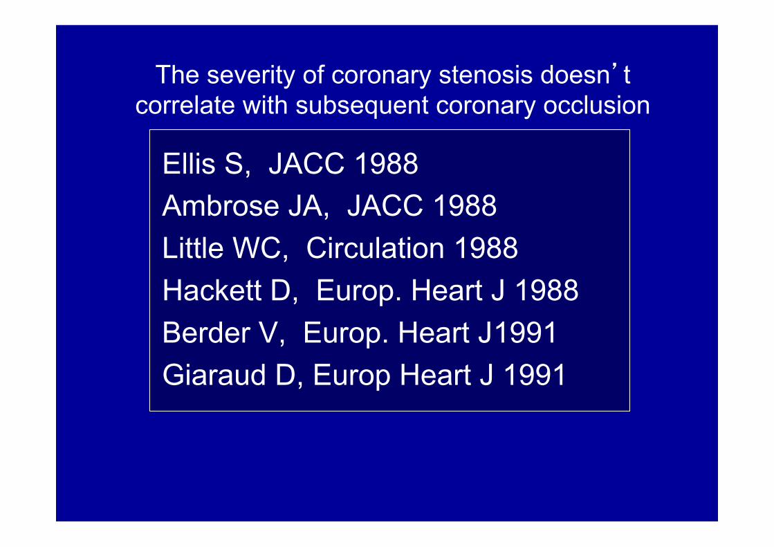
The severity of coronary stenosis doesn’t correlate with subsequent coronary occlusion
Ellis S, JACC 1988 Ambrose JA, JACC 1988 Little WC, Circulation 1988 Hackett D, Europ. Heart J 1988 Berder V, Europ. Heart J 1991 Giaraud D, Europ Heart J 1991
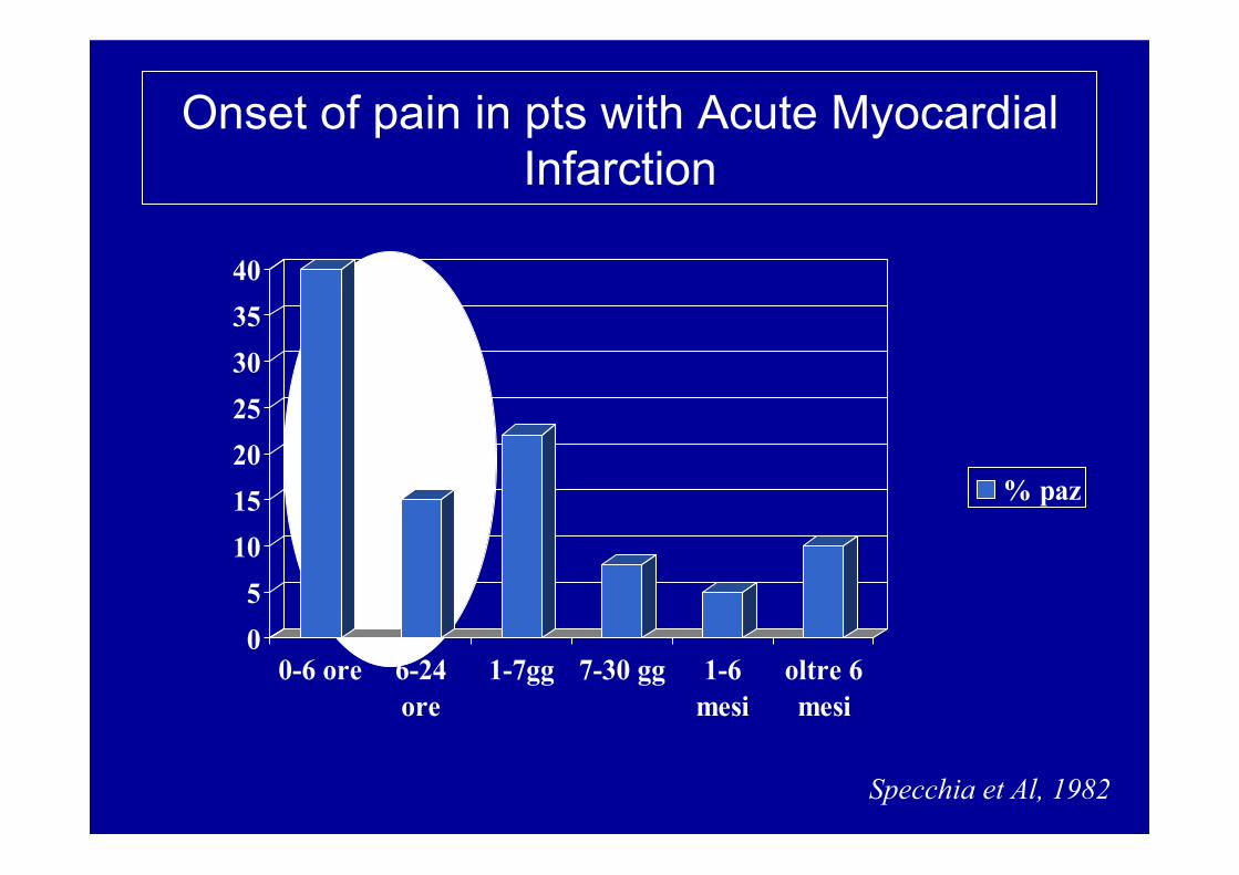
Onset of pain in pts with Acute Myocardial Infarction
05
10152025303540
0-6 ore 6-24ore
1-7gg 7-30 gg 1-6mesi
oltre 6mesi
% paz
Specchia et Al, 1982
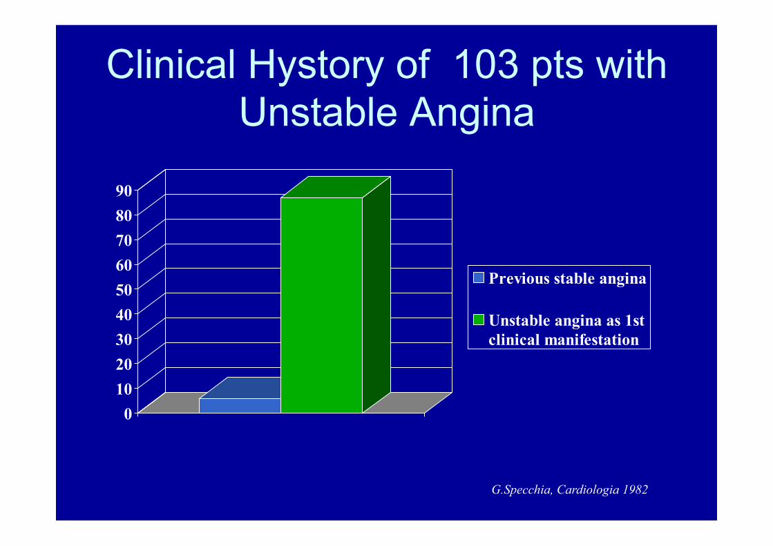
Clinical Hystory of 103 pts with Unstable Angina
0102030405060708090
1° Trim.
Previous stable angina
Unstable angina as 1stclinical manifestation
G.Specchia, Cardiologia 1982
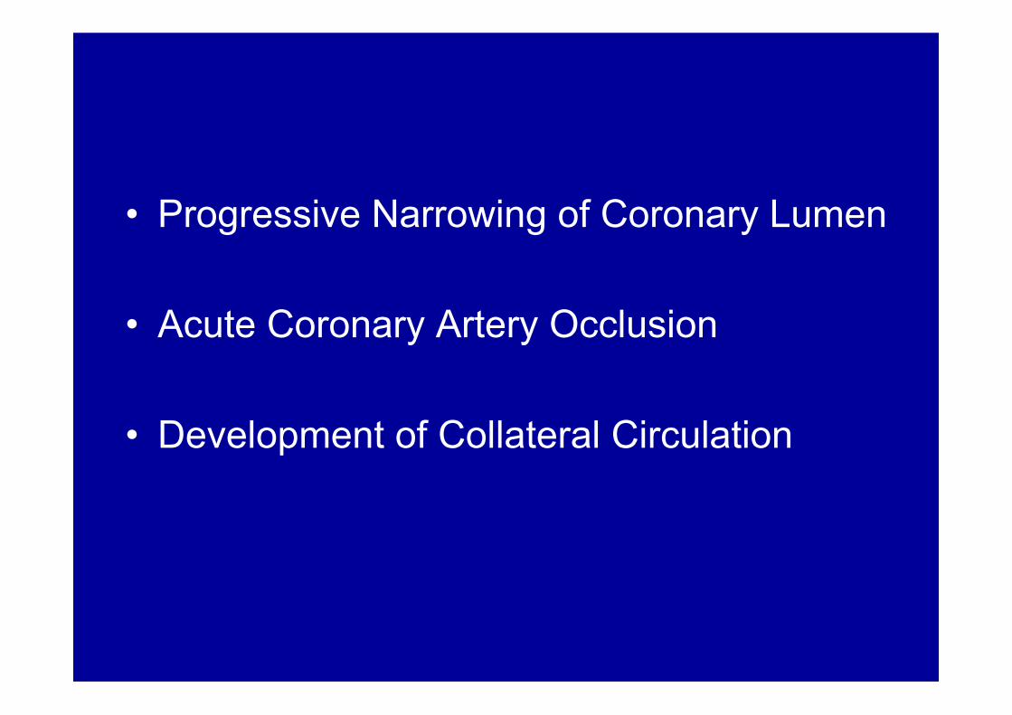
• Progressive Narrowing of Coronary Lumen
• Acute Coronary Artery Occlusion
• Development of Collateral Circulation

Athero Process Collateral Circulation
Angina or Silent Ischemia
+ -
Angina/ischemia threshold

Athero Process Collateral Circulation
Plaque Instability Coronary Occlusion
Small MI Nothing
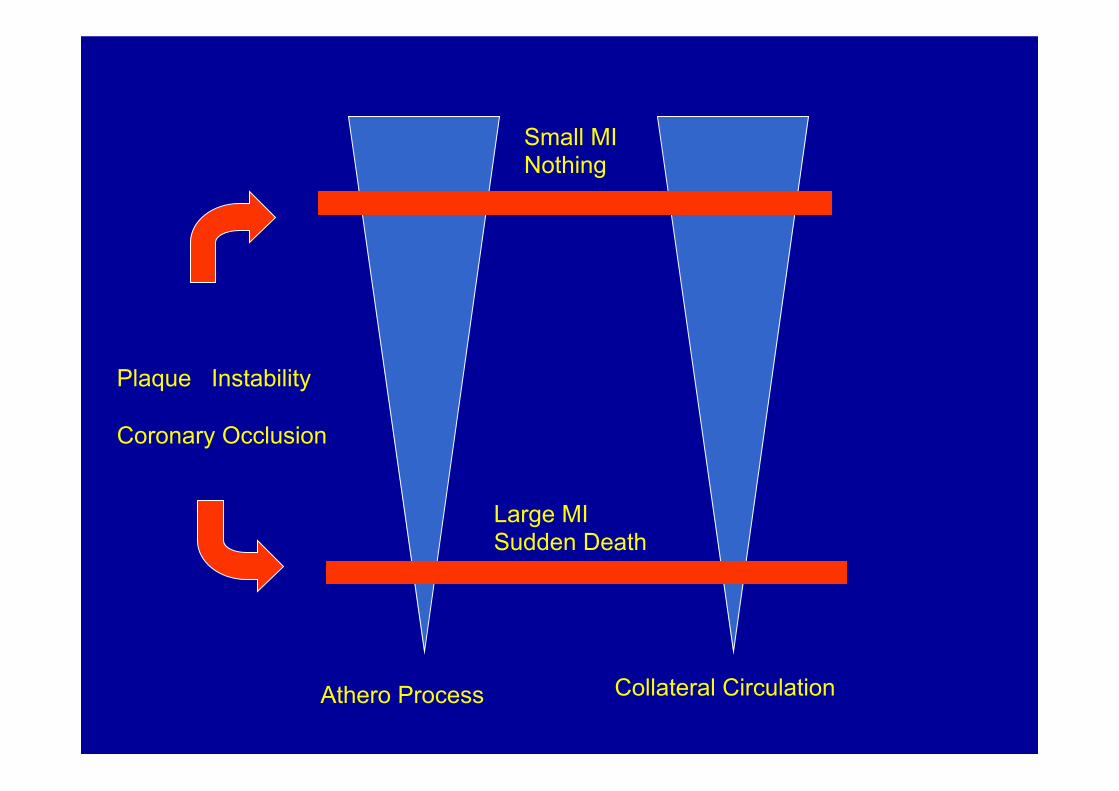
Athero Process Collateral Circulation
Plaque Instability Coronary Occlusion
Large MI Sudden Death
Small MI Nothing

Unanswered questions
• Real correlations between known risk factors and presence/severity of CAD
• Identification of strong genetic determinants in the development of CAD
• The severity of stenosis or its functional consequences: the same information?
• Feasible methods to identify vulnerable plaques • Which is the real trigger of inflammation and
instability
Unanswered questions
• The frequent discrepancy between the time of reperfusion and severity of subsequent scar in pts with AMI : only due to amount of collateral circulation?
• Anginal syndromes in Pts without angiographic evidence of coronary disease
• Gender related characteristics of CAD • Is CAD regression possible ?

T H E E N D
Today’s Problems in trans-catheter Revascularization
• CABG or STENTING for LM or 3-vessel • disease? • Opening chronic total occlusion? • The best TIME for treating Ischemic Related Artery in
ACS • Antithrombotic therapy and Excess of Bleeding • Late STENT Thrombosis, in particular with DES • Keeping the success of revascularization with the Time • Revascularization improves quality of life: does
Revascularization prolong also survival? • Is there a future for stem cells in pts with advanced
coronary disease ? in pts with LV disfunction?
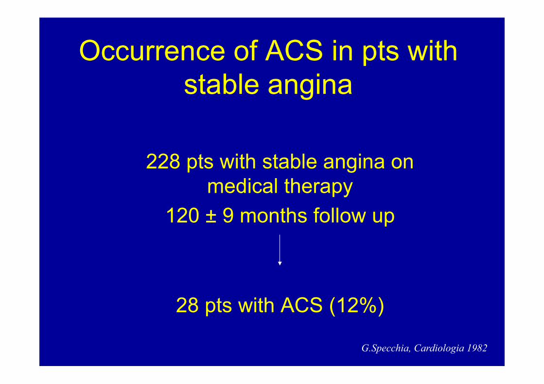
Occurrence of ACS in pts with stable angina
228 pts with stable angina on medical therapy
120 ± 9 months follow up
28 pts with ACS (12%) G.Specchia, Cardiologia 1982


Today’s Problems in Surgery
• Ventricular Remodelling associated or not with CABG
• Surgery in pts on Clopidogrel IIb/IIIa long infusion?
ISCHEMIC VALVE DISEASE
Clinical Aspects
G.Specchia
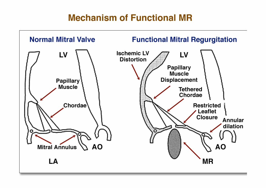
Ischemic Mitral Regurgitation
• Mitral Insufficiency as a result of myocardial infarction or ischemia (papillary muscles damage and/or changes in left ventricular geometry and remodeling)
• By definition Mitral Leatflets are normal.
Ischemic Mitral Regurgitation Prevalence
• Increasing Survival in Ischemic Heart Disease Increasing number of pts with IMR
• 15-50% of pts with AMI develop systolic murmur or ECHO findings of MR
• 10-20% of pts with chronic symptomatic CAD have MR, often moderate (~7%) or severe (~4%)

Ischemic Mitral Regurgitation • ACUTE IMR : papillary muscle rupture (rare 1-5% of
death in AMI) with mitral leatflets prolapse; • Annular dilatation • Discoordination of normal syncronous papillary
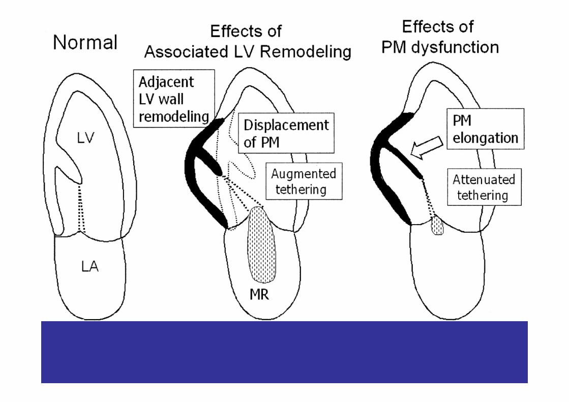
muscle contraction : infarcted Papillary Muscle elongation while uninfarcted Papillary Muscle contracts earlier and vigorously
• CHRONIC IMR : Left Ventricle Remodeling, Annular dilatation, Papillary Muscle fibrosis and atrophy, with leatflet tethering and restriction of leatflet motion
• Mitral Valve prolapse ( Rare, probably antedate)
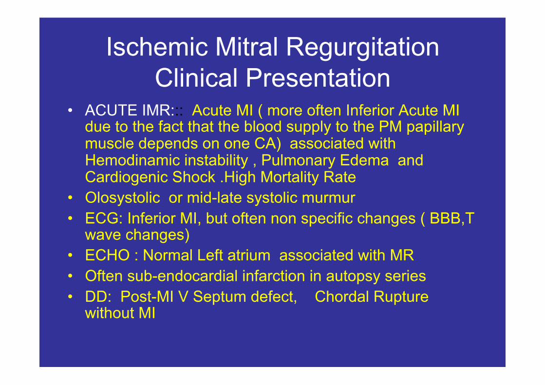
Ischemic Mitral Regurgitation Clinical Presentation
• ACUTE IMR::: Acute MI ( more often Inferior Acute MI due to the fact that the blood supply to the PM papillary muscle depends on one CA) associated with Hemodinamic instability , Pulmonary Edema and Cardiogenic Shock .High Mortality Rate
• Olosystolic or mid-late systolic murmur • ECG: Inferior MI, but often non specific changes ( BBB,T
wave changes) • ECHO : Normal Left atrium associated with MR • Often sub-endocardial infarction in autopsy series • DD: Post-MI V Septum defect, Chordal Rupture
without MI
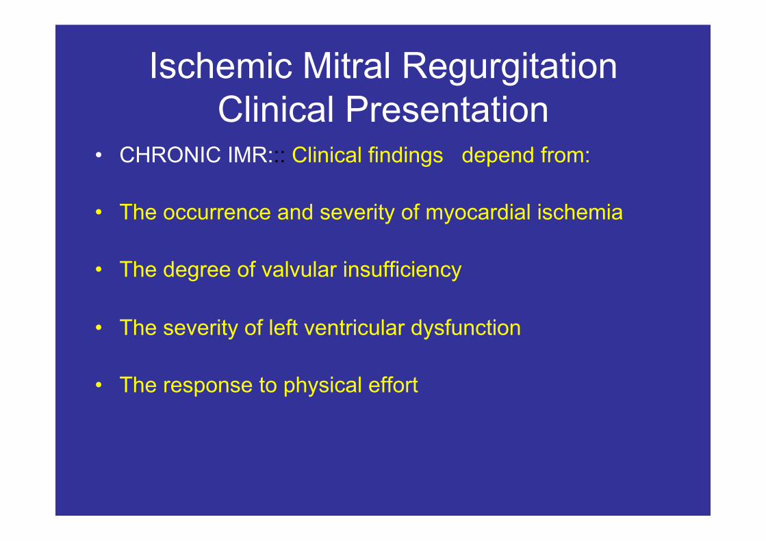
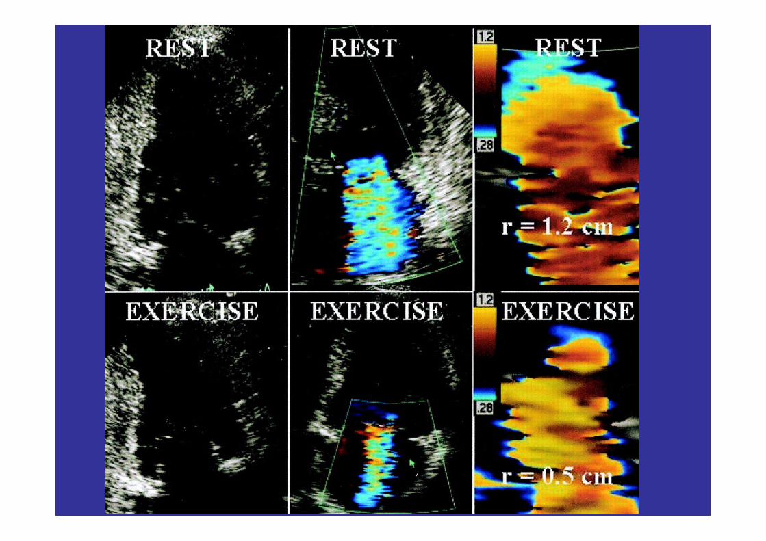
Ischemic Mitral Regurgitation Clinical Presentation
• CHRONIC IMR::: Clinical findings depend from:
• The occurrence and severity of myocardial ischemia
• The degree of valvular insufficiency
• The severity of left ventricular dysfunction
• The response to physical effort

P. Lamcellotti et Al, JACC2003

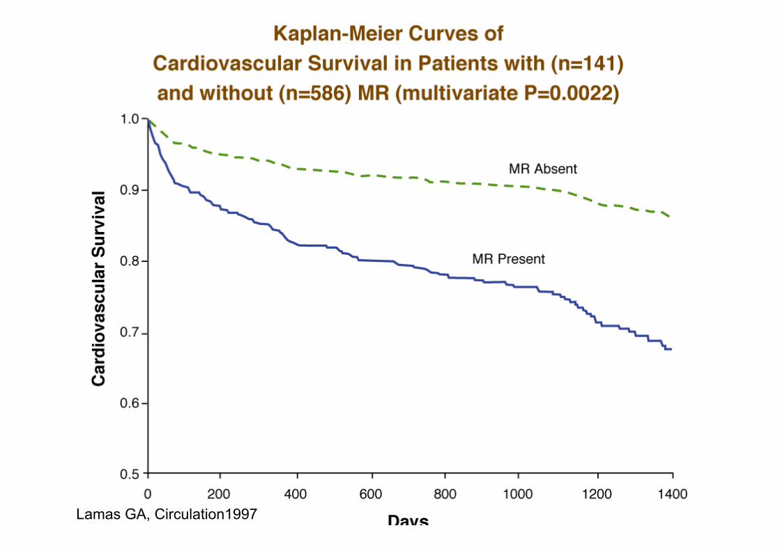
Lamas GA, Circulation1997
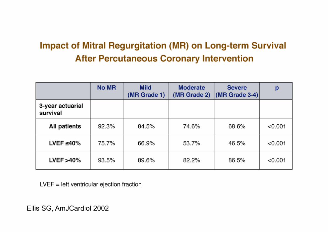
Ellis SG, AmJCardiol 2002
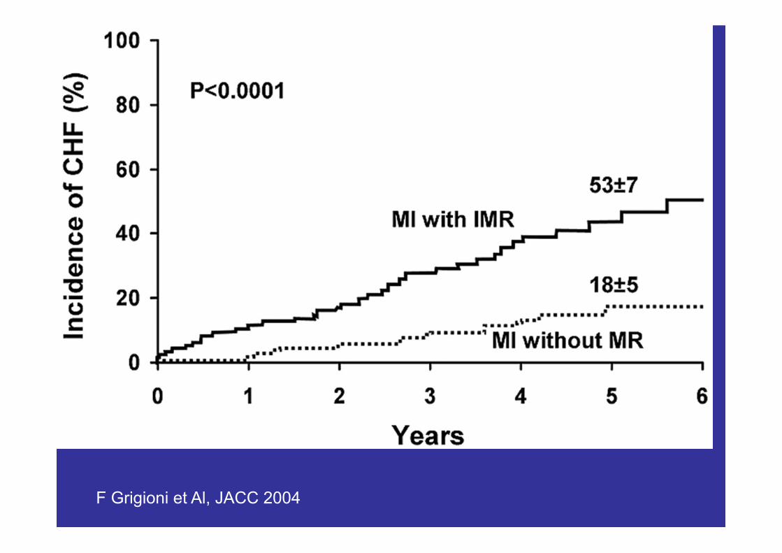
F Grigioni et Al, JACC 2004
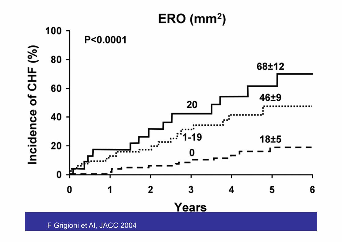
F Grigioni et Al, JACC 2004
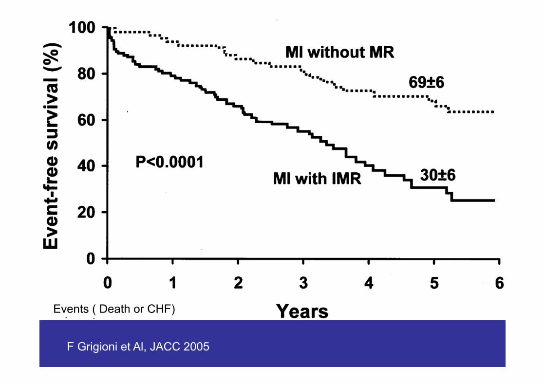
F Grigioni et Al, JACC 2005
Events ( Death or CHF)

F Grigioni et Al, JACC 2004
Events:CHF or Cardia Death

Pellizzon, G. G. et al. J Am Coll Cardiol 2004;43:1368-1374
Multivariate predictors of one-year mortality

• In AMI acute MR can cause early death (1-5% of deaths).
• MR represent a frequent complication in survivors after AMI. Early after MI 15-50% of pts show systolic murmur ,which in many pts disappears by the time
• In pts who undergo PPCI ~ 20% have MR which persists after the acute phase, In others MR may develop later
• Pathological changes that lead to acute or chronic IMR are multiple, involving LV remodelling, annular dilatation, papillary muscle displacement , dysfunction or rupture, chordal thetering, leaflet prolapse or restriction
• Lysis and PPCI did not change significantly the frequency of post-MI MR. In fact the improved survival in pts with AMI has resulted in increasing number of pts with post-MI chronic MR and subsequent LV dysfunction and HF.
• Post-MI pts with MR have a poorer prognosis in comparison with post-MI pts without MR
• Waiting for an effective and applicable, catheter-based percutaneous approach, surgery (urgent or elective) represents in most of the cases of IMR the only therapeutic strategy available today

END

Trials in ACS (UA or NSTEMI) Combined end-point in
Conservative Arms
0
5
10
15
20
25
TIMI IIIBVANQWISHMATEFRISC 2TIMI 18VINORITA 3ICTUS

Copyright ©2004 American College of Cardiology Foundation. Restrictions may apply.
Pellizzon, G. G. et al. J Am Coll Cardiol 2004;43:1368-1374
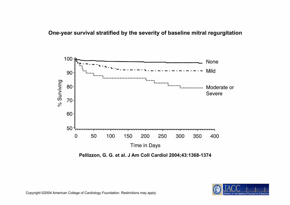
One-year survival stratified by the severity of baseline mitral regurgitation
Copyright ©2004 American College of Cardiology Foundation. Restrictions may apply.
Pellizzon, G. G. et al. J Am Coll Cardiol 2004;43:1368-1374
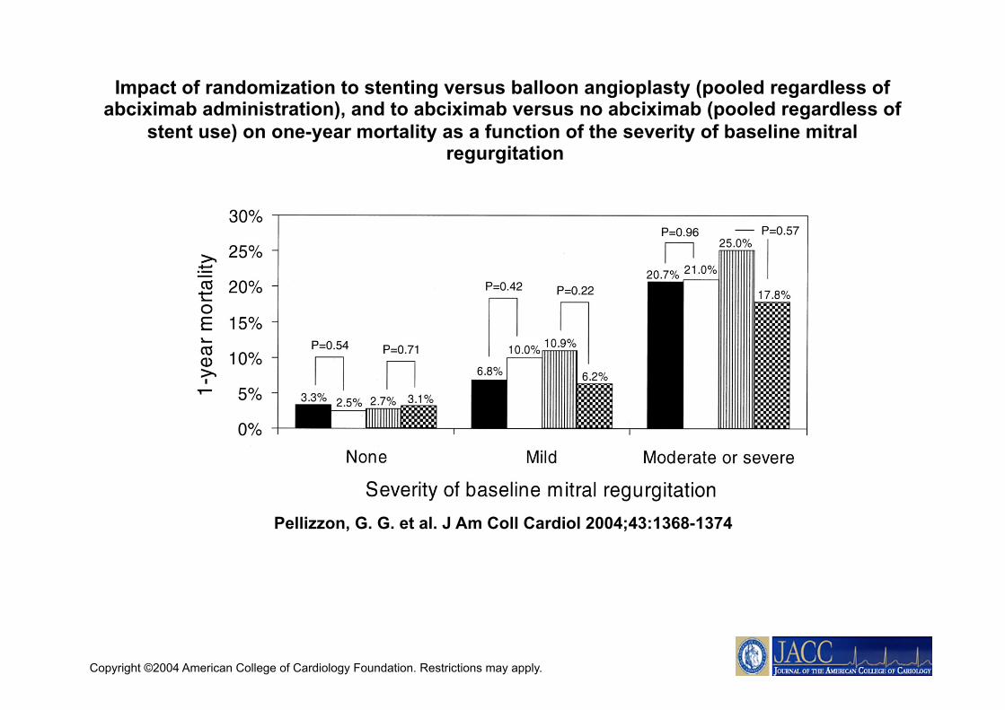
Impact of randomization to stenting versus balloon angioplasty (pooled regardless of abciximab administration), and to abciximab versus no abciximab (pooled regardless of
stent use) on one-year mortality as a function of the severity of baseline mitral regurgitation

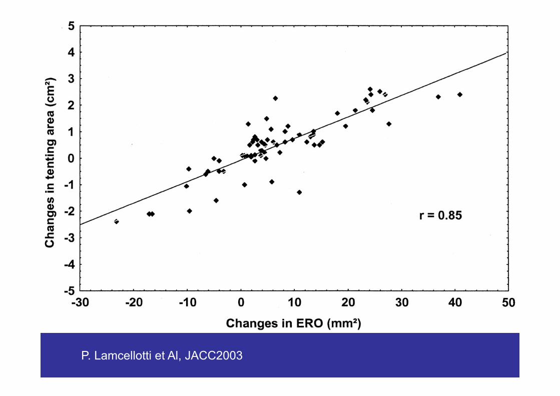
P. Lamcellotti et Al, JACC2003
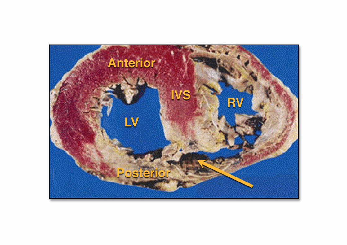
Cardiac Rupture Clinical Aspect
G.Specchia

Left Ventricle Rupture
Infrequent complication ( 2-4%)
High Mortality (5-24% of all in-hospital deaths)
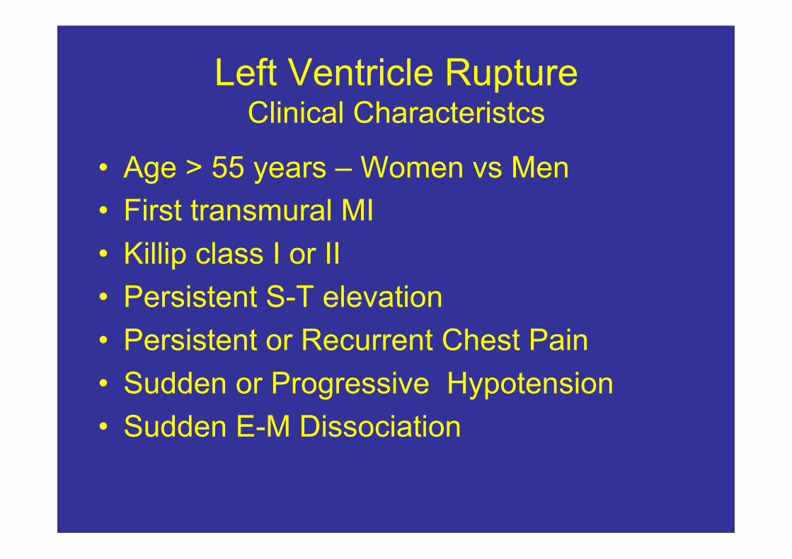
Left Ventricle Rupture Clinical Characteristcs
• Age > 55 years – Women vs Men • First transmural MI • Killip class I or II • Persistent S-T elevation • Persistent or Recurrent Chest Pain • Sudden or Progressive Hypotension • Sudden E-M Dissociation
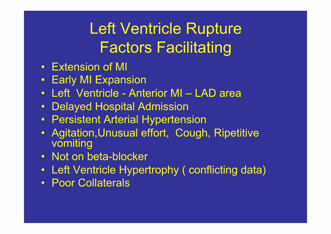
Left Ventricle Rupture Factors Facilitating
• Extension of MI • Early MI Expansion • Left Ventricle - Anterior MI – LAD area • Delayed Hospital Admission • Persistent Arterial Hypertension • Agitation,Unusual effort, Cough, Ripetitive
vomiting • Not on beta-blocker • Left Ventricle Hypertrophy ( conflicting data) • Poor Collaterals

During the reperfusion era the frequency of acute cardiac rupture has declined
Left ventricular free wall rupture occurs in less than 2% of cases,
Left ventricular septum or papillary muscle rupture in less than 1% of cases
Fibrinolytic therapy more than 14 hours after onset of symptoms represents a risk factor
The most important determinants in preventing rupture are successful early reperfusion
The highest risk is within the first 24 hours after MI.
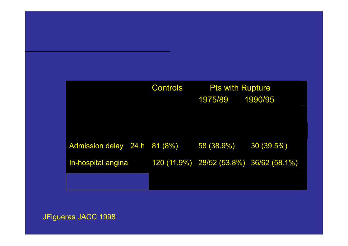
1978–1989 (n = 149)
Controls Pts with Rupture 1975/89 1990/95
Admission delay 24 h 81 (8%) 58 (38.9%) 30 (39.5%)
In-hospital angina 120 (11.9%) 28/52 (53.8%) 36/62 (58.1%)
JFigueras JACC 1998

In Hospital Deaths in AMI pts treated with thrombolysis
00,20,40,60,81
1,21,41,61,82
0-3hours
3-6hours
6-9hours
9-12hours
CardiacRupture
Mauri F et Al, G.It Cardiol 1987

Multivariable analysis of patients treated with Thrombolitic agents , experiencing cardiac rupture
RC Becker et Al, JACC 1999
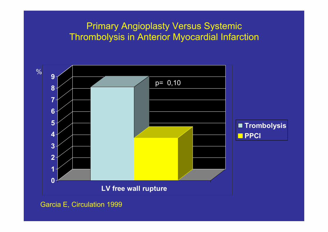
Primary Angioplasty Versus Systemic Thrombolysis in Anterior Myocardial Infarction
0123456789
LV free wall rupture
TrombolysisPPCI
p= 0,10
Garcia E, Circulation 1999
%
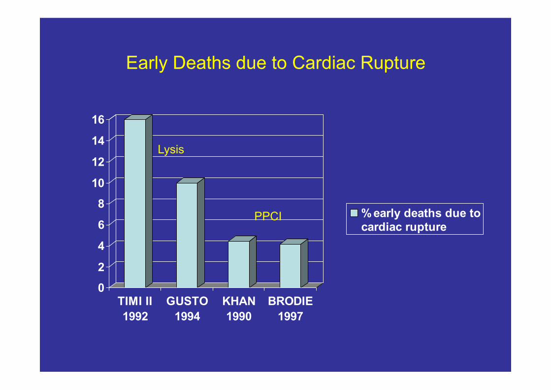
Early Deaths due to Cardiac Rupture
0
2
4
6
8
10
12
14
16
TIMI II1992
GUSTO1994
KHAN1990
BRODIE1997
% early deaths due tocardiac rupture
Lysis
PPCI
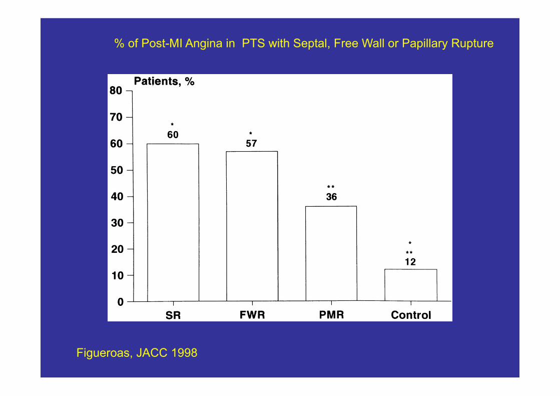
Figueroas, JACC 1998
% of Post-MI Angina in PTS with Septal, Free Wall or Papillary Rupture
J Figueroas Jacc 1998
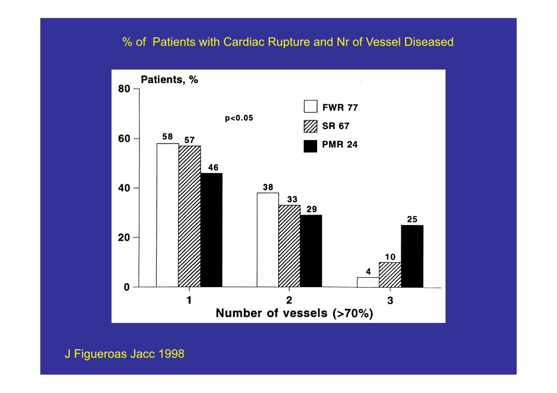
% of Patients with Cardiac Rupture and Nr of Vessel Diseased

J Figueroas Jacc 1998
% of Pts with Cardiac Rupture and IRA
Left Ventricle Rupture Clinical Presentation
• ACUTE: Acute Tamponade with Hypotension- Cardiogenic Shock - Electro-mechanical Dissociation
• SUB-ACUTE: Moderate to Severe Pericardial Effusion with or without Cardiac Tamponade and Hemodinamic Progressive Deterioration
Silent Subacute Free Wall Rupture
• No hemodynamic compromise • Pericardial effusion identified on a routine
ECHO • Attribution to a peri- or post- MI pericarditis • Initial healing by fibrin deposit and subsequent
definite healing
• Delayed development of pseudo-aneurism • Re-rupture , cardiac tamponade and Death

END

Cardiac Rupture in Pts treated with Thrombolitic Agents
0,000,200,400,600,801,001,201,401,601,802,00
Delay of treatment
0-3 hours3-6 hours6-9 hours9-12 hours
% o
f pts
with
car
diac
rupt
ure
F Mauri et Al, G. It.Cardiol 1987
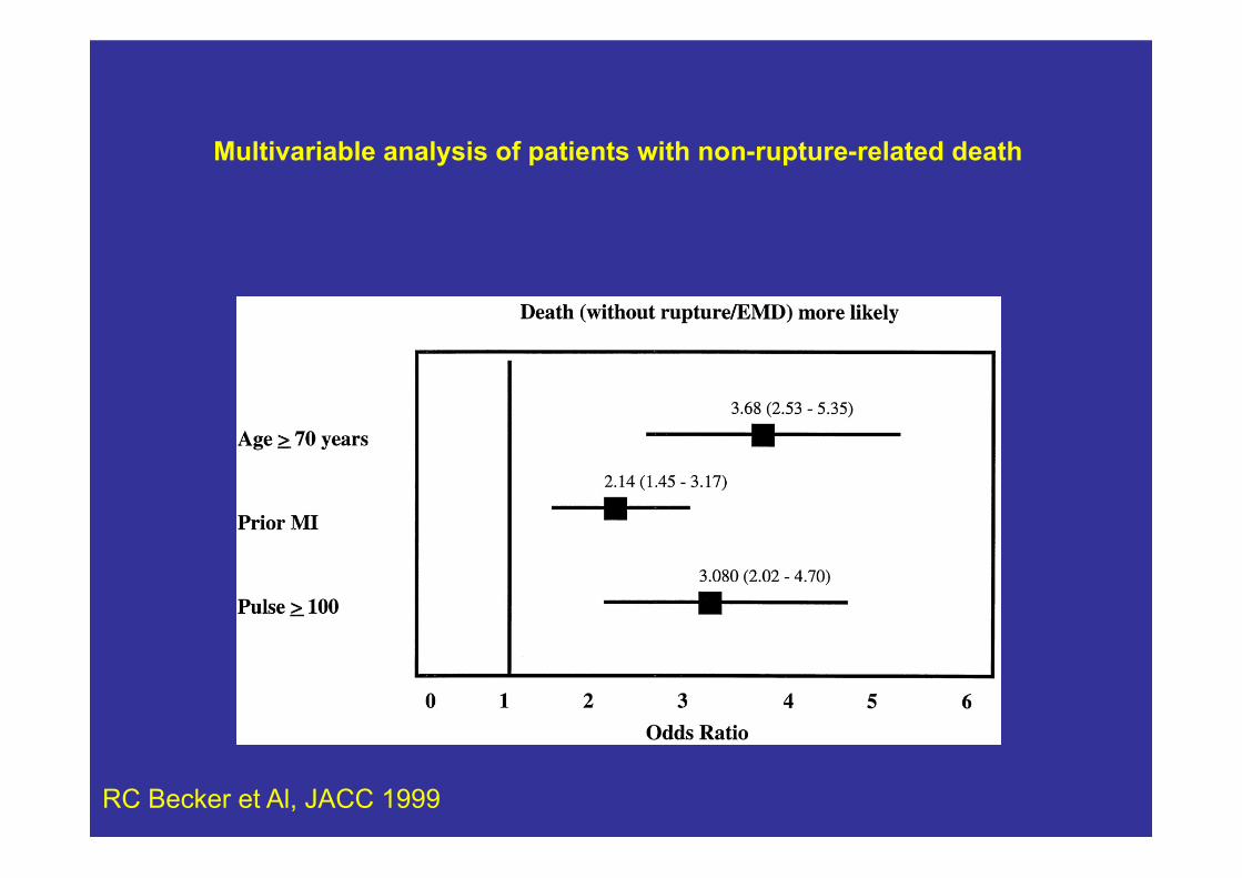
Multivariable analysis of patients with non-rupture-related death
RC Becker et Al, JACC 1999