Coronary Arterial Drug-Eluting Stent: From Structure to Clinical
30
10 Coronary Arterial Drug-Eluting Stent: From Structure to Clinical Tim Wu and Stephen McCarthy Biomedical Engineering and Biotechnology, University of Massachusetts Lowell, MA, USA 1. Introduction 1.1 Cardiovascular diseases and Percutaneous Coronary Intervention (PCI) Coronary Artery Disease (CAD) is the chronic blocking or narrowing of the coronary arteries caused by neointimal hyperplasia of the arterial wall. It has been the number one killer in the United States since 1900 and remains a common cause of death in the Western world despite therapeutic advances. Approximately 14 million Americans have CAD, 500,000 people die from acute myocardial infarction, and one million more survive but with a 1.5 to 15 times greater risk of mortality or morbidity than the rest of the population each year. The annual health care cost of the disease is in excess of $112 billion 1 . The current levels of predictors of heart disease risk, such as obesity, diabetes, and smoking, suggests that this will continue to be a significant public health problem for the foreseeable future 2 . The options for CAD treatments include medications, percutaneous coronary arterial interventions (PCI, including angioplasty and stenting), or coronary artery bypass graft surgery (CABG). In general, patients with coronary narrowing that does not limit coronary artery blood flow receive medications and lifestyle modification to help prevent progression. If a patient has coronary atherosclerosis that limits blood flow in the coronary arteries, balloon angioplasty and stenting can be offered. In patients with multiple areas of coronary artery narrowing or blockage, CABG is generally recommended. 1.1.1 Coronary Artery Bypass Graft Surgery (CABG) Of those patients with coronary artery disease, about 10% undergo CABG surgery. Patients with severe narrowing or blockage of the left main coronary artery or those with disease involving two or three coronary arteries are generally considered for bypass surgery. In a CABG, the surgeon uses a portion of a healthy vessel (either an artery or vein) from the leg, chest, or arm to create a detour or bypass around the blocked portion of the coronary artery. Depending on how many coronary arteries (and main branches) are blocked, patients typically receive 1 to 5 bypasses. The most commonly used bypass vessels are the saphenous vein from the leg, the internal mammary artery from the chest, www.intechopen.com
Transcript of Coronary Arterial Drug-Eluting Stent: From Structure to Clinical
10
Coronary Arterial Drug-Eluting Stent: From Structure to Clinical
Tim Wu and Stephen McCarthy Biomedical Engineering and Biotechnology,
University of Massachusetts Lowell, MA, USA
1. Introduction
1.1 Cardiovascular diseases and Percutaneous Coronary Intervention (PCI)
Coronary Artery Disease (CAD) is the chronic blocking or narrowing of the coronary
arteries caused by neointimal hyperplasia of the arterial wall. It has been the number one
killer in the United States since 1900 and remains a common cause of death in the Western
world despite therapeutic advances. Approximately 14 million Americans have CAD,
500,000 people die from acute myocardial infarction, and one million more survive but
with a 1.5 to 15 times greater risk of mortality or morbidity than the rest of the population
each year. The annual health care cost of the disease is in excess of $112 billion 1. The
current levels of predictors of heart disease risk, such as obesity, diabetes, and smoking,
suggests that this will continue to be a significant public health problem for the
foreseeable future 2.
The options for CAD treatments include medications, percutaneous coronary arterial
interventions (PCI, including angioplasty and stenting), or coronary artery bypass graft
surgery (CABG). In general, patients with coronary narrowing that does not limit coronary
artery blood flow receive medications and lifestyle modification to help prevent
progression. If a patient has coronary atherosclerosis that limits blood flow in the coronary
arteries, balloon angioplasty and stenting can be offered. In patients with multiple areas of
coronary artery narrowing or blockage, CABG is generally recommended.
1.1.1 Coronary Artery Bypass Graft Surgery (CABG)
Of those patients with coronary artery disease, about 10% undergo CABG surgery.
Patients with severe narrowing or blockage of the left main coronary artery or those with
disease involving two or three coronary arteries are generally considered for bypass
surgery. In a CABG, the surgeon uses a portion of a healthy vessel (either an artery or
vein) from the leg, chest, or arm to create a detour or bypass around the blocked portion
of the coronary artery. Depending on how many coronary arteries (and main branches)
are blocked, patients typically receive 1 to 5 bypasses. The most commonly used bypass
vessels are the saphenous vein from the leg, the internal mammary artery from the chest,
www.intechopen.com

Coronary Artery Diseases
198
and the radial artery from the arm. During a CABG, a heart-lung machine artificially
maintains circulation while the surgeon operates on the heart. CABG operations require
general anesthesia and typically 4 to 7 days in the hospital. It may take up to 3 months to
fully recover from the surgery.
1.1.2 Percutaneous Interventions (PCIs)
Percutaneous Interventions (PCIs), which include percutaneous transluminal coronary
artery angioplasty (PTCA) and coronary artery stenting (CAS) are the major therapies for
CAD treatment. PTCA involves insertion of an expandable balloon catheter against a
primary atherosclerotic plaque or secondary restenotic lesion to increase vessel patency
and blood flow. Clinical stenting was introduced in 1986 with the Wallstent to repair
abrupt closure after PTCA and has revolutionized interventional cardiology 3. In CAS,
the stent (a tiny metal scaffolding) functions to brace the vessel wall and reduce the risk of
restenosis following angioplasty. CAS reduces the rate of angiographic and clinical
restenosis compared to PTCA alone. This results in a significant reduction in the
frequency of major adverse cardiac events (MACE) after PCI driven mainly by a reduction
in target vessel revascularization (TVR). CAS not only increases procedural success rates,
but also increases the safety of procedures by decreasing the need for emergency
CABG surgery. As a result, stents are currently utilized in over 85% of the two million
PCIs in US and the total direct cost for those life-saving procedures is over $5 billion
annually.
1.2 Problems in current DESs
Since the introduction of the stent, there exist two serious complications: namely,
thrombosis (subacute and late-stage) and in-stent restenosis (ISR). While subacute
thrombosis has been controlled with the use of dual anti-platelet therapy, late-stage
thrombosis occurring in drug-eluting stents (DESs) has been a big concern recently.
Restenosis remains among the most challenging problems in interventional cardiology. The
recent development of DESs is a major breakthrough as a potential solution for the
restenosis problem.
1.2.1 In-stent Restenosis (ISR)
Restenosis, the re-narrowing of an opened artery after stenting or PTCA procedure, is due
to a proliferative response of the intima, a layer of cells that lines the lumen of the vessel
composed of connective tissue and smooth muscle cells (SMC). In restenosis, vascular
neointimal hyperplasia results in complete blockage of the original artery and insufficient
oxygenation of cardiac tissue, leading to cardiac arrhythmia or cardiac arrest. Restenosis
has been the biggest problem in PCI until the recent successful development of DESs.
Initially, the restenosis rate in PTCA procedures was over 50% within six months post
balloon dilation. Stenting lowers this rate to 20-30%. Both Sirolimus (Cypher®, Cordis Inc,
Fla) and Paclitaxel (Taxus®, Boston Scientific Inc, Natick, MA) coated DESs can
significantly reduce the rate of restenosis to <10%4-6. However, restenosis in patients with
high risk such as small vessels, diabetes, and long diffusion diseased arteries still remains
www.intechopen.com

Coronary Arterial Drug-Eluting Stent: From Structure to Clinical
199
unacceptably high (30%-60% in bare metal stents [BMSs] and 6%-18% in DESs)7-9 despite
DES implantation.
ISR formation is a multi-factorial, sequential process. Although the detailed mechanism of the arterial tissue response to an implanted stent is still under investigation, it is generally believed that three stages are involved in the process. 1) Thrombotic Phase (day 0-3): this phase is the initial response of the artery tissue to stent implantation characterized with rapid activation, adhesion, aggregation and deposition of platelets and neutrophils to form a thrombus at the injured site. 2) Recruitment Phase: this phase occurs between days 3 to 8 and is characterized by an intensive inflammatory cell infiltration. In this phase, inflammatory cells are activated and infiltrate into the injured vessel wall. Subsequently, the recruited inflammation cells provide the key stimulus for subsequent smooth muscle cell (SMC) proliferation and migration. 3) Proliferate Phase: This phase lasts 1 to 3 months depending on the thickness of the residual thrombus and the rate of growth. At this stage, inflammation cells colonize the residual thrombus, forming a “cap” across the mural thrombus. The cells progressively proliferate, resorbing residual thrombus until all thrombus is gone and is replaced by the neointima tissue 10-12.
The process of neointimal growth, which consists of SMCs, extracellular matrix, and macrophages recruited over a period of several weeks, is similar to the process of tumor tissue growth 13. This pathologic similarity between tumor cell growth and benign neointimal formation has led to the discovery of anti-tumor drugs such as Paclitaxel as effective agents for the treatment of ISR 12, 13.
1.2.2 Stent thrombosis
In spite of restenosis remaining a clinical problem in approximately 10% of patients with DES implantation, it can often be successfully treated with repeat implantation. The greatest concern, however, has been for stent thrombosis which is associated with a high rate of myocardial infarction and death. The rate of early stent thrombosis (less than 30 days following implantation) appears similar in both BMSs and DESs (Cypher® and Taxus®) 14. However, late stent thrombosis has been increasingly reported beyond 12 months following DES implantation with the greatest risk occurring as a result of premature discontinuation of anti-platelet therapy 15-19. Although the precise mechanism of late stage stent thrombosis is unknown, it is generally believed that the combination of delayed endothelialization due to anti-proliferative therapy and persistence of the nonerodable polymer contribute to a hypersensitivity reaction, possibly with some residual active drug that may not be eluted 20, 21.
2. The structure and components of a drug eluting stent
Drug eluting stents are comprised of three major parts: 1) drug delivery platform (metal stent) to hold the drug and provide supporting forces for the collapsed artery; 2) drug coating polymer to attach the drug on the stent surface; 3) anti restenosis drugs released from the stent to inhibit restenosis formation after stent implantation. We review each component systemically in order to have an anatomic understanding of drug eluting stent.
www.intechopen.com

Coronary Artery Diseases
200
2.1 Stent platforms
2.1.1 Stent materials
2.1.1.1 Non degradable material
316L stainless steel: 316L stainless steel (316L SS) is the most commonly used metal material in stents due to its excellent mechanical properties and corrosion resistance and is used in the first generation DESs, Cypher (sirolimus-eluting stent, Cordis, Warren, NJ) and Taxus (paclitaxel-eluting stent, Boston Scientific, Natick, MA). However, its ferromagnetic nature and low density make it a non-MRI compatible and poorly visible fluoroscopic material. Furthermore, the release of nickel, chromate and molybdenum ions is likely to provoke immune reactions and inflammatory responses, which may result in ISR22. Therefore, a number of materials have been used as coatings to improve its visibility and biocompatibility.
Co-Cr: Co-Cr alloy exhibits superior radial strength and improved radiopacity compared to 316L SS. It allows for thinner stent struts, which is a significant advantage because the thickness of stent struts is a important factor on the reduction of restenosis in BMSs23, 24. At the same time, it reduces device profile and consequently improves its deliverability to the target lesion. Co-Cr platforms are employed in the second generation DES, Xience V (everolimus-eluting stent, Abott Vascular, CA) and Endeavor (zotarolimus-eluting stent, Medtronic Vascular, Santa Rosa, CA).
Ta: The highly stable surface oxide layer, preventing electron exchange between the metal and the absorbed biological species25, 26, make Ta an excellent corrosion resistant material. Ta has been coated on 316L SS to improve corrosion properties and biocompatibility of 316L SS27. Its high density and non-ferromagnetic properties28,29 make Ta an excellent fluoroscopically visible and MRI compatible material. However, its poor mechanical properties make Ta stents have higher rates of recoil compared to 316L SS based stents and result in enhanced neointimal formation30. Although no Ta based stents have been approved by FDA for general use to date, a bare Ta stent has been used by Cordis® (Johnson & Johnson, USA) in clinical trials and released commercially in Japan, Canada and Europe31.
Ti: Although Ti has excellent biocompatibility and corrosion resistance, pure Ti is not commonly used for making stents due to its low tensile strength and ductility. However, Ti alloys might be optimum for stents. Ti-nitride-oxide coating on 316L SS was biologically inert, reducing platelet and fibrinogen deposition, and neointimal hyperplasia32. Promising results were reported in clinical trials by the Titan stent (Hexacath, France) using this coating33,34. Moreover, Ti-based Ta and niobium alloys showed excellent haemocompatibility35, indicating they have potential applications for stents. An extensively used Ti alloys in stents is Ni-Ti.
Ni-Ti: Ni-Ti alloy is also a common material for stents which has good biocompatibility, radial force and shape memory. However, the release of nickel ions and their toxic effects on tissues have been reported36, 37. In addition, it is suspected of producing mild inflammation when contacting monocytes38. In order to solve these problems, the surface of Ni-Ti is passivated to increase the Ti oxide concentration and decrease the Ni concentration39, 40, and it is coated by some materials such as polyurethane41, Ti nitride42 and polycrystalline oxides43 to improve the corrosion resistance. Another problem is that the Ni-Ti alloy does
www.intechopen.com

Coronary Arterial Drug-Eluting Stent: From Structure to Clinical
201
not have adequate visibility under fluoroscopy44 though it can be visualized by MRI45. Ni-Ti is used by ACT-OneTM (Progressive Angioplasty Systems, USA)46, Paragon (Progressive Angioplasty Systems, USA)47, and Radius (Scimed, USA)48 in clinical trials.
Pt-Ir: The bare stents made from Pt-Ir alloy of 90% platinum and 10% iridium have been implanted in animal models, showing excellent radiopacity and a reduction in both thrombosis and neointimal proliferation with less inflammatory reactions. However, their recoiling percentage was much higher (16%) than the 316L SS stents (5%)49, 50. Though Pt-Ir alloy stents have been proved safe and effective in a human clinical trial51, the biocompatibility and haemocompatibility of Pt-Ir alloys needs be further investigated.
2.1.1.2 Biodegradable metallic materials
Besides the aforementioned non-degradable metallic stents, a new concept is biodegradable metallic stent. The recently used biodegradable metallic materials are pure Fe52 and Mg alloys53.
Pure Fe: The degradation of Fe was completed through the oxidation of Fe into ferrous and ferric irons which were dissolved into biological media54. It is reported that ferrous ions could reduce the proliferation rate of vascular smooth muscle cell in vitro by influencing growth-related gene expression and therefore may antagonize restenosis in vivo54. The pure Fe (more than 99.5%) stents with strut thickness varied from 100 to 120µm have been implanted successfully in rabbit and porcine and endothelialization was observed52, 55. However, further investigation is needed on the modification of the composition and design of the stent to expedite the degradation process55.
Mg alloys: Mg and its alloys has been used as orthopedic materials56, but it is a new principle to apply these materials to coronary stents53. Unlike pure Fe, pure Mg is not suitable for making stents due to mechanical and corrosion properties. There are two Mg alloys, AE2153 and WE4357, used for making stents. These materials are radiolucent and thus cannot be imaged by X-rays. However, they can be visualized by intravascular ultrasound and MRI58. The Lekton Magic stent (Biotronik, Switzerland) is made from WE43, which is safe and can provide sufficient support to the collapsed arteries within its degradation time (2-3 months)59,60. It has been implanted in porcine models, and the preliminary clinical data showed promising results that after one-month implantation of magic stents, 18 out of 20 patients had normal flow while 30%-40% restenosis occurred in the other 2 patients57. However, mixed results were found in a human study using a Biotronik’s Mg absorbable metal stents61,62, indicating further evaluation is needed.
2.1.2 Stent geometric design
Self-expanding vs. balloon-expandable: Basically, there are two stent types: the self-expanding and the balloon-expandable stents. The self-expanding stent is made of a coil, mesh or zigzag configuration that is constrained with an outer sheath. The stent elastically springs open to a predetermine size after the sheath is removed. The balloon-expandable stent is crimped and premounted on a balloon, and expanded by the inflation of the balloon whose diameter determines the size of expanded stent. Though the self-expanding stent will expand to a calculated size automatically, it is not recommended and seldom used at present because it continues to expand in the weeks after deployment and thus leads to the
www.intechopen.com

Coronary Artery Diseases
202
stent migration to the adventia and the stent cross-sectional area increasing accompanied by medial compression63, 64.
Coil vs. Tube: Generally, the balloon-expandable stent is classified into two groups: the tube and the coil design. The tubular stent is cut from a steel tube or a flat sheet of metal which is then rolled and welded to form a tube. The coil design is composed of a continuously wound wire or a series of flat sheet coils, which has a greater strut width with gaps and fewer or no connections between struts compared with the tubular stent65. These characteristics make coil stents more flexible but have poor radial strength and the wide gap allows tissue prolapse66. As a result, coil design is being replaced by the tube design gradually.
Slotted tube vs. Modular: Tubular balloon-expandable stents also can be divided into two types: slotted tube and modular design. Both of them are cut from a tube of metal. A clinical study showed slotted tube stents resisted restenosis more than the modular stents (22.1% vs 25.2%)67. The slotted tube stent also has two kinds: one is closed cell design, and the other one is open cell design. The former one is loser to the original concept of slotted tubes, while the latter is more like the modular stent. In a clinical study, less platelet activation was found in the implantation of closed cell stents than the open cell stents, which may have resulted from a greater degree of tissue prolapsed with the open cell stents68.
Long vs. Short: It has been proven that stent length is associated with restenosis rate and clinical events (mainly target lesion revascularization): 20 mm length stent resulted in 22.6% angiographic restenosis rate; 27 mm length stent led to 36% angiographic restenosis rate; and 43 mm length stent produced 67.5% angiographic restenosis rate69. Similar results were found in another clinical study that showed the angiographic restenosis rates were 20.8% and 37.3%, respectively, in the implantation of 16 mm and ≥ 32 mm BMSs, and the angiographic restenosis rates were 7.2% and 10.3% for the implantation of the same length of DESs70. Therefore, stent length is still a problem in DESs. On the one hand, the DESs can reduce restesnosis in both long and short lesions compared with BMSs, but a long stent is expected to cover all the diseased area. On the other hand, a long DES may increase the occurrence of restenosis.
Wide vs. Narrow: As with stent length, the stent width is another problem in both BMSs and DESs. The same study mentioned above showed the angiographic rates were 40.8% in the 2.5 mm diameter vs. 12.9% in the 3.5 mm diameter BMSs, and 8.8% vs. 5.5% in the same diameter DESs70. Although the study suggested that the wide diameter stent is more favorable than the narrow one, the width is still a problem which has not been eliminated by DESs.
More struts vs. less: An animal study, of stainless steel stents with different configurations implanted in rabbit iliac arteries, indicated that the stents with 12 struts per cross section had 50% to 60% less mural thrombus and 2-fold less neointimal area than those of identical stents with only 8 struts per cross section71. However, another animal study showed the neointimal growth is not related to the number of struts, but associated with the stretching of arteries and the injury depth caused by the implantation of stents72. Therefore, further research is needed to evaluate the impact of the number of struts in a stent on thrombosis and restenosis.
www.intechopen.com

Coronary Arterial Drug-Eluting Stent: From Structure to Clinical
203
Thick vs. thin struts: As discussed above, the injury depth and the stretching of the coronary arteries have a great impact upon neointimal growth72, and the two parameters are vary inversely with the thickness of the stent struts. Thinner struts slice into tissue more easily and hence induce more deep injury to the coronary arteries, but unlike thicker struts, they cause less angulations and stretching of the arterial wall. The effect of strut thickness on restenosis incidence was evaluated in both ISAR-STEREO and ISAR-STEREO 2 trials23, 24. In the ISAR-STEREO trial, 326 patients were implanted with thin-strut stents (strut thickness is 50 µm) while in the other 325 patients were assigned to receive thick-strut stents (strut thickness is 140 µm) randomly. The results showed the incidence of angiographic restenosis was 15.0% in the thin-strut group vs. 25.8% in the thick-strut group (P=0.003), clinical restenosis was also significantly reduced, with a reintervention rate of 8.6% among thin-strut patients vs 13.8% among thick-strut patients (relative risk, 0.62; 95% CI, 0.39 to 0.99; P=0.03), and no difference was observed in the combined 1-year rate of death and myocardial infarction (MI)23. In the ISAR-STEREO 2 trial, a total of 611 patients with symptomatic coronary artery disease were randomly assigned to receive two different kind of stents in both design and strut thickness: the thin-strut ACS RX Multilink stent with interconnected ring design (Guidant, Advanced Cardiovascular Systems, Santa Clara, California) (strut thickness 50μm) were implanted into 309 patients while the thick-strut BX Velocity stent with closed cell design (Cordis Corp., Miami, Florida) (strut thickness 140μm) were implanted into 302 patients. The results showed the incidence of angiographic restenosis was 17.9% in the thin-strut group vs. 31.4% in the thick-strut group (p < 0.001), a target-vessel revascularization (TVR) due to restenosis was required in 12.3% of the thin-strut group vs. 21.9% of the thick-strut group (p = 0.002), and no significant difference was observed in the combined incidence of death and MI at one year24. Therefore, the stents with thinner struts is preferred for the design of new stents as they can reduce angiographic and clinical restenosis more than those with thicker struts.
Square vs. round strut cross-section: Another note worthy stent geometry is the shape of the strut cross-section. An in vitro study showed the migration distance of human aortic endothelial cells along the surface of the flat pieces of 316L stainless steel with the sharpest edge angles of the bottom surfaces (35°) was significantly greater than with larger angles (70°, 90°, and 140°, P < 0.05) under static and flow conditions, indicating the edge angle of stent struts does influence the rate of endothelialization. The stents with smaller edge angles and slopes facilitate endothelialization as compared with the larger angles73. In addition, when compared to those with smaller edge angles, the stents with larger angels have sharper edges in the direction of blood flow, which may interfere with blood by slicing the blood cells. Therefore, the square strut cross-section is not recommended and the round strut cross-section without corners or sharp edges is popular at present.
Rough vs. smooth surface: The stent surface topography is thought to play an important role in stent performance. Though some studies showed a smooth, polished surface reduced thrombus adhesion and neointimal growth74, it does not mean extremely smooth surfaces are optimal for re-endothelialization. Recently, an in vitro study suggested that a stent surface with a microscopic pattern of parallel grooves disposed in the direction of blood flow leads to a faster re-endothelialization process than the stent with smooth surface75. In addition, in a recent animal study, a similar result was observed a stent with a microscopic parallel grooves accelerated the endothelialization rate significantly 1 week after implantation in porcine carotid arteries compared to smooth stents76, 77. In order to
www.intechopen.com

Coronary Artery Diseases
204
determine the relationship between the stent surface topography and outcome in patients undergoing implantation of stents with rough and smooth surfaces, a clinical trial of 200 patients with significant stenosis in native coronary vessels were randomly assigned in a double-blind study to receive either a rough or a smooth-surface stent. The study indicated rough stent surface does not increase late lumen loss after stent implantation as compared with a conventional smooth stent surface, and the angiographic restenosis rates were 25% with rough-surface stents vs 35% with smooth surface stents (P=0.19)78. Therefore, a relatively rough surface is better for stent performance than a smooth one.
Symmetry of Deployment: Previous studies suggested that the degree of restenosis is related to the symmetry of stent expansion and increased neointimal thickening was found in the unevenly deployed stents71, 79. In the presence of calcified lesions, the stents usually were expanded asymmetrically80, 81. It is believed that the symmetry of stent deployment might be influenced by the balloon folding patterns82. Currently, trefoil or tetrafoil folding is common, which prevents excessive expansion of one side of the stent83.
2.2 Drug delivery vehicles – Coating polymer
Polymers are large molecule compounds consisting of repeating structural units typically connected by covalent chemical bonds, which can be broadly classified into nonbiodegradable and biodegradable polymers when used as drug carriers in DESs.
2.2.1 Non biodegradable polymers
Both the first and the second generation of DESs are using nonbiodegradable polymers to control the drug-elution profile. In the first generation of DES, Cypher (sirolimus-eluting stent, Cordis, Warren, NJ) used polyethylene-co-vinyl acetate (PEVA)/poly-n-butyl methacrylate (PBMA) to delivery sirolimus, while Taxus (paclitaxel-eluting stent, Boston Scientific, Natick, MA) used polystyrene-b-isobutylene-b-styrene (SIBS) to delivery paclitaxel. Both stents can reduce restenosis significantly compared to BMSs70,84-86, however, an increase in the rate of MI and mortality was reported in patients following 18 months to 3 years after the implantation of Cypher or Taxus87-90. In the second generation of DES, Xience V (everolimus-eluting stent, Abott Vascular, CA) employed a fluoropolymer to carry everolimus, while Endeavor (zotarolimus-eluting stent, Medtronic Vascular, Santa Rosa, CA) employed phosphorylcholine (PC) to carry zotarolimus. Compared with the respective BMS, the target vessel revascularization (TVR) was reduced significantly by both Endeavor and Xience V stents91,92. Xience V stents reduced angiographic late loss without an increase of stent thrombosis compared with Taxus stents in the SPIRIT III study93, whereas Endeavor stents showed a higher incidence of restenosis compared with Cypher stents in the ENDEAVOR III trial94. However, both Endeavor and Taxus stents had an equivalent target lesion revascularization rate in a later trial95. In addition, there were no cases of very late stent thrombosis in earlier ENDEAVOR trials over 4 years indicating Endeavor stents are safe in the long-term96.
2.2.2 Biodegradable polymers
As nonbiodegradable polymers may lead to inflammatory response97, the biodegradable polymers applied for stent coatings to delivery drugs are being investigated. The most
www.intechopen.com

Coronary Arterial Drug-Eluting Stent: From Structure to Clinical
205
commonly used polymers now are polylactic acid (PLA), polyglycolic acid (PGA) and their copolymer, polylactic-co-glycolic acid (PLGA)98, 99, which are fully metabolized to water and carbon dioxide and excreted via the respiratory system. Despite a series of promising preliminary data that was reported100-103, the development of biodegradable polymers in DES is still challenging. On the one hand, the degradation of polymers are affected by a variety of factors104 such as the pH, the polymer’s size, molecular weight and crystallinity, etc, making the drug release control difficult. On the other hand, the accumulated acidic products from polymer degradation may result in significant inflammatory response of the vessel wall and therefore lead to restenosis.
2.2.3 Other materials and methods
Other stents that do not use a polymer at all are still under development. For example, a titanium–nitric oxide alloy has been applied to stainless steel stents with encouraging results, including decreased platelet adhesion and neointimal hyperplasia compared with BMS34. A microporous stainless steel stent (Yukon, Translumina, Germany) allows for dose-adjustable, multiple drugs, and on-site coating105 [63]. The system is therapeutically effective with rapamycin106. A nanoporous hydroxyapetite (a biocompatible crystalline derivative of calcium phosphate) coating, which can be impregnated with anti-restenotic drugs, is currently under development107. A stainless steel stent coated with nanoporous aluminium oxide and tacrolimus showed disappointing results, however, with evidence of particle debris shed from the coating contributing to increased neointimal hyperplasia108. An interesting drug delivery system developed recently is composed of magnetic nanoparticles (MNPs) loaded with endothelial cells and a 304 grade stainless steel109. The endothelial cells were loaded on polylactide modified MNPs and then moved by a magnetic field gradient towards the stent surface after injection, which enabled artificial endothelialization and repeated dosing, showing promising future. However, further evaluation in animal studies and clinical trials is required.
2.3 Therapeutic agents
2.3.1 Sirolimus and its analogues
Sirolimus: Sirolimus (rapamycin), a macrocyclic lactone used in the Cypher stents, binds to FK-binding protein 12 and subsequently inhibits the mammalian target of rapamycin (mTOR). The mTOR prevents the degradation of p27kip1, a cyclin-depengdent kinase inhibitor, thereby inhibits the migration and proliferation of SMCs110, 111. However, the mTOR is also a downstream target of the phosphatidylinositol-3 kinase pathway, which inhibits the tissue factor in endothelial cells and monocytes112-114. Therefore, the inhibition of mTOR by sirolimus leads to increasing expression and activity of tissue factor in endothelial cells112.
Zotarolimus: The former name of zotarolimus is ABT-578, which is one of the sirolimus analogues developed by Abbott laboratories. The extremely lipophilic property and low water solubility115 make zotarolimus a better candidate for DES than sirolimus. It has been employed in Medtronic’s Endeavor stents which was approved by FDA in 2008 and showed better endothelialization than Cypher at 14 days post implantation116. It is also used in Zomaxx stents developed by Abbott laboratories117.
www.intechopen.com

Coronary Artery Diseases
206
Everolimus: Everolimus is another sirolimus analogue, which is also used as an immunosuppressive agent 118. As with sirolimus, it inhibits smooth muscle cell proliferation and in-stent neointimal growth119, 120. However, compared to sirolimus, everolimus has a better pharmacokinetic profile and bioavailability when used in organ transplant121. When it is used in intracoronary elution, everolimus absorbs to local tissue more rapidly and has a longer celluar residence time and activity122. It has been used in another FDA approved stent, Xience V, which also showed better endothelialization than Cypher at 14 days post implantation of stents116.
Biolimus A9: Biolimus A9 has similar immunosuppressive potency to sirolimus, but it is absorbed by vessel walls more readily and enters cells more rapidly123, 124. It has been investigated in two PLA coated stainless steel stents, Biomatrix (Biosensors International, Singapore) and Nobori (Terumo, Japan) 124-126. The biolimus-eluting stent has been proven to be safe and effective in reducing neointimal proliferation when compared to BMSs and Taxus in the Nobori 1 trial126.
2.3.2 Paclitaxel and its analogues
Paclitaxel: Paclitaxel, a lipophilic diterpenoid, binds to β-subunit of the tubulin heterodimer, promoting tubulin polymerization and cell cycle arrest, thus inhibiting the migration and proliferation of SMCs127, 128. Notwithstanding, an important regulator of endothelial and monocytic tissue factor induction113, 114, c-Jun NH2-terminal kinase, is also activated by paclitaxel129, 130, and, consequently, enhances the activity of tissue factor in endothelial cells 130.
Coroxane: To improve the solubility of paclitaxel and reduce non-drug-related toxicties131, a protein-engineered nanoparticle albumin bound paclitaxel (nab-paclitaxel) named Coroxane was developed by Abraxis Bioscience Inc.. The phase 1 study has been conducted showing 10-30 mg/m2 doses of the drug are safe for humans132. The phase II study is ongoing.
Docetaxel: Docetaxel is a semi-synthetic analogue of paclitaxel used for the treatment of ovarian, breast and non-small cell lung cancer133. Compared to paclitaxel, docetaxel has better anti-proliferative properties134, however, it has dose-dependent cytotoxicity135.
2.3.3 Others
Tacrolimus: Tacrolimus (FK-506 or Fujimycin or Prograf), a hydrophobic macrolide immunosuppressant drug, is a T cell inhibitor resulting in cell apoptosis by holding cells in the G0 phase of the cell cycle and has a different mechanism from that of sirolimus136, 137 . Moreover, unlike the mTOR inhibitors and paclitaxel, tacrolimus does not increase expression of tissue factor since it has a preferential effect on SMCs as suppressed endothelial cells112, 130, 138, 139. Although some studies showed that tacrolimus-eluting stents can significantly reduce neointimal proliferation108, 140, a study on Janus, a new design of tacrolimus-eluting stent (Sorin Biomedica Cardio, Italy), indicated the performance of Janus was no better than a BMS141. Other tacrolimus-eluting stents and their long term outcomes are still under investigation142.
Pimecrolimus: Pimecrolimus (SDZ ASM 981), an analogue of ascomycin, has similar effects and mechanism of tacrolimus. It has received substantial interest for its significant anti-inflammatory activity, immunomodulatory capabilities and low systemic immunosuppressive potential143.
www.intechopen.com

Coronary Arterial Drug-Eluting Stent: From Structure to Clinical
207
Curcumin: Curcumin, a component of turmeric, exhibits a variety of biological activities such as anti-proliferative activity, anti-inflammatory, anti-oxidant activity, wound healing ability, and anti-microbial activity144-146. Thus, curcumin is an excellent candidate in the application of coronary stents to inhibit the proliferation of SMCs and prevent inflammatory response, and hence prevent the occurrence of restenosis.
Resveratrol: Resveratrol (3, 5, 4′- trihydroxystilbene), is a phytoalexin found in various plants, such as grapes, berries, peanuts and wines, and has many biological activities including anti-fungal, anti-bacterial, anti-viral, anti-oxidant, and anti-inflammatory activity147, 148. It is most important, however, that resveratrol has the potential to be used as a therapeutic agent in DES to inhibit thrombosis and restenosis because of its ability to block the aggregation of human platelets and prevent the proliferation of vascular smooth muscle cells149-152.
CD 34 antibody: Contrary to SMC inhibition, a novel prohealing technology is used in the GenousTM stent (Orbus Neich, Fort Lauderdale, Florida), which is a stainless steel stent coated with murine monoclonal antihuman CD34 antibodies that attracts endothelial progenitor cell to enhance re-endothelialization. The reported results indicate that the GenousTM stent is effective and promising153-156.
Anti-VEGF: Another similar concept is the bevacizumab-eluting stent (Biocompatibles Ltd., London, UK)157. Bevacizumab, a specific anibody of vascular endothelial growth factor (VEGF), was coated on the surface of BiodivYsio stent to inhibit the development of the vaso vasorum and thereby promote atheromatous plaque stability. Further investigation is in progress.
Drug Combinations: Besides developing new individual drugs to inhibit thrombosis and restenosis, combining the known effective drugs together on the stents is a novel method. For example, pimecrolimus and paclitaxel were loaded together on Symbio stents; heparin and sirolimus were coated on Synchronnium stents; genistein were combined with sirolimus158.
3. Current commercially available and investigational DESs
Currently, four drug-eluting stents have been approved by the FDA for the U.S. market including: Sirolimus coated Cypher® stent (Cordis, Inc.), Paclitaxel coated Taxus® stent (Boston Scientific, Inc.), Zotarolimus coated Endeavour® stent(Medtronic, Minneapolis, MN) and Everolimus coated Xience® Stent (Abbott, Redwood city, California). Cook, MIV Therapeutics, Inc. and a few others including MicroMedical Group, Inc. have been working hard to enter the stent market as well. The table below provides a summary of currently available and investigational DESs and their status.
Among currently available nonbiodegradable DESs, both Cypher® and Taxus® stents have been well- accepted by clinical cardiologists and patients. The incidences of in-stent restenosis in both stents are effectively confined to less than 10%. However, the common problem faced with both stents is late-stage thrombosis which occurs at approximately 0.4% after one year of implantation. Endeavour® and Xience® are relatively new to the market, and therefore their long-term clinical safety and efficacy need to be further investigated.
Cypher® and Taxus® stents, the first generation of DESs, are constructed from 316L stainless steel, while recently approved Endeavour® and Xience® stents are made from
www.intechopen.com

Coronary Artery Diseases
208
Cobalt chrome. All four approved DESs are coated with nonbiodegradable polymers. Nonbiodegradable polymers were used rather than biodegradable polymers primarily due to the bio-incompatibility issues of existing biodegradable polymers.
FDA CE Mark
Taxus® Paclitaxel Nir® 132 316LSS SIBS BSC Approved
Cypher® Sirolimus BxVelocity 140 316LSS PEVA/PBMA Cordis, J&J Approved
Endeavour® Zotarolimus Driver 91 Cobalt PC Medtronic Approved
Xience ® Everolimus ML-VISION® 81 Cobalt Fluropolymer Abbott Approved
BioMatrix Biolimus A9 S stent N/A 316LSS PLA Biosensor Int yes
Nobori Biolimus A9 Nobori Stent 112 316lss PLA TERUMO yes
Xtent Biolimus A9 NX™ DES PLA Xtent inv.
BioFreedom Biolimus A9 112 316LL Biosensor Int inv.
Janus Tacrolimus 110 316ll Sorin yes
Yukon Sirolimus 87 316ll Yukon yes
Genous Anti-CD 34 antibody 100 316ll OrbusNeich yes
JACTax Stent Paclitaxol JACTax Stent 316L Labcoat inv.
MIV Siroliumus Miv 87 Cobalt Hap MIV therapeutics
Brand Name
No polymer--microporous
No Polymer
PLA with microdroplet
Platforms
No polymer--Rreservoir
Polymers
Manufacturers Generations Drugs Strut
thickness MaterialsPlatforms Nondegrad.
Current Status
Biodegrad.
First
Second
PEVA: Poly(ethylene-co-vinyl acetate; PBMA: polybutyl methacrylate; PC: Phosphorylcholine; PLA: Poly(acitic acid); PTFE: Polyetrafluoroethylene; SIBS: styrene-b-isobutylene-b-
styrene; PLGA: Poly(Lactide-co-Glycolide); ACP: amorphous calcium phosphate; N/A : Not available, BVS: bioabsorable everolimus-eluting stent, Inv.: Investigation
New
Generation
and Pipeline
Cobalt--adjustable length
No polymer-Texture
Table 1. The current commercially available and investigational DESs and their status
3.1 Cypher® stent
Cypher® stent has 8-33 mm in length and is offered in 2.5-3.5 mm diameters. Its coating
matrix is composed of three nonbiodegradable polymers: Parylene C coated primer layer for
improving adhesion of polymer to the metal surface, followed by a layer of polyethylene-co-
vinyl acetate (PEVA)/poly n-butyl methacrylate (PBMA) copolymer containing the active
agent of Sirolimus. The top layer is a drug-free coating of PBMA to control drug release and
to prevent a burst effect. The Cypher® stent releases 50% of its Sirolimus content during the
first week after implantation and 85% of the drug over 30 days. All of the Sirolimus is eluted
over 90 days.
3.2 Taxus® stent
Taxus® stent comes in lengths of 8-28mm and diameters of 2.5-3.75mm. The stent is coated with non-biodegradable poly (styrene-b-isobutylene-b-styrene), known as TransLute polymer. The stent provides a burst release of Paclitaxel during the initial 48 hours followed by slow release over the next 10 days and no further release after 30 days.
3.3 Endeavour® DES
Endeavour® stent is a cobalt chrome stent with strut thickness of 91 µm (originally S660 stent). The stent was coated with 4.3 µm of phosphorylcholine polymer and the immunosuppressive drug: Zotarolimusan an analog of Sirolimus.
www.intechopen.com

Coronary Arterial Drug-Eluting Stent: From Structure to Clinical
209
3.4 Xience® DES
Xience® stent is the most recently FDA approved DES. The stent is made by using Abbott’s cobalt chrome Multilink® Vision stent as a platform, coated with 7.6um of nonbiodegradable fluropolymer and another Sirolimus analogue drug, Everolimus.
4. Fully Biodegradable Drug Eluting Stent (BDES)
Despite a tremendous amount of capital invested in developing fully biodegradable stents to overcome the drawbacks existing in current nonbiodegradable stents, only five fully biodegradable stents have been investigated in humans so far:
1. The Igaki-Tamai bioabsorbable stent (Igaki Medical Planning Company, Kyoto, Japan) 2. Biodegradable everolimus eluting XIENCE V stent (BVS, Abbott Vascular, Santa Clara,
CA, USA). 3. Tyrosine polycarbonate biodegradable stent (Reva Medical Inc, San Diego, CA) 4. Salicylate-based polyanhydride ester biodegradable sirolimus-eluting stent (Ideal™
Bioabsorbable Therapeutics Inc., CA, USA), and 5. Biodegradable magnesium alloy stent (Biotronik AG, Berlin, German)
Table 2 below summarizes the physical, chemical and structural characteristics of these five stents (159).
4.1 The Igaki-Tamai Bioabsorbable Stent
Igaki-Tamai Bioabsorbable Stent is the first absorbable stent that was implanted in humans. It is constructed from poly-L-lactic acid (PLLA). The stent is designed in a zig-zag helical coil structure with a 170um strut thickness. The stent is delivered by a balloon-mounted self-expanding sheathed system with warmed contrast medium. As PLLA is radiolucent, gold markers at each end provide radio-opacity for stent identification. The stent does not release any antiproliferative drug. In the preliminary, first-in-man prospective, nonrandomized clinical trial that enrolled 50 patients, a 4-year follow-up of all the patients (100%) revealed a low complication rate with 1 in-hospital stent thrombosis causing a Q-wave MI, 1 non cardiac death, and 18% repeat PCI and no surgical revascularization. Although there have been no further human coronary implants and the focus is now on a peripheral application, this stent is important in the history of PCI with absorbable stents.
4.2 Biodegradable everolimus-eluting XIENCE V stent
XIENCE Stent has a backbone of circumferential hoops of crystalline PLA, with a strut thickness of 150μm, linked directly together or held by straight bridges achieving a radial strength similar to the Multi-link Vision metal stent. At both ends of the stent, two adjacent radio-opaque metal markers facilitate correct fluoroscopic positioning. The stent was coated with everolimus( 8.2 μg/mm) and amorphous PLA matrix in the ratio of 1:1. Eighty percent of the coated drug is released in 30 days, which is similar to the release patterns of the Xience V™ and Cypher® DESs. The stent has been evaluated in an observational first-in-man study of 30 patients with single de novo coronary artery lesions. At the 2-year follow-up, the device has been shown to be safe with no cardiac deaths, target lesion revascularizations, nor stent thromboses and only one non-Q wave MI. In-stent late loss amounted to 0.43 ± 0.44 mm at 6 months and 0.48 ± 0.28 mm at 2 years. Binary restenosis
www.intechopen.com

Coronary Artery Diseases
210
was observed in 7.7% of patients (2/26) at 6 months and in 0% of patients at 2 years (0/19). One third of stent struts had resolved after 2 years as assessed by optical coherence tomography. Although these data are encouraging, the small number of patients enrolled in the trial and the simplicity of the lesions treated means that a great deal of work remains to be done before the device can be considered for routine use in clinical practice. The stent is currently commercially available in Europe.
4.3 Tyrosine polycarbonate biodegradable REVA stent
The REVA stent is made from monomeric units of the common amino acid L-tyrosine, chemically modified to incorporate iodine molecules so as to make this stent radiopaque. This polymer degrades to carbon dioxide and water in a similar fashion to PLA. The stent struts were designed with a “slide and lock’” feature that enables strut thickness to be reduced without compromising radial integrity. The RESORB first-in-man trial showed unfavorable outcomes between 4 and 6 months post-implantation with a higher-than-anticipated target lesion revascularization rate driven mainly by reduced stent diameter
4.4 Polyanhydride ester (PAE) polymeric stent
The PAE stent is made by incorporating salicylic acid into the backbone of a PAE polymer. The stent surface is coated with Sirolimus with complete elution of the drug by 30 days. The stent offers the advantages of being absorbable and providing anti-inflammatory and antiproliferative properties. In the first-in-man Whisper trial, a stent with strut thickness of 200um and a crossing profile of 2.0 mm with a stent-to-artery coverage of 65% was implanted in 8 patients. Because of the higher-than-expected intimal hyperplasia in this FIM study, a subsequent design with thinner struts, a higher dose of Sirolimus, and a lower percent wall coverage is currently under investigation.
4.5 Absorbable magnesium stent
The AMS stent is composed of 93% magnesium and 7% rare-earth metals. It degrades within the body over a 2-to-3-month time frame, forming inorganic salts containing calcium, chloride, oxide, sulfates and phosphates. The AMS stent has been evaluated in an observational study (PROGRESS) of 71 patients with an angiographic and IVUS assessment at 4 months and clinical follow-up at 1 year. The relatively high in-stent late loss of 1.08 ± 0.49 mm at 4 months translated into a TLR rate of 48.4% at 1 year. The main contributors to restenosis, as detected by IVUS at 4 months, were a decrease of external elastic membrane volume and neointimal hyperplasia, but only small remnants of the stent material were observed at 4 months.
5. Future Development of DES
Ideal DES, from a clinical aspect, should possess the following characteristics: a remarkable ease of use, unparalleled efficacy, impeccable safety, and restoration of conduit vessel physiology. However, from an engineering point of view, it is impossible to incorporate all parameters to make an “apple-pie” DES. Therefore the next generation of DES will be continuously developed by optimizing these three components through a variety ways including the new material selection, new platform design, and new drug incorporation, etc.
www.intechopen.com

Co
ron
ary
Arte
rial D
rug
-Elu
ting
Ste
nt: F
rom
Stru
ctu
re to
Clin
ica
l
211
StentsStrut
Material
Coating
MaterialDesign
Absorption
Products
Drug
Elution
Stent
RadiopacityDepolyment
Strut
Thickness
(um)
Crossing
Profile
(mm)
Stent-to-
Artery
Ratio(%)
Duration
Radial
Support
Absorption
Time
Igaki-Tamai PLLA Nil
Zig-Zag
helical coils
with straight
bridge
Lactic
acid, CO2,
and H2O
NilGold
marker
Self
Expanding
& heated
balloon
170 ? 24 6 mo 2y
Bioabsorable
Magnesium
Alloy
Metal
Magnesium
Alloy
Nil
Sinusoidal
in-phase
hoop linked
by straight
bridge
Not
applicableNil Nil Balloon 165 1.2 10
Days or
weeks< 4mo
BVS 1.0 PLLA PDLA
out-of-
phase hoop
with straight
& direct
link
Lactic
acid, CO2
and H2O
EverolimusPlatinum
markerBalloon 156 1.4 25 weeks 2y
BVS 1.1 PLLA PDLA
In-phase
hoop with
straight and
direct link
lactic acid,
CO2, and
H2O
EverolimusPlatinum
markerBalloon 156 1.4 25 3 mo 2y
REVA
Tyrosine-
Derived
Polycarbona
te
NilSide and
lock
Amino
acids
ethanol
and CO2
Nil
Idodine
impregnate
d
Balloon 200 1.7 55 3-6 mo 2y
BTI
Bioabsorable
Therapeutics
Incoporated
Salicylate
linker
Salicylate
different
linker
Tuber with
laser cut
void
Salicylate,
CO2, and
H2O
Sirolimus
&
Salicylate
Nil Balloon 200 2 65 3 mo 6 mo
Tab
le 2. Co
mp
arison
of th
e Preo
terties Am
on
g 5 A
bso
rable S
tents
ww
w.intechopen.com
Coronary Artery Diseases
212
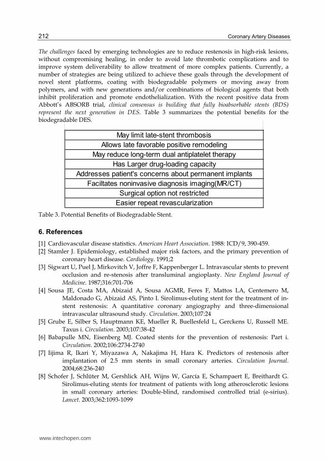
The challenges faced by emerging technologies are to reduce restenosis in high-risk lesions, without compromising healing, in order to avoid late thrombotic complications and to improve system deliverability to allow treatment of more complex patients. Currently, a number of strategies are being utilized to achieve these goals through the development of novel stent platforms, coating with biodegradable polymers or moving away from polymers, and with new generations and/or combinations of biological agents that both inhibit proliferation and promote endothelialization. With the recent positive data from Abbott’s ABSORB trial, clinical consensus is building that fully bioabsorbable stents (BDS) represent the next generation in DES. Table 3 summarizes the potential benefits for the biodegradable DES.
Faciltates noninvasive diagnosis imaging(MR/CT)
Surgical option not restricted
Easier repeat revascularization
May limit late-stent thrombosis
Allows late favorable positive remodeling
May reduce long-term dual antiplatelet therapy
Has Larger drug-loading capacity
Addresses patient's concerns about permanent implants
Table 3. Potential Benefits of Biodegradable Stent.
6. References
[1] Cardiovascular disease statistics. American Heart Association. 1988: ICD/9, 390-459. [2] Stamler J. Epidemiology, established major risk factors, and the primary prevention of
coronary heart disease. Cardiology. 1991;2 [3] Sigwart U, Puel J, Mirkovitch V, Joffre F, Kappenberger L. Intravascular stents to prevent
occlusion and re-stenosis after transluminal angioplasty. New England Journal of
Medicine. 1987;316:701-706 [4] Sousa JE, Costa MA, Abizaid A, Sousa AGMR, Feres F, Mattos LA, Centemero M,
Maldonado G, Abizaid AS, Pinto I. Sirolimus-eluting stent for the treatment of in-stent restenosis: A quantitative coronary angiography and three-dimensional intravascular ultrasound study. Circulation. 2003;107:24
[5] Grube E, Silber S, Hauptmann KE, Mueller R, Buellesfeld L, Gerckens U, Russell ME. Taxus i. Circulation. 2003;107:38-42
[6] Babapulle MN, Eisenberg MJ. Coated stents for the prevention of restenosis: Part i. Circulation. 2002;106:2734-2740
[7] Iijima R, Ikari Y, Miyazawa A, Nakajima H, Hara K. Predictors of restenosis after implantation of 2.5 mm stents in small coronary arteries. Circulation Journal. 2004;68:236-240
[8] Schofer J, Schlüter M, Gershlick AH, Wijns W, García E, Schampaert E, Breithardt G. Sirolimus-eluting stents for treatment of patients with long atherosclerotic lesions in small coronary arteries: Double-blind, randomised controlled trial (e-sirius). Lancet. 2003;362:1093-1099
www.intechopen.com

Coronary Arterial Drug-Eluting Stent: From Structure to Clinical
213
[9] Muramatsu T, Tsukahara R, Ho M, Ito Y, Ishimori H, Hirano K, Nakano M, Matsushita M, Leung W. Clinical outcome of stent implantation in small coronary arteries using different type of coronary stents. J Invasive Cardiol. 2001;13:634-639
[10] Welt FGP, Rogers C. Inflammation and restenosis in the stent era. Arterioscler. Thromb.
Vasc. Biol. 2002;22:1769-1776 [11] Shah PK. Inflammation, neointimal hyperplasia, and restenosis: As the leukocytes roll,
the arteries thicken. Circulation. 2003;107:2175-2177 [12] Bennett MR. In-stent stenosis: Pathology and implications for the development of drug
eluting stents. Heart 2003;89:218-224 [13] Cotran RS, Kumar V, Collins T, Robbins SL. Robbins pathologic basis of disease. 6th Ed.
Philadelphia: WB Saunders; 1999. [14] Ong ATL, Hoye A, Aoki J, Van Mieghem CAG, Rodriguez Granillo GA, Sonnenschein
K, Regar E, McFadden EP, Sianos G, Van Der Giessen WJ, De Jaegere PPT, De Feyter P, Van Domburg RT, Serruys PW. Thirty-day incidence and six-month clinical outcome of thrombotic stent occlusion after bare-metal, sirolimus, or paclitaxel stent implantation. J Am Coll Cardiol. 2005;45:947-953
[15] Iakovou I, Schmidt T, Bonizzoni E, Ge L, Sangiorgi GM, Stankovic G, Airoldi F, Chieffo A, Montorfano M, Carlino M, Michev I, Corvaja N, Briguori C, Gerckens U, Grube E, Colombo A. Incidence, predictors, and outcome of thrombosis after successful implantation of drug-eluting stents. JAMA. 2005;293:2126-2130
[16] Ong ATL, McFadden EP, Regar E, De Jaegere PPT, Van Domburg RT, Serruys PW. Late angiographic stent thrombosis (last) events with drug-eluting stents. J Am Coll
Cardiol. 2005;45:2088-2092 [17] Wang F, Stouffer GA, Waxman S, Uretsky BF. Late coronary stent thrombosis: Early vs.
Late stent. Catheterization and Cardiovascular Interventions. 2002;55:142-147 [18] Wenaweser P, Rey C, Eberli FR, Togni M, Tuller D, Locher S, Remondino A, Seiler C,
Hess OM, Meier B. Stent thrombosis following bare-metal stent implantation: Success of emergency percutaneous coronary intervention and predictors of adverse outcome. European Heart Journal. 2005;26:1180-1187
[19] McFadden EP, Stabile E, Regar E, Cheneau E, Ong ATL, Kinnaird T, Suddath WO, Weissman NJ, Torguson R, Kent KM. Late thrombosis in drug-eluting coronary stents after discontinuation of antiplatelet therapy. Lancet. 2004; 364:1519- 1521
[20] Virmani R, Guagliumi G, Farb A, Musumeci G, Grieco N, Motta T, Mihalcsik L, Tespili M, Valsecchi O, Kolodgie FD. Localized hypersensitivity and late coronary thrombosis secondary to a sirolimus-eluting stent should we be cautious? Circulation. 2004;109:701-705
[21] Joner M, Finn AV, Farb A, Mont EK, Kolodgie FD, Ladich E, Kutys R, Skorija K, Gold HK, Virmani R. Pathology of drug-eluting stents in humans:: Delayed healing and late thrombotic risk. Journal of the American College of Cardiology. 2006;48:193-202
[22] Köster R, Vieluf D, Kiehn M, Sommerauer M, Kähler J, Baldus S, Meinertz T, Hamm CW. Nickel and molybdenum contact allergies in patients with coronary in-stent restenosis. Lancet. 2000;356:1895-1897
www.intechopen.com

Coronary Artery Diseases
214
[23] Kastrati A, Mehilli J, Dirschinger J, Dotzer F, Schühlen H, Neumann F-J, Fleckenstein M, Pfafferott C, Seyfarth M, Schömig A. Intracoronary stenting and angiographic results : Strut thickness effect on restenosis outcome (isar-stereo) trial. Circulation. 2001;103:2816-2821
[24] Pache J, Kastrati A, Mehilli J, Schühlen H, Dotzer F, Hausleiter J, Fleckenstein M, Neumann F-J, Sattelberger U, Schmitt C, Müller M, Dirschinger J, Schömig A. Intracoronary stenting and angiographic results: Strut thickness effect on restenosis outcome (isar-stereo-2) trial. Journal of the American College of Cardiology. 2003;41:1283-1288
[25] Zitter H, Plenk Jr H. The electrochemical behavior of metallic implant materials as an indicator of their biocompatibility. Journal of Biomedical Materials Research. 1987;21:881-896
[26] Johnson PF, Bernstein JJ, Hunter G, Dawson WW, Hench LL. An in vitro and in vivo analysis of anodized tantalum capacitive electrodes: Corrosion response, physiology, and histology. Journal of Biomedical Materials Research. 1977;11:637-656
[27] Macionczyk F, Gerold B, Thull R. Repassivating tantalum/tantalum oxide surface modification on stainless steel implants. Surface & Coatings Technology. 2001;142:1084-1087
[28] Teitelbaum GP, Raney M, Carvlin MJ, Matsumoto AH, Barth KH. Evaluation of ferromagnetism and magnetic resonance imaging artifacts of the strecker tantalum vascular stent. CardioVascular and Interventional Radiology. 1989;12:125-127
[29] Matsumoto AH, Teitelbaum GP, Barth KH, Carvlin MJ, Savin MA, Strecker EP. Tantalum vascular stents: In vivo evaluation with mr imaging. Radiology. 1989;170:753-755
[30] Barth KH, Virmani R, Froelich J, Takeda T, Lossef SV, Newsome J, Jones R, Lindisch D. Paired comparison of vascular wall reactions to palmaz stents, strecker tantalum stents, and wallstents in canine iliac and femoral arteries. Circulation. 1996;93:2161-2169
[31] Ozaki Y, Keane D, Nobuyoshi M, Hamasaki N, Popma JJ, Serruys PW. Coronary lumen at six-month follow-up of a new radiopaque cordis tantalum stent using quantitative angiography and intracoronary ultrasound. The American Journal of
Cardiology. 1995;76:1135-1142 [32] Windecker S, Mayer I, De Pasquale G, Maier W, Dirsch O, De Groot P, Wu Y-P, Noll G,
Leskosek B, Meier B, Hess OM. Stent coating with titanium-nitride-oxide for reduction of neointimal hyperplasia. Circulation. 2001;104:928-933
[33] Mosseri M, Tamari I, Plich M, Hasin Y, Brizines M, Frimerman A, Miller H, Jafari J, Guetta V, Solomon M. Short-and long-term outcomes of the titanium-no stent registry. Cardiovascular Revascularization Medicine. 2005;6:2-6
[34] Windecker S, Simon R, Lins M, Klauss V, Eberli FR, Roffi M, Pedrazzini G, Moccetti T, Wenaweser P, Togni M, Tuller D, Zbinden R, Seiler C, Mehilli J, Kastrati A, Meier B, Hess OM. Randomized comparison of a titanium-nitride-oxide-coated stent with a stainless steel stent for coronary revascularization: The tinox trial. Circulation. 2005;111:2617-2622
[35] Biehl V, Wack T, Winter S, Seyfert UT, Breme J. Evaluation of the haemocompatibility of titanium based biomaterials. Biomolecular Engineering. 2002;19:97-101
www.intechopen.com

Coronary Arterial Drug-Eluting Stent: From Structure to Clinical
215
[36] Heintz C, Riepe G, Birken L, Kaiser E, Chakf N, Morlock M, Delling G, Imig H. Corroded nitinol wires in explanted aortic endografts: An important mechanism of failure? Journal of Endovascular Therapy. 2001;8:248-253
[37] Berger-Gorbet M, Broxup B, Rivard C, Yahia LH. Biocompatibility testing of niti screws using immunohistochemistry on sections containing metallic implants. Journal of
Biomedical Materials Research. 1996;32:243 - 248 [38] Wataha JC, Lockwood PE, Marek M, Ghazi M. Ability of ni-containing biomedical
alloys to activate monocytes and endothelial cells in vitro. Mater Res. 1999;45:251-257
[39] Shabalovskaya SA. On the nature of the biocompatibility and on medical applications of niti shape memory and superelastic alloys. Biomedical materials and engineering. 1996;6:267-289
[40] Maitz MF, Shevchenko N. Plasma-immersion ion-implanted nitinol surface with depressed nickel concentration for implants in blood. Journal of Biomedical Materials
Research Part A. 2006;76:356–365 [41] Mazumder MM, De S, Trigwell S, Ali N, Mazumder MK, Mehta JL. Corrosion resistance
of polyurethane-coated nitinol cardiovascular stents. Journal of Biomaterials Science,
Polymer Edition. 2003;14:1351-1362 [42] Starosvetsky D, Gotman I. Corrosion behavior of titanium nitride coated ni-ti shape
memory surgical alloy. Biomaterials. 2001;22:1853-1859 [43] Shih CC, Lin SJ, Chung KH, Chen YL, Su YY. Increased corrosion resistance of stent
materials by converting current surface film of polycrystalline oxide into amorphous oxide. Journal of Biomedical Materials Research. 2000;52:323-332
[44] Schürmann K, Vorwerk D, Kulisch A, Stroehmer-Kulisch E, Biesterfeld S, Stopinski T, Günther RW. Experimental arterial stent placement: Comparison of a new nitinol stent and wallstent. Investigative radiology. 1995;30:412-420
[45] Adams GJ, Baltazar U, Karmonik C, Bordelon C, Lin PH, Bush RL, Lumsden AB, Morrisett JD. Comparison of 15 different stents in superficial femoral arteries by high resolution mri ex vivo and in vivo. Journal of Magnetic Resonance Imaging. 2005;22:125-135
[46] Nakamura S, Degawa T, Nishida T, Anzai H, Mitsuo K, Sakatani H, Tsunoda T, Ui K, Yabuki S, Yamaguchi T. Preliminary experience of act-onetm coronary stent implantation. J. Am. Coll. Cardiol. 1996;27:53
[47] Holmes DR, Lansky A, Kuntz R, Bell MR, Buchbinder M, Fortuna R, O'Shaughnessy CD, Popma J. The paragon stent study: A randomized trial of a new martensiti nitinol stent versus the palmaz-schatz stent for treatment of complex native coronary arterial lesions. The American Journal of Cardiology. 2000;86:1073-1079
[48] Isshiki T, Eto K, Ochini M, Milani H, Kondo K, Takoshita S, Sato T. Nitinol radius stent induces less platelet aggregation than stainless steel palmaz-schatz/glanturco-rubin ii stents. JACC. 1998;31:312
[49] Hijazi ZM, Homoud M, Aronovitz MJ, Smith JJ, Faller GT. A new platinum balloon-expandable stent (angiostent) mounted on a high pressure balloon: Acute and late results in an atherogenic swine model. The Journal of invasive cardiology. 1995;7:127-134
www.intechopen.com

Coronary Artery Diseases
216
[50] Bhargava B, Scheerder ID, Ping QB, Yanming H, Chan R, Kim HS, Kollum M, Cottin Y, Leon MB. A novel platinum-iridium, potentially gamma radioactive stent: Evaluation in a porcine model. Catheterization and Cardiovascular Interventions. 2000;51:364-368
[51] Foti R, Tamburino C, Galassi AR, Russo G, Nicosia A, Grassi R, Monaco A, Azzarelli S, Mammana C, Calvi V. Safety, feasibility and efficacy of a new single-wire stent in the treatment of complex coronary lesions: The angiostent. Cardiologia. 1998;43:725-730
[52] Peuster M, Wohlsein P, Brugmann M, Ehlerding M, Seidler K, Fink C, Brauer H, Fischer A, Hausdorf G. A novel approach to temporary stenting: Degradable cardiovascular stents produced from corrodible metal-results 6-18 months after implantation into new zealand white rabbits. Heart. 2001;86:563-569
[53] Heublein B, Rohde R, Kaese V, Niemeyer M, Hartung W, Haverich A. Biocorrosion of magnesium alloys: A new principle in cardiovascular implant technology? Heart
2003;89:651-656 [54] Mueller PP, May T, Perz A, Hauser H, Peuster M. Control of smooth muscle cell
proliferation by ferrous iron. Biomaterials. 2006;27:2193-2200 [55] Peuster M, Hesse C, Schloo T, Fink C, Beerbaum P, von Schnakenburg C. Long-term
biocompatibility of a corrodible peripheral iron stent in the porcine descending aorta. Biomaterials. 2006;27:4955-4962
[56] Staiger MP, Pietak AM, Huadmai J, Dias G. Magnesium and its alloys as orthopedic biomaterials: A review. Biomaterials. 2006;27:1728-1734
[57] Di Mario C, Griffiths H, Goktekin O, Peeters N, Verbist JAN, Bosiers M, Deloose K, Heublein B, Rohde R, Kasese V. Drug-eluting bioabsorbable magnesium stent. Journal of Interventional Cardiology. 2004;17:391-395
[58] Eggebrecht H, Rodermann J, Hunold P, Schmermund A, Bose D, Haude M, Erbel R. Novel magnetic resonance-compatible coronary stent: The absorbable magnesium-alloy stent. Circulation. 2005;112:e303-304
[59] Sharkawi T, Cornhill F, Lafont A, Sabaria P, Vert M. Intravascular bioresorbable polymeric stents: A potential alternative to current drug eluting metal stents. Journal of Pharmaceutical Sciences. 2007;96:2829 - 2837
[60] Bosiers M, Deloose K, Verbist J, Peeters P. Will absorbable metal stent technology change our practice? Journal of cardiovascular surgery. 2006;47:393-397
[61] Zartner P, Cesnjevar R, Singer H, Weyand M. First successful implantation of a biodegradable metal stent into the left pulmonary artery of a preterm baby. Catheterization and Cardiovascular Interventions. 2005;66:590-594
[62] Schranz D, Zartner P, Michel-Behnke I, Akinturk H. Bioabsorbable metal stents for percutaneous treatment of critical recoarctation of the aorta in a newborn. Catheterization and Cardiovascular Interventions. 2006;67:671-673
[63] Waale WHE, Van Der Veen FH, Leeuwen C, Lankveld M, Havenith M, Bar FW, Oppen J, Wellens HJJ. Modulation of healthy pig coronary arteries by self-expanding stents. Journal of Interventional Cardiology. 1996;9:45-52
[64] Bramwell O, Leon MB. Acute and chronic effects of self-expanding nitinol stents in porcine coronary arteries. Coronary Artery Disease. 1997;8:45-46
www.intechopen.com

Coronary Arterial Drug-Eluting Stent: From Structure to Clinical
217
[65] Mosseri M, Rozenman Y, Mereuta A, Hasin Y, Gotsman MS. New indicator for stent covering area. Catheterization and cardiovascular diagnosis. 1998;44:188 - 192
[66] Leon MB, Popma JJ, O'haughnessy C, Dean LS, Lansky AJ, Fry ETA, Curran MJ. Quantitative angiographic outcomes after gianturco-roubin stent implantation in complex lesion subsets. Circulation. 1997;96: I-593
[67] Heuser R, Lopez A, Kuntz R, Reduto L, Badger R, Coleman P, Whitlow P, Iannone LA, Safian R, Yeung A. Smart: The microstent's ability to limit restenosis trial. Catheterization and Cardiovascular Interventions. 2001;52:269 - 277
[68] Gurbel PA, Callahan KP, Malinin AI, Serebruany VL, Gillis J. Could stent design affect platelet activation? Results of the platelet activation in stenting (past) study. Journal
of Invasive Cardiology. 2002;14:584-589 [69] Foley DP, Pieper M, Wijns W, Suryapranata H, Grollier G, Legrand V, de Scheerder I,
Hanet C, Puel J, Mudra H. The influence of stent length on clinical and angiographic outcome in patients undergoing elective stenting for native coronary artery lesions; final results of the magic 5l study. European Heart Journal. 2001;22:1585-1593
[70] Stone GW, Ellis SG, Cox DA, Hermiller J, O'Shaughnessy C, Mann JT, Turco M, Caputo R, Bergin P, Greenberg J. A polymer-based, paclitaxel-eluting stent in patients with coronary artery disease. N. Engl. J. Med. 2004;350:221-231
[71] Garasic JM, Edelman ER, Squire JC, Seifert P, Williams MS, Rogers C. Stent and artery geometry determine intimal thickening independent of arterial injury. Circulation. 2000;101:812-818
[72] Gunn J, Arnold N, Chan KH, Shepherd L, Cumberland DC, Crossman DC. Coronary artery stretch versus deep injury in the development of in-stent neointima. Heart. 2002;88:401-405
[73] Hamuro M, Palmaz JC, Sprague EA, Fuss C, Luo J. Influence of stent edge angle on endothelialization in an in vitro model. Journal of Vascular and Interventional
Radiology. 2001;12:607-611 [74] De Scheerder I, Verbeken E, Van Humbeeck J. Metallic surface modification. Seminars in
interventional cardiology 1998;3:139-144 [75] Palmaz JC, Benson A, Sprague EA. Influence of surface topography on
endothelialization of intravascular metallic material. Journal of Vascular and
Interventional Radiology. 1999;10:439-444 [76] Fuss C, Sprague EA, Bailey SR, Palmaz JC. Surface micro grooves (mg) improve
endothelialization rate in vitro and in vivo. Am J Cardiol. 2001;37:70A [77] Palmaz JC, Bailey S, Marton D, Sprague E. Influence of stent design and material
composition on procedure outcome. Journal of Vascular Surgery. 2002;36:1031-1039 [78] Dibra A, Kastrati A, Mehilli J, Pache J, von Oepen R, Dirschinger J, Schomig A.
Influence of stent surface topography on the outcomes of patients undergoing coronary stenting: A randomized double-blind controlled trial. Catheterization and
Cardiovascular Interventions. 2005;65:374 - 380 [79] Rogers C, Edelman ER. Endovascular stent design dictates experimental restenosis and
thrombosis. Circulation. 1995;91:2995-3001
www.intechopen.com
Coronary Artery Diseases
218
[80] Albrecht D, Kaspers S, Fussl R, Hopp HW, Sechtem U. Coronary plaque morphology affects stent deployment: Assessment by intracoronary ultrasound. Catheterization
and cardiovascular diagnosis. 1996;38:229 - 235 [81] Henneke KH, Regar E, Konig A, Werner F, Klauss V, Metz J, Theisen K, Mudra H.
Impact of target lesion calcification on coronary stent expansion after rotational atherectomy. American Heart Journal. 1999;137:93-99
[82] Narracott AJ, Lawford PV, Gunn JPG, Hose DR. Balloon folding affects the symmetry of stent deployment: Experimental and computational evidence. Engineering in Medicine and Biology Society, 2007. EMBS 2007. 29th Annual International Conference of
the IEEE. 2007:3069-3073 [83] Narracott AJ, Hose DR, Lawford PV, Gunn J. Measurement of the symmetry of in vitro
stent expansion: A stereo-photogrammetric approach. Journal of medical engineering
& technology. 2003;27:59-70 [84] Morice MC, Serruys PW, Sousa JE, Fajadet J, Ban Hayashi E, Perin M, Colombo A,
Schuler G, Barragan P, Guagliumi G. A randomized comparison of a sirolimus-eluting stent with a standard stent for coronary revascularization. N. Engl. J. Med. 2002;346:1773-1780
[85] Moses JW, Leon MB, Popma JJ, Fitzgerald PJ, Holmes DR, O'Shaughnessy C, Caputo RP, Kereiakes DJ, Williams DO, Teirstein PS. Sirolimus-eluting stents versus standard stents in patients with stenosis in a native coronary artery. N. Engl. J. Med. 2003;349:1315-1323
[86] Stone GW, Ellis SG, Cannon L, Mann JT, Greenberg JD, Spriggs D, O'Shaughnessy CD, DeMaio S, Hall P, Popma JJ, Koglin J, Russell ME, for the TVI. Comparison of a polymer-based paclitaxel-eluting stent with a bare metal stent in patients with complex coronary artery disease: A randomized controlled trial. JAMA. 2005;294:1215-1223
[87] Aziz S, Morris JL, Perry RA. Late stent thrombosis associated with coronary aneurysm formation after sirolimus-eluting stent implantation. J. Invasive. Cardiol. 2007;19:E96-98
[88] Camenzind E. Treatment of in-stent restenosis--back to the future? N. Engl. J. Med. 2006;355:2149-2151
[89] Camenzind E, Steg PG, Wijns W. Stent thrombosis late after implantation of first-generation drug-eluting stents: A cause for concern. Circulation. 2007;115:1440-1455
[90] Pfisterer ME. The basket-late-study. Basel stent cost-effectiveness trial--late thrombotic events trial. Herz. 2006;31:259
[91] Tsuchida K, Piek JJ, Neumann FJ, Van Der Giessen WJ, Wienler M, Zeiher AM, Grube E, Haase J, Thuesen L, Hamm CW. One-year results of a durable polymer everolimus-eluting stent in fife novo coronary narrowings(the spirit first trial). EuroIntervention. 2005;1:266-272
[92] Fajadet J, Wijns W, Laarman GJ, Kuck KH, Ormiston J, Munzel T, Popma JJ, Fitzgerald PJ, Bonan R, Kuntz RE. Randomized, double-blind, multicenter study of the endeavor zotarolimus-eluting phosphorylcholine-encapsulated stent for treatment of native coronary artery lesions: Clinical and angiographic results of the endeavor ii trial. Circulation. 2006;114:798-806
www.intechopen.com

Coronary Arterial Drug-Eluting Stent: From Structure to Clinical
219
[93] Stone GW, Midei M, Newman W, Sanz M, Hermiller JB, Williams J, Farhat N, Mahaffey KW, Cutlip DE, Fitzgerald PJ, Sood P, Su X, Lansky AJ, for the SIIII. Comparison of an everolimus-eluting stent and a paclitaxel-eluting stent in patients with coronary artery disease: A randomized trial. JAMA. 2008;299:1903-1913
[94] Kandzari DE, Leon MB, Popma JJ, Fitzgerald PJ, O'Shaughnessy C, Ball MW, Turco M, Applegate RJ, Gurbel PA, Midei MG. Comparison of zotarolimus-eluting and sirolimus-eluting stents in patients with native coronary artery disease a randomized controlled trial. J. Am. Coll. Cardiol. 2006;48:2440-2447
[95] Slottow TLP, Steinberg DH, Waksman R. Overview of the 2007 food and drug administration circulatory system devices panel meeting on patent foramen ovale closure devices. Circulation. 2007;116:677-682
[96] Meredith IT, Ormiston J, Whitbourn R, Kay IP, Muller D, Popma JJ, Cutlip DE, Fitzgerald PJ. Four-year clinical follow-up after implantation of the endeavor zotarolimus-eluting stent: Endeavor i, the first-in-human study. Am. J. Cardiol. 2007;100:56-61
[97] Finn AV, Nakazawa G, Joner M, Kolodgie FD, Mont EK, Gold HK, Virmani R. Vascular responses to drug eluting stents: Importance of delayed healing. Arterioscler.
Thromb. Vasc. Biol. 2007;27:1500-1510 [98] Hollinger JO. Preliminary report on the osteogenic potential of a biodegradable
copolymer of polyactide (pla) and polyglycolide (pga). J. Biomed. Mater. Res. 1983;17:71-82
[99] Gunatillake PA, Adhikari R. Biodegradable synthetic polymers for tissue engineering. Eur Cells Mat. 2003;5:1-16
[100] Ge J, Qian J, Wang X, Wang Q, Yan W, Yan Y, Fan B, Ge L, Liu X. Effectiveness and safety of the sirolimus-eluting stents coated with bioabsorbable polymer coating in human coronary arteries. Catheter Cardiovasc Interv. 2007;69:198 - 202
[101] Lee CH, Lim J, Low A, Zhang XL, Kyaing TT, Chan MY, Wong HB, Lim YT, Tan HC. Sirolimus-eluting, bioabsorbable polymer-coated constant stent (curatm) in acute st-elevation myocardial infarction: A clinical and angiographic study (curami registry). J Invasive Cardiol. 2007;19:182-185
[102] Aoki JO, Andrew T. L., Abizaid A, Tien HP, Bonnier H, Mcclean DR, Verheye S, Belardi J, Condado JA, Pieper M, Sousa JE, Bressers M, Symons J, Litvack F, Sianos G, Serruys PW. One-year clinical outcome of various doses and pharmacokinetic release formulations of paclitaxel eluted from an erodable polymer—insight in the paclitaxel in-stent controlled elution study (pisces). EuroIntervention. 2005;1:165-172
[103] Serruys PW, Sianos G, Abizaid A, Aoki J, den Heijer P, Bonnier H, Smits P, McClean D, Verheye S, Belardi J, Condado J, Pieper M, Gambone L, Bressers M, Symons J, Sousa E, Litvack F. The effect of variable dose and release kinetics on neointimal hyperplasia using a novel paclitaxel-eluting stent platform: The paclitaxel in-stent controlled elution study (pisces). J. Am. Coll. Cardiol. 2005;46:253-260
[104] Middleton JC, Tipton AJ. Synthetic biodegradable polymers as orthopedic devices. Biomaterials. 2000;21:2335-2346
www.intechopen.com

Coronary Artery Diseases
220
[105] Wessely R, Hausleiter J, Michaelis C, Jaschke B, Vogeser M, Milz S, Behnisch B, Schratzenstaller T, Renke-Gluszko M, Stöver M, Wintermantel E, Kastrati A, Schömig A. Inhibition of neointima formation by a novel drug-eluting stent system that allows for dose-adjustable, multiple, and on-site stent coating Arteriosclerosis,
Thrombosis, and Vascular Biology. 2005;25:748-753 [106] Mehilli J, Kastrati A, Wessely R, Dibra A, Hausleiter J, Jaschke B, Dirschinger J,
Schömig A. Randomized trial of a nonpolymer-based rapamycin-eluting stent versus a polymer-based paclitaxel-eluting stent for the reduction of late lumen loss Circulation. 2006;113:273-279
[107] Rajtar A, Kaluza GL, Yang Q, Hakimi D, Liu D, Tsui M, Lien M, Smith D, Clubb FJ, Troczynski T. Hydroxyapatite-coated cardiovascular stents. EuroIntervention. 2006;2:113-115
[108] Kollum M, Farb A, Schreiber R, Terfera K, Arab A, Geist A, Haberstroh J, Wnendt S, Virmani R, Hehrlein C. Particle debris from a nanoporous stent coating obscures potential antiproliferative effects of tacrolimus-eluting stents in a porcine model of restenosis. Catheter Cardiovasc Interv. 2005;64:85-90
[109] Polyak B, Fishbein I, Chorny M, Alferiev I, Williams D, Yellen B, Friedman G, Levy RJ. High field gradient targeting of magnetic nanoparticle-loaded endothelial cells to the surfaces of steel stents. Proc. Natl. Acad. Sci. U.S.A. 2008;105:698-703
[110] Sun J, Marx SO, Chen HJ, Poon M, Marks AR, Rabbani LRE. Role for p27kip1 in vascular smooth muscle cell migration. Circulation. 2001;103:2967-2972
[111] Tanner FC, Boehm M, Akyurek LM, San H, Yang ZY, Tashiro J, Nabel GJ, Nabel EG. Differential effects of the cyclin-dependent kinase inhibitors p27kip1, p21cip1, and p16ink4 on vascular smooth muscle cell proliferation. Circulation. 2000;101:2022-2025
[112] Steffel J, Latini RA, Akhmedov A, Zimmermann D, Zimmerling P, Luscher TF, Tanner FC. Rapamycin, but not fk-506, increases endothelial tissue factor expression implications for drug-eluting stent design. Circulation. 2005;112:2002-2011
[113] Steffel J, Luscher TF, Tanner FC. Tissue factor in cardiovascular diseases molecular mechanisms and clinical implications. Circulation. 2006;113:722-731
[114] Guha M, Mackman N. The phosphatidylinositol 3-kinase-akt pathway limits lipopolysaccharide activation of signaling pathways and expression of inflammatory mediators in human monocytic cells. Journal of Biological Chemistry. 2002;277:32124-32132
[115] Seabra‐Gomes R. Percutaneous coronary interventions with drug eluting stents for
diabetic patients. Heart. 2006;92:410–419
[116] Joner M, Nakazawa G, Finn AV, Quee SC, Coleman L, Acampado E, Wilson PS, Skorija K, Cheng Q, Xu X, Gold HK, Kolodgie FD, Virmani R. Endothelial cell recovery between comparator polymer-based drug-eluting stents. Journal of the American
College of Cardiology. 2008;52:333-342 [117] Lasave LI, Costa JdR, Abizaid AA, Feres F, Tanajura LF, Staico R, Abizaid AA, Beraldo
P, Sousa AMR, Sousa JEMR. A three-dimensional intravascular ultrasound comparison between the new zotarolimus-eluting stent (zomaxx™) and the non-drug-eluting trimaxx™ stent. Journal of Invasive Cardiology. 2007;19:303-308
www.intechopen.com

Coronary Arterial Drug-Eluting Stent: From Structure to Clinical
221
[118] Dunn C, Croom KF. Everolimus: A review of its use in renal and cardiac
transplantation. Drugs. 2006;66:547-570
[119] Farb A, John M, Acampado E, Kolodgie FD, Prescott MF, Virmani R. Oral everolimus
inhibits in-stent neointimal growth. Circulation. 2002;106:2379-2384
[120] Waksman R, Pakala R, Baffour R, Hellinga D, Seabron R, Kolodgie F, Virmani R.
Optimal dosing and duration of oral everolimus to inhibit in-stent neointimal
growth in rabbit iliac arteries. Cardiovascular Revascularization Medicine. 2006;7:179-
184
[121] Patel JK, Kobashigawa JA. Everolimus: An immunosuppressive agent in
transplantation. Expert opinion on pharmacotherapy. 2006;7:1347-1355
[122] Grube E, Buellesfeld L. Everolimus for stent-based intracoronary applications. Reviews
in cardiovascular medicine. 2004;5:S3-S8
[123] Grube E, Buellesfeld L. Biomatrix biolimus a9-eluting coronary stent: A next-
generation drug-eluting stent for coronary artery disease. Expert Review of Medical
Devices. 2006;3:731-741
[124] Costa RA, Lansky AJ, Abizaid A, Müeller R, Tsuchiya Y, Mori K, Cristea E, Leon MB,
Eduardo Sousa J, Schmidt T. Angiographic results of the first human experience
with the biolimus a 9 drug-eluting stent for de novo coronary lesions. American
Journal of Cardiology. 2006;98:443-446
[125] Grube E, Hauptmann KE, Buellesfeld L, Lim V, Abizaid A. Six-month results of a
randomized study to evaluate safety and efficacy of a biolimus a 9 eluting stent
with a biodegradable polymer coating. EuroIntervention. 2005;1:53-57
[126] Chevalier B, Serruys PW, Silber S, Garcia E, Suryapranata H, Hauptmann K, Wijns W,
Schuler G, Fath-Ordoubadi F, Worthley SG, Thuesen L, Meredith IT, Bressers M,
Nagai H, Paunovic D. Randomised comparison of noboritm, biolimus a9-eluting
coronary stent with ataxus®, paclitaxel-eluting coronary stent in patients with
stenosis in native coronary arteries: The nobori 1 trial. EuroIntervention. 2007;2:426-
434
[127] Sollott SJ, Cheng L, Pauly RR, Jenkins GM, Monticone RE, Kuzuya M, Froehlich JP,
Crow MT, Lakatta EG, Rowinsky EK. Taxol inhibits neointimal smooth muscle cell
accumulation after angioplasty in the rat. Journal of Clinical Investigation.
1995;95:1869-1876
[128] Abal M, Andreu JM, Barasoain I. Taxanes: Microtubule and centrosome targets, and
cell cycle dependent mechanisms of action. Current Cancer Drug Targets. 2003;3:193-
203
[129] Wang TH, Wang HS, Ichijo H, Giannakakou P, Foster JS, Fojo T, Wimalasena J.
Microtubule-interfering agents activate c-jun n-terminal kinase/stress-activated
protein kinase through both ras and apoptosis signal-regulating kinase pathways.
Journal of Biological Chemistry. 1998;273:4928-4936
[130] Stahli BE, Camici GG, Steffel J, Akhmedov A, Shojaati K, Graber M, Luscher TF,
Tanner FC. Paclitaxel enhances thrombin-induced endothelial tissue factor
expression via c-jun terminal nh2 kinase activation. Circulation Research. 2006;
99:149-155
www.intechopen.com

Coronary Artery Diseases
222
[131] Green MR, Manikhas GM, Orlov S, Afanasyev B, Makhson AM, Bhar P, Hawkins MJ.
Abraxane®, a novel cremophor®-free, albumin-bound particle form of paclitaxel for
the treatment of advanced non-small-cell lung cancer. Annals of Oncology.
2006;17:1263-1268
[132] Margolis J, McDonald J, Heuser R, Klinke P, Waksman R, Virmani R, Desai N, Hilton D. Systemic nanoparticle paclitaxel (nab-paclitaxel) for in-stent restenosis i (snapist-i): A first-in-human safety and dose-finding study. Clin. Cardiol. 2007;30:165-170
[133] Clarke SJ, Rivory LP. Clinical pharmacokinetics of docetaxel. Clinical Pharmacokinetics. 1999;36:99-114
[134] Yasuda S, Noguchi T, Gohda M, Arai T, Tsutsui N, Nakayama Y, Matsuda T, Nonogi H. Local delivery of low-dose docetaxel, a novel microtubule polymerizing agent, reduces neointimal hyperplasia in a balloon-injured rabbit iliac artery model. Cardiovascular Research. 2002;53:481-486
[135] Silvestrini R, Zaffaroni N, Orlandi L, Oriana S. In vitro cytotoxic activity of taxol and taxotere on primary cultures and established cell lines of human ovarian cancer. Stem Cells. 1993;11:528-535
[136] Gottschalk AR, Quintans J. Apoptosis in b lymphocytes: The wehi-231 perspective. Immunology and Cell Biology. 1995;73:8-16
[137] Halloran PF. Immunosuppressive drugs for kidney transplantation. New England
Journal of Medicine. 2004;351:2715-2729 [138] Mohacsi PJ, Tuller D, Hulliger B, Wijngaard PL. Different inhibitory effects of
immunosuppressive drugs on human and rat aortic smooth muscle and endothelial cell proliferation stimulated by platelet-derived growth factor or endothelial cell growth factor. Journal of Heart and Lung Transplantation. 1997;16:484-492
[139] Matter CM, Rozenberg I, Jaschko A, Greutert H, Kurz DJ, Wnendt S, Kuttler B, Joch H, Grünenfelder J, Zünd G, Tanner FC, Lüscher T. Effects of tacrolimus or sirolimus on proliferation of vascular smooth muscle and endothelial cells. Journal of
Cardiovascular Pharmacology. 2006;48:286-292 [140] Grube E, Buellesfeld L. Rapamycin analogs for stent-based local drug delivery.
Everolimus- and tacrolimus-eluting stents. Herz. 2004;29:162-166 [141] Morice MC, Bestehorn HP, Carrie D, Macaya C, Aengevaeren W, Wijns W, Dubois C,
De Winter R, Verheye S, Hoffmann S. Direct stenting of de novo coronary stenoses with tacrolimus-eluting versus carbon-coated carbostents. The randomized jupiter ii trial. EuroIntervention. 2006;2:45-52
[142] Tanimoto S, W vdG, H vB, Sorop O, Kukreja N, Fukaya K, Nishide T, Nakano R, Maeda H, Serruys PW. Mahoroba™: Tacrolimus eluting coronary stent. EuroIntervention. 2007;3:149-153
[143] Gupta AK, Chow M. Pimecrolimus: A review. Journal of the European Academy of
Dermatology & Venereology. 2003;17:493-503 [144] Dorai T, Aggarwal BB. Role of chemopreventive agents in cancer therapy. Cancer
letters. 2004;215:129-140 [145] Gupta KK, Bharne SS, Rathinasamy K, Naik NR, Panda D. Dietary antioxidant
curcumin inhibits microtubule assembly through tubulin binding. FEBS Journal. 2006;273:5320-5332
www.intechopen.com

Coronary Arterial Drug-Eluting Stent: From Structure to Clinical
223
[146] Ruby AJ, Kuttan G, Babu KD, Rajasekharan KN, Kuttan R. Anti-tumour and antioxidant activity of natural curcuminoids. Cancer letters. 1995;94:79-83
[147] Docherty JJ, Fu MM, Tsai M. Resveratrol selectively inhibits neisseria gonorrhoeae and neisseria meningitidis. Journal of Antimicrobial Chemotherapy. 2001;47:243-244
[148] de la Lastra C, Villegas I. Resveratrol as an antioxidant and pro-oxidant agent: Mechanisms and clinical implications. Biochemical Society Transactions. 2007;35:1156-1160
[149] Mnjoyan ZH, Fujise K. Profound negative regulatory effects by resveratrol on vascular smooth muscle cells: A role of p53–p21waf1/cip1 pathway Biochemical and Biophysical
Research Communications. 2003;311:546-552 [150] Olas B, Wachowicz B. Resveratrol, a phenolic antioxidant with effects on blood platelet
functions. Platelets. 2005;16:251-260 [151] Poussier B, Cordova AC, Becquemin JP, Sumpio BE. Resveratrol inhibits vascular
smooth muscle cell proliferation and induces apoptosis. Journal of Vascular Surgery. 2005;42:1190-1197
[152] Pace-Asciak CR, Hahn S, Diamandis EP, Soleas G, Goldberg DM. The red wine phenolics trans-resveratrol and quercetin block human platelet aggregation and eicosanoid synthesis: Implications for protection against coronary heart disease. Clinica Chimica Acta. 1995;235:207-219
[153] Aoki J, Serruys PW, van Beusekom H, Ong ATL, McFadden EP, Sianos G, van der Giessen WJ, Regar E, de Feyter PJ, Davis HR. Endothelial progenitor cell capture by stents coated with antibody against cd34 the healing-fim (healthy endothelial accelerated lining inhibits neointimal growth-first in man) registry. Journal of the
American College of Cardiology. 2005;45:1574-1579 [154] Duckers HJ, Silber S, De Winter R, Heijer PD, Rensing B, Rau M, Mudra H, Benit E,
Verheye S, Wijns W. Circulating endothelial progenitor cells predict angiographic and intravascular ultrasound outcome following percutaneous coronary interventions in the healing-ii trial: Evaluation of an endothelial progenitor cell capturing stent. EuroIntervention. 2007;3:67-75
[155] Duckers HJ, Soullie T, Peter DH, Rensing B, Robbert DW, Rau M, Mudra H, Silber S, Benit E, Velheye S, Wiins W, Serruys PW. Accelerated vascular repair following percutaneous coronary intervention by capture of endothelial progenitor cells promotes regression of neointimal growth at long term follow-up : Final results of the healing ii trial using an endothelial progenitor cell capturing stent (genous r stent)™. EuroIntervention. 2007;3:350-358
[156] Co M, Tay E, Lee CH, Poh KK, Low A, Lim J, Lim IH, Lim YT, Tan HC. Use of endothelial progenitor cell capture stent (genous bio-engineered r stent) during primary percutaneous coronary intervention in acute myocardial infarction: Intermediate-to long-term clinical follow-up. American Heart Journal. 2008;155:128-132
[157] Stefanadis C, Toutouzas K, Stefanadi E, Kolodgie F, Virmani R, Kipshidze N. First experimental application of bevacizumab-eluting pc coated stent for inhibition of vasa vasorum of atherosclerotic plaque: Angiographic results in a rabbit atheromatic model. Hellenic Journal of Cardiology. 2006;47:7-10
www.intechopen.com

Coronary Artery Diseases
224
[158] Kukreja N, Onuma Y, Daemen J, Serruys PW. The future of drug-eluting stents. Pharmacological Research. 2008;57:171-180
[159] Yosinobu Onuma and Patrick W. Serruys. Bioresorbable Scaffold : The Advent of a New Era in Percutaneous Coronary and Peripheral Revascularization? Circulation
2011, 123:779-797
www.intechopen.com

Coronary Artery DiseasesEdited by Dr. Illya Chaikovsky
ISBN 978-953-51-0238-0Hard cover, 332 pagesPublisher InTechPublished online 07, March, 2012Published in print edition March, 2012
InTech EuropeUniversity Campus STeP Ri Slavka Krautzeka 83/A 51000 Rijeka, Croatia Phone: +385 (51) 770 447 Fax: +385 (51) 686 166www.intechopen.com
InTech ChinaUnit 405, Office Block, Hotel Equatorial Shanghai No.65, Yan An Road (West), Shanghai, 200040, China
Phone: +86-21-62489820 Fax: +86-21-62489821
This book has "wide geography" both literally and figuratively. First of all, this book brings togethercontributions from around the world, both from post-industrial countries and developing world. This is natural,because coronary artery disease is becoming pandemic worldwide. CAD is the single most frequent cause ofdeath in developed countries, causes about 1 in every 5 deaths. Mortality from cardiovascular disease ispredicted to reach 23.4 million in 2030. Moreover, in the developing world, cardiovascular disease tends toaffect people at a younger age and thus could negatively affect the workforce and economic productivity. Themorbidity, mortality, and socioeconomic importance of CAD make its diagnosis and management fundamentalfor all practicing physicians. On another hand, the book widely represents "geography" of CAD itself, i.e. manyvarious aspects of its pathophysiology, epidemiology, diagnosis, treatment are touched in this book. This bookdoes not pretend on complete and integral description of the Coronary artery disease. Rather, it containsselected issues on this complex multifactorial disease. Nevertheless, we hope that readers will find CoronaryArtery Disease useful for clinical practice and further research.
How to referenceIn order to correctly reference this scholarly work, feel free to copy and paste the following:
Tim Wu and Stephen McCarthy (2012). Coronary Arterial Drug-Eluting Stent: From Structure to Clinical,Coronary Artery Diseases, Dr. Illya Chaikovsky (Ed.), ISBN: 978-953-51-0238-0, InTech, Available from:http://www.intechopen.com/books/coronary-artery-diseases/coronary-arterial-drug-eluting-stent-from-its-structure-to-clinical

© 2012 The Author(s). Licensee IntechOpen. This is an open access articledistributed under the terms of the Creative Commons Attribution 3.0License, which permits unrestricted use, distribution, and reproduction inany medium, provided the original work is properly cited.