Cori Lecture
21
C ARL F. C ORI and G ERTY T. C ORI Polysaccharide phosphorylase Nobel Lectures, December 11, 1947 Part I - by Carl F. Cori Polysaccharide phosphorylase is characterized as an enzyme which can break or make an a-I-4-glucosidic bond at the termination (non-reducing end) of a glycogen or starch chain. The process is illustrated below: The interaction of phosphate with the terminal glucosidic bond results in the formation of glucose-1-phosphate and the loss of a chain unit; in the reverse reaction the glucose part of glucose-1-phosphate is added as a new chain unit and phosphate is set free. This reversible enzymatic polymeriza- tion occurs with little change in free energy, as may be calculated from the equilibrium constant. The reaction which involves expenditure of energy in the conversion of glucose to glycogen is the hexokinase reaction, the forma- tion of glucose-6-phosphate from glucose and adenosine triphosphate.
-
Upload
miguel-ramos -
Category
Documents
-
view
232 -
download
1
description
Cori lecture
Transcript of Cori Lecture

CARL F. CORI and GERTY T. CORI
Polysaccharide phosphorylase
Nobel Lectures, December 11, 1947
Part I - by Carl F. Cori
Polysaccharide phosphorylase is characterized as an enzyme which can breakor make an a-I-4-glucosidic bond at the termination (non-reducing end) ofa glycogen or starch chain. The process is illustrated below:
The interaction of phosphate with the terminal glucosidic bond results inthe formation of glucose-1-phosphate and the loss of a chain unit; in thereverse reaction the glucose part of glucose-1-phosphate is added as a newchain unit and phosphate is set free. This reversible enzymatic polymeriza-tion occurs with little change in free energy, as may be calculated from theequilibrium constant. The reaction which involves expenditure of energy inthe conversion of glucose to glycogen is the hexokinase reaction, the forma-tion of glucose-6-phosphate from glucose and adenosine triphosphate.

P O L Y S A C C H A R I D E P H O S P H O R Y L A S E 187
For the discussion which follows it is important to note that the phosphoryl-ated hexoses can enter into the following enzymatic equilibria (Table 1)
Table 1. Enzymatic equilibria at pH 7 at 30º.
Resting mammalian muscle contains about 0.003 mole of hexose-6-phos-phate (5 glucose-6- and + fructosed-phosphate) per kilo. Assuming thatthe mutase reaction is also close to equilibrium in a resting muscle, we cancalculate the concentration of glucose-1-phosphate to be expected as 0.0001mole per kilo or less than 0.2 per cent of the total acid soluble phosphatecontent of muscle. It seems clear that the detection and isolation of this inter-mediate could not have been accomplished without a separation of phos-phorylase activity from that of the other enzymes.
Formation of hexose-6-phosphate - The discovery of polysaccharide phos-phorylase and glucose-1-phosphate can be traced to systematic work on theformation of hexosed-phosphate in muscle. Of particular importance wasthe fact that the method used for the determination of hexosed-phosphateconsisted of two independent measurements, one based on the reducingpower of the compound and the other on its phosphate content and thatthere was generally good agreement between these two measurements. Inthis manner it was found that a number of procedures led to an increase inthe hexosed-phosphate content of muscle, among which may be listed,anaerobiosis, injection of epinephrine in intact animals, incubation of isolatedfrog muscle in Ringer’s solution containing epinephrine, and electric stim-ulation of mammalian or frog muscle.
Balance experiments during aerobic recovery ofpreviously stimulated andisolated frog muscle indicated that the hexose-6-phosphate which disap-peared was in large part reconverted to glycogen; hence it was made prob-able that the reaction, glycogen -+ glucose-6-phosphate, was reversible. Thenext step was the finding that the increase in hexosed-phosphate in isolatedfrog muscle incubated anaerobically with epinephrine was accompanied by

188 1 9 4 7 C . F . C O R I A N D G . T . C O R I
a corresponding decrease in inorganic phosphate (average of 6 experimentsper 100 g muscle, + 14 mg ester P, - 16 mg inorganic P). Phosphocreatineand adenosine triphosphate (ATP) remained unchanged, suggesting thatthey were not involved in the formation of hexose-6-phosphate, but sincetheir regeneration through lactic acid formation was not excluded, the ex-periments were repeated with muscles poisoned with iodoacetate. The resultswere the same as with unpoisoned muscle and it was therefore concludedthat hexosed-phosphate was formed from glycogen by ester&cation withinorganic phosphate:
These findings were presented in 1935 at the 15th International Physio-logical Congress and were discussed at that time with Professor Pamaswho then stated that he had under consideration experiments with muscleextract. Prior to that time it has been assumed that glycogen reacted withATP to form hexose diphosphate. Parnas and Baranowski found that a dis-appearance of inorganic phosphate could be demonstrated in a cell-freeextract of muscle which did not contain phosphocreatine or ATP. This was
ofimportance because it established beyond doubt the participation of in-organic phosphate in the splitting of glycogen, a process which has been aptlycalled "phosphorolysis" by Parnas. However, the mechanism of phosphor-olysis remained unknown until glucose-r-phosphate had been isolated.
Formation of glucose-1-phosphate - The following experiments led to thedetection and isolation of glucose-1-phosphate. Minced frog muscle was ex-tracted three times with 20 volumes of cold distilled water, a procedurewhich removed most of the acid-soluble phosphates normally present inmuscle, but did not remove glycogen. When the washed residue was in-cubated anaerobically at 20º in isotonic phosphate buffer at pH 7.2, somehexose monophosphate was formed. On addition of a catalytic amount ofmuscle adenylic acid, the formation of hexose monophosphate was verymarkedly increased. When phosphate was replaced by isotonic KCl, no esterformation occurred. The glucose part of the ester could have come onlyfrom glycogen, and the phosphate part only from the added inorganic phos-phate, thus confirming the reaction postulated for intact muscle.
After short periods of incubation there was much more organic phosphatepresent in the hexose monophosphate fraction than corresponded to thereducing power of hexose-6-phosphate. Such a discrepancy had not been

P O L Y S A C C H A R I D E P H O S P H O R Y L A S E 189
encountered before in analyses of the hexose monophosphate fraction, andsince the discrepancy became smaller or disappeared completely after longerperiods of incubation, the formation of a precursor of glucosed-phosphatewas suspected. Short hydrolysis in NH2SO4 at 100º (conditions under whichhexosed-phosphate is not hydrolyzed) revealed the presence of a compoundwhich yielded equivalent amounts of fermentable sugar and inorganic phos-phate.
A representative experiment is shown in Table 2. Comparison of the sec-ond, third and the last columns shows that the additional organic phosphatepresent before hydrolysis is accounted for by this new compound. Further-more, the disappearance of this compound in the third hour of incubation( - 0 . 7 4 m i l l i moles) is accounted for by a corresponding gain in reducing power ("hexose") before hydrolysis (+0.67 millimoles).
Table 2. Formation of glucose-1-phosphate in minced and washed frog muscle in-cubated in phosphate buffer plus adenylic acid.
The water-soluble, alcohol-insoluble barium salts (hexose monophosphate fraction)were isolated and analyzed for phosphate and reducing power before and after hydrol-
ysis in N H2SO4 for 10 minutes at 100º.AU-values are given in milimoles per 100 g muscle.
* The sugar formed after hydrolysis was completely fermentable, while hexose-6phosphate under the conditions chosen, was not fermented by the live yeast.
The new phosphate ester was isolated as the crystalline brucine salt in alarge-scale experiment similar to that shown in Table 2 and identified asglucose-1-phosphate.
When glucose-1-phosphate was added to a cell-free frog or rabbit muscleextract, it was converted rapidly to glucose-6-phosphate by an enzymewhich was named phosphoglucomutase. It was due to the leaking out ofthe mutase that glucose-1-phosphate accumulated in washed and minced

190 1 9 4 7 C . F . C O R I A N D G . T . C O R I
frog muscle. Mutase is greatly enhanced in its activity by magnesium ions.In order to demonstrate the formation of glucose-I-phosphate from gly-cogen and inorganic phosphate in muscle extract, it was necessary to removemagnesium ions by dialysis.
An experiment which shows the effect of magnesium ions as well as ofadenylic acid is given in Table 3. Addition of magnesium ions to the dialyzedextract had no effect on the total amount of ester formed, but it preventedthe accumulation of glucose-I-phosphate.
Experiments similar to those shown in Table 3 were performed with
Table 3. Formation of glucose-1-phosphate in dialyzed (17 hours) rabbit muscleextract.
Incubated for 60 minutes at 24º after addition of glycogen and inorganic phosphate.All values are given in micromoles per 10 cc. of extract:
dialyzed extracts of other mammalian tissues (brain, heart, liver, kidney) andof yeast. In all of these the formation of glucose-I-phosphate could be dem-onstrated, pointing to a wide distribution of the enzyme phosphorylase.Hanes has described the occurrence of this enzyme in higher plants, partic-ularly in tubers and seeds. In general, the enzyme is present in tissues and cellswhich contain glycogen or starch.
Properties and synthesis of glucose-l-phosphate - The ester, having no freereducing group, does not react with alkaline copper solutions or with hypo-iodite and is resistant to the action of strong alkali. Complete hydrolysis oc-curs in 10 minutes at 100º in 0.1 N HCl or H2SO4 and equivalent amounts offree glucose and inorganic phosphate are formed. The quantitative determi-nation of the ester is based on this property. The neutral barium and potas-sium salts of the ester are sparingly soluble in 66 per cent alcohol. A crys-talline dipotassium salt, containing 2 H2O, has been described by Kiessling.
The ester has been synthesized by a condensation of α−tetraacetyl glucose-

P O L Y S A C C H A R I D E P H O S P H O R Y L A S E 191
I-bromide with trisilver phosphate. An intermediate product, tri- (tetraace-tyl glucose-I)-phosphate, is formed which yields glucose-I-phosphate onhydrolysis in 0.2 N HCl in methyl alcohol. The synthetic, like the naturalproduct, is the α-isomer. The p-isomer is obtained by substituting dibenzylphosphate or "monosilver" phosphate as the phosphorylating agent; it is notacted upon by phosphorylase. Neither are the synthetically obtained dl--iso-mers of mannose-I- or galactose-I-phosphate.
Reversibility - The first clue for a possible reversibility of the reaction, gly-cogen + phosphate -+ glucose-I-phosphate, came from the observation thataddition of glucose-I-phosphate to a reaction mixture containing enzyme,glycogen and phosphate was strongly inhibitory, while glucosed-phosphatehad only a weak inhibitory effect on the formation of glucose-I-phosphate.Further investigation showed that conditions for reversibility were unfavor-able because the concentration of glucose-I-phosphate could not be main-tained, owing to the activity of phosphoglucomutase even in electrodialyzedextracts and at pH 6.5 (which is less favorable for its action than for the actionof phosphorylase). It became clear that a separation of the two enzymes wasnecessary in order to investigate reversibility. A partial separation was firstachieved by adsorption of phosphorylase on aluminium hydroxide, fol-.lowed by elution with disodium phosphate and dialysis to remove inorganicphosphate. When glucose-I-phosphate was added to this enzyme prepara-tion, inorganic phosphate was set free and a polysaccharide was formed inequivalent amounts, showing the reversibility of the reaction. IndependentlyKiessling had prepared a protein fraction from yeast juice by fractionationwith 0.3 saturated ammonium sulfate which also catalyzed the reaction in areversible manner, while some qualitative observations with yeast extractbased on iodine colors had been made earlier by Schäffner and Specht.
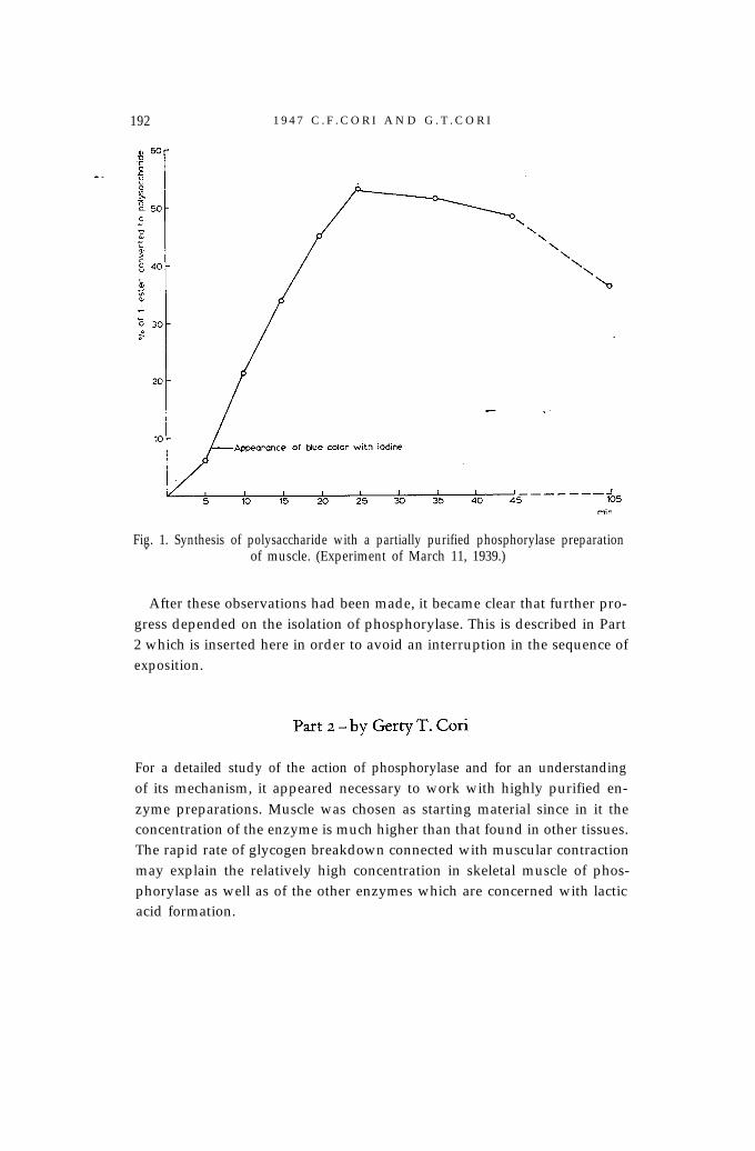
An original experiment with a partially purified preparation of musclephosphorylase is reproduced here (Fig. 1) because it is instructive in relationto later developments. The curve in Fig. 1 shows a definite lag period; thepolysaccharide which was formed gave a blue color with iodine and the reac-tion did not attain a true equilibrium owing to incomplete separation ofphosphorylase from phosphoglucomutase. Reversibility could also be dem-onstrated with phosphorylase preparations of heart and brain. The iodinecolor of the newly formed polysaccharide was brown to reddish purplerather than blue as with muscle phosphorylase. Preparations of liver phos-phorylase formed a polysaccharide which could not be distinguished fromglycogen in iodine color and other properties.
192 1 9 4 7 C . F . C O R I A N D G . T . C O R I
Fig. 1. Synthesis of polysaccharide with a partially purified phosphorylase preparation.of muscle. (Experiment of March 11, 1939.)
After these observations had been made, it became clear that further pro-gress depended on the isolation of phosphorylase. This is described in Part2 which is inserted here in order to avoid an interruption in the sequence ofexposition.
For a detailed study of the action of phosphorylase and for an understandingof its mechanism, it appeared necessary to work with highly purified en-zyme preparations. Muscle was chosen as starting material since in it theconcentration of the enzyme is much higher than that found in other tissues.The rapid rate of glycogen breakdown connected with muscular contractionmay explain the relatively high concentration in skeletal muscle of phos-phorylase as well as of the other enzymes which are concerned with lacticacid formation.

P O L Y S A C C H A R I D E P H O S P H O R Y L A S E 193
The primer action of glycogen - The protein fraction of a muscle extract,precipitated by less than 0.5 saturation with (NH4)2SO4, showed a markedrise in phosphorylase activity per unit of protein over the unfractionatedstarting material. This was however the case only when the enzyme wascatalyzing the reaction toward the right:
(1)
When enzyme activity was tested in the opposite direction a puzzling dif-ficulty was encountered. Activity set in only after a lag period; refractiona-tion of the enzyme increased this lag period from minutes to hours and insome preparations completely abolished the activity toward polysaccharideformation. A similar observation was made by Kiessling with yeast phos-phorylase and led him to conclude that he had separated two enzymes, oneconcerned with glycogen synthesis, one with its breakdown.
Liver phosphorylase, upon salt fractionation, was found to retain activitytoward polysaccharide synthesis. Such preparations always contained tracesof glycogen, while the purified muscle enzyme was free of glycogen. Thisobservation offered a clue. Addition of glycogen to the reaction mixture inas low a concentration as 10 mg per cent led to immediate activity of musclephosphorylase preparations, seemingly inactive when tested without gly-
Fig. 2. Rate of conversion of glucose-1-phosphate to polysaccharide in the presence ofcrystalline muscle phosphorylase and increasing amounts of glycogen.
194 1 9 4 7 C . F . C O R I A N D G . T . C O R I
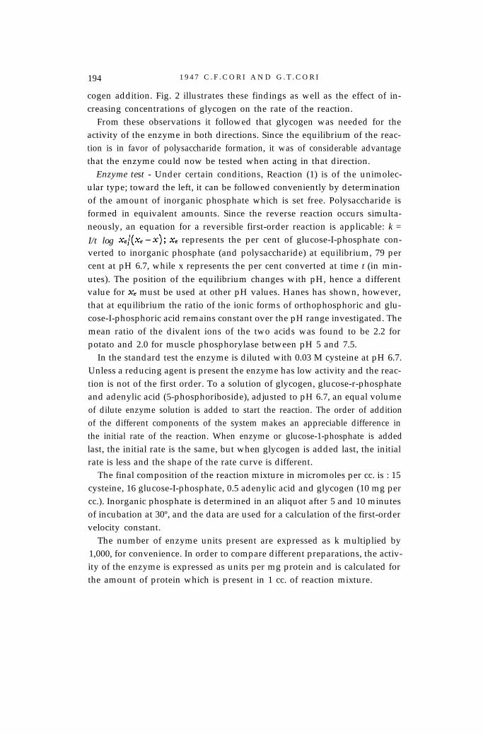
cogen addition. Fig. 2 illustrates these findings as well as the effect of in-creasing concentrations of glycogen on the rate of the reaction.
From these observations it followed that glycogen was needed for theactivity of the enzyme in both directions. Since the equilibrium of the reac-tion is in favor of polysaccharide formation, it was of considerable advantagethat the enzyme could now be tested when acting in that direction.
Enzyme test - Under certain conditions, Reaction (1) is of the unimolec-ular type; toward the left, it can be followed conveniently by determinationof the amount of inorganic phosphate which is set free. Polysaccharide isformed in equivalent amounts. Since the reverse reaction occurs simulta-neously, an equation for a reversible first-order reaction is applicable: k =
I/t log &/(Xe - x); xe represents the per cent of glucose-I-phosphate con-verted to inorganic phosphate (and polysaccharide) at equilibrium, 79 percent at pH 6.7, while x represents the per cent converted at time t (in min-utes). The position of the equilibrium changes with pH, hence a differentvalue for x, must be used at other pH values. Hanes has shown, however,that at equilibrium the ratio of the ionic forms of orthophosphoric and glu-cose-I-phosphoric acid remains constant over the pH range investigated. Themean ratio of the divalent ions of the two acids was found to be 2.2 forpotato and 2.0 for muscle phosphorylase between pH 5 and 7.5.
In the standard test the enzyme is diluted with 0.03 M cysteine at pH 6.7.Unless a reducing agent is present the enzyme has low activity and the reac-tion is not of the first order. To a solution of glycogen, glucose-r-phosphateand adenylic acid (5-phosphoriboside), adjusted to pH 6.7, an equal volumeof dilute enzyme solution is added to start the reaction. The order of additionof the different components of the system makes an appreciable difference inthe initial rate of the reaction. When enzyme or glucose-1-phosphate is addedlast, the initial rate is the same, but when glycogen is added last, the initialrate is less and the shape of the rate curve is different.
The final composition of the reaction mixture in micromoles per cc. is : 15cysteine, 16 glucose-I-phosphate, 0.5 adenylic acid and glycogen (10 mg percc.). Inorganic phosphate is determined in an aliquot after 5 and 10 minutesof incubation at 30º, and the data are used for a calculation of the first-ordervelocity constant.
The number of enzyme units present are expressed as k multiplied by1,000, for convenience. In order to compare different preparations, the activ-ity of the enzyme is expressed as units per mg protein and is calculated forthe amount of protein which is present in 1 cc. of reaction mixture.

P O L Y S A C C H A R I D E P H O S P H O R Y L A S E 195
The pH optimum of the reaction is between 6.5 and 6.8 and the temper-ature optimum is close to 38º. The conversion of glucose-r-phosphate (pK2
6.1) to orthophosphate (pK2 6.8) results in a shift in pH. This change is smalland has a neglible effect on the rate during the early course of the reaction.The pH can be kept constant by the addition of various buffers but they alldecrease the activity of the enzyme.
Crystalline muscle phosphorylase has an activity of about 3,000 units permg protein at pH 6.7 and 30º. Calculated for the initial rate of the reaction,this corresponds to a turnover number of 40,000 molecules of glucose-Iphosphate per molecule of enzyme (mol. wt. 400,000) per minute.
Phosphorylase a and b - It had been observed in experiments with dialyzedand aged muscle extracts that the rate of phosphorylase activity was in-creased 10 times or more by the addition of adenylic acid in low concentra-tions. Later it was found that phosphorylase preparations obtained by precip-itation of a fresh-water extract of rabbit muscle at 0.41 saturation with(NH4)2S04, when freshly dissolved, had in the absence of added adenylicacid as high as 66 per cent of the full activity (i.e. activity in the presenceof added adenylic acid). When the enzyme solution was kept for 1 hour at25º its activity without adenylic acid dropped to zero; at this point addi-tion of adenylic acid to the reaction mixture brought back the originalactivity.
These observations led to the conclusion that there exist two forms ofphosphorylase, one active (phosphorylase a) and one inactive (phosphorylaseb) without adenylic acid addition and that muscle tissue contains a factor,soon shown to be an enzyme, which converts the a into the b form. Theassumption that this enzyme removes adenylic acid from the a form seemedjustified at this point and the enzyme was designated as PR (prosthetic groupremoving enzyme). Later work, however, failed to establish the presence ofadenylic acid in phosphorylase a and the nature of the prosthetic group whichis removed by the PR enzyme remains obscure. There is evidence that anorganic phosphate which is dialyzable and difficult to hydrolyse in hot acidis split off from phosphorylase a by PR.
In order to preserve the a form, it is necessary to prevent action of the PRenzyme. In vivo this is accomplished by avoiding as much as possible stim-ulation of the muscles before or during excision; in vitro, by the earliestseparation of phosphorylase from the PR enzyme.
Preparation of crystalline phosphorylase a - The procedure described belowis based on experience gained in a large number of preparations. Information

196 1 9 4 7 C . F . C O R I A N D G . T . C O R I
concerning the various steps is given which has not been included in theoriginal description of the method.
A rabbit is killed by injection of amytal and the muscles of the hind legsand back are rapidly excised and weighed. The following steps are carriedout in a cold room at 4º. The muscles are passed through an ordinary meatgrinder and extracted with one volume of water for about 10 minutes (ex-traction in a blendor is to be avoided since it leads to conversion of the a tothe b form). After the residue is separated by straining it through gauze it isre-extracted with another volume of water. The combined extracts are fil-tered through cotton and coarse filter paper; this should be accomplished in1-2 hours. It is unnecessary to obtain a completely clear extract. The pH ofthe extract is measured with a glass electrode and if it is above 6.2, it isadjusted to that pH by adding 0.05 N HCl with stirring. The extract is thendialyzed in cellophane tubes (diameter 2.3 cm) against running tap water of4-10º for 3-4 hours. The turbid extract is collected in a beaker and 0.05 NHCl is added to bring the pH to 5.7. A flocculent precipitate forms whichcontains most of the PR enzyme and only a small amount of phosphorylase.This is the case only in rabbit muscle. In other species this separation is lesscomplete and so far the enzyme has only been crystallized from rabbit muscle.The isoelectric precipitate is removed by centrifugation followed by filtra-tion. Table 4 shows that no purification is achieved in this step, which never-theless, because of the separation from the PR enzyme, is essential for thesuccess of the preparation.
To the filtrate which is red and must be perfectly clear, sodium glycero-phosphate or KHCO3 is added in substance until the pH is 6.8. Then theextract is precipitated by bringing it to 0.41 saturation with a (NH4)2S04
Table 4. Preparation of crystalline muscle phosphorylase.
Total

P O L Y S A C C H A R I D E P H O S P H O R Y L A S E 197
solution saturated at room temperature and neutralized to pH 6.8. Overnightthe flocculent precipitate settles to the bottom and most of the supernatantfluid can be decanted. Finally the precipitate, which contains the enzyme isseparated by centrifugation, preferably at high speed (10,000 r.p.m.). Thewell-drained precipitate is dissolved in water, about 3 to 4 ml per 100 gmuscle used originally. Table 4 shows that with a small loss the enzyme hasnow been purified about 20 times and in this particular experiment was 39per cent pure.
The solution, which is slightly turbid, is transferred to a cellophane tube(diameter 1.3 cm) and after a short dialysis (1/2 to 1 hour) against coldrunning tap water, the tube is transferred to a cysteine solution (0.005 M)brought to pH 6.6 to 6.8 with 0.03 M sodium glycerophosphate. Buffersother than glycerophosphate have been used successfully, while no crystalswere obtained when glutathione was substituted for cysteine or when cys-teine was omitted. Dialysis is continued at 0º, against several changes of thesame cysteine-buffer solution, until most of the sulfate has been eliminatedfrom the enzyme solution. Precipitation of the enzyme sets in before this isthe case; the precipitate is sometimes, but not always, crystalline. It is sepa-rated by centrifugation, the supernatant fluid is carefully drained off and
Fig. 3. Crystalline phosphorylase prepared from rabbit muscle ( x 130).

1 9 4 7 C . F . C O R I A N D G . T . C O R I
the precipitate is stirred into a 0.01 to 0.03 M cysteine-glycerophosphatesolution of pH 6.6 to 6.8 at 30 to 35º. The volume should be about the sameas in the first crystallization. Solution of most of the protein should berapidly achieved by vigorous stirring with a glass rod, however foamingshould be avoided: the material is then centrifuged at room temperature(25º) at 10,000 r.p.m. for about 5 minutes. If a considerable amount of pro-tein remains undissolved, it can be re-extracted. The small insoluble residueconsists mostly of cystine crystals. The supernatant fluid should be perfectlyclear and almost colorless. It is immediately transferred to an ice bath.Crystals appear rapidly, the rate depending, among other factors, on theconcentration of enzyme in the solution. In order to obtain the large crystalsshown in Fig. 3 it is necessary to use a dilute solution and slow cooling.
Table 4 shows that only 2 per cent of the protein in the crude extract wasphosphorylase; 60 per cent of the enzyme was recoveredin the first crystals.Recrystallization causes a slight rise in specific activity. Eventually the moth-er liquor has almost the same specific acitivity as the crystals.
During summer, when the temperature in St. Louis is well over 30º onmany days, the phosphorylase content of the muscles drops to such a lowlevel that crystallization becomes impossible. When the rabbits are kept for
about 1 week at 13º the level rises sufficiently to obtain crystals.
Table 5. Comparison of properties of phosphorylase a and b.

P O L Y S A C C H A R I D E P H O S P H O R Y L A S E 199
Phosphorylase b was prepared by letting purified PR enzyme act on twicecrystallized phosphorylase a. The solution was then brought slowly to 0.28saturation with (NH4)2S04. The enzyme crystallized in the form of rhom-boid plates. Table 5 gives a comparison of the properties of the two forms ofthe enzyme.
Determination of phosphorylase a and b in mixtures is based on two par-allel activity determinations, one without and one with the addition of aden-ylic acid in 10-3 M concentration. In the former case the a form has 66per cent of its full activity, while the b form is inactive; in the latter case bothforms are fully active.
Conversion of phosphorylase a to b, in vitro - The PR enzyme originally ob-tained by isoelectric precipitation of dialyzed muscle extract, was consid-erably purified by fractionation with (NH4)2SO4. It was shown that its activ-ity was greatly increased in the presence of cysteine and that Mn++ ions hadan activating effect. The conversion of the a to the b form follows the first-order reaction rate equation. PR enzyme units could thus be expressed interms of the first-order velocity constant.
A conversion of phosphorylase a to b could also be effected by crystallinetrypsin at pH 6 to 6.2, at which pH the proteolytic acitivity of trypsin is keptat aminimum. This conversion is not a first-order reaction and is not ac-celerated by Mn++ ions. Work by E. Krebs (unpublished) indicates that phos-phorylases b obtained by PR and trypsin may not be identical.
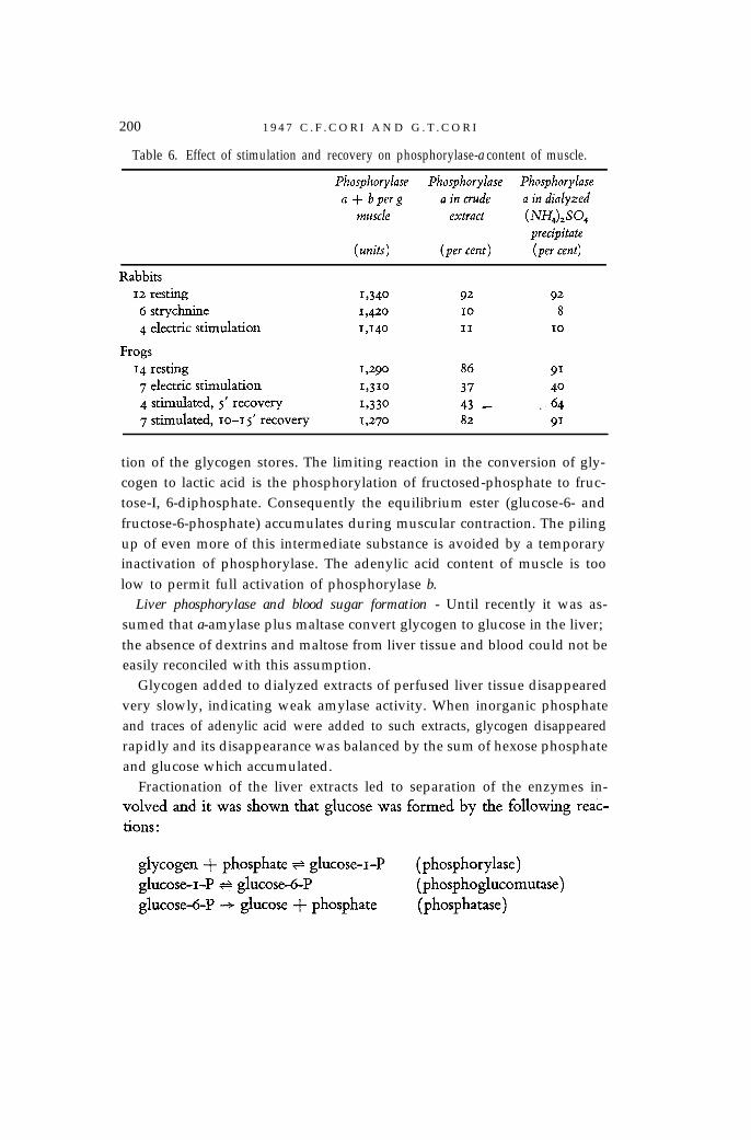
Conversion of phosphoryluse a to b, in vivo - Extracts of resting musclescontain predominantly phosphorylase a. During strong muscular contractionproduced by strychnine or electric stimulation a large part of the enzyme isconverted to the b form, presumably through the action of the PR enzymein vivo. The sum of phosphorylase a and b does not differ significantly inresting and stimulated muscles. Determinations were carried out in crude,and crude dialyzed extracts and in the precipitate obtained with (NH4)2SO4
at 0.41 saturation (Table 6). No crystals of phosphorylase a were obtainedwhen its concentration fell below 25 per cent of the total phosphorylase.
Experiments on frogs gave results very similar to those obtained on rab-bits. When electric stimulation was followed by a rest period of 10-15 min-utes before the muscles were excised, the extract contained again prepon-derantly phosphorylase a. Conversion of the b to the a form has so far onlybeen observed in vivo and nothing is known about its mechanism.
The progressive conversion of phosphorylase a to b in muscle as contrac-tion continues might represent a regulatory mechanism, preventing exhaus-
200 1 9 4 7 C . F . C O R I A N D G . T . C O R I
Table 6. Effect of stimulation and recovery on phosphorylase-a content of muscle.
tion of the glycogen stores. The limiting reaction in the conversion of gly-cogen to lactic acid is the phosphorylation of fructosed-phosphate to fruc-tose-I, 6-diphosphate. Consequently the equilibrium ester (glucose-6- andfructose-6-phosphate) accumulates during muscular contraction. The pilingup of even more of this intermediate substance is avoided by a temporaryinactivation of phosphorylase. The adenylic acid content of muscle is toolow to permit full activation of phosphorylase b.
Liver phosphorylase and blood sugar formation - Until recently it was as-sumed that a-amylase plus maltase convert glycogen to glucose in the liver;the absence of dextrins and maltose from liver tissue and blood could not beeasily reconciled with this assumption.
Glycogen added to dialyzed extracts of perfused liver tissue disappearedvery slowly, indicating weak amylase activity. When inorganic phosphateand traces of adenylic acid were added to such extracts, glycogen disappearedrapidly and its disappearance was balanced by the sum of hexose phosphateand glucose which accumulated.
Fractionation of the liver extracts led to separation of the enzymes in-

P O L Y S A C C H A R I D E P H O S P H O R Y L A S E 201
The phosphatase seems to be specific for glucose-6-phosphate; its pH opti-mum is within the physiological range. The glucose formed diffuses into theblood stream and the phosphate can react again with glycogen.
It is the absence of phosphatase from skeletal muscle tissue which explainsthe fact that muscle does not contribute glucose to the blood. In the kidney,both the glycogen and phosphorylase content are very low and its contribu-tion of glucose to the blood through the above system seems to be insignif-icant.
Plant phosphorylases - When Hanes first described the occurrence of phos-phorylase in higher plants, he pointed out that there existed a close par-allelism between the action of an enzyme system prepared from peas andpotatoes and what was then known about the action of the correspondingenzyme system from animal tissues and from yeast. Hanes fractionated pota-to extract with ammonium sulfate and noted an initial lag period in theformation of starch from glucose-I-phosphate. In conformity with the resultsobtained with the animal enzyme he found that this lag period was abol-ished by the addition of a small amount of starch. Green and Stumpf foundtheir purified potato phosphorylase preparations completely inactive in thedirection of synthesis, unless a small amount of starch was added. Weibulland Tiselius showed that under certain conditions the reaction catalyzed by
Table 7. Concentration of substrates at which phosphoryiases show half maximalactivity.
* Calculated per mole end-group, assuming that glycogen contains 10 per cent andstarch 4.5 per cent end-groups.
202 1 9 4 7 C . F . C O R I A N D G . T . C O R I
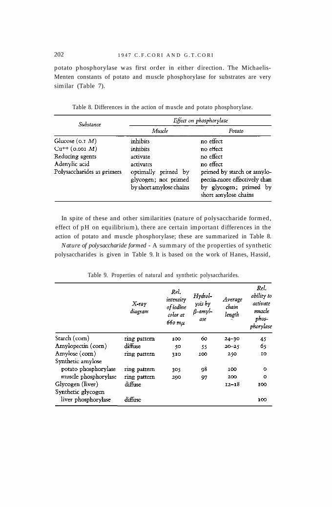
potato phosphorylase was first order in either direction. The Michaelis-Menten constants of potato and muscle phosphorylase for substrates are verysimilar (Table 7).
Table 8. Differences in the action of muscle and potato phosphorylase.
In spite of these and other similarities (nature of polysaccharide formed,effect of pH on equilibrium), there are certain important differences in theaction of potato and muscle phosphorylase; these are summarized in Table 8.
Nature of polysaccharide formed - A summary of the properties of syntheticpolysaccharides is given in Table 9. It is based on the work of Hanes, Hassid,
Table 9. Properties of natural and synthetic polysaccharides.

P O L Y S A C C H A R I D E P H O S P H O R Y L A S E 203
Bear, Cori and others. It seems clear that muscle and potato phosphorylaseform a linear polysaccharide which closely resembles the amylose compo-nent of natural starch in all its properties.
In order to form a branched polysaccharide of the type of amylopectin orglycogen, a second enzyme is needed which forms a-1-6 glucosidic linkagesat the points of branching. Such an enzyme has been found in both animaland plant tissues, but the mechanism of its action is not clearly understood.When crystalline muscle phosphorylase plus a second enzyme (called thebranching factor and obtained from liver or heart) were allowed to act onglucose-1-phosphate, an autocatalytic type of curve was obtained (Fig.4).Traces of glycogen, introduced by the liver preparation, were present, butnot enough to prime muscle phosphorylase when acting alone.
Fig. 4. Enzymatic synthesis of a branched polysaccharide from glucose-I-phosphate. InCurve B crystalline muscle phosphorylase was acting simultaneously with a supplemen-tary enzyme prepared from liver. In Curve A the liver enzyme preparation was in-
activated by heating before being added to muscle phosphorylase.
As shown in the last column of Table 9, branched and linear polysaccha-rides differ markedly in their activating effect on muscle phosphorylase. Theautocatalytic type of curve would result from the increasing number ofactivating end-groups formed during the synthesis of a branched polysac-charide. It was suggested that a combined action of both enzymes was nec-essary for the formation of glycogen or amylopectin. Haworth and collab-orators have, however, reported the conversion of amylose to amylopectin

204 1 9 4 7 C . F . C O R I A N D G . T . C O R I
by the action of an enzyme prepared from potato, a sort of cross-linking en-zyme which would break the long amylose chains into smaller fragmentsand rearrange them in a laminated pattern.
Connected with the problem of formation of a-r-6 linkages is that of theirdisruption. Neither LY- nor p-amylase can split this linkage. Recently an en-zyme present in muscle has been investigated in this laboratory which incombination with phosphorylase is able to cause an almost complete deg-radation of glycogen. Phosphorylase alone cannot degrade branched poly-saccharides beyond the branch points. In fact, the limit dextrin formed fromamylopectin or glycogen by either potato or muscle phosphorylase is largerthan that formed by ,%amylase. The question whether the same enzyme isinvolved in the formation and disruption of the 1-6 linkage has not beensettled.
Theory of the action of phosphorylase - In the phosphorolysis of giycogen orstarch the reactants are (1) inorganic phosphate, and (2) the terminal glucoseunit of the polysaccharide chains. The chain molecule is thereby shortened,one glucose unit at a time, but the concentration of terminal glucose unitsremains the same until the limit of degradation is reached.
The theory of the action of phosphorylase is based on the assumption thatthe terminal glucose unit is also a reactant in the reverse direction; in this casethe polysaccharide chains would be lengthened by successive additions ofnew glucose units from glucose-1-phosphate, again without any change inthe concentration of the terminal glucose units. The reaction catalyzed byphosphorylase in the direction of increasing chain length might therefore beexpressed as follows.
Terminal glucose (of n chain units) + glucose-r-phosphate + terminalglucose (of n + 11 chain units) + phosphate, etc.
The following observations are in accord with this formulation. (1) Muscleas well as potato phosphorylase remain inactive- when glucose-1-phos-phate alone is added; they require the further addition of polysaccharide asan essential reactant before new polysaccharide can be formed. (2) When in-creasing amounts of glycogen or starch are added, the rate of polysaccharideformation from glucose-1-phosphate increases in a manner which is char-acteristic for a reacting molecule (see Fig. 2.) (3) Branched polysaccharidesare more strongly activating than linear polysaccharides, the effect beingroughly proportional to the number of end-groups present. (4) The equilib-rium of the reaction is independent of the concentration of polysaccharide.This would follow from the fact that the concentration of one of the two

P O L Y S A C C H A R I D E P H O S P H O R Y L A S E 205
reactants in either direction, the terminal glucose units, remains constant.Hence this term cancels out when the usual mass-law expression is writtenand the equilibrium constant, K = (phosphate)/(glucose-I-phosphate). (5)For the same reason, the reaction in either direction, although involvingtwo reactants, has been found to be kinetically of the first order.
Several experiments have been devised to test this theory. The reaction,glucose-r-phosphate + polysaccharide + phosphate, in the presence of 32P,should lead to an incorporation of 32P in glucose-I-phosphate. Such an ex-change occurred in the presence but not in the absence of polysaccharideprimer, showing that the latter was an essential reactant for polysaccharidesynthesis from glucose-1-phosphate.
A further consequence of the theory presented above is that the length ofthe newly formed polysaccharide chains should depend on the ratio of molarconcentrations, glucose-I-phosphate which reacted/terminal glucose units ofadded primer. When this ratio is large, long chains, when it is small, shortchains should be formed. This has been shown to be the case by a numberof investigators, who used iodine color as a measure of chain length. A com-parison has also been made of the chain length calculated from the aboveratio and that actually found by end-group assay, with fair agreement.
A special case arises when the limit dextrin of glycogen (formed by ex-haustive treatment with phosphorylase) is used as primer and an amount ofglucose-1-phosphate is added which would allow no more than one chainunit to be added for each primer end-group. At the start the reaction canproceed only in the direction of synthesis, since there are no end-groupspresent in the limit dextrin which can be removed by phosphorolysis. As thereaction proceeds, cleavable end-groups are added, but the rate of phosphorol-ysis close to the limit of degradation is very slow. If polysaccharide syn-thesis consists in the addition of glucose units to primer end-groups, onewould expect under these special conditions a shift in the equilibrium to theside of polysaccharide formation and a dependence of the equilibrium on theconcentration of added primer. Hestrin who carried out this experiment(unpublished) found both these predictions fulfilled.
There is thus experimental support for the idea that polysaccharides act asprimers in the direction of synthesis because they enter stoichiometricallyinto the reaction catalyzed by phosphorylase.
206 1 9 4 7 C . F . C O R I A N D G . T . C O R I
References to the literature quoted will be found in the following articles:1. C. F. Cori and G. T. Cori, J. Biol. Chem., 116 (1936) 169; Proc. Soc. Exptl. Biol.
Med., 34 (1936) 702; 39 (1938) 327.2. C. F. Cori, S. P. Colowick, and G. T. Cori, J. Biol. Chem., 121 (1937) 465; 123
(1938) 375; 124 (1938) 543.3. C. F. Cori, G. Schmidt, and G. T. Cori, Science, 89 (1939) 464.4. A. A. Green, C. F. Cori, and G. T. Cori, J. Biol. Chem., 151 (1943) 21; 158 (1945)
5. M. A. Swanson and C. F. Cori, J. Biol. Chem., 172 (1948) 797.6. M. Cohn and G. T. Cori, J. Biol. Chem., 175 (1948) 89.