core.ac.uk · 2018-01-10 · iii Table of Contents...
195
USING COMPENSATION-ATTENUATED GENETICS TO UNDERSTAND UNDERLYING NETWORKS GOVERNING CELLULAR ROBUSTNESS WANG SIHUI (B.Sc. (Hons)), NUS A THESIS SUBMITTED FOR THE DEGREE OF DOCTOR OF PHILOSOPHY NUS GRADUATE SCHOOL FOR INTEGRATIVE SCIENCES AND ENGINEERING NATIONAL UNIVERSITY OF SINGAPORE 2013
Transcript of core.ac.uk · 2018-01-10 · iii Table of Contents...

USING COMPENSATION-ATTENUATED GENETICS TO UNDERSTAND UNDERLYING NETWORKS GOVERNING CELLULAR ROBUSTNESS
WANG SIHUI
(B.Sc. (Hons)), NUS
A THESIS SUBMITTED
FOR THE DEGREE OF DOCTOR OF PHILOSOPHY
NUS GRADUATE SCHOOL FOR INTEGRATIVE SCIENCES AND ENGINEERING
NATIONAL UNIVERSITY OF SINGAPORE
2013

i
Declaration
I hereby declare that the thesis is my original work and it has been written by me in
its entirety. I have duly acknowledged all the sources of information which have
been used in the thesis.
This thesis has also not been submitted for any degree in any university previously.
22/11/13 ------------------------------------- Wang Sihui

ii
Acknowledgements
I would like to express my gratitude to my supervisor, Associate Professor Davis Ng
T.W., for his guidance and advice throughout the course of this research project. I
would also like to thank Dr. Guillaume Thibault for helpful discussion and his
willingness to share his knowledge and expertise.
Special thanks go to members of the Cell Stress and Homeostasis group, Dr. Rupali
Prasad, Dr. Shinichi Kawaguchi, Mr Anthony Tran, Mr Xu Cheng Chao, Ms Liu Ying
and Ms Nassira Bedford, for their assistance these years in one way or another.
I am grateful for the scholarship awarded by NUS Graduate School for Integrative
Sciences and Engineering, without which this journey would not have been possible.
I would also like to extend my thanks to the academic and technical staff at Temasek
Life Sciences Laboratory for their invaluable help in my research.

iii
Table of Contents Declaration .................................................................................................................................. i
Acknowledgements.................................................................................................................... ii
Abstract ..................................................................................................................................... vi
List of Tables ............................................................................................................................ vii
List of Figures .......................................................................................................................... viii
List of Abbreviations ................................................................................................................. xi
Chapter 1.Introduction .............................................................................................................. 1
1.1.The secretory pathway in eukaryotes.............................................................................. 1
1.1.1. Protein folding in the secretory pathway ................................................................ 1
1.1.2. Protein quality control ........................................................................................... 16
1.2. The unfolded protein response .................................................................................... 19
1.2.1. Sensing ER stress .................................................................................................... 21
1.2.2. UPR activation and regulation ............................................................................... 25
1.3. The UPR compensatory mechanism masks the phenotype of a loss in gene function 46
1.4. Using genetic screening as a means to dissect molecular pathways ........................... 48
1.5. Thesis Rationale ............................................................................................................ 51
Chapter 2. Materials and Methods .......................................................................................... 52
2.1. Yeast Strains, Media, and Cell Culture .......................................................................... 52
2.1.1. Yeast strains ........................................................................................................... 52
2.1.2. Cell culture and media ........................................................................................... 56
2.2. General molecular and biochemical techniques .......................................................... 58
2.2.1. Plasmids ................................................................................................................. 58
2.2.2. Primers used in this study ...................................................................................... 61
2.2.3. Reagents and Antibodies ....................................................................................... 62
2.2.4. Cell Labeling and Immunoprecipitation (Pulse chase analysis) ............................. 62
2.2.5. Growth Assay (Spotting) ........................................................................................ 63
2.2.6. Assessment of CPY folding using MalPEG conjugation to cysteine sulfhydryl groups .............................................................................................................................. 64
2.2.7. Western Analysis .................................................................................................... 64
2.2.8. Quantitative PCR .................................................................................................... 65
2.2.9. DNA Microarray ..................................................................................................... 66
2.3. Synthetic Lethality Screen ............................................................................................. 67
2.3.1. UV Mutagenesis ..................................................................................................... 67
iv
2.3.2. Determining kill rate .............................................................................................. 67
2.3.3. Screening for temperature-sensitive synthetic lethal mutants ............................. 68
2.3.4. Identifying Recessive Mutants ............................................................................... 69
2.3.5. Cloning and Sequencing Temperature-sensitive Mutants ..................................... 69
Chapter 3. Genetic screening for temperature-sensitive mutants displaying synthetic lethality with an ire1 null mutation ......................................................................................... 73
3.1. Introduction .................................................................................................................. 73
3.2. Genetic screening ......................................................................................................... 73
3.2.1. Screening by colony colour phenotype.................................................................. 74
3.2.2. Screening by counter-selection using 5-fluoroorotic acid (5-FOA) ....................... 77
3.2.3. Screening by temperature sensitivity .................................................................... 79
3.3. Cloning by complementation ........................................................................................ 81
3.4. Secondary screen for biosynthetic and ERAD mutants ................................................ 82
3.5. Results of genetic screens ............................................................................................. 82
3.5.1. Summary of colony colour assay ........................................................................... 83
3.5.2. Summary of 5-FOA screen ..................................................................................... 86
3.5.3. Summary of TS screen ............................................................................................ 88
3.6. Discussion...................................................................................................................... 90
Chapter 4. The UPR buffers against a lethal pdi1 dysfunction ................................................ 92
4.1. Introduction .................................................................................................................. 92
4.2. Generating the pdi1-2 mutant in the W303 background ............................................. 93
4.3. Characterization of the pdi1-2 mutant ......................................................................... 96
4.3.1. pdi1-2Δire1 displays conditional lethality and retains endogenous proteins in the ER ..................................................................................................................................... 96
4.3.2. Pdi1pts is stable at the restrictive temperature .................................................... 99
4.3.3. ER-retention of endogenous proteins is due to misfolding and not a general trafficking defect ............................................................................................................ 100
4.3.4. Effect of pdi1 mutation on ERAD of misfolded substrates .................................. 102
4.4. UPR induction via Ire1p/Hac1p is necessary for viability of pdi1-2 ............................ 106
4.5. Oxidation is not defective in pdi1-2 ............................................................................ 110
4.6. Other members of the PDI family are dispensable for pdi1-2 survival with UPR induction ............................................................................................................................ 113
4.7. High-copy suppressor screen identified NOP56 as a suppressor ............................... 116
4.8. Microarray analysis .................................................................................................... 120
4.9. Verification of microarray data by quantitative PCR .................................................. 126

v
4.10. Expression of KAR2 suppresses defects in pdi1-2Δire1 ............................................ 128
4.11. Co-expression of KAR2 interacting factors has no additive effect over KAR2 expression alone ................................................................................................................ 131
4.12. The Hsp70-like Lhs1p alone does not compensate for defects in pdi1-2Δire1 ........ 135
4.13. KAR2 and pdi1ts work synergistically in pdi1-2Δire1 ................................................. 138
4.14. Characterizing the functional interaction between KAR2 and pdi1ts........................ 141
4.15. Discussion ................................................................................................................. 144
Chapter 5. Characterization of other mutants from the screen ............................................ 152
5.1. Secondary screen for protein biogenesis defect ........................................................ 152
5.2. Secondary screen for ERAD defect ............................................................................. 159
5.3. tssl36 is a tip20 mutant ............................................................................................... 163
5.4. tssl30 causes enhanced ERAD of CPY*........................................................................ 167
5.5. Discussion.................................................................................................................... 169
Chapter 6. Conclusion and future direction........................................................................... 171
7. References ......................................................................................................................... 173

vi
Abstract
The unfolded protein response (UPR) is a homeostatic mechanism in cells which is
activated in response to accumulation of unfolded/misfolded proteins in the
endoplasmic reticulum (ER). The Ire1/Hac1 signaling pathway relays the UPR signal
and activates a transcriptional programme which helps restore equilibrium in the ER
by alleviating ER stress. Using compensation-attenuated genetics, a novel allele of
protein disulfide isomerase (PDI), pdi1-2, was isolated. Pdi1p is an essential protein
in Saccharomyces cerevisiae involved in the catalytic oxidation, reduction and
isomerization of disulfide bonds in secretory and membrane proteins. pdi1-2 is
inviable in the absence of the UPR, but UPR activation suppressed lethality and
compensated for defects in the biogenesis of endogenous proteins, CPY and Gas1p.
Microarray analysis suggested that the UPR is modulated over time and shows
plasticity in its output in response to different types of stress. Surprisingly, PDI family
members that are UPR target genes were dispensable for suppression of lethality in
pdi1-2, suggesting they are not functionally interchangeable. pdi1-2 is oxidation-
competent, suggesting that the CPY folding defect may be due to a defect in its
chaperone function. Upregulation of the Hsp70 chaperone Kar2p and its Hsp40
cofactors by the UPR helped buffer the lethal pdi1 dysfunction. Interestingly, co-
expression of KAR2 and pdi1ts synergistically restored cell viability and CPY
maturation to a level comparable to the UPR. It is likely that KAR2 specifically
compensates for the chaperone defect in pdi1-2 during protein folding. This suggests
that different chaperone networks in the ER can buffer one another during ER stress,
and may work in synergy to contribute to cellular robustness.

vii
List of Tables
Table 1. List of UPR target genes and their functional categories .............................. 28 Table 2. List of genes synthetic lethal with Δire1/Δhac1 based on SGA analysis ........ 47 Table 3. Yeast strains used in this study ...................................................................... 52 Table 4. Components of yeast culture media .............................................................. 57 Table 5. Plasmids used in this study ............................................................................ 58 Table 6. List of primers used in this study ................................................................... 61 Table 7. List of high-copy suppressor plasmids isolated and the genes encoded ..... 116 Table 8. List of genes encoded by the complementing plasmid ............................... 165

viii
List of Figures
Figure 1. Overview of protein folding in the ER and the chaperones involved. ............ 2
Figure 2. Co-translational and post-translational translocation .................................... 4
Figure 3. Domain organization of Hsp70 ....................................................................... 6
Figure 4. Domain organization of Hsp40 ....................................................................... 9
Figure 5. Substrate-binding cycle of Kar2p .................................................................. 11
Figure 6. Domain organization of PDI family members ............................................... 15
Figure 7. The Hrd1 and Doa10 complexes involved in ERAD ....................................... 18
Figure 8. The UPR signaling pathway in S.cerevisiae ................................................... 29
Figure 9. The three branches of the UPR in higher eukaryotes. .................................. 35
Figure 10. Diagrammatic representation of the ER stress response in higher
eukaryotes ................................................................................................................... 41
Figure 11. Schematic representation of the BCL2 family of proteins under resting
conditions and during ER stress ................................................................................... 44
Figure 12. Steps in the secretory pathway defined by temperature-sensitive yeast sec
mutants deficient in protein secretion ........................................................................ 49
Figure 13. Diagrammatic representation of the yeast adenine biosynthesis pathway
...................................................................................................................................... 74
Figure 14. Primary genetic screen using colony colour phenotype ............................ 76
Figure 15. Primary genetic screen by counter-selection using 5-FOA ......................... 78
Figure 16. Primary genetic screen by temperature sensitivity .................................... 80
Figure 17. Workflow of genetic screen using colony colour and the number of
mutants obtained at each step .................................................................................... 84
Figure 18. Workflow of genetic screen using 5-FOA and the number of mutants
obtained at each step .................................................................................................. 87
Figure 19. Workflow of genetic screen using temperature sensitivity and the number
of mutants obtained at each step ................................................................................ 89
Figure 20. pdi1-2 contains a L476S point mutation in the a' domain of PDI1 ............. 92
Figure 21. Integrating the ts allele into the W303 genome ........................................ 95
Figure 22. pdi1-2 is inviable at the restrictive temperature in the absence of the UPR
...................................................................................................................................... 96

ix
Figure 23. The ER forms of CPY and Gas1p accumulate in pdi1-2Δire1 at the
restrictive temperature................................................................................................ 98
Figure 24. Pdi1pts is stable at the restrictive temperature .......................................... 99
Figure 25. The retention of CPY and Gas1p in the ER is not due to a general ER-golgi
transport defect ......................................................................................................... 100
Figure 26. CPY is misfolded in pdi1-2 Δire1 at the restrictive temperature .............. 102
Figure 27. CPY* is stabilized in the pdi1 mutant and the UPR does not fully
compensate for this defect ........................................................................................ 103
Figure 28. ERAD of PrA* is affected in pdi1-2Δire1 ................................................... 104
Figure 29. ERAD of a non-glycosylated substrate is similarly affected in pdi1-2Δire1
.................................................................................................................................... 105
Figure 30. The UPR is induced in pdi1-2 at the restrictive temperature ................... 106
Figure 31. Viability is mediated by Ire1p/Hac1p signaling branch ............................ 107
Figure 32. UPR activation fixes the defect in CPY maturation ................................... 108
Figure 33. Deletion of the lumenal domain of IRE1 abolished suppression of lethality
.................................................................................................................................... 109
Figure 34. Oxidation is not defective in pdi1-2Δire1 ................................................. 111
Figure 35. Addition of diamide did not improve oxidative protein folding ............... 112
Figure 36. Deletion of PDI family members has no effect on viability when the UPR is
activated. ................................................................................................................... 114
Figure 37. The UPR sufficiently compensates for the defect in CPY processing in the
absence of PDI family members. ............................................................................... 115
Figure 38. Isolates from high-copy suppressor screen .............................................. 118
Figure 39. High-copy plasmids HC5 and HC7 partially suppressed cell lethality ....... 119
Figure 40. UPR target genes are differentially induced in pdi1-2, compared to WT
.................................................................................................................................. ..123
Figure 41. Microarray analysis identified UPR genes differentially upregulated in
pdi1-2 at various time points after shifting to the restrictive temperature..............125
Figure 42. Correlation between qPCR data with microarray data ............................ 127
Figure 43. Expression of KAR2 suppresses lethality .................................................. 129
Figure 44. KAR2 improved CPY maturation in vivo .................................................... 130

x
Figure 45. Coexpression of KAR2 cofactors has no significant suppression of pdi1-2
lethality over expression of KAR2 alone. ................................................................... 132
Figure 46. Coexpression of KAR2 with its cofactors does not improve CPY maturation
over expression of KAR2 alone .................................................................................. 134
Figure 47. Expression of Lhs1p does not compensate for defects in pdi1-2Δire1 .... 137
Figure 48. Synergy between KAR2 and PDIts.............................................................. 140
Figure 49. A functional KAR2 is required for suppression of cell lethality in pdi1-
2Δire1. ........................................................................................................................ 142
Figure 50. Mutations in pdi1-2 and kar2 are synthetic lethal ................................... 143
Figure 51. Secondary screen for defects in biogenesis of CPY and Gas1p ................ 155
Figure 52 . Bioprocessing of CPY in mutant strains ................................................... 157
Figure 53. Bioprocessing of Gas1p in mutant strains. ............................................... 158
Figure 54. Secondary screen for ERAD mutants using CPY* ..................................... 160
Figure 55. CPY* degradation is partially affected by putative ERAD mutants isolated
from the screen. ......................................................................................................... 162
Figure 56. Protein processing of CPY, Gas1p and invertase are affected in tssl36 ... 164
Figure 57. tssl30 is extremely thermolabile ............................................................... 167
Figure 58. UPR activation inhibits enhanced degradation of CPY* ........................... 168

xi
List of Abbreviations
AIR P-ribosylamino imidazole bp Base pair(s) BSA Bovine serum albumin bZIP Basic leucine zipper CAIR P-ribosylamino imidazolecarboxylate cDNA Complementary DNA cLD Core lumenal domain CPY Carboxypeptidase Y Da Dalton DMSO Dimethylsulfoxide DNA Deoxyribonucleic acid dNTP Deoxyribonucleoside triphosphate DTT Dithiothreitol ENaC Epithelial sodium channel ER Endoplasmic reticulum ERAD ER-associated degradation FRET Fluorescence resonance energy transfer GPI Glycophosphatidylinositol HLE Helix-loop element HPL Hyper-phosphorylated loop HRP Horseradish peroxidase HSP Heat shock protein MalPEG Methoxypolyethylene glycol maleimide MHC Major histocompatibility complex mRNA Messenger ribonucleic acid NEF Nucleotide exchange factor NEM N-ethylmaleimide PBS Phosphate buffered saline PCR Polymerase chain reaction PDI Protein disulfide isomerase PEG Polyethylene glycol ppαF prepro-alpha factor PrA Proteinase A qPCR Quantitative polymerase chain reaction RNA Ribonucleic acid rpm Revolutions per minute SBD Substrate binding domain SDS-PAGE Sodium dodecyl sulfate polyacrylamide gel electrophoresis SGA Synthetic genetic array SRP Signal recognition particle Tm Tunicamycin UPR Unfolded protein response UPRE Unfolded protein response element UV Ultraviolet WT Wild type
1
Chapter 1.Introduction
1.1.The secretory pathway in eukaryotes
As organisms evolve from prokaryotes to eukaryotes, there is increasing complexity
in cellular layout, structure, and function. One of the hallmarks of eukaryotes is the
compartmentalization of the cell into distinct subcellular organelles, each with its
own tailor-made environment that has been optimized for its specific function. The
secretory pathway in eukaryotes consists of various organelles that come together to
perform the important task of producing soluble proteins that are secreted and
allow communication or interaction with the external milieu.
The secretory pathway consists of the rough endoplasmic reticulum (ER), ER exit
sites, the ER to golgi intermediate compartment, the golgi complex and the
subsequent transport of secretory vesicles. The pathway is modulated by
intracellular and extracellular stimuli and responds by changing its secretory capacity
accordingly to deal with the demands of cell growth, survival and homeostasis
(Farhan and Rabouille 2011).
1.1.1. Protein folding in the secretory pathway
The ER is the main site where folding and processing of secretory and membrane
proteins take place. It has been estimated that a third of cellular proteins pass
through the ER. As such, the ER can be regarded as the protein folding factory of the
cell. Similar to an actual factory where manufacturing of specific products and
assessment of their quality occur, the ER provides a conducive environment specially

2
equipped for protein folding, as well as a quality control system that maintains the
integrity of folded proteins (Fig. 1).
Figure 1. Overview of protein folding in the ER and the chaperones involved Schematic representation of the chaperones and cofactors involved in folding and quality control of secretory and membrane proteins in the ER. Chaperones are involved in various steps of protein biogenesis including translocation, folding, post-translational modifications (glycosylation, disulfide bond formation) and protein quality control (Verghese et al. 2012). Detailed discussion of the chaperones and their functions is found in the text.
Nascent polypeptide chains synthesized by ribosomes are translocated into the ER
lumen through the Sec61 translocon found on the ER membrane. In yeast and
mammalian cells, this process can occur co-translationally or post-translationally (fig.
2). In co-translational translocation, N-terminal ER-targeting signal sequences found
on nascent polypeptide chains that are being translated are recognized by the signal
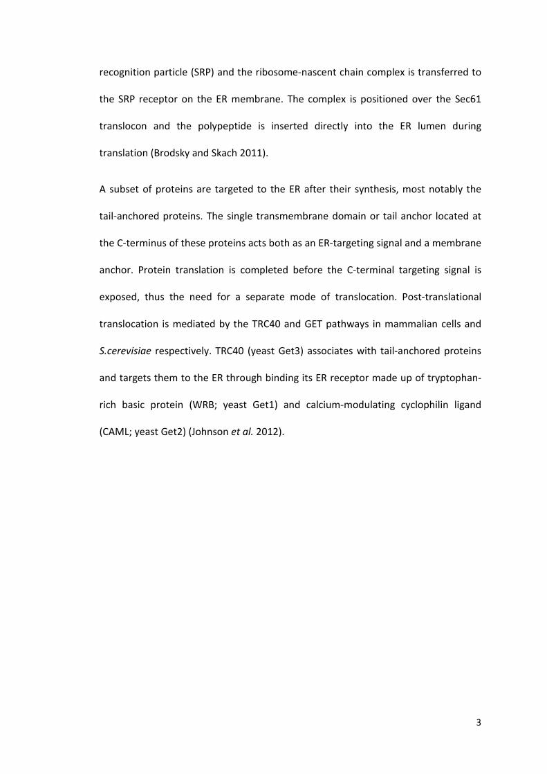
3
recognition particle (SRP) and the ribosome-nascent chain complex is transferred to
the SRP receptor on the ER membrane. The complex is positioned over the Sec61
translocon and the polypeptide is inserted directly into the ER lumen during
translation (Brodsky and Skach 2011).
A subset of proteins are targeted to the ER after their synthesis, most notably the
tail-anchored proteins. The single transmembrane domain or tail anchor located at
the C-terminus of these proteins acts both as an ER-targeting signal and a membrane
anchor. Protein translation is completed before the C-terminal targeting signal is
exposed, thus the need for a separate mode of translocation. Post-translational
translocation is mediated by the TRC40 and GET pathways in mammalian cells and
S.cerevisiae respectively. TRC40 (yeast Get3) associates with tail-anchored proteins
and targets them to the ER through binding its ER receptor made up of tryptophan-
rich basic protein (WRB; yeast Get1) and calcium-modulating cyclophilin ligand
(CAML; yeast Get2) (Johnson et al. 2012).

4
Figure 2. Co-translational and post-translational translocation Two modes of translocation can occur in living cells. In co-translational translocation, the SRP recognizes and binds ER targeting sequences on nascent polypeptides, and localizes the ribosome complex to the ER via binding to the SRP receptor. The nascent polypeptide is transported across the Sec61 channel while being translated. In the post-translational mode, translated polypeptides in the cytosol are targeted to and imported into the ER via the TRC40 (mammalian) or GET (yeast) pathway.
As nascent polypeptide chains enter the ER lumen, they encounter a network of
chaperones, co-chaperones, and folding enzymes which prevent their aggregation
and help them attain their correct native structures. This is achieved by a series of
reactions that occur in the ER including signal peptide cleavage, N-linked
glycosylation, disulfide bond formation and the addition of glycophosphatidylinositol
(GPI)-anchor (Araki and Nagata 2012). There are three main classes of molecular
chaperones in the ER - the heat shock protein (HSP) family, the glycoprotein
chaperones, and the protein disulfide isomerase (PDI) family, each contributing to
significant aspects of protein biogenesis. Here, we will mainly focus on the ER
chaperones found in the budding yeast, Saccharomyces cerevisiae.

5
1.1.1.1. HSP family of chaperones
The HSPs are a highly conserved group of proteins that were initially discovered to
be upregulated in response to elevated temperature, and this phenomenon was
subsequently termed the heat shock response. The heat shock response was first
observed as patterns of puffing activity in the polytene chromosomes of Drosophila,
at regions where increased transcriptional activity were occurring (Shamaei-Tousi et
al. 2007). HSPs were later found to be induced under other types of stress conditions
as well, such as exposure to heavy metals and cytotoxic chemicals (Neuhaus-
Steinmetz and Rensing 1997), oxidative insults, ischaemia/reperfusion and
hemorrhagic shock (De Maio 1999).
Members of the HSPs are classified according to their molecular weight. In the ER of
the budding yeast, two main groups of HSPs - Hsp70 and Hsp40, are present to
maintain protein homeostasis. Each member plays a distinct role in protein folding,
with some performing multiple functions in the ER. These are discussed in detail
below.
1.1.1.1.1. ER Hsp70s in S.cerevisiae - Kar2p and Lhs1p
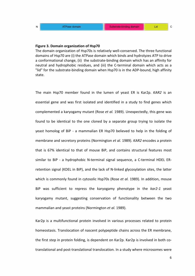
Hsp70s have similar domain architecture consisting of a N-terminal ATPase domain,
a substrate-binding domain, and a C-terminal α-helix-rich domain that acts as a "lid"
for the substrate-binding domain (Fig. 3). The ATPase activity is inherently weak and
requires stimulation through interaction of Hsp70s with other cofactors like the
Hsp40s (Kampinga and Craig 2010). This increases the affinity of Hsp70s for their
substrates.
6
Figure 3. Domain organization of Hsp70 The domain organization of Hsp70s is relatively well-conserved. The three functional domains of Hsp70 are (i) the ATPase domain which binds and hydrolyzes ATP to drive a conformational change, (ii) the substrate-binding domain which has an affinity for neutral and hydrophobic residues, and (iii) the C-terminal domain which acts as a "lid" for the substrate-binding domain when Hsp70 is in the ADP-bound, high affinity state.
The main Hsp70 member found in the lumen of yeast ER is Kar2p. KAR2 is an
essential gene and was first isolated and identified in a study to find genes which
complemented a karyogamy mutant (Rose et al. 1989). Unexpectedly, this gene was
found to be identical to the one cloned by a separate group trying to isolate the
yeast homolog of BiP - a mammalian ER Hsp70 believed to help in the folding of
membrane and secretory proteins (Normington et al. 1989). KAR2 encodes a protein
that is 67% identical to that of mouse BiP, and contains structural features most
similar to BiP - a hydrophobic N-terminal signal sequence, a C-terminal HDEL ER-
retention signal (KDEL in BiP), and the lack of N-linked glycosylation sites, the latter
which is commonly found in cytosolic Hsp70s (Rose et al. 1989). In addition, mouse
BiP was sufficient to repress the karyogamy phenotype in the kar2-1 yeast
karyogamy mutant, suggesting conservation of functionality between the two
mammalian and yeast proteins (Normington et al. 1989).
Kar2p is a multifunctional protein involved in various processes related to protein
homeostasis. Translocation of nascent polypeptide chains across the ER membrane,
the first step in protein folding, is dependent on Kar2p. Kar2p is involved in both co-
translational and post-translational translocation. In a study where microsomes were

7
prepared from a temperature-sensitive kar2-159 yeast mutant shifted to the non-
permissive temperature, both the precursor of a yeast mating pheromone, ppαF,
and the precursor of secretory invertase, validated substrates for post-translational
and co-translational translocation respectively, were unable to be translocated
(Brodsky et al. 1995). This is in agreement with a previous study in which depletion
of Kar2p resulted in the accumulation of these precursors on the cytosolic side of the
ER membrane (Vogel et al. 1990).
In post-translational translocation, efficient precursor translocation through the
Sec61p translocon requires the interaction of Kar2p with Sec63p, a member of the
Hsp40 family. This interaction is mediated by the lumenal DnaJ domain of Sec63p
and the ATPase domain of Kar2p (Lyman and Schekman 1995). In addition, Kar2p
also acts as a "molecular ratchet" to aid in the transport of ER-targeted precursor
proteins through the translocon. Using a soluble translocation system in vitro, Kar2p
was shown to bind ppαF in a Sec63p-dependent manner. Binding of Kar2p on the
lumenal side of the ER membrane minimized the backward movements of the
inserted nascent chain through the translocon due to Brownian motion. As the
nascent chain emerges on the lumenal side, more molecules of Kar2p bind, thus
favoring the forward movement of the polypeptide and its eventual translocation
into the ER lumen. Interestingly, replacement of Kar2p with antibodies targeting
different parts of ppαF also resulted in translocation, albeit at a lower efficiency.
This reinforced the "Brownian ratchet" theory in which the passive movement of a
polypeptide through the translocon, coupled with a binding partner, Kar2p, that

8
prevents backsliding, is sufficient to drive movement across the ER membrane
(Matlack et al. 1999).
Kar2p is also believed to play roles in protein folding and ER-associated degradation
(ERAD). It was shown to be involved in the maturation of a well-characterized
endogenous glycoprotein carboxypeptidase Y (CPY). When Kar2 function was
compromised using temperature-sensitive kar2 mutants, CPY folding was disrupted
and aggregates of CPY accumulated in the ER (Simons et al. 1995). ERAD of a
misfolded mutant form of CPY, CPY*, was shown to be dependent on Kar2 and was
stabilized in the kar2-113 mutant strain (Plemper et al. 1997). It is likely that Kar2
acts as a general chaperone for different folding substrates during normal biogenesis
and under proteotoxic stress, and that its specificity for different functions is
determined by its interaction with different Hsp40 cofactors in the ER (Vembar et al.
2010).
Another member of the Hsp70 family in the yeast ER is Lhs1p. Lhs1p is a non-
essential protein that shares 24% amino acid identity with Kar2p. Deletion of LHS1
resulted in a partial translocation defect for various proteins including Kar2p, CPY,
proteinase A (PrA), protein disulfide isomerase (PDI), invertase and ppαF, suggesting
its involvement in protein translocation (Baxter et al. 1996). However, unlike Kar2p,
Lhs1p is only required for post-translational import (Craven et al. 1996, Hamilton and
Flynn 1996). The activities of Lhs1p and Kar2p are coupled; Lhs1p stimulates Kar2p
by acting as its nucleotide exchange factor while Kar2p reciprocally activates the
ATPase domain of Lhs1p (Steel et al. 2004). In addition, Lhs1p was shown to aid in
the refolding and solubilization of heat-denatured pro-CPY and Hsp150Δβlactamase,

9
but not in the folding of newly synthesized proteins, suggesting a chaperone role
during heat stress (Saris et al. 1997).
1.1.1.1.2. ER Hsp40s in S.cerevisiae - Sec63p, Jem1p, and Scj1p
Hsp40s are crucial for the function of Hsp70s in vivo. Hsp70s participate in a diverse
range of cellular functions, but their activities require the stimulation of their
intrinsically weak ATPase domains by Hsp40s. Interaction with different Hsp40s
determines the specificity of Hsp70 for their localization, function, and client
substrates (Kampinga and Craig 2010). All Hsp40s contain the evolutionarily
conserved J-domain, which mediates interaction with the ATPase domain of Hsp70s
(Fig. 4).
Figure 4. Domain organization of Hsp40 Hsp40s show high diversity in their structures. The only conserved domain is the J domain, which mediates interaction with Hsp70. Other domains which may be present are: (i) a glycine/phenylalanine rich G/F region which stabilizes its interaction with Hsp70, (ii) a zinc-finger domain which binds substrates, and (iii) a variable C-terminal region important for substrate binding and may provide specificity.
In the budding yeast, three Hsp40s are present - Sec63p, Scj1p and Jem1p. Sec63p,
an essential integral membrane protein found on the ER membrane, is required for
protein translocation into the ER lumen. The C-terminus of Sec63p faces the cytosol,
while the N-terminus J-domain is located in the ER lumen, where it recruits Kar2p to
the Sec61p translocation machinery and stimulates the ATPase domain of Kar2p
(Feldheim et al. 1992, Corsi and Schekman 1997). Disrupting Sec63p-Kar2p
interaction by mutating a conserved residue in the J-domain of Sec63p caused

10
impaired translocation (Lyman and Schekman 1995), reinforcing the role of Sec63p
in Kar2p's translocation function.
Jem1p is a non-essential transmembrane ER protein with its J-domain facing the ER
lumen. Deletion of JEM1 caused a defect in nuclear fusion during mating (Nishikawa
and Endo 1997), while overexpression of Jem1p suppressed the karyogamy defect of
the kar2-1 mutant (Brizzio et al. 1999). These data suggest a genetic interaction
between Jem1p and Kar2p and it is likely that the pair cooperates to promote
nuclear fusion. It was also reported that Jem1p deletion caused stabilization of
lumenal ERAD substrates, CPY* and ppαF, but not that of a membrane protein
(Nishikawa et al. 2001). Jem1p, together with another Hsp40, Scj1p, were found to
be involved in the ubiquitination and degradation of the epithelial sodium channel
(ENaC) without Kar2p, suggesting that Hsp40s can target some substrates for ERAD
independently of their Hsp70s (Buck et al. 2010).
Scj1p, a non-essential ER lumenal Hsp40, functions together with Kar2p to mediate
protein maturation. Deletion of SCJ1 sensitized the cells to tunicamycin (Tm)
treatment (which inhibits N-linked glycosylation) or conditions resulting in
underglycosylation of proteins, and induced the unfolded protein response (UPR)
(Schlenstedt et al. 1995, Silberstein et al. 1998). Similar to a Δjem1 mutant, yeast
lacking Scj1p showed stabilization of lumenal ERAD substrates and EnaC (Nishikawa
et al. 2001, Buck et al. 2010).
1.1.1.1.3. Nucleotide exchange factor in S.cerevisiae - Sil1
Substrate binding by Kar2p in the ER is enhanced by its interaction with Hsp40s
which increases its substrate-binding affinity. Another protein, the nucleotide

11
exchange factor (NEF), Sil1p, plays an equally important role in this substrate
interaction cycle (Fig. 5) by promoting substrate release from Kar2p via the exchange
of ADP for ATP, which decreases the substrate-binding affinity. This frees Kar2p for
another round of substrate binding, thus maintaining a pool of Kar2p for its various
cellular functions, including translocation (Kabani et al. 2000, Tyson and Stirling 2000,
Kabani et al. 2002). The deletion of SIL1 is synthetic lethal with a LHS1 deletion
(Tyson and Stirling 2000), which is expected given that both proteins act as NEFs of
Kar2p.
Figure 5. Substrate-binding cycle of Kar2p The binding and release of unfolded proteins to Kar2p is regulated by ATP. Interaction of Kar2p with Hsp40s (Scj1p, Jem1p, Sec63p) stimulates its ATPase domain and results in the hydrolysis of ATP to ADP. This increases Kar2p's affinity for its substrates. Replacement of ADP with ATP by nucleotide exchange factors (Sil1p, Lhs1p) reduces its substrate-binding affinity and leads to release of the folded protein. This reactivates Kar2p for the next cycle of protein folding.

12
1.1.1.2. ER glycoprotein chaperone - Cne1p
Cne1p, an ER integral membrane protein, shares 24% identity and 31% similarity at
the amino acid level with the mammalian glycoprotein chaperone, calnexin (Parlati
et al. 1995). Calnexin acts as a molecular chaperone and retains glycoproteins in the
ER to ensure their proper folding (Ellgaard et al. 1999), and its yeast homolog, Cne1p,
is believed to be involved in the folding of glycoproteins and their quality control
(Parlati et al. 1995). Cne1p possesses a conserved lectin domain which has been
shown to bind monoglucosylated oligosaccharide, and a P- (proline-rich) domain that
was shown to be required for Cne1p's ability to suppress aggregation and promote
refolding of heat-denatured citrate synthase (Xu et al. 2004a, Xu et al. 2004b). In
addition, a study demonstrated that the chaperone activity of Cne1p was inhibited
by association with Mpd1p, a member of the PDI family of proteins, while the
reductive activity of Mpd1p was enhanced by this interaction, suggesting possible
functional interactions between the network of folding factors (Kimura et al. 2005).
1.1.1.3. PDI family of oxidoreductases
In contrast to cytosolic proteins, many secretory and membrane proteins contain
intramolecular disulfide bonds which help stabilize their tertiary or quaternary
structures (Verghese et al. 2012). The ER constitutes a unique environment for the
folding of such proteins as it is an oxidising compartment unlike other organelles in
the cell, and it houses a family of protein disulfide isomerases that catalyze the
formation, reduction, and isomerization of disulfide bonds. Yeast ER contains five
PDIs, of which only Pdi1p is essential (Farquhar et al. 1991). These PDI family
proteins are characterized by the presence of at least one thioredoxin-like domain.
13
1.1.1.3.1. PDI1
Pdi1p is an essential and abundant ER-resident protein that performs multiple roles
in the ER lumen. As an oxidoreductase, it catalyzes native disulfide bond formation in
secretory and transmembrane proteins. Yeast Pdi1p shares 30% identity with
mammalian PDIs (Tachikawa et al. 1991), and contains four thioredoxin-like domains
- a, b, b', and a', of which the a and a' domains contain the catalytically active CGHC
motif. The solved crystal structure of full-length yeast Pdi1p showed that the protein
adopts a twisted "U" shape, with the a and a' domains forming the arms and the b
and b' domains forming the base. A flexible x-linker joins the b' and a' domains,
allowing flexibility in the a' domain. A C-terminal extension, whose deletion reduced
in vitro Pdi1p activity by half, is found opposite the a' active site (Tian et al. 2006).
There are conflicting evidence in the literature regarding the essential function of
Pdi1p, but a study by Xiao et al. demonstrated that even in a strain deleted for all
homologs of Pdi1p in the yeast ER, isomerase-deficient mutants of Pdi1p that were
oxidation-competent still supported wild-type growth, suggesting that oxidation is
the essential function of yeast Pdi1p (Xiao et al. 2004).
In vivo, Pdi1p is a major substrate of Ero1p, an essential thiol oxidase that maintains
proper redox balance in the ER. Ero1p reoxidizes Pdi1p that has been reduced in
oxidative protein folding, making it competent for transferring disulfide bonds to
folding proteins. In turn, Pdi1p regulates the activity of Ero1p either by reducing or
oxidizing its regulatory bonds (Kim et al. 2012).
Besides its redox function, Pdi1p also forms a complex with the mannosidase, Htm1p,
and targets misfolded glycoprotein for ERAD (Gauss et al. 2011). Its chaperone and
14
redox activity were shown to be required for the ERAD of apolipoprotein B (ApoB)
and CPY* respectively. The b′ domain of Pdi1p is believed to mediate its chaperone
activity, and deletion of this domain reduced ApoB degradation (Grubb et al. 2012)
as well as disrupted the export of a cysteine-free misfolded secretory protein for
degradation (Gillece et al. 1999). Several studies have demonstrated that
mammalian PDI acts as a chaperone in the folding of cysteine-free proteins in vitro
(Cai et al. 1994, Song and Wang 1995) , but studies using yeast Pdi1p showed no
such activity (Katiyar et al. 2001).
1.1.1.3.5. Homologs of PDI1
In budding yeast, there are four non-essential homologs of PDI1 - MPD1, MPD2,
EUG1, and EPS1. All of them contain at least one thioredoxin-like domain and are
soluble ER lumenal proteins with the exception of Eps1p, which contains a
transmembrane domain and localizes to the ER membrane (Tachibana and Stevens
1992, Tachikawa et al. 1995, Tachikawa et al. 1997, Wang and Chang 1999). The
domain organization and active cysteine sites of the PDI family members are shown
in Figure 6. Mpd1p, Mpd2p, and Eps1p each has one thioredoxin domain containing
the active site CXXC motif, while Eug1p has two thioredoxin domains with CXXS
motifs (Norgaard et al. 2001).

15
Figure 6. Domain organization of PDI family members Diagram showing the domain organization of the 5 PDI family members (PDI1, MPD1, MPD2, EUG1 and EPS1) in S.cerevisiae, and the location of the CXXC active-site motif. Each member has at least one thioredoxin-like domain containing the CXXC motif, with the exception of EUG1 which has CXXS motifs. All PDI family members are ER-lumenal proteins except EPS1, which consists of a transmembrane domain (TMD) and is found in the ER membrane.
When overexpressed, each homolog has the ability to suppress the inviability caused
by PDI1 deletion and partially suppress the defect in CPY maturation (Tachibana and
Stevens 1992, Tachikawa et al. 1995, Tachikawa et al. 1997, Wang and Chang 1999),
but this required the presence of the other homologs, suggesting that their functions
are not interchangeable. Only Mpd1p seemed to be able to carry out the essential
function of Pdi1p, as overexpression of Mpd1p could suppress a strain deleted for all
other members of the PDI family (Norgaard et al. 2001).
With the exception of EPS1, all other genes of the PDI family are upregulated by the
UPR (Travers et al. 2000), suggesting their importance in protein folding and
homeostasis. Eps1p was shown to be involved in the ERAD of a misfolded plasma
membrane substrate, Pma1-D378N, and displayed genetic interactions with other

16
components of the ERAD machinery, like CDC48, UBC6 and UBC7, suggesting its role
in the ERAD pathway (Wang and Chang 2003).
1.1.2. Protein quality control
Protein synthesis is a fundamental process required for cellular turnover, growth and
survival. In the ER where synthesis of secretory and membrane proteins occur,
quality control mechanisms ensure that only properly folded proteins reach their
target destination. As proteins frequently misfold, it is imperative that these
mechanisms identify, retain, and degrade misfolded proteins before they form
protein aggregates and cause cellular toxicity. Most proteins that fail to fold are
retained in the ER and targeted for ERAD (Ellgaard and Helenius 2003, Araki and
Nagata 2011, Thibault and Ng 2012).
Misfolded proteins are recognized based on the location of their lesions, in addition
to other ERAD determinants such as exposed hydrophobic regions, N-linked glycan
signals (Xie et al. 2009), and O-mannosylation (Xu et al. 2013). Two main complexes
make up the ERAD machineries (Fig. 7) - the Hrd1 complex and the Doa10 complex.
Membrane proteins with lesions in their cytosolic domains are targeted to the Doa10
complex (ERAD-C) while misfolded soluble proteins or membrane proteins with
lesions in their lumenal or transmembrane domains (ERAD-L/ERAD-M) are targeted
to the Hrd1 complex. Together with E2 ubiquitin-conjugating enzymes, Ubc1p,
Ubc6p and Ubc7p, the E3 ubiquitin ligases, Hrd1p and Doa1p, mediate ubiquitination
of the ERAD substrates. This step is required for subsequent retrotranslocation of
the substrates into the cytosol (Thibault and Ng 2012).

17
Retrotranslocation of ERAD substrates is necessary as the ubiquitin-proteasome
system responsible for degradation of these proteins are located in the cytosol
and/or the nucleus. The identity of the retrotranslocon remains controversial,
though studies suggested that the channel could be made up of Sec61p, Der1p, or
Hrd1p (Meusser et al. 2005, Thibault and Ng 2012). The force needed for protein
dislocation is believed to come from the AAA-ATPase Cdc48p, in conjunction with
Npl4p and Ufd1p which bind ubiquitinated proteins (Meusser et al. 2005).
Retrotranslocated substrates are finally recognized and degraded by the 26S
proteosome. In some cases, misfolded proteins can bypass ERAD and be degraded
via macroautophagy (Ding and Yin 2008).

18
Figure 7. The Hrd1 and Doa10 complexes involved in ERAD (Thibault and Ng 2012) Schematic representation of the Hrd1 and Doa10 ERAD machineries in S.cerevisiae. The two E3 ligases are shown in complexes with their known interacting partners. The Doa10 complex is responsible for ERAD-C, whereby membrane proteins with lesions in their cytosolic domains are recognized and targeted for degradation. The Hrd1 complex on the other hand, recognizes lesions in soluble lumenal proteins and the lumenal domains of membrane proteins (ERAD-L), as well as lesions in transmembrane domains (ERAD-M).

19
1.2. The unfolded protein response As discussed earlier, the ER is vital for the folding and processing of secretory and
transmembrane proteins that pass through the secretory pathway. It plays a pivotal
role in ensuring that proteins fold into their native structures, and that unfolded/
misfolded proteins are recognized, retained, and targeted for degradation by quality
control machineries. To ensure that misfolded and unfolded proteins do not
accumulate and lead to cell toxicity, the ER regulates its folding capacity to meet the
folding requirements of the cell. The balance between nascent protein influx and
functional protein output can be perturbed by both endogenous and exogenous
stresses. These include nutrient deprivation, changes in ER redox potential and ER
calcium levels, chemical insults that disrupt protein folding (e.g. DTT and
tunicamycin), increased protein trafficking through the ER (due to differentiation),
genetic mutation, and pathogenic infection (Rutkowski and Kaufman 2004, Carrara
et al. 2013). Under such circumstances, the ER turns on a network of signaling
pathways collectively termed the unfolded protein response (UPR) in an attempt to
restore ER homeostasis.
The UPR was initially characterized by Kozutsumi et al. who discovered that
expression of misfolded forms of influenza virus haemagglutinin (HA) in simian cells
induced the expression of BiP and GRP94, both major ER proteins, while wild-type
HA did not (Kozutsumi et al. 1988). Other groups have also shown that these same
proteins were induced under different conditions of stress, including glucose
starvation, treatment with drugs that inhibit glycosylation, with calcium ionophores
20
or with amino acid analogues (Hightower 1980, Chang et al. 1987). These results
suggest that a signaling pathway must exist between the ER lumen and the nucleus.
The gene required for this pathway was subsequently identified in Saccharomyces
cerevisiae as IRE1 and cloned. IRE1 was shown to be essential for cell survival under
stress conditions that cause ER accumulation of unfolded proteins. Δire1 mutants
were also unable to induce transcription of KAR2 (yeast homolog of BiP) and PDI1 -
two folding genes usually upregulated in response to increased unfolded proteins in
the ER, suggesting IRE1's role in ER to nucleus signaling (Cox et al. 1993).
The UPR is conserved in eukaryotes and has evolved in complexity in metazoans to
cope with the increasing demand in secretory functions of higher order organisms.
For example, there is one UPR signal transducer (IRE1) in S. cerevisiae, two (ire-
1/IRE1 and pek/PERK) in Caenorhabditis elegans and Drosophila melanogaster, and
three (IRE1, PERK and ATF6) in mammals (Mori 2009). This emphasizes the
importance of the UPR in buffering organisms against imbalances in ER function.
Studies by various groups over the past two decades have helped elucidate the
mechanisms of UPR signaling. Stress-induced accumulation of unfolded proteins in
the ER lumen is detected by transmembrane sensors on the ER membrane. In yeast,
the UPR is mediated by Ire1p (Cox et al. 1993), the sole signal transducer. In
mammals, three different signaling branches of the UPR are present, each mediated
by a unique stress sensor. The mode of UPR activation is discussed in greater detail
below. Ultimately, the UPR aims to increase ER folding capacity by ER expansion,
increasing the number of chaperones and folding factors, increasing degradation of

21
misfolded proteins, and decreasing protein load through translation attenuation.
When ER homeostasis fails to be restored, apoptosis is initiated.
1.2.1. Sensing ER stress
Since folding of secretory and transmembrane proteins occur primarily in the ER,
perturbations to the ER impede their processing. ER stress is sensed by the cell via
the detection of these unfolded proteins in the lumen. The exact mode of UPR
activation has been debated. Currently, biochemical and structural studies give
evidence for two models: (i) a Kar2/BiP- dependent competition model (Bertolotti et
al. 2000) and (ii) direct peptide-binding model (Credle et al. 2005, Gardner and
Walter 2011).
1.2.1.1. Ire1p as the sole stress sensor in yeast
In yeast, a type I transmembrane kinase/endoribonuclease, Ire1p, acts as the sole ER
stress sensor. The Kar2/BiP-dependent model suggests that Kar2p binds to the
lumenal domain of Ire1p in unstressed conditions and keeps it as an inactive
monomer. Upon ER stress unfolded proteins compete for Kar2p binding, resulting in
Kar2p's dissociation from Ire1p (Kimata et al. 2003). Ire1p then forms high-order
oligomers and is activated, transmitting the signal through activation of the cytosolic
domains. This is supported by studies which showed that BiP-UPR sensor complexes
present in unstressed cells dissociate upon induction of ER stress. In addition, BiP
overexpression was observed to attenuate UPR signaling (Carrara et al. 2013).
However, subsequent studies have implicated Kar2/BiP as an adjustor rather than an
on/off switch of the UPR (Kimata et al. 2004, Pincus et al. 2010). Deletion of the BiP
binding site of Ire1 preserved its ER stress-inducibility, but rendered the cells

22
hypersensitive to ethanol and high temperature, suggesting BiP is not a determinant
of switching the UPR on but plays a regulatory role in modulating the stress response
(Kimata et al. 2004). BiP binding also prevented Ire1 from responding to low levels of
stress, and aided in Ire1 deactivation after ER stress has been alleviated, suggesting
BiP fine-tunes the dynamics of the UPR to ensure the output matches the severity of
the stress encountered (Pincus et al. 2010).
The crystal structure of the lumenal domain of yeast Ire1 revealed a conserved core
lumenal domain (cLD) that possesses a unique fold and was shown to be essential
for UPR activation by unfolded proteins. In addition, Ire1 dimers form a deep groove
reminiscent of the peptide-binding pocket seen in major histocompatibility
complexes (MHCs), consisting of a base made up of a β-sheet and lined on the sides
by α-helices. It was proposed that this groove binds unfolded proteins, similar to the
binding of unstructured peptides by MHCs (Credle et al. 2005). Consistent with the
structural studies, recent advances demonstrated that the cLD of yeast Ire1 binds
unfolded proteins in vivo and a variety of peptides made up primarily of basic and
hydrophobic residues in vitro. Mutating three hydrophobic amino acid residues
found on the floor of the groove reduced binding of a misfolded protein to the cLD
and a concomitant decrease in UPR signaling and reduced survival after UPR
induction (Gardner and Walter 2011). X-ray crystallography also defined two
interfaces at opposing ends of the cLD whose mutations impaired Ire1 activation.
This implied that dimerization at either interface is insufficient for activation, and
activation may require the formation of higher-order linear oligomers (Credle et al.
2005). Indeed, it was later discovered that oligomerization is essential for Ire1p
23
function in yeast (Korennykh et al. 2009, Gardner and Walter 2011) and that
mammalian Ire1 similarly forms oligomers (Li et al. 2010).
Taken together, studies to date suggest that in yeast, ER stress is sensed by the
direct binding of accumulated unfolded proteins to the cLD of Ire1p and promotes
clustering of Ire1p. Kar2p plays a modulatory role in this process by tuning the extent
of UPR activation to be on par with the severity of the ER stress.
1.2.1.2. Ire1, PERK, and ATF6 stress sensors in metazoans
In higher eukaryotes the number of ER stress sensors has increased in proportion
with the increased demand and complexity of multicellular organisms. In mammals
there are three signal transducers of the UPR - Ire1, PERK, and ATF6, each mediating
a distinct branch of the UPR program.
1.2.1.2.1. Ire1
Ire1 is conserved from yeast to mammals. However in contrast to the crystal
structure of yeast Ire1 dimers, the crystal structure of human Ire1α dimers showed
that the MHC-like groove formed at the interface is too small for peptide binding as
the flanking α-helices are too close together. High-order oligomers were also not
observed in the crystal lattice (Zhou et al. 2006). This seems to lend support to the
Kar2/BiP-dependent model of ER stress-sensing but subsequent studies showed that
human Ire1 did form oligomers and that this high-order assembly was required for
Ire1 activation, as demonstrated by mutagenesis analysis (Li et al. 2010, Gardner and
Walter 2011). How can we explain this difference in structure? It was hypothesized
that the human Ire1 dimer represented the inactive state of the sensor domain.
Binding of unfolded proteins to accessible surface pockets of the dimer may induce a

24
conformational change in the sensor domain via movement of the α-helices which
would expand the peptide-binding groove and induce oligomerization (Korennykh
and Walter 2012). More structural evidence is required in support of this hypothesis,
but current evidence supports a conserved mode of ER stress sensing between yeast
and mammalian Ire1.
1.2.1.2.2. PERK
PERK is a type I transmembrane protein mediating the second branch of UPR
signaling in metazoans. It consists of a lumenal domain that shares 47% sequence
homology to the lumenal domain of Ire1, and a cytosolic kinase domain that is
similar to known eIF2α kinases - interferon-inducible RNA-dependent protein kinase
(PKR) and haem-regulated eIF2α kinase (HRI) (Harding et al. 1999). The structure of
the lumenal sensor domain of PERK remains unknown, though it is postulated to
sense unfolded proteins through a mechanism very similar or identical to that of Ire1.
Indeed, the lumenal domain of C.elegans PERK could replace the lumenal domain of
S.cerevisiae Ire1p and function in UPR signaling in vivo (Liu et al. 2000). The lumenal
domains of mammalian PERK and Ire1 were also shown to be functionally
interchangeable. Similar to Ire1, PERK associates with BiP in unstressed cells but not
under stress conditions. ER stress also induced formation of PERK oligomers
(Bertolotti et al. 2000). These data suggest that PERK senses ER stress using a
mechanism similar to that of Ire1.

25
1.2.1.2.3. ATF6
ATF6 is a 90 kDa type II transmembrane protein that mediates the third branch of
the UPR. It consists of an N-terminal cytosolic segment containing a basic leucine
zipper domain, and a C-terminal lumenal domain (Haze et al. 1999). The mechanism
through which ATF6 senses ER stress is still unknown, though studies have shown
that the lumenal domain is essential and sufficient for sensing ER stress and its
subsequent transport to the golgi apparatus (Chen et al. 2002, Sato et al. 2011). The
lumenal domain displays no sequence homology to other proteins but associates
with BiP in unstressed cells, so BiP dissociation in the presence of unfolded proteins
may contribute to its activation. The presence of intra- and intermolecular disulfide
bonds in the lumenal domain could be indicative of a role in sensing the redox
condition of the ER (Walter and Ron 2011).
1.2.2. UPR activation and regulation
After unfolded proteins in the ER are detected via the lumenal sensor domains of
UPR transducers, the transducers themselves are activated and this initiates a series
of downstream events which culminate in increased folding capacity of the ER
through upregulation of chaperones and folding factors, increased degradation of
misfolded proteins through upregulation of genes involved in ER-associated
degradation (ERAD), global translation attenuation to decrease protein load, and ER
expansion via upregulation of genes involved in phospholipid synthesis (Chakrabarti
et al. 2011). When ER stress is alleviated and homeostasis is restored, UPR signaling
is attenuated. The mechanisms of UPR activation is well-studied while that of its
regulation is not as well-characterized. These are discussed in detail below.

26
1.2.2.1. Activation via splicing of HAC1 mRNA in yeast
In yeast, binding of unfolded proteins activates Ire1p, causing them to cluster and
form oligomers (Korennykh et al. 2009). This promotes the assembly of the cytosolic
kinase and endoribonuclease domains into high-order oligomers which are stabilized
by three distinct interfaces - IF1, IF2, and IF3, formed by kinase/kinase and
endoribonuclease/endoribonuclease interactions (Korennykh and Walter 2012). The
kinase domain undergoes trans- autophosphorylation, serving as its own substrate.
Oligomerization directly activates the endoribonuclease domain through
stabilization of the helix-loop element (HLE) that constitutes the endoribonuclease
active site (Korennykh et al. 2009). The residues in the HLE are critical for RNA
cleavage, suggesting that oligomerization completes the endoribonuclease active
site via HLE stabilization (Lee et al. 2008).
The activated endoribonuclease domain of Ire1p then cleaves the inactive cytosolic
HAC1 mRNA in an unconventional, spliceosome-independent manner at two splice
junctions. tRNA ligase Trl1p (Rlg1p) then joins the two exons after the 252bp intron
has been spliced (Sidrauski et al. 1996, Sidrauski and Walter 1997). Unspliced HAC1
mRNA is found associated with polyribosomes in the cytosol but its translation is
stalled by the binding of the intron to the 5' untranslated region. Splicing by Ire1p
relieves this inhibition and produces functional Hac1p (Ruegsegger et al. 2001) (Fig.
8).
Hac1p, a potent basic leucine zipper (bZIP) transcription factor, subsequently
translocates to the nucleus where it binds to the promoters of many UPR target
genes and upregulates their transcription. Transcription activation also involves the

27
SAGA histone acetyltransferase complex comprising Gcn5p, Ada2p, Ada3p and
Ada5p (Spt2p), which is believed to modify chromatin and make UPR target genes
more accessible for transcription activation. Splicing of HAC1 mRNA in vivo was
shown to require ADA5. Ire1p interacts physically with Gcn5p and Ada5p, and
deletion of Ada5p has been shown to abolish the UPR, implicating this complex in
UPR activation (Welihinda et al. 1997, Welihinda et al. 1999, 2000).
DNA microarray analysis determined the transcriptional profile of the UPR,
identifying 381 genes that were upregulated in response to dithiothreitol (DTT) or
tunicamycin (Tm) treatment based on a stringent set of criteria. Both chemicals
induce ER protein misfolding specifically - DTT is a reducing agent that prevents
disulfide bond formation while Tm inhibits N-linked glycosylation. 208 out of the 381
genes were previously characterized and these genes are involved in a diverse array
of cell functions including protein folding and modifications, phospholipid
metabolism, translocation, vesicular transport, cell wall biosynthesis, vacuolar
protein targeting, and ERAD (Travers et al. 2000). A subset of these genes and the
functional pathways they are involved in are listed in Table 1.

28
Table 1. List of UPR target genes and their functional categories (Travers et al. 2000)

29
Figure 8. The UPR signaling pathway in S.cerevisiae The sole signal transducer of the UPR in yeast is Ire1p, a transmembrane serine/threonine kinase and endoribonuclease. Dissociation of Kar2p or unfolded protein- binding to its lumenal domain causes the formation of high-order oligomers. The activated endoribonuclease domain then performs an unconventional splicing of HAC1 mRNA. Active Hac1p is synthesized and enters the nucleus where it acts as a potent transcriptional activator of UPR target genes. Adapted from (Korennykh and Walter 2012)
30
Successful amelioration of ER stress and restoration of ER homeostasis result in UPR
attenuation through reduced HAC1 mRNA splicing, and it appears that
downregulation of the UPR response is necessary for cell survival. A kinase-defective
Ire1-D828A mutant that was able to activate the UPR in response to Tm treatment
but had sustained levels of HAC1 mRNA even after Tm removal displayed increased
sensitivity to ER stress compared to wild-type (WT) cells (Chawla et al. 2011).
Similarly, cells containing Ire1(D797N, K799N), a kinase-inactive mutant that is
impaired in deactivation, showed severe growth defect compared to WT cells when
exposed to Tm treatment despite normal splicing of HAC1 mRNA (Rubio et al. 2011).
Evidence from these studies pointed to the importance of a functional kinase
domain of Ire1p in attenuating the activity of the endoribonuclease domain. Indeed,
mass spectrometry analysis identified a 28-amino acid hyper-phosphorylated loop
(HPL) in the carboxy-terminal end of the kinase domain postulated to be
phosphorylated via trans-autophosphorylation. Deletion of the HPL in WT cells
resulted in the persistence of Ire1p oligomeric foci but had no additional effect in
cells with phosphoryl-transfer-deficient Ire1 (Rubio et al. 2011). This suggests that
phosphorylation by Ire1p of its kinase domain is required for disassembly of Ire1p
oligomers and subsequent signaling attenuation. In addition, negative homeostatic
regulation of the UPR may be achieved in part through dephosphorylation of Ire1p.
Three phosphomimetic mutants - Ire1-S840D/S841D/T844D mutated at residues in
the activation loop previously described to be phosphorylated, showed continued
presence of spliced HAC1 mRNA despite removal of ER stress, suggesting that
dephosphorylation at these sites were required for Ire1p deactivation (Chawla et al.
2011). This corroborated previous studies which showed that (i) dose-dependent

31
cell-cycle regulator 2 (Dcr2) phosphatase bound to Ire1-S840E/S841E (mimics
phosphorylated Ire1) in vivo and was able to dephosphorylate Ire1 in vitro (Guo and
Polymenis 2006), and (ii) Ptc2p - a serine/threonine phosphatase interacted
specifically with phosphorylated Ire1 through its kinase interaction domain and
dephophorylated Ire1p in vitro (Welihinda et al. 1998).
Regulation of the UPR also occurs at other levels of the pathway. Recently, an in vitro
proteomics screen identified Ypt1 as one of the binding partners of HAC1 mRNA.
Ypt1 is essential and a member of the Rab family of GTPases that is involved in ER to
golgi trafficking in the secretory pathway. In vivo, Ypt1 was shown to bind to
unspliced HAC1 mRNA in the absence of ER stress and promote its decay. Also,
decreased Ypt1 expression led to increased levels of both unspliced and spliced
HAC1 mRNA and a delayed attenuation of the UPR after ER stress was removed
(Tsvetanova et al. 2012), suggesting Ypt1's involvement in regulating UPR dynamics
at the posttranscriptional level of HAC1. The exact mechanism however, remains to
be elucidated.
Studies also point to regulation at the level of Ire1p's lumenal sensor domain by
Kar2p. Kar2p binds to the lumenal domain of Ire1p and provides a threshold for UPR
activation by keeping Ire1p in its inactive monomeric form and providing a barrier to
its oligomerization. Ire1p mutants where the Kar2p binding site has been deleted
showed normal HAC1 splicing and foci formation in response to unfolded proteins.
However, cells carrying these mutants responded to low levels of stress which did
not activate the UPR in WT cells. Fluorescence resonance energy transfer (FRET)
analysis also demonstrated that mutant Ire1p oligomers stay associated for an
32
extended period of time after stress removal (Kimata et al. 2004, Pincus et al. 2010).
Therefore, Kar2p modulates the UPR by preventing aberrant signaling when there
are mild fluctuations in the levels of unfolded proteins in the cell and promotes
efficient de-oligomerization of Ire1p and UPR deactivation when ER function has
been restored.
1.2.2.2. Activation of the UPR in higher eukaryotes
There are three signaling branches of the UPR in higher eukaryotes (Fig. 9). While the
Ire1 branch is conserved, two additional branches mediated by PERK and ATF6 allow
modulation of the UPR program so that the cell can tune the stress response to deal
appropriately with presumably more diverse stressors in more complex organisms.
1.2.2.2.1. Ire1-dependent XBP1 splicing
Similar to S.cerevisiae, activation of Ire1 in higher eukaryotes by unfolded proteins
results in the formation of higher-order oligomers and trans-autophosphorylation of
the cytosolic kinase domain, leading to activation of the endoribonuclease domain.
Two Ire1 homologs are found in mammals, namely Ire1α and Ire1β. Ire1α is
expressed in different tissues while Ire1β is exclusive to the intestinal epithelia
(Chakrabarti et al. 2011). Ire1 catalyzes the unconventional splicing of a 26-
nucleotide intron from XBP1 mRNA (XBP1u) to produce spliced XBP1 mRNA (XBP1s).
An as yet unidentified tRNA ligase joins the two segments and XBP1s gets translated
to a more potent transcriptional activator due to the addition of a strong activation
domain after the splicing event (Yoshida et al. 2001). XBP1s homodimers, in
conjunction with co-regulators like nuclear-factor Y (NF-Y), upregulate expression of
UPR target genes (Chakrabarti et al. 2011).
33
1.2.2.2.2. PERK-dependent eIF2α phosphorylation By virtue of the homology of its lumenal domain to that of Ire1, PERK is most likely
activated in a similar fashion to that of Ire1. BiP dissociation and oligomerization of
PERK's lumenal domain promotes the trans-autophosphorylation of its cytoplasmic
kinase domain at multiple sites, in particular T980 on the kinase activation loop. The
crystal structure of PERK's kinase domain suggests that phosphorylation at T980
helps to stabilize the activation loop as well as a helix in the C-terminal loop to
facilitate subsequent eIF2α binding (Cui et al. 2011). Activated PERK phosphorylates
eIF2α at S51, blocking GDP/GTP nucleotide exchange by eIF2B, and preventing
initiation of translation (Harding et al. 1999). Translation attenuation by PERK
decreases the influx of proteins into the secretory pathway, effectively reducing the
protein load in the ER.
However, some genes can bypass this inhibition of 5' cap-initiated translation by
initiating translation via internal ribosome entry sites found in their 5' untranslated
regions (Komar and Hatzoglou 2011). One such gene is ATF4, whose expression and
translation is upregulated upon eIF2α phosphorylation. ATF4 acts as a transcription
factor that regulates many genes involved in amino acid metabolism and transport
and redox chemistry. Consequently, Perk -/- cells unable to activate ATF4
accumulated endogenous reactive oxygen species during ER stress, and Atf4 -/- cells
were more susceptible to amino acid depletion and oxidative stress (Harding et al.
2000, Harding et al. 2003). Therefore, the PERK branch may play a role in mounting a
protective response against oxidative insults.

34
In addition, ATF4 also induces expression of C/EBP homologous protein (CHOP), a
member of the CCAAT/enhancer-binding protein (C/EBP) transcription factor family
(Fawcett et al. 1999). CHOP is present at low levels in the cytosol of unstressed cells,
but is greatly induced in response to ER stress. Induction of CHOP is pro-apoptotic;
studies showed that it increased the transcription of pro-apoptotic BCL-2 interacting
mediator of cell death (BIM) and decreased the expression of anti-apoptotic Bcl-2.
Overexpression of CHOP also led to translocation of pro-apoptotic Bax from the
cytosol to the mitochondria (Oyadomari and Mori 2004, Merksamer and Papa 2010).
1.2.2.2.3. ATF6-dependent gene transcription
The 90 kDa ER transmembrane precursor protein, ATF6 (p90ATF6), translocates from
the ER to the golgi via trafficking by COPII vesicles upon activation by unfolded
proteins (Schindler and Schekman 2009). There, it is proteolytically cleaved by site-1
and site-2 proteases (S1P and S2P). S1P removes the lumenal domain of p90ATF6
while S2P cleaves the transmembrane anchor to liberate the 50 kDa N-terminal
cytoplasmic domain (p50ATF6) which then localizes to the nucleus. p50ATF6 is a bZIP
transcription factor that upregulates many ER-resident proteins involved in stress
response (Walter and Ron 2011).

35
Figure 9. The three branches of the UPR in higher eukaryotes. The UPR in higher eukaryotes is activated via three distinct branches mediated by the transmembrane sensors - Ire1, PERK, and ATF6. There is evidence that unfolded proteins bind directly to the lumenal sensor domains of Ire1 and PERK, while the mechanism through which ATF6 detects unfolded proteins remains unclear. When activated, Ire1 assembles into high-order oligomers while PERK forms dimers. Signaling through Ire1 occurs via the splicing of Xbp1 mRNA and its subsequent translation to a functional transcription factor. Signaling through PERK results in translational attenuation via eIF2α phosphorylation. Signaling through ATF6 occurs via the cleavage and release of cytosolic ATF6 [ATF6(N)] which translocates to the nucleus and acts as a transcription activator. Collectively, these three branches of the UPR modify transcriptional and translational programs to increase ER capacity and reduce ER protein load so as to alleviate ER stress and restore ER homeostasis. (Korennykh and Walter 2012)

36
1.2.2.2.4. Regulation and cross-talk between the three UPR signaling branches
In contrast to the budding yeast where only the Ire1 pathway of the UPR is present,
higher eukaryotes have three UPR signaling branches which are hypothesized to
modify the stress response according to the type of stress and the needs of the cell.
Unsurprisingly, this involves regulation within each signaling pathway as well as
cross-talk between the different branches.
All three UPR signal transducers - Ire1, PERK, and ATF6, are associated with BiP in
unstressed conditions. As discussed earlier, evidence points to BiP as a fine-tuner of
UPR dynamics in the Ire1 signaling branch by keeping Ire1 in its inactive monomeric
state and increasing the threshold for oligomerization, and providing a stabilizing
interaction that promotes subsequent de-oligomerization and deactivation (Pincus et
al. 2010). A similar mechanism may be occuring in the PERK signaling branch since
PERK senses ER stress and is activated in a manner similar to Ire1. Together, BiP
modulates the sensitivity and duration of the UPR for optimal ER homeostasis.
In the ATF6 signaling branch, the role of BiP is less clear since ATF6 operates
differently from Ire1 or PERK. It was hypothesized that BiP binding is important for
ATF6 regulation and BiP is not simply titrated away by unfolded proteins as proposed
by the competition model of UPR activation. An ER stress-responsive sequence was
identified within a specific region of ATF6's lumenal domain that was required for ER
stress-induced BiP release. Furthermore, native ATF6-BiP complexes did not
dissociate when ATP was added, suggesting that other factors stabilize the complex
(Shen et al. 2005a). These lend support to an active dissociation model of BiP.

37
PERK signaling is regulated in various ways. Translation inhibition mediated by PERK-
induced eIF2α phosphorylation is abrogated late in ER stress by ATF4-induced
expression of GADD34. GADD34 activates and targets the type I protein
serine/threonine phosphatase (PP1) to the ER where it dephosphorylates eIF2α and
initiates translation recovery (Kojima et al. 2003, Ma and Hendershot 2003). This is
necessary so that UPR target genes that are induced by the Ire1/ATF6 branches for
restoration of ER homeostasis can be synthesized.
In addition, the activity of PERK is negatively regulated by P58IPK - an inhibitor of the
interferon-induced double-stranded RNA-activated protein kinase, which is induced
via ATF6 during ER stress. P58IPK inhibits PERK's activity by binding to its cytosolic
kinase domain and preventing phosphorylation of eIF2α by PERK. This ensures that
PERK-induced translational block is transient and translation can resume to allow ER
recovery further on in the stress response (Yan et al. 2002, van Huizen et al. 2003).
Signaling through the ATF6 pathway is in part dependent on the PERK pathway. PERK
and its downstream effector ATF4 were shown to be required for increased ATF6
expression during ER stress to ensure the continued availability of this transducer for
cleavage. Microarray analysis showed that ATF4 facilitates ER to golgi transport of
ATF6 by upregulating genes that are involved in this transport process. Consequently,
depletion of PERK in the liver resulted in reduced UPR response, both translationally
and transcriptionally, leading to decreased expression of protein folding factors,
disrupted lipid metabolism, and enhanced apoptosis (Teske et al. 2011). Interestingly,
PERK negatively regulates itself via induction of ATF6, since ATF6 induces P58IPK
which inhibits PERK.

38
Interactions with other proteins may regulate the activity of ATF6. One of the gene
products of XBP1s, Wolfram syndrome 1 (WFS1), encodes a transmembrane protein,
Wolframin, that was found to target ATF6 to the proteosome and promote its
degradation by the ubiquitin-proteosome pathway in rodent and human cell lines
(Fonseca et al. 2010). The UPR effector XBP1u, which is itself a UPR target, was
shown to accumulate later on in the stress response and the protein encoded by the
this unspliced form negatively regulates ATF6 signaling by directly associating with
active ATF6 and targeting it for proteosomal degradation (Yoshida et al. 2009).
Regulation of Ire1 signaling occurs at multiple points. Ire1 appears to interact with
various factors that affect its activity. Cytosolic heat shock protein 72 (Hsp72) was
shown to associate with the cytosolic domain of Ire1α and enhanced XBP1 splicing,
increased expression of its target genes, and inhibited ER stress-induced apoptosis.
This appeared to be a direct regulation as Hsp72 increased the endoribonuclease
activity of recombinant Ire1α in vitro (Gupta et al. 2010). In mammals, the Src-
homology domain-containing protein Nck1 has been implicated as a negative
regulator of Ire1α through their physical interaction. Deletion of Nck1 enhanced ERK-
1 activation (downstream of Ire1 activation) and cell survival (Nguyen et al. 2004).
ASK1-interacting protein (AIP1) has also been shown to interact with Ire1 through its
PH domain. This ER stress-induced association facilitated Ire1 dimerization and
subsequent activation. AIP1 knockout mice displayed impaired ER stress-induced
signaling in the Ire1 branch, suggesting AIP1 plays a role in Ire1-mediated UPR
response (Luo et al. 2008). In addition, Jun activation domain-binding protein-1
(JAB1) was found to bind Ire1α at the linker region between its transmembrane and

39
kinase domains, and dissociated upon ER stress. Mutant JAB1 that constitutively
bound Ire1α decreased UPR signaling, implicating JAB1 as a negative regulator (Oono
et al. 2004).
Several studies have provided a link between Ire1α and the Bcl-2 family of proteins
normally involved in apoptosis. Recently, two BH3-only proteins, BIM and PUMA,
were reported to interact with Ire1α through the BH3 domain, control XBP1 splicing
in vivo, and potentiate Ire1α/XBP1 signaling. Premature inactivation of Ire1α
signaling, decreased XBP1 expression, and reduced target gene upregulation were
observed when BIM and PUMA were deleted. This regulation involved BCL-2 and was
disrupted by the expression of BAD (Bcl-2 associated death promoter) (Rodriguez et
al. 2012). A previous study reported a similar observation with Bcl-1 family proteins -
BAX and BAK. BAX and BAK formed a complex with Ire1α's cytosolic domain that was
essential for Ire1α/XBP1 signaling (Hetz et al. 2006). Taken together, these findings
suggest that these pro-apoptotic members of the Bcl-2 family of proteins may act as
sensors to detect the level of ER dysfunction; they modulate pro-survival Ire1α
signaling under reasonable levels of ER stress but trigger apoptosis when the damage
is deemed irreversible.
Modulation of the Ire1 branch also occurs through regulation of Xbp1. The protein
encoded by the unspliced XBP1u mRNA appears to negatively regulate Ire1 signaling.
It was shown to shuttle between the cytoplasm and the nucleus and could bind to
active Xbp1s in the cytosol. This Xbp1u-Xbp1s complex was rapidly degraded by the
proteosome, thus decreasing the availability of Xbp1s for transcriptional activation
of UPR targets (Yoshida et al. 2009). The activity of Xbp1s can be altered by its post-

40
translational modifications. Phosphorylation by p38 mitogen-activated protein
kinase (p38 MAPK) and acetylation by p300 enhanced nuclear translocation and
increased protein stability and transcriptional activity of Xbp1s respectively, whereas
deacetylation by SIRT1 and sumoylation attenuated its activity (Wang et al. 2011,
Hetz 2012). Providing the first link between regulation of the UPR by microRNA, miR-
30c-2-3p was recently shown to negatively regulate expression of XBP1 (Byrd et al.
2012). miR30c-2-3p is induced by the PERK signaling branch of the UPR,
demonstrating once again the integration of the various signaling branches
1.2.2.3. Remodeling of the cell during stress
The cell undergoes massive remodeling during the initial stages of ER stress due to
the induction of hundreds of genes regulated by the UPR. Transcriptional induction
comes about due to activation of signaling cascades as well as their possible cross-
talk with other stress-related signaling pathways in the cell. Changes in the protein
profile of the cell can also be a result of non-transcriptional changes.
On the whole, the cell is able to sense the nature and severity of the ER stress and
respond accordingly based on the integration of information from multiple signaling
pathways. This produces two outcomes: (i) the cell restores cellular homeostasis
and reaches a new steady state, or (ii) the cell fails to alleviate the stress and dies by
apoptosis (Fig. 10).

41
Figure 10. Diagrammatic representation of the ER stress response in higher eukaryotes. Whether the cell survives or dies by apoptosis during ER stress depends on the balance between different cellular responses activated by the UPR. To combat acute ER stress, translation attenuation and regulated Ire1-dependent decay (RIDD) occur to provide immediate short-term protection. If stress persists, transcriptional induction of UPR targets by the three UPR signaling pathways help improve protein folding, ERAD, and general ER homeostasis in a bid to alleviate ER stress. At the same time, apoptotic signaling pathways are also activated. The cellular outcome depends on which of these signals are ablated or perpetuated, resulting either in a new homeostatic state in the cell, or cell death.
1.2.2.3.1. UPR-induced gene induction
The global transcriptional scope of the UPR has been characterized in many
eukaryotes including S.cerevisiae, C.elegans and mammals (Travers et al. 2000,
Okada et al. 2002, Shen et al. 2005b). Common to all organisms or cell types studied
is the induction of several categories of genes - those encoding protein chaperones,

42
protein folding factors, proteins involved in vesicle trafficking and secretion, proteins
involved in ERAD, and proteins involved in lipid metabolism. In the budding yeast,
gene upregulation is induced by the sole transducer Ire1p, whereas in higher
eukaryotes, different UPR transducers appear to upregulate different subsets of
genes (Okada et al. 2002, Lee et al. 2003). Ultimately, these actions increase the
capacity of the protein folding machinery, improves the folding of proteins, targets
misfolded or slow folding proteins for degradation, and increases the size of the ER
through membrane proliferation.
1.2.2.3.2. Regulated Ire1-dependent decay (RIDD)
PERK signaling-induced eIF2α phosphorylation results in a halt in protein translation,
reducing the load of secretory proteins entering the ER and decreasing the burden
on the folding machinery. In recent years, an alternative mode for reducing protein
load was reported. This mechanism involves the degradation of pre-existing mRNAs
by the endoribonuclease domain of Ire1, and is termed regulated Ire1-dependent
decay (RIDD). RIDD is conserved from the fission yeast, Schizosaccharomyces pombe,
to metazoans, but is interestingly absent in the budding yeast. S.pombe has no Hac1
/XBP1 ortholog, and appears to alleviate ER stress mainly through RIDD (Kimmig et al.
2012).
In Drosophila S2 cells, a subset of mRNAs containing an ER-targeting signal in their N-
termini localized to the ER membrane where they were rapidly degraded (Hollien
and Weissman 2006). This was similarly observed in mouse fibroblasts and required
the nuclease activity of Ire1. In addition, unlike XBP1 splicing which can be induced in
the absence of stress using an activated Ire1 mutant, RIDD requires both ER stress

43
induction and Ire1 activity to be activated (Hollien et al. 2009). ER localization was
found to be vital for RIDD, and was sufficient to induce degradation of a normally
unaffected mRNA. Majority of ER-associated mRNAs were degraded faster during ER
stress and this depended on Ire1 activity, suggesting RIDD as the default mRNA
decay pathway (Gaddam et al. 2013). These studies suggest that RIDD complements
the transcriptional UPR program and serves as an efficient way to make space for
UPR target gene synthesis in the ER, thus aiding to restore ER homeostasis.
1.2.2.3.3. ER stress-induced apoptosis
The UPR signaling branches convey both pro-survival and pro-apoptotic signals
simultaneously. For example, Ire1 activation induces gene expression that enables
adaptation to stress, yet at the same time, activates pro-apoptotic Ire1-JNK (JUN N-
terminal kinase) signaling through the recruitment of TNFR-associated factor 2
(Traf2), and the subsequent activation of apoptosis signal-regulating kinase 1 (ASK1).
Similarly, PERK signaling results in translational attenuation which efficiently reduces
protein folding burden during ER stress, increased transcription of ATF4 that
upregulates pro-survival genes controlling redox chemistry, amino acid metabolism,
and protein folding, but also induces CHOP, which positively regulates pro-apoptotic
members of the BCL-2 family (Hetz 2012).
It is thus likely that the cell integrates a barrage of signals from different pathways in
assessing the homeostatic state of the cell. When adaptive responses fail, apoptosis
is preferred and initiated. Apoptosis is mainly mediated by the BCL-2 family of
proteins. Pro-apoptotic BAX and BAK are usually sequestered by BCL-2 and kept
inactive while BH3-only protein, BIM, is bound to dynein in the cytosol. Under

44
chronic ER stress, phosphorylation of BIM and BCL-2 due to Ire1-mediated JNK
signaling activates BIM and liberates BAX and BAK. PERK-induced CHOP expression
also reduces BCL-2 transcription but increases BIM transcription. The pro-apoptotic
proteins - BIM, BAX, and BAK, insert into the mitochondrial membrane and causes
the release of cytochrome c, activation of the caspase cascade, and finally cell death
(Szegezdi et al. 2006) (Fig. 11).
Figure 11. Schematic representation of the BCL2 family of proteins under resting conditions and during ER stress (Szegezdi et al. 2006) The BCL2 family consists of both pro-apoptotic and anti- apoptotic members. Under non-stressed conditions, pro-apoptotic Bax and Bak are kept inactive by Bcl-2 at the ER and mitochondria membranes, while Bim (BH3) is sequestered and bound to dynein in the cytosol. During ER stress, activation of the Ire1 and PERK signaling pathways activates JNK and induces CHOP respectively. JNK inhibits Bcl-2 by phosphorylation while CHOP reduces Bcl-2 expression. JNK also releases Bim by phosphorylation. Together, these actions activate pro-apoptotic Bax, Bak and Bim, resulting in the release of cytochrome c, caspase activation, and cell death.

45

46
1.3. The UPR compensatory mechanism masks the phenotype of a loss in gene function The UPR is an important and complex stress response that monitors multiple aspects
of the secretory pathway. In response to disturbances in the secretory pathway,
especially in the ER where protein folding and maturation takes place, the UPR turns
on the synthesis of many genes in the pathway to compensate for the loss in ER
homeostasis. In the budding yeast where Ire1p is the sole signal transducer of the
UPR, almost 400 genes are transcriptionally upregulated when the UPR is activated.
This is good news for the cell as it helps the cell to survive fluctuations in both its
external and internal environment.
This massive gene upregulation on the other hand, poses a problem when we are
trying to study the functions of genes in the secretory pathway. The loss of function
of a gene understandably causes physiological stress, and most likely leads to the
induction of stress responses in the cell. Similarly, compensatory mechanisms like
the UPR are activated when perturbation of ER homeostasis occurs due to a loss of
gene function. For example, ERD2, which encodes the integral membrane receptor
that binds the ER-retention signal, is essential for the retrieval of ER-resident
proteins and its deletion causes inviability. Interestingly, mutations in ERD2 which
disrupts this essential function does not affect cell growth nor cause any observable
phenotype. It was subsequently discovered that erd2 mutations displayed synthetic
lethality with Δire1. IRE1 is a non-essential gene but erd2 Δire1 mutants were
inviable, suggesting that the UPR was actively compensating for the loss of ERD2-
mediated ER retrieval. The study showed that the UPR compensated by upregulating
ER-resident proteins so that their levels were sufficient for normal ER function

47
despite the non-retrieval (Beh and Rose 1995). This demonstrates that the
compensatory mechanisms of the UPR can complicate the analysis of a loss of gene
function, and whatever phenotype that we observe is a combination of two effects
resulting from the induction of stress response(s) and the mutation/deletion of a
gene.
In the budding yeast genome which contains about 6000 genes, only 20% are
essential for viability. The remaining non-essential genes can be individually deleted
without detrimental effects, suggesting the broad extent of buffering against genetic
perturbations within the cell. In a particular study, a genome-wide genetic
interaction map was constructed in S.cerevisiae using the synthetic genetic array
(SGA) methodology, which examined 5.4 million gene-gene pairs for interactions that
displayed synthetic lethality. From the dataset, more than 100 genes involved in
different processes were found to be synthetic lethal with the deletion of the sole
UPR transducer IRE1 (Δire1) or its downstream effector, HAC1 (Δhac1; Table 2),
suggesting the extensive buffering provided by the UPR (Costanzo et al. 2010).
Table 2. List of genes synthetic lethal with Δire1/Δhac1 based on SGA analysis (Costanzo et al. 2010). Genes in bold are synthetic lethal with both Δire1 and Δhac1. Adapted from (Thibault et al. 2011)

48
Taken together, such studies suggest that to obtain a clearer idea of the in vivo
functions of a gene product, it is necessary to eliminate the compensation that arises
with its disruption of function. Protein folding processes in the ER are intricately
linked to the UPR. A proper understanding of the genes involved in protein folding
necessitates the elimination of the UPR, which, in a normal functioning cell, is non-
essential. However, results from the SGAs uncovered the dependency of the cell on
the UPR for buffering against ER disequilibium. The synthetic lethal interactions
between a large number of ER genes with the UPR requires genetic strategies to be
devised for the study of these genes.
1.4. Using genetic screening as a means to dissect molecular pathways
The budding yeast S.cerevisiae provides an excellent model for genetic studies due
to the ease of manipulation and the fact that major cellular mechanisms and
metabolic pathways are conserved from yeast to higher eukaryotes. Genetic screens
carried out by the Schekman group more than thirty years ago helped dissect the
yeast secretory pathway based on studies of a large number of sec mutants isolated
from the screens (Novick and Schekman 1979, Novick et al. 1980, Ferro-Novick et al.
1984, Deshaies and Schekman 1987, Kaiser and Schekman 1990). These
temperature-sensitive (ts) mutants accumulated precursor forms of secretory
proteins, e.g. invertase, intracellularly at the restrictive temperature (37⁰C) along
different steps in the secretory pathway, allowing identification and characterization
of the genes involved in the pathway. The sec genes were subsequently grouped into
five different classes based on their functions (Fig. 12) (Lodish H 2000).

49
Figure 12. Steps in the secretory pathway defined by temperature-sensitive yeast sec mutants deficient in protein secretion (Lodish H 2000) Extensive studies by the Schekman group helped identify genes involved in the secretory pathway and elucidate their cellular functions. These SEC genes are classified into 5 classes, based on the phenotype of their mutants as shown in the figure. Mutants of each class caused accumulation of secreted proteins in different compartments of the cells, suggesting their involvement in various aspects of protein flux through the secretory pathway.
In addition, several other genetic screens have uncovered mutants that led to the
identification of factors involved in different pathways. Though these screens were
not exhaustive, they provided a starting point to look for other genes that may be
involved in the same pathway. For example, the gpi1, gpi2 and gpi3 mutants were
identified from a colony screen for cells blocked in [3H]inositol incorporation into
proteins, which led to the elucidation of the steps involved in the synthesis of
glycosyl phosphatidylinositol (GPI) membrane anchors in yeast (Leidich et al. 1994,
Leidich et al. 1995). Similarly, DER1 was identified and cloned from a genetic screen
designed to identify genes involved in the degradation of misfolded lumenal proteins.
Ethylmethanesulfonate-mutagenized yeast cells that had a defect in the degradation
of mutated versions of CPY (denoted CPY*) and proteinase A (denoted PrA*)

50
specifically accumulated these proteins and were selected for. Der1 was found to be
an ER-resident protein involved in ERAD of misfolded/unassembled secretory
proteins (Knop et al. 1996).

51
1.5. Thesis Rationale
Genes involved in the secretory pathway have crucial functions and perturbations
result in activation of and compensation by the UPR. Traditional genetic analysis
often neglects the contributions of compensatory pathways to the phenotype of a
genetic mutant. One of the aims of this project is to enable analysis of gene
functions when the UPR is attenuated, in particular, genes involved in protein folding
in the ER. To do that, I devised a genetic screen to identify conditional mutants that
are temperature-sensitive and display synthetic lethality with an ire1- null mutation
at the restrictive temperature. These mutants allow us to dissect molecular functions
in the absence of compensatory mechanisms of the UPR.
A bigger question is how the UPR compensates for physiologic stress. In contrast to
chemical-induced ER stress, mutations in different genes of the secretory pathway
cause stresses of varying severity. Yet, in the budding yeast, there is only one UPR
signaling pathway, unlike in mammalian systems. How will the UPR respond to
defects of different nature? Does the UPR induce all its gene targets regardless of
the type of stress, or is its output regulated? Using this genetic system, I aim to gain
insights into the kinetics and mechanisms of UPR compensation in relation to
physiologic stressors.

52
Chapter 2. Materials and Methods
2.1. Yeast Strains, Media, and Cell Culture
2.1.1. Yeast strains
Table 3. Yeast strains used in this study
WSY371 MATa, trp1-1, his3-11, ura3-1 , can1-100, ade2-1 , leu2-3,112 P.Walter (UCSF)
Strains Genotype _____ Source
WSY372 MATa, pdi1-2, ire1::KANMX, trp1-1, his3-11, ura3-1 , can1-100, ade2-1 , leu2-3,112 This study WSY230 MATa, ire1::KANMX, trp1-1, his3-11, ura3-1 , can1-100, ade2-1 , leu2-3,112 This study WSY475 MATa,pdi1-2, trp1-1, his3-11, ura3-1 , can1-100, ade2-1, leu2-3,112 This study WSY412 MATα, mpd1, mpd2, eug1, eps1, trp1-1, his3-11,15, ura3-1 , can1-100, ade2-1 , leu2-3,112, (pWS20) This study WSY418 MATα, pdi1, mpd1, eug1, trp1-1, his3-11,15, ura3-1 , can1-100, ade2-1 , leu2-3,112, (pWS48) This study WSY419 MATα, pdi1, mpd2, eug1, trp1-1, his3-11,15, ura3-1 , can1-100, ade2-1 , leu2-3,112, (pWS48) This study WSY420 MATα, mpd1, mpd2, trp1-1, his3-11,15, ura3-1 , can1-100, ade2-1 , leu2-3,112, (pWS48) This study WSY421 MATα, mpd1, mpd2, eug1, trp1-1, his3-11,15, ura3-1 , can1-100, ade2-1 , leu2-3,112, (pWS48) This study DNY421 MATa, ade2ade3ire1, trp1-1, his3-11, ura3-1 , can1-100, ade2-1 leu2-3,112, (pWS8) Ng (2005) DNY420 MATα, ade2ade3ire1, trp1-1, his3-11, ura3-1 , can1-100, ade2-1 leu2-3,112 Ng (2005) WSY376 MATa, pdi1-2, ire1::KANMX, trp1-1, his3-11, ura3-1 , can1-100, ade2-1 , leu2-3,112 (pWS10) This study WSY377 MATa, pdi1-2, ire1::KANMX, trp1-1, his3-11, ura3-1 , can1-100, ade2-1 , leu2-3,112 (pWS32) This study WSY454 MATa, pdi1-2, ire1::KANMX, trp1-1, his3-11, ura3-1 , can1-100, ade2-1 , leu2-3,112 (pGT110) This study WSY17 MATa, tssl1,ade2ade3ire1, trp1-1, his3-11, ura3-1 , can1-100, ade2-1 leu2-3,112 This study WSY19 MATa, tssl2, ade2ade3ire1, trp1-1, his3-11, ura3-1 , can1-100, ade2-1 leu2-3,112 This study WSY18 MATa, tssl3, ade2ade3ire1, trp1-1, his3-11, ura3-1 , can1-100, ade2-1 leu2-3,112 This study WSY21 MATa, tssl4, ade2ade3ire1, trp1-1, his3-11, ura3-1 , can1-100, ade2-1 leu2-3,112 This study WSY26 MATa, tssl5, ade2ade3ire1, trp1-1, his3-11, ura3-1 , can1-100, ade2-1 leu2-3,112 This study WSY27 MATa, tssl6, ade2ade3ire1, trp1-1, his3-11, ura3-1 , can1-100, ade2-1 leu2-3,112 This study WSY28 MATa, tssl7, ade2ade3ire1, trp1-1, his3-11, ura3-1 , can1-100, ade2-1 leu2-3,112 This study WSY29 MATa, tssl8, ade2ade3ire1, trp1-1, his3-11, ura3-1 , can1-100, ade2-1 leu2-3,112 This study

53
Strains Genotype _____ Source
WSY30 MATa, tssl9, ade2ade3ire1, trp1-1, his3-11, ura3-1 , can1-100, ade2-1 leu2-3,112 This study WSY31 MATa, tssl10, ade2ade3ire1, trp1-1, his3-11, ura3-1 , can1-100, ade2-1 leu2-3,112 This study WSY32 MATa, tssl11, ade2ade3ire1, trp1-1, his3-11, ura3-1 , can1-100, ade2-1 leu2-3,112 This study WSY33 MATa, tssl12, ade2ade3ire1, trp1-1, his3-11, ura3-1 , can1-100, ade2-1 leu2-3,112 This study WSY34 MATa, tssl13, ade2ade3ire1, trp1-1, his3-11, ura3-1 , can1-100, ade2-1 leu2-3,112 This study WSY35 MATa, tssl14, ade2ade3ire1, trp1-1, his3-11, ura3-1 , can1-100, ade2-1 leu2-3,112 This study WSY36 MATa, tssl15, ade2ade3ire1, trp1-1, his3-11, ura3-1 , can1-100, ade2-1 leu2-3,112 This study WSY37 MATa, tssl16, ade2ade3ire1, trp1-1, his3-11, ura3-1 , can1-100, ade2-1 leu2-3,112 This study WSY38 MATa, tssl17, ade2ade3ire1, trp1-1, his3-11, ura3-1 , can1-100, ade2-1 leu2-3,112 This study WSY39 MATa, tssl18, ade2ade3ire1, trp1-1, his3-11, ura3-1 , can1-100, ade2-1 leu2-3,112 This study WSY40 MATa, tssl19, ade2ade3ire1, trp1-1, his3-11, ura3-1 , can1-100, ade2-1 leu2-3,112 This study WSY41 MATa, tssl20, ade2ade3ire1, trp1-1, his3-11, ura3-1 , can1-100, ade2-1 leu2-3,112 This study WSY42 MATa, tssl21, ade2ade3ire1, trp1-1, his3-11, ura3-1 , can1-100, ade2-1 leu2-3,112 This study WSY43 MATa, tssl22, ade2ade3ire1, trp1-1, his3-11, ura3-1 , can1-100, ade2-1 leu2-3,112 This study WSY44 MATa, tssl23, ade2ade3ire1, trp1-1, his3-11, ura3-1 , can1-100, ade2-1 leu2-3,112 This study WSY45 MATa, tssl24, ade2ade3ire1, trp1-1, his3-11, ura3-1 , can1-100, ade2-1 leu2-3,112 This study WSY49 MATa, tssl25, ade2ade3ire1, trp1-1, his3-11, ura3-1 , can1-100, ade2-1 leu2-3,112 This study WSY50 MATa, tssl26, ade2ade3ire1, trp1-1, his3-11, ura3-1 , can1-100, ade2-1 leu2-3,112 This study WSY51 MATa, tssl27, ade2ade3ire1, trp1-1, his3-11, ura3-1 , can1-100, ade2-1 leu2-3,112 This study WSY52 MATa, tssl28, ade2ade3ire1, trp1-1, his3-11, ura3-1 , can1-100, ade2-1 leu2-3,112 This study WSY241 MATa, tssl29, his3Δ1, leu2Δ0, met15Δ0, ura3Δ0 This study WSY245 MATa, tssl30, his3Δ1, leu2Δ0, met15Δ0, ura3Δ0 This study WSY242 MATa, tssl31, his3Δ1, leu2Δ0, met15Δ0, ura3Δ0 This study WSY248 MATa, tssl32, his3Δ1, leu2Δ0, met15Δ0, ura3Δ0 This study WSY249 MATa, tssl33, his3Δ1, leu2Δ0, met15Δ0, ura3Δ0 This study WSY250 MATa, tssl34, his3Δ1, leu2Δ0, met15Δ0, ura3Δ0 This study WSY251 MATa, tssl35, his3Δ1, leu2Δ0, met15Δ0, ura3Δ0 This study WSY252 MATa, tssl36, his3Δ1, leu2Δ0, met15Δ0, ura3Δ0 This study WSY232 MATa, ire1::KANMX, his3Δ1, leu2Δ0, met15Δ0, ura3Δ0 Thermo- Scientific WSY302 MATa, his3Δ1, leu2Δ0, met15Δ0, ura3Δ0 Thermo- Scientific WSY263 MATa, tssl29, his3Δ1, leu2Δ0, met15Δ0, ura3Δ0, (pWS2) This study WSY264 MATa, tssl30, his3Δ1, leu2Δ0, met15Δ0, ura3Δ0, (pWS2) This study WSY265 MATa, tssl31, his3Δ1, leu2Δ0, met15Δ0, ura3Δ0, (pWS2) This study

54
Strains Genotype _____ Source
WSY258 MATa, tssl32, his3Δ1, leu2Δ0, met15Δ0, ura3Δ0, (pWS2) This study WSY259 MATa, tssl33, his3Δ1, leu2Δ0, met15Δ0, ura3Δ0, (pWS2) This study WSY260 MATa, tssl34, his3Δ1, leu2Δ0, met15Δ0, ura3Δ0, (pWS2) This study WSY261 MATa, tssl35, his3Δ1, leu2Δ0, met15Δ0, ura3Δ0, (pWS2) This study WSY262 MATa, tssl36, his3Δ1, leu2Δ0, met15Δ0, ura3Δ0, (pWS2) This study WSY286 MATa, tssl30, his3Δ1, leu2Δ0, met15Δ0, ura3Δ0, (pWS2, pWS18) This study WSY270 MATa, tssl36, his3Δ1, leu2Δ0, met15Δ0, ura3Δ0, (pWS5) This study WSY115 MATa, pdi1-2, ire1::KANMX, trp1-1, his3-11, ura3-1 , can1-100, ade2-1 , leu2-3,112, (pSW5) This study WSY401 MAT (pWS14, pWS2) This study
a, trp1-1, his3-11, ura3-1 , can1-100, ade2-1 , leu2-3,112
WSY402 MATa, ire1::KANMX, trp1-1, his3-11, ura3-1 , can1-100, ade2-1 , leu2-3,112, (pWS14, pWS2) This study WSY403 MATa, pdi1-2, ire1::KANMX, trp1-1, his3-11, ura3-1 , can1-100, ade2-1 , leu2-3,112, (pWS14, pWS2) This study WSY404 MATa, pdi1-2, ire1::KANMX, trp1-1, his3-11, ura3-1 , can1-100, ade2-1 , leu2-3,112, (pWS18, pWS2) This study WSY455 MAT (pWS13) This study
a, trp1-1, his3-11, ura3-1 , can1-100, ade2-1 , leu2-3,112
WSY456 MATa, ire1::KANMX, trp1-1, his3-11, ura3-1 , can1-100, ade2-1 , leu2-3,112, (pWS13) This study WSY457 MATa, pdi1-2, ire1::KANMX, trp1-1, his3-11, ura3-1 , can1-100, ade2-1 , leu2-3,112, (pWS13) This study WSY458 MATa, pdi1-2, trp1-1, his3-11, ura3-1 , can1-100, ade2-1 , leu2-3,112, (pWS13) This study WSY539 MATa, pdi1-2, ire1::KANMX, trp1-1, his3-11, ura3-1 , can1-100, ade2-1 , leu2-3,112, (pJC835) This study WSY596 MATa, pdi1-2, ire1::KANMX, trp1-1, his3-11, ura3-1 , can1-100, ade2-1 , leu2-3,112, (pGT201) This study WSY590 MAT (pWS3, pWS15) This study
a, trp1-1, his3-11, ura3-1 , can1-100, ade2-1 , leu2-3,112
WSY591 MATa, ire1::KANMX, trp1-1, his3-11, ura3-1 , can1-100, ade2-1 , leu2-3,112, (pWS3, pWS15) This study WSY592 MATa, pdi1-2, ire1::KANMX, trp1-1, his3-11, ura3-1 , can1-100, ade2-1 , leu2-3,112, (pWS3, pWS15) This study WSY593 MATa, pdi1-2, trp1-1, his3-11, ura3-1 , can1-100, ade2-1 , leu2-3,112, (pWS3, pWS15) This study WSY594 MATa, ero1-1,ura3-52, ade2-101, leu2-3, (pWS3, pWS15) This study WSY376 MATa, pdi1-2, ire1::KANMX, trp1-1, his3-11, ura3-1 , can1-100, ade2-1 , leu2-3,112, (pWS10) This study WSY454 MATa, pdi1-2, ire1::KANMX, trp1-1, his3-11, ura3-1 , can1-100, ade2-1 , leu2-3,112, (pGT110) This study WSY534 MATa, pdi1-2, ire1::KANMX, trp1-1, his3-11, ura3-1 , can1-100, ade2-1 , leu2-3,112, (pWS33) This study WSY411 MATa, pdi1-2, ire1::KANMX, trp1-1, his3-11, ura3-1 , can1-100, ade2-1 , leu2-3,112, (pWS26) This study WSY481 MATa, pdi1-2, ire1::KANMX, trp1-1, his3-11, ura3-1 , can1-100, ade2-1 , leu2-3,112, (pWS14, pWS15, pWS49, pWS50) This study WSY482 MAT (pWS14, pWS15, pWS49, pWS50) This study
a, trp1-1, his3-11, ura3-1 , can1-100, ade2-1 , leu2-3,112,
WSY483 MATa, ire1::KANMX, trp1-1, his3-11, ura3-1 , can1-100, ade2-1 , leu2-3,112, (pWS14, pWS15, pWS49, pWS50) This study WSY484 MATa, pdi1-2, ire1::KANMX, trp1-1, his3-11, ura3-1 , can1-100, ade2-1 , leu2-3,112, (pWS14, pWS15, pWS49, pWS50) This study

55
Strains Genotype _____ Source
WSY485 MATa, pdi1-2, trp1-1, his3-11, ura3-1 , can1-100, ade2-1, leu2-3,112, (pWS14, pWS15, pWS49, pWS50) This study WSY486 MATa, pdi1-2, ire1::KANMX, trp1-1, his3-11, ura3-1 , can1-100, ade2-1 , leu2-3,112, (pWS10, pWS15, pWS49, pWS50) This study WSY487 MATa, pdi1-2, ire1::KANMX, trp1-1, his3-11, ura3-1 , can1-100, ade2-1 , leu2-3,112, (pWS51, pWS15, pWS49, pWS50) This study WSY488 MATa, pdi1-2, ire1::KANMX, trp1-1, his3-11, ura3-1 , can1-100, ade2-1 , leu2-3,112, (pWS52, pWS15, pWS49, pWS50) This study WSY489 MATa, pdi1-2, ire1::KANMX, trp1-1, his3-11, ura3-1 , can1-100, ade2-1 , leu2-3,112, (pWS53, pWS15, pWS49, pWS50) This study WSY490 MATa, pdi1-2, ire1::KANMX, trp1-1, his3-11, ura3-1 , can1-100, ade2-1 , leu2-3,112, (pWS51, pWS9, pWS49, pWS50) This study WSY491 MATa, pdi1-2, ire1::KANMX, trp1-1, his3-11, ura3-1 , can1-100, ade2-1 , leu2-3,112, (pWS52, pWS9, pWS49, pWS50) This study WSY492 MATa, pdi1-2, ire1::KANMX, trp1-1, his3-11, ura3-1 , can1-100, ade2-1 , leu2-3,112, (pWS53, pWS9, pWS49, pWS50) This study WSY493 MATa, pdi1-2, ire1::KANMX, trp1-1, his3-11, ura3-1 , can1-100, ade2-1 , leu2-3,112, (pWS51, pWS9, pWS49, pWS54) This study WSY494 MATa, pdi1-2, ire1::KANMX, trp1-1, his3-11, ura3-1 , can1-100, ade2-1 , leu2-3,112, (pWS53, pWS9, pWS49, pWS54) This study WSY495 MATa, pdi1-2, ire1::KANMX, trp1-1, his3-11, ura3-1 , can1-100, ade2-1 , leu2-3,112, (pWS51, pWS9, pWS49, pWS55) This study WSY531 MATa, pdi1-2, ire1::KANMX, trp1-1, his3-11, ura3-1 , can1-100, ade2-1 , leu2-3,112, (pWS15, pWS56) This study WSY513 MATa, pdi1-2, ire1::KANMX, trp1-1, his3-11, ura3-1 , can1-100, ade2-1 , leu2-3,112, (pWS9, pWS56) This study WSY500 MATa, pdi1-2, ire1::KANMX, trp1-1, his3-11, ura3-1 , can1-100, ade2-1 , leu2-3,112, (pWS48, pWS10) This study WSY530 MATa, pdi1-2, ire1::KANMX, trp1-1, his3-11, ura3-1 , can1-100, ade2-1 , leu2-3,112, (pWS48, pWS14) This study WSY409 MATa, pdi1-2, ire1::KANMX, trp1-1, his3-11, ura3-1 , can1-100, ade2-1 , leu2-3,112, (pWS15) This study WSY639 MATa, pdi1-2, ire1::KANMX, trp1-1, his3-11, ura3-1 , can1-100, ade2-1 , leu2-3,112, (pWS9) This study WSY633 MATa, pdi1-2, ire1::KANMX, trp1-1, his3-11, ura3-1 , can1-100, ade2-1 , leu2-3,112, (pWS57) This study WSY635 MATa, pdi1-2, ire1::KANMX, trp1-1, his3-11, ura3-1 , can1-100, ade2-1 , leu2-3,112, (pWS58) This study WSY638 MATa, pdi1-2, ire1::KANMX, trp1-1, his3-11, ura3-1 , can1-100, ade2-1 , leu2-3,112, (pWS59) This study WSY599 MATα, kar2-113, ura3-52, ade2-101, trp1-Δ1 DNY90 WSY600 MATα, kar2-203, ura3-52, ade2-101, leu2-3,112, trp1-Δ1 DNY91 WSY601 MATa, kar2-159, ura3-52, ade2-101, leu2-3,112 DNY92 WSY602 MATa, kar2-1,ura3-52, ade2-101, leu2-3,112 SMY409 WSY603 MATa, kar2-133, ura3-52, ade2-101, leu2-3,112 SMY468 WSY618 MATα, kar2-113, pdi1-2, ura3-52, ade2-101, trp1-Δ1 This study WSY620 MATα, kar2-203, pdi1-2, ura3-52, ade2-101, leu2-3,112, trp1-Δ1 This study WSY622 MATa, kar2-159, pdi1-2, ura3-52, ade2-101, leu2-3,112 This study WSY624 MATa, kar2-1,pdi1-2, ura3-52, ade2-101, leu2-3,112 This study WSY626 MATa, kar2-133, pdi1-2, ura3-52, ade2-101, leu2-3,112 This study _________________________________________________________________________________

56
2.1.2. Cell culture and media
Yeast strains were grown in YPD or synthetic complete (SC) media (Table 4). Where
plasmid selection is required, SC medium lacking the corresponding amino acid(s) is
used. All strains were cultured at 25 ⁰C and allowed to grow to A600 reading of
between 0.2 to 0.5 to prevent stress induction of the unfolded protein response
especially in temperature-sensitive strains. Temperature-sensitive strains were
shifted to the restrictive temperature of 37 ⁰C when necessary for the indicated time
period. Δire1 strains are inositol auxotrophs and were grown in cell culture medium
supplemented with inositol (50 µg/mL).

57
Table 4. Components of yeast culture media
_____________________________________________________________________
YPD medium (1 L) Bacto™ yeast extract 10 g Bacto™ peptone 20 g Dextrose 20 g Bacto™ agar (for plates) 20 g SC medium (1 L) Yeast nitrogen base 7 g Dextrose 20 g Bacto™ agar (for plates) 20 g Adenine sulphate 6 mg Uracil 3 mg Tryptophan 8 mg Histidine 6 mg Arginine 2 mg Tyrosine 3 mg Leucine 8 mg Isoleucine 3 mg Lysine 3 mg Phenylalanine 5 mg Glutamic acid 10 mg Aspartic acid 10 mg Valine 15 mg Threonine 20 mg Serine 20 mg _____________________________________________________________________
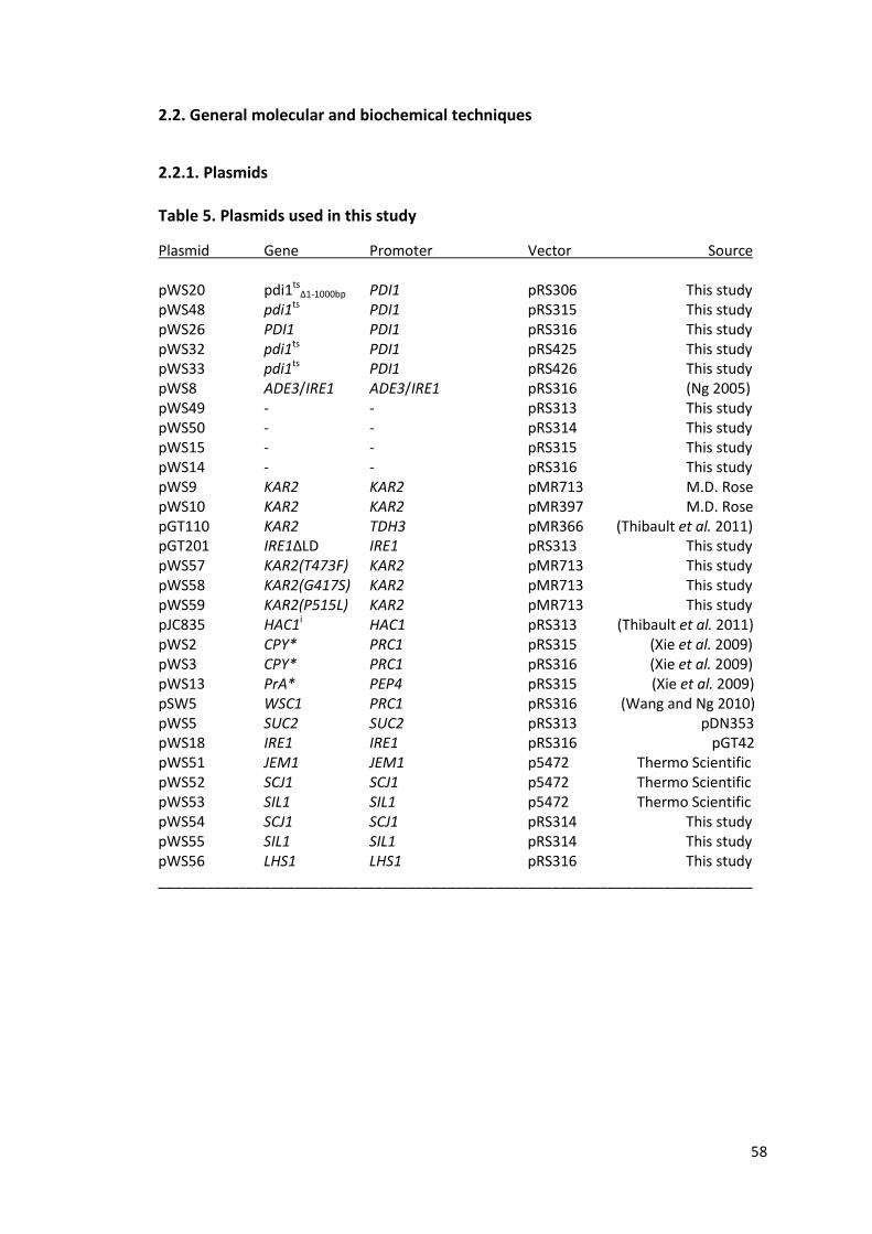
58
2.2. General molecular and biochemical techniques
2.2.1. Plasmids
Table 5. Plasmids used in this study
Plasmid Gene Promoter Vector Source
pWS20 pdi1tsΔ1-1000bp PDI1 pRS306 This study
pWS48 pdi1ts PDI1 pRS315 This study pWS26 PDI1 PDI1 pRS316 This study pWS32 pdi1ts PDI1 pRS425 This study pWS33 pdi1ts PDI1 pRS426 This study pWS8 ADE3/IRE1 ADE3/IRE1 pRS316 (Ng 2005) pWS49 - - pRS313 This study pWS50 - - pRS314 This study pWS15 - - pRS315 This study pWS14 - - pRS316 This study pWS9 KAR2 KAR2 pMR713 M.D. Rose pWS10 KAR2 KAR2 pMR397 M.D. Rose pGT110 KAR2 TDH3 pMR366 (Thibault et al. 2011) pGT201 IRE1ΔLD IRE1 pRS313 This study pWS57 KAR2(T473F) KAR2 pMR713 This study pWS58 KAR2(G417S) KAR2 pMR713 This study pWS59 KAR2(P515L) KAR2 pMR713 This study pJC835 HAC1i HAC1 pRS313 (Thibault et al. 2011) pWS2 CPY* PRC1 pRS315 (Xie et al. 2009) pWS3 CPY* PRC1 pRS316 (Xie et al. 2009) pWS13 PrA* PEP4 pRS315 (Xie et al. 2009) pSW5 WSC1 PRC1 pRS316 (Wang and Ng 2010) pWS5 SUC2 SUC2 pRS313 pDN353 pWS18 IRE1 IRE1 pRS316 pGT42 pWS51 JEM1 JEM1 p5472 Thermo Scientific pWS52 SCJ1 SCJ1 p5472 Thermo Scientific pWS53 SIL1 SIL1 p5472 Thermo Scientific pWS54 SCJ1 SCJ1 pRS314 This study pWS55 SIL1 SIL1 pRS314 This study pWS56 LHS1 LHS1 pRS316 This study __________________________________________________________________________
59
Plasmids were constructed using standard molecular cloning techniques (Joseph
Sambrook 2001) and sequenced for verification before use. All restriction enzymes
were purchased from New England Biolabs and used according to manufacturer's
protocols. Primers used are listed in Table 6.
pWS20 : A 1.6 kB truncated fragment of pdi1ts was amplified using primers WSO67
and WSO68. This fragment consists of the C-terminal 569bp of pdi1ts and an
additional 1000bp downstream of the stop codon. The fragment was inserted into
the pRS306 integrating plasmid after digestion with EcoRI and KpnI.
pWS26 : A 2 kB fragment comprising the PDI1 promoter and gene was removed
from pDN372 by digestion with SmaI and XhoI and inserted into the pRS316 vector.
pWS48 : This was constructed by carrying out site-directed mutagenesis using
pDN372 as the template, according to the protocol in the QuikChange® Site-Directed
Mutagenesis kit (Stratagene). The point mutation that gave rise to the pdi1-2 allele
was introduced using the primers WSO30 and WSO31.
pWS32 : This was subcloned from pWS48 by cutting out the insert using SmaI and
XhoI and ligating the fragment containing the PDI1 promoter and the pdi1-2 allele
into the pRS425 vector.
pWS57, pWS58, pWS59 : These were constructed by site-directed mutagenesis using
pWS9 as template according to instructions from the QuikChange® Site-Directed
Mutagenesis kit (Stratagene). pWS57 contains the T473F mutation in the encoded
KAR2 gene introduced by primers WSO212 and WSO213. pWS58 contains the G417S
mutation in the encoded KAR2 gene introduced by primers WSO214 and WSO215,

60
while pWS59 contains the P515L mutation in the encoded KAR2 gene introduced by
primers WSO216 and WSO217.
pWS54 : A 2.3 kB fragment comprising the SCJ1 gene and proximal promoter was
amplified using the primers WSO 128 and WSO 129. After digestion with BamHI and
SmaI, the fragment was ligated into the pRS314 vector.
pWS55 : The SIL1 gene and its proximal promoter was amplified using the primers
WSO124 and WSO125. The 2.4 kB fragment was digested using BamHI and SmaI and
inserted into the pRS314 vector.
pWS56 : A 3.6 kB fragment consisting of the LHS1 gene and its proximal promoter
was amplified using the primers WSO136 and WSO137. The fragment was digested
with BamHI and NotI and inserted into the pRS314 vector.

61
2.2.2. Primers used in this study
Primers used and their sequences are listed in Table 3.
Table 6. List of primers used in this study
WSO10 CCGCCCAGTCCTGCTCGCTT Primer Sequence (5'--> 3')
WSO11 GAAGTGGCGAGCCCGATCTT WSO30 GATCCTTGGACTCTTCATTCGACTTCATCAAGG WSO31 CCTTGATGAAGTCGAATGAAGAGTCCAAGGATC WSO67 CGGAATTCCGTTTGACGAATTGAGCGAC WSO68 GGGGTACCGACGCTTTGTTTGACCTCAG WSO124 CGGGATCCCAAGTTAGAAGAACCACCAG WSO125 TCCCCCGGGATTACCACGGCAGGGCAGTT WSO126 CCGCTCGAGCTGACTATGTCCCCTTTGGT WSO127 TCCCCCGGGTCGAACGTGCTTATGACTCC WSO128 CGGGATCCAGAAGATGCAGAGATAGCGG WSO129 TCCCCCGGGCTTGATATGGATGGGGAGCA WSO136 CGGGATCCGCAACTGCCTTTTATCACGG WSO137 ATAAGAATGCGGCCGCCATCTCTCGAAATTGCCCCA WSO143 GTTCAACTGTCGAGACCTTTGGC WSO144 ACGTCCGGACTCATGCTAGAAG WSO145 ATGGTAAGAAGGCCTCCAAGGG WSO146 GGATAAGACACCAGCTTGAACGG WSO147 TGGATTCTGAGGTTGCTGCTTTGG WSO148 ACCTTGGTGTCTTGGTCTACCG WSO149 TGGAAGCCTTGCCCTACTTTGG WSO150 GCAAACCGCTTTCGACGTATTGG WSO151 CGACAGTAATCAACTCGGCTGACG WSO152 ACTTGGATTCGACTCGGTGGTC WSO153 GCAGGCAAATCGATTCGTTACACC WSO154 ACTGTACCGTTAGAGGGCCTTG WSO155 TCACAGGACATTGCCAGATTGGAG WSO156 GTAGCCGTGCTTGTAATCATGCG WSO157 TGGTGCGGCCATTGTAAGAAGC WSO158 GCAGCAACTTGGACTACACCATCC WSO165 TACCGTGAAGGTACTGCCAAGC WSO166 TGTCGTCATGTGTCTTCCCAAC WSO175 ACCCDCTATCCACATCTTTCCG WSO176 CCTGACTGAGCTAAATAGCAACCC WSO212 CCCAAATTTTCTCTTTTGCCGTTGACAACCAACCAACCG WSO213 CGGTTGGTTGGTTGTCAACGGCAAAAGAGAAAATTTGGG WSO214 CCAGCA CCAAGAGGTG TACTTCAAAT TGAAGTCACA TTTGC WSO215 GCAAATGTGACTTCAATTTGAAGTACACCTCTTGGTGCTGG WSO216 GATGAAGCTG TTGCATACTCTGCAGCCGTT CAAGC WSO217 GCTTGAACGGCTGCAGAGTATGCAACAGCTTCATC ________________________________________________________________________

62
2.2.3. Reagents and Antibodies
Anti-CPY antibody was a gift from Reid Gilmore (University of Massachusetts,
Worcester, MA). Anti-Gas1p antibody was raised against amino acids 40 to 289 of
Gas1p (Davis Ng). Anti-hemagglutinin mouse monoclonal antibody (HA.11) was
purchased from Covance Research Products Inc. Anti-Pdi1p was a gift from Karin
Römisch (Saarland University, Germany). Diamide was purchased from Sigma-Aldrich.
2.2.4. Cell Labeling and Immunoprecipitation (Pulse chase analysis)
Precultures of yeast strains were diluted and grown overnight at 25 ⁰C in SC medium
lacking methione and cysteine, and the amino acid (s) required for plasmid selection
(where applicable). When cell cultures have reached an A600 reading of 0.2-0.5, 9OD
units of cells were pelleted at 1500 g for 10 min and resuspended in 2.7 mL of SC
medium. For metabolic labeling of cells at 37 ⁰C, resuspended cells were pre-
incubated at 37 ⁰C for 15 min before the addition of 110 µCi of [35S]
methione/cysteine. After a 5 min or 10 min pulse (depending on the protein
assayed), cold methione/cysteine were added to a final concentration of 2 mM. 900
µL of cell culture was removed at each designated time point and added to
trichloroacetic acid (TCA) to achieve a final concentration of 10% TCA. Samples were
bead-beaten twice for 30 s, with a 5 min incubation on ice in between. Following
which, the lysates were centrifuged at full speed for 10 min at 4 ⁰C , and the
supernatant removed by aspiration. The pellet was resuspended in 150 µL
resuspension solution [Tris-HCl pH 11, 1 mM PMSF) and boiled at 100 ⁰C for 10 min.
After centrifuging at full speed for 10 min at 4 ⁰C, the supernatant was transferred to
a new tube and used for immunoprecipitation (IP). For experiments where protein

63
processing was analyzed, 50 µL of lysate was added to 700 µL IP Solution II with
protease inhibitors [50 mM Tris pH 7.4, 150 mM NaCl, 1% Triton X-100, 0.01% NaN3,
1mM PMSF, 0.5 uL protease inhibitor cocktail (Sigma Aldrich)] and the appropriate
antibody. For experiments where substrate degradation was analyzed, total TCA-
precipitable counts for the samples were measured using the scintillation counter
(Beckman Coulter) and normalized by adjusting the lysate volumes used for IP.
Samples were placed on a rotator for 2 h at 4 ⁰C. After incubation, samples were
centrifuged for 20 min at full speed. Proteins were immunoprecipitated by the
addition of the supernatant to 30 µL of Protein A Sepharose beads and subsequent
incubation for 1 h at 4 ⁰C. Beads were then washed thrice with IP Solution I (20 mM
Tris pH 7.4, 150 mM NaCl, 1% Triton X-100, 0.02% NaN3, 0.2% SDS) and once with
phosphate-buffered solution (PBS; 137 mM NaCl, 2.7 mM KCl, 10 mM Na2HPO4, 1.8
mM KH2PO4). 10 µL of 2x protein loading buffer (0.2% bromophenol blue, 0.2M DTT,
20% glycerol) was added to each sample, and the samples boiled for 10 min at 100
⁰C to elute the proteins from the beads. Proteins were separated by SDS-PAGE and
visualized by autoradiography using the Typhoon™ 9200 phosphorimager.
Quantification was carried out using the ImageQuant™ TL software (GE Healthcare
Life Sciences).
2.2.5. Growth Assay (Spotting)
Yeast strains were grown to log phase in either SC or SC- dropout media. Cells were
diluted to an OD600 of 0.2 and ten-fold serial dilutions were performed. Diluted cells
were then spotted on either SC or SC-selective plates. The plates were placed at the

64
permissive (25⁰C) and non-permissive (37⁰C) temperatures for 2-3 days till colonies
have formed sufficiently.
2.2.6. Assessment of CPY folding using MalPEG conjugation to cysteine sulfhydryl groups Strains were grown to A600 of 0.2- 0.5 at 25 ⁰C, following which 9OD units of each
strain were preincubated at 37 ⁰C for 15 min in the presence or absence of 10 mM
DTT, before the addition of 110 µCi of [35S] methione/cysteine. After a 5 min pulse,
cold methione/cysteine were added to a final concentration of 2 mM and samples
were removed at each 15 min time points. TCA was added to a final concentration of
10% and cells were bead-beaten twice for 30 s with a 5 min incubation on ice in
between. Subsequently, the lysates were centrifuged at full speed for 10 min at 4 ⁰C,
and the supernatant removed by aspiration. The pellet was washed once with
acetone to fully remove the TCA, before resuspension in Buffer A (100 mM Tris
pH7.4, 2% SDS). Where indicated, samples were treated with 5 mM
methoxypolyethyleneglycol 5000 Maleimide (MalPEG; Fluka). Samples were then
heated at 100 ⁰C for 10 min, before incubation on ice for 1h. During which, samples
were heated at 100 ⁰C for 5 min after every 15 min on ice. After centrifuging at full
speed for 10 min at 4 ⁰C, the supernatant was transferred to a new tube a nd used
for immunoprecipitation. IP is performed as described above.
2.2.7. Western Analysis
Proteins were separated using SDS-PAGE at 200 V using pre-cast Mini-PROTEAN®
TGX™ gels (BioRad), and transferred to nitrocellulose membrane using the Trans-
Blot® Turbo™ transfer system (BioRad). The membrane was blocked in blocking

65
buffer (5 % non-fat milk in PBS, 0.1 % Tween-20) for 30 min at 37 ⁰C with constant
shaking. After which, the membrane was probed with the antibody against the
target protein, diluted in 0.5 % non-fat milk in PBST (PBS, 0.1 % Tween-20) for 1 h at
room temperature or overnight at 4 ⁰C. The membrane was then washed thrice for
10 min at room temperature using PBST with constant agitation. Subsequently the
membrane was probed with the appropriate secondary antibody -- α-mouse IgG
(Pierce Biotechnology) or α-rabbit IgG (Jackson ImmunoResearch Laboratories, Inc.) ,
diluted 1: 10 000 in 0.5 % non-fat milk in PBST (PBS, 0.1 % Tween-20) for 1 h at room
temperature. The membrane was washed thrice for 10 min at room temperature
using PBST before incubation with the SuperSignal® West Pico chemiluminescent
substrate (Thermo Scientific) for 5 min. The membrane was then exposed to film (GE
Healthcare) and protein bands were visualized after developing the films using the
M35 X-OMAT processor (Kodak).
2.2.8. Quantitative PCR
Cell cultures were grown to A600 of 0.2- 0.5 at 25 ⁰C in SC medium. Cells were
removed for the 0 h time point before cultures were shifted to the restrictive
temperature of 37 ⁰C. Cell cultures were periodically diluted with pre-warmed SC
medium to keep A600 at less than 0.5. At each time point (0 h, 1 h, 8 h, 48 h), cells
were pelleted by centrifugation at 1500 g for 10 min; cell pellets were washed twice
with ice-cold PBS. Total RNA was extracted and purified using the RNeasy Mini Kit
(Qiagen) according to manufacturer's instructions. cDNA synthesis was performed
using the SuperScript®III First Strand Synthesis System (Invitrogen) following
manufacturer's protocol. Samples comprising cDNA, gene-specific primers, and the

66
Power SYBR® Green PCR Master Mix (Invitrogen) were prepared and qPCR was
carried out using the 7500 Fast Real-Time PCR system (Applied Biosystems). Data
was analyzed using the 7500 Fast System SDS software (Applied Biosystems), using
the expression level of actin as an internal control.
2.2.9. DNA Microarray
Cell cultures were grown to A600 of 0.2- 0.5 at 25 ⁰C in SC medium. Cells were
removed for the 0 h time point before cultures were shifted to the restrictive
temperature of 37 ⁰C. Cell cultures were periodically diluted with pre-warmed SC
medium to keep A600 at less than 0.5. At each time point (0 h, 1 h, 8 h, 48 h), cells
were pelleted by centrifugation at 1500 g for 10 min; cell pellets were washed twice
with ice-cold PBS. Total RNA was extracted and purified using the RNeasy Mini Kit
(Qiagen) according to manufacturer's instructions. Quality control of the RNA was
carried out using the Agilent RNA Nano 6000 Chip (Agilent Technologies). Probes
were prepared using the Low Input Quick Amp Labeling System with 100 ng of total
RNA as starting material according to manufacturer's protocol, which included the
One-Colour Microarray-Based Gene Expression Analysis Protocol Version 6.5 (Agilent
Technologies), and later hybridized on a customized Agilent GE 8X60K array. Arrays
were scanned using a high resolution DNA Microarray Scanner (Model G2505C,
Agilent Technologies) and the data were analyzed using the GeneSpring GX software
(Agilent Technologies), and the open source softwares Cluster 3.0 (de Hoon et al.
2004) and Java Treeview (Saldanha 2004).

67
2.3. Synthetic Lethality Screen
2.3.1. UV Mutagenesis
DNY421 cells were cultured overnight at 25 ⁰C in SC Ura-dropout media to select for
the plasmid encoding IRE1/ADE3. The next day when cell culture has reached mid-
log phase, 50 A600 OD units of cells were harvested and centrifuged at 1000g for 5
min. The supernatant is decanted and the cell pellet washed once with 50 mL of ice-
cold sterile water. The cells were then pelleted and resuspended in 25 mL of 0.9 %
KCl. The cell suspension was transferred to a sterile 100 mm Petri dish containing a
sterile stir bar and placed on a magnetic stirrer to keep the suspension
homogeneous. A hand-held UV lamp (254 nm, 4 W) was clamped in position 15 cm
above the petri dish using a retort stand. UV mutagenesis was carried out in a
darkroom equipped with a low intensity red light to prevent photoactivable DNA
repair mechanisms from turning on. Before starting, a 1 mL aliquot of the cell
suspension was removed. The cells are then irradiated with sequential 30 s pulses of
short-wave UV light. 1 mL of the cells was removed after each 30 s pulse and
transferred to a new tube. A total of six pulses were administered.
2.3.2. Determining kill rate
To determine the kill rate after each pulse of irradiation, 100 µL was removed from
each 1 mL aliquot and a series of ten-fold dilutions were performed (10-1 - 10-5).
From the 10-3 to 10-5 dilutions for each time point, 100 µL was plated onto YPD
plates. The plates were then wrapped in aluminium foil to keep out light and placed
at 25 ⁰C for 3 days to allow colonies to grow. The kill rate was determined by

68
comparing the colony numbers at each mutagenic dose to that of the non-
mutagenized control.
2.3.3. Screening for temperature-sensitive synthetic lethal mutants
Remaining cell suspension from the 1 mL aliquot was innoculated in 9 mL of YPD
media in a culture tube. The tubes were wrapped in aluminium foil and left to rotate
on a roller drum for 18 h to stabilize the mutations. The mutagenized cells were then
stored at 4 ⁰C to stop their growth till they were plated. After the kill rate has been
determined, the kill rate of 75 % (corresponding to 120 s UV irradiation) was
deemed to be ideal for the genetic screen. A ten-fold dilution series was performed
using cells that had been treated with a 120 s dose of UV. 100 µL from each dilution
(10-4 - 10-6) was plated on YPD plates and incubated at 25 ⁰C for 3 days to determine
the cell titre. After the cell titre was determined, mutagenized cells were spread on
low-adenine SC plates, at a density of 300 colony-forming units per plate. The plates
were incubated at 25 ⁰C for 2 days before they were replica-plated onto a second set
of low-adenine SC plates. These plates were incubated at 37 ⁰C for 2 days to allow
colonies to form and develop the red pigmentation. After colonies have grown to a
sufficient size on both sets of plates, the two corresponding plates at both
temperatures were compared. Colonies that were non-sectoring (completely red) at
37 ⁰C but showed a sectoring red/white phenotype at 25 ⁰C were selected and
restreaked onto two sets of plates containing the same media to filter out the false
positives. The two sets of plates were incubated at 25 ⁰C and 37 ⁰C respectively.
Mutant strains that displayed the required phenotypes at each temperature were
stored in glycerol at -80 ⁰C while false positives were discarded.

69
2.3.4. Identifying Recessive Mutants
2.3.4.1. Backcrossing, Sporulation, and Tetrad Dissection
To identify recessive mutants, mutant strains were crossed with DNY 420, the MATα
strain isogenic to the parental strain. The strains were allowed to mate 5 h at room
temperature before some of the cells were streaked out on a thin YPD plate. The
diploid zygotes were picked using a yeast tetrad dissection microscope (Axioskop 40,
Zeiss) and allowed to grow for two days on the YPD plate before they were streaked
out at 37 ⁰C. Mutants whose diploids grew at 37 ⁰C were recessive.
Next, diploids were inoculated in 3 mL of sporulation media (3 g/L KOAc, 0.2 g/L
raffinose) and left to sporulate for 60-72 h on a roller drum at 25 ⁰C. 300 µL of the
culture was centrifuged and the cell pellet treated with 20 µL of zymolyase (1 mg/mL;
US Biological) to digest the ascus cell wall of the tetrads. After incubation for 12 min
at room temperature, 180 µL of 1M sorbitol was added to dilute out the zymolyase.
30 µL of this mixture was then spreaded onto a thin YPD plate and yeast tetrads
were picked and dissected. Mutants synthetic lethal with IRE1 will show a 2
sectoring : 2 red colony segregation pattern at 37 ⁰C.
2.3.5. Cloning and Sequencing Temperature-sensitive Mutants
2.3.5.1. High-Efficiency Lithium Acetate Transformation
Cells were cultured overnight at 25 ⁰C in 50 mL of SC medium to achieve a A600
reading of 0.4 - 0.8 the next day. Cells were then centrifuged at 1500 g for 5 min and
the supernatant decanted. The cell pellet was washed once with 20 mL of lithium
acetate mix (0.1 M LiOAc, TE pH 7.4) and the sample centrifuged again at 1500g for 5
min. The cell pellet is resuspended in 400 µL of lithium acetate mix and kept at 4 ⁰C

70
till ready for use. The transformation reaction was set up and consisted of: (i) 700 µL
PLATE mix (10 mM Tris pH7.4, 1 mM EDTA, 0.1 M LiOAc, 40% PEG), (ii) 10 µL salmon
sperm carrier DNA that had been boiled and cooled on ice, (iii) 1 µL YCp50 plasmid
library (3 µg/µL), and (iv) 100 µL of competent yeast cells. The mixture was vortexed
gently and incubated at 25 ⁰C for 30 - 60 min. After that, 100 µL of DMSO was added
before the cells were incubated at 30 ⁰C for 22 min. This presents as heatshock for
the cells as the yeast strains are temperature-sensitive. Cells were then centrifuged
at 14 000 rpm for 20 s and the supernatant removed. 1 mL of SC medium was used
to resuspend the cells before they were placed on a rotator for 1 h at 25 ⁰C for
recovery. Subsequently, cells were centrifuged at 14 000 rpm for 20 s to remove the
supernatant. They were resuspended in 1 mL SC-URA medium for plasmid selection.
1 % of the cell culture was plated on a SC-URA plate and left at 25 ⁰C to check
transformation efficiency, while the rest of the culture was plated on five SC-URA
plates and incubated at 37 ⁰C for 2 days to let colonies develop.
2.3.5.2. Extraction of Plasmid DNA
Yeast strain containing the plasmid was grown overnight in SC medium lacking the
amino acid required for plasmid selection. 1.5 mL of the cell culture was centrifuged
at 14 000 rpm for 1 min and the cell pellet resuspended in 200 µL of breaking buffer
(2 % Triton X-100, 1 % SDS, 100 mM NaCl, 10 mM Tris pH 8.0, 1 mM EDTA pH 8.0).
The cell suspension was added to 200 µL of 25:24:1 phenol/chloroform/isoamyl
alcohol with 200 µL of 0.5 mm zirconium beads and bead-beaten twice for 30 s, with
a 5 min incubation on ice in between. The cell lysate was transferred to a new tube

71
and centrifuged at 14 000 rpm for 5 min. 50 µL of the aqueous layer containing the
plasmid was removed carefully and stored at -20 ⁰C until needed.
2.3.5.3. Transformation using electroporation
1 µL of the extracted plasmid was added to 50 µL of electrocompetent E.coli cells on
ice. The mixture was transferred to an electrocurvette (Biorad). Electroporation was
carried out using a micropulser (BioRad). After which, transformed bacterial cells
were added to 950 µL of LB medium and left to recover for 1 h at 37 ⁰C on a shaker.
The cells were then pelleted, resuspended in 150 µL of LB medium, and plated on a
LB plate containing the antibiotic for plasmid selection. Colonies were allowed to
grow at 37⁰C overnight.
2.3.5.4. Extraction of yeast genomic DNA
Yeast strains were cultured in 2 mL of SC media overnight. Yeast genomic DNA was
extracted using the Yeast DNA Extraction Kit (Thermo Scientific) according to
manufacturer's protocol. Briefly, cells were centrifuged at 14 000 rpm for 1 min and
the supernatant removed. The pellet was resuspended in 800 µL Y-PER reagent and
incubated at 65 ⁰C for 10 min. Samples were then centrifuged at 14 000 rpm for 1
min and the supernatant discarded. The pellet was then resuspended in 400 µL each
of DNA Releasing Reagent A and B and the mixture was incubated at 65 ⁰C for 10 min.
After that, 200 µL of Protein Removal Reagent was added to each sample and the
samples were centrifuged at 14 000 rpm for 5 min. The supernatant was transferred
to a new tube containing 600 µL of isopropanol and the samples inverted a few
times to achieve homogeneous mixing. The samples were centrifuged at 14 000 rpm
for 10 min and the supernatant removed. The DNA pellet was washed with 1.5 mL of
72
70 % ethanol . The DNA was once again pelleted by centrifugation at 14 000 pm for 1
min and the ethanol was removed. After air-drying, the DNA pellet was dissolved in
50 µL of TE buffer and stored at -80 ⁰C.
2.3.5.5. Sequencing
Samples to be sequenced were prepared using the BigDye® Terminator v3.1
sequencing kit and sequencing was carried out using the 3730xl DNA analyzer
(Applied Biosystems). Complementing plasmids were sequenced using the primers
WSO10 and WSO11 to identify the sequences flanking the genomic DNA inserts;
genes were sequenced using their respective primers (Table 6).

73
Chapter 3. Genetic screening for temperature-sensitive mutants displaying synthetic lethality with an ire1 null mutation
3.1. Introduction
Different approaches have been adopted to improve our understanding of the UPR,
specifically to identify genes or cell functions that are physiologically linked to the
UPR. A previous study developed a genetic screen to isolate synthetic lethal mutants
that were dependent on UPR activation for viability (Ng 2005). Our genetic screen
was designed however, to isolate a special group of conditional mutants that could
provide further insights to the UPR. These mutants are temperature-sensitive and
display synthetic lethality with the ire1 null phenotype at the restrictive temperature
of 37 ⁰C, i.e, they require UPR activation for survival at 37 ⁰C but not at the
permissive temperature of 25 ⁰C. In addition, these mutants allow the UPR to be
uncoupled from the loss in gene function, which allows us to study a specific
mutation without the accompanying compensation.
3.2. Genetic screening
For this project, I used different strategies to screen for the conditional mutants. The
screens exploit the fact that IRE1 is non-essential in S.cerevisiae and is not required
for viability under conditions that do not trigger ER stress. The principle of each
screen is described in detail in the following sections.

74
3.2.1. Screening by colony colour phenotype
The first screen I did make use of a yeast colony-colour sectoring assay. This assay
takes advantage of the red-colony phenotype of ade2 cells on media limiting in
adenine. ADE2 encodes an enzyme in the adenine biosynthetic pathway in
S.cerevisiae which converts the red intermediate, P-ribosylamino imidazole (AIR), to
P-ribosylamino imidazolecarboxylate (CAIR). An ade2 mutation producing a non-
functional enzyme results in the accumulation of AIR, giving rise to red colonies. An
ade3 mutation blocks the adenine synthesis pathway at a point before the
accumulation of the red pigment. Since production of AIR requires the ADE3 gene, a
yeast strain containing the ade2ade3 mutations forms white colonies as AIR is not
produced (Fig. 13).
Figure 13. Diagrammatic representation of the yeast adenine biosynthesis pathway ADE2 and ADE3 are two of the enzymes that catalyze intermediate reactions in the yeast adenine biosynthesis pathway. Production of the red intermediate, AIR, is dependent on ADE3 while ADE2 converts AIR to CAIR. An ade2 mutation causes accumulation of AIR, giving rise to a red colouration; an ade3 mutation inhibits production of AIR, producing no red colouration.
For this screen I made use of parental strain DNY421, which is an ade2ade3Δire1
strain that has been transformed with pDN336, a yeast CEN/ARS vector with a URA3
selectable marker encoding wild-type ADE3 and IRE1 genes. As both ADE3 and IRE1
are non-essential, this strain gives rise to red and white sectoring colonies on
adenine-limiting media as the plasmid is lost at a frequency of 10-2 per cell division
due to mitotic mis-segregation (Guthrie 1991). This represents a mixture of cells with

75
(red) and without (white) the plasmid. Based on this principle, any mutation that
renders the IRE1 gene essential will result in the formation of red colonies as only
colonies retaining the ADE3-IRE1 plasmid can survive. Since the screen aimed to look
for temperature-sensitive synthetic lethal mutants, mutants which sectored at the
permissive temperature of 25 ⁰C ( IRE1 is not required for survival) but were red at
the restrictive temperature of 37 ⁰C (IRE1 is essential for survival) were isolated. This
allowed us to visually screen for mutants that required the plasmid for survival (Fig.
14).

76
Figure 14. Primary genetic screen using colony colour phenotype Schematic representation of the genetic screen based on colony colour phenotype. UV mutagenesis of the parental strain generates different types of mutant alleles. The conditional mutants of interest display a sectoring phenotype at 25⁰C and appear red at 37⁰C.

77
3.2.2. Screening by counter-selection using 5-fluoroorotic acid (5-FOA)
The screen was subsequently repeated using a Δire1 strain in the BY4741
background, transformed with the centromeric pRS316-IRE1 plasmid which encodes
both IRE1 and URA3. This strain does not contain the ade2, ade3 mutations and
precludes the use of the colony colour assay. Instead, the desired mutant strains
were identified by counter-selection using 5-FOA. Mutagenized cells that require the
UPR for survival at the restrictive temperature will retain the plasmid and express
both URA3 and IRE1. The URA3 gene encodes orotidine 5-phosphate decarboxylase,
an enzyme involved in the synthesis of pyrimidine ribonucleotides. This enzyme
converts 5-FOA to 5-fluorodeoxyuridine, which is toxic to the cell. As a result,
mutagenized cells replica-plated onto 5-FOA-containing solid media will die at the
restrictive temperature if their genomic mutations render the UPR essential for
viability (Fig. 15).

78
Figure 15. Primary genetic screen by counter-selection using 5-FOA Schematic representation of the genetic screen based on counter-selection using 5-FOA. The conditional mutants of interest generated by UV mutagenesis are viable at 25⁰C but inviable at 37⁰C, due to counter-selection against colonies expressing URA3.

79
3.2.3. Screening by temperature sensitivity
The last approach that I took was to screen directly for temperature-sensitive
colonies based on their growth at the restrictive temperature. Mutagenized Δire1
cells were plated at 25⁰C and allowed to grow till very small colonies form. They
were then replica-plated and grown at 37⁰C. Colonies that grew at 25⁰C but failed to
grow at 37⁰C were selected and transformed with a plasmid encoding IRE1 to check
which of these temperature sensitive mutants displayed synthetic lethality with the
ire1 null mutation. Mutants that were synthetic lethal can now grow at the
restrictive temperature as the UPR can be induced, whereas mutants that were
merely temperature sensitive will still be inviable (Fig. 16).

80
Figure 16. Primary genetic screen by temperature sensitivity Schematic representation of the genetic screen based on temperature sensitivity. TS mutants generated by UV mutagenesis are transformed with an IRE1-encoding plasmid. Conditional mutants of interest that were inviable at 37 ⁰C are now viable when expressing IRE1.

81
3.3. Cloning by complementation
Since the mutant strains identified are conditional mutants, the gene that is mutated
in each strain can be identified by complementation. The mutant strains (without a
functional IRE1) were transformed with the centromeric YCp50 shuttle based yeast
genomic library (Rose et al. 1987) and incubated at the restrictive temperature.
Colonies carrying putative plasmids containing the complementing gene were viable
at the restrictive temperature. These colonies were selected and the plasmids they
contained were then extracted. The complete sequence of the genomic insert in the
plasmid was determined by sequencing the vector-insert junctions using appropriate
primers and submitting the junction sequences to the Saccharomyces Genome
Database (www.yeastgenome.org). In this way, the genes present in this insert are
known and the complementing gene can be determined either by deletion analysis
of the complementing plasmid or by sequencing each corresponding gene
individually from the genomic DNA of the mutant strain to verify and locate the
mutation.
However, it is important to note that even though most genes are represented in the
YCp50 genomic library at a frequency of about 0.1%, some yeast genes may be
under-represented for various reasons. As amplification of the genomic library
requires propagation in E.coli, genes, like KAR2, which are toxic to E.coli will be found
at a much lower frequency in the library (Rose et al. 1987).

82
3.4. Secondary screen for biosynthetic and ERAD mutants
A secondary screen was carried out to screen mutants for defects in secretory
protein synthesis and ERAD. Two well characterized endogenous proteins –
carboxypeptidase Y (CPY), and Gas1p, a GPI-anchored plasma membrane protein,
were used to detect biosynthetic mutants using pulse chase analysis. The post-
translational modifications and trafficking of these proteins through the secretory
pathway can be visualized on a gel with their characteristic mobility shifts. This assay
can detect defects in protein translocation, N-linked glycosylation, O-linked
glycosylation, GPI-anchor biosynthesis, ER to golgi transport, and transport out of
the golgi (Ng 2005). ERAD mutants were identified using pulse chase analysis of a
model ERAD substrate CPY*, which is a misfolded version of CPY. CPY* is rapidly
degraded in a wild type strain. Any perturbation to ERAD function in mutant strains
will result in the accumulation of CPY*. A detailed description of the secondary
screens and their results are discussed in Chapter 5.
3.5. Results of genetic screens
Here, I will briefly describe the various steps involved in the screens and provide a
summary of the number of mutants obtained at each step. The characterization of
the mutants are presented in Chapter 5. In this thesis, I focused on one interesting
mutant that was isolated from the screen, and this mutant will be discussed in detail
in Chapter 4.

83
3.5.1. Summary of colony colour assay
About 30 000 mutants were screened using the colony colour phenotype, of which
28 of them were temperature-sensitive and displayed synthetic lethality with an ire1
deletion. Mutagenized cells were plated on SC-low adenine plates at both 25⁰C and
37⁰C, and incubated for three days. After which, colonies were visually inspected and
those sectoring at 25 ⁰C but were non-sectoring and red at 37 ⁰C were selected.
About 5% of the colonies passed this initial screening. These mutants were then
restreaked to obtain single colonies to further verify their colony colour and only
about 11% of these mutants were putative ts-synthetic lethal mutants. Next, to
certify that these mutants were indeed dependent on IRE1 for viability, the IRE1-
encoding plasmid was dropped from the mutants and these mutants were
restreaked at 37 ⁰C. Of these, 17% were inviable and represented the true ts-
synthetic lethal mutants that I was interested to find (Fig. 17).

84
Figure 17. Workflow of genetic screen using colony colour and the number of mutants obtained at each step A total of 30 000 colonies were screened for their colony colour phenotype, of which about 5% appeared to be putative mutants of interest. Further screening revealed most of these to be false positives, and only 0.09% of the total colonies screened were true ts-synthetic lethal mutants.

85
These mutants were subsequently cloned by complementation using the YCp50
yeast genomic library. While identifying and characterizing the mutants, I
unexpectedly discovered that the ire1 null strain itself displayed defects in protein
biogenesis as well as ERAD when pre-incubated at the restrictive temperature for a
prolonged period of time (1h). This was not apparent during shorter pre-incubation
period (15-30 min) or at a lower temperature (30⁰C). Unfortunately, this complicated
our secondary screen since the ire1 null strain was our control strain, and mutants
that were not as thermolabile required longer pre-incubation at 37 ⁰C before pulse
chase analysis could be carried out.
This observation suggested that there may be an inherent mutation present in the
strain which manifests itself after a prolonged shift to 37 °C, as the Δire1 strain of
another strain background (BY4741) behaved like wild-type after a 1 h shift to 37 °C.
In addition, the screen appeared to be biased as several mutants carried genomic
mutations in YOS9 and MPD1, while others could not be cloned using the yeast
genomic library. This could also be a result of an inherent mutation which
predisposes the screen to be enriched for certain mutants. To overcome these
difficulties, I changed my strategy and used counter-selection by 5-FOA as well as
temperature sensitivity for screening.

86
3.5.2. Summary of 5-FOA screen
The genetic screen was repeated using a Δire1 strain of BY4741 background, which
was first verified to behave like wild-type after 1h incubation at 37 ⁰C. For this screen,
counter-selection of the desired mutant strains using 5-FOA was carried out.
Mutagenized cells were plated on 5-FOA-containing SC-plates, incubated at 25 ⁰C for
two days, replica-plated and then incubated at 37 ⁰C for two days to allow colonies
to form. By visual inspection, colonies that survived at 25 ⁰C but not at 37 ⁰C were
selected. Out of the 18 000 colonies screened, about 10% passed this initial selection.
For these mutants, the corresponding colonies growing on the 25 ⁰C plate were
selected and restreaked at 37 ⁰C to verify their temperature sensitivity. 24% of these
were true ts mutants. They were retransformed with a plasmid encoding IRE1 and
incubated at 37 ⁰C. True ts-synthetic lethal mutants should now grow at 37 ⁰C as the
UPR can be induced. Five mutants were identified out of the 18 000 screened (Fig.
18).
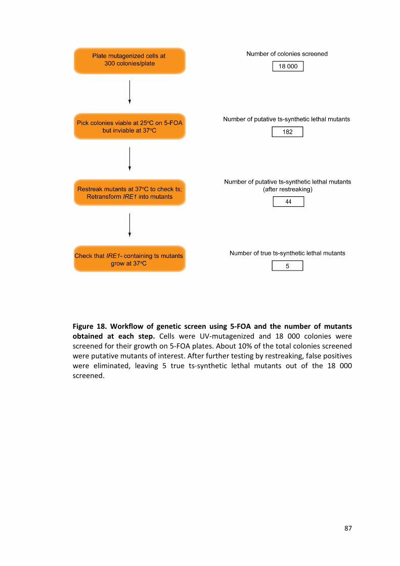
87
Figure 18. Workflow of genetic screen using 5-FOA and the number of mutants obtained at each step. Cells were UV-mutagenized and 18 000 colonies were screened for their growth on 5-FOA plates. About 10% of the total colonies screened were putative mutants of interest. After further testing by restreaking, false positives were eliminated, leaving 5 true ts-synthetic lethal mutants out of the 18 000 screened.

88
3.5.3. Summary of TS screen
In addition to screening using 5-FOA, a more straight-forward method to obtain the
ts-synthetic lethal mutants was used. Mutagenized cells were first screened for
temperature sensitivity, followed by synthetic lethality with Δire1. 9 000 colonies
were plated at 25 ⁰C and subsequently replica-plated at 37 ⁰C. Of these, 8%
appeared to be ts by visual inspection. These mutants were restreaked at 37 ⁰C to
verify that they were indeed ts. 10% of these were true ts mutants. They were then
transformed with an IRE1-encoding plasmid and grown at 37 ⁰C. Mutants that were
viable at 37 ⁰C were ts-synthetic lethal with an ire1 null mutation. Ultimately, three
mutants were identified out of the 9 000 colonies screened (Fig. 19).

89
Figure 19. Workflow of genetic screen using temperature sensitivity and the number of mutants obtained at each step. 9 000 colonies were screened for their viability at 37 ⁰C. 0.8% of these colonies were found to be true ts mutants after verification. To check their synthetic lethality with an ire1 null mutation, these mutants were transformed with an IRE1-encoding plasmid and 3 mutants of interest were identified.

90
3.6. Discussion
Screening for the temperature-sensitive, synthetic lethal mutants was impeded by
the high percentage of false positives. This problem was common to all three
strategies used for screening, and could be attributed to various factors. In the case
of the colony colour assay, many colonies that appeared red at 37⁰C during the initial
screen displayed various degrees of sectoring when they were restreaked at 37⁰C on
new agar plates. This could be explained by the fact that temperature-sensitive
mutants may display a phenotype that is intermediate between their respective WT
and knockout strains, as only certain aspects of their gene function were disrupted
by the temperature shift. As such, holding on to the IRE1 plasmid (thus allowing UPR
activation) may be preferable for cell growth, but may not be essential. Colonies that
initially appeared red at 37⁰C may belong to this group of mutants . When these
mutants were subsequently restreaked, the larger number of colonies growing per
mutant allowed us to observe some degree of sectoring in the colonies, suggesting
they were not strictly synthetic lethal. I also observed that many of the mutants
obtained after restreaking were synthetically sick, but not lethal, with the ire1-null
mutation, which explained the small number of true ts-synthetic lethal mutants
isolated.
False positives from the 5-FOA and temperature-sensitivity screens could be due to
inaccuracies during replica-plating. As replica-plating was carried out when the size
of the colonies on the master plate was relatively small, it is possible that some
colonies were not transferred sufficiently for growth on the new plate. On hindsight,
heat stress from shifting the cells to 37⁰C could have played a role in the retainment

91
of the IRE1 plasmid in the mutants, even when it was not essential for growth. Heat
stress was previously shown to not induce the UPR (Cox and Walter 1996), but
studies in our laboratory have subsequently shown that some induction of the UPR
occurs.
The screen is far from being exhaustive. To be able to identify ts-synthetic lethal
mutants that cover a broad range of functions in the secretory pathway in a feasible
manner, we have to greatly increase the number of mutants in the initial screen,
while finding a way to keep false positives at a minimum.

92
Chapter 4. The UPR buffers against a lethal pdi1 dysfunction
4.1. Introduction
From the screen using colony colour phenotype, I isolated a mutant that was found
to carry a point mutation in PDI1, which encodes the ER-resident protein disulfide
isomerase essential for the formation of disulfide bonds in secretory and membrane
proteins. This is the first time a pdi1 mutant has been isolated from a forward
genetic screen. This allele has a single base change that results in an amino acid
change from leucine to serine at position 476, located in the a' domain of Pdi1p (Fig.
20). The pdi1 mutant is subsequently referred to as pdi1-2.
Figure 20. pdi1-2 contains a L476S point mutation in the a' domain of PDI1 The crystal structure of S.cerevisiae Pdi1p (PDB ID: 2B5E) adapted to show the location of the two catalytic sites (purple spheres) and the position of the L476S point mutation (yellow). Pdi1p contains four thioredoxin-like domains - a, b, b', a', of which the a and a' domains contain the active sites. The point mutation is found in the a' domain.

93
pdi1-2Δire1 showed a strong defect in CPY and Gas1p processing during the
secondary screen, suggesting a likely defect in protein folding. This defect is
apparent after a mere 15 min shift to the restrictive temperature, indicating that the
pdi1 allele is very thermolabile. pdi1-2 is viable when the UPR is activated, suggesting
that the essential function of Pdi1p is still intact. As PDI1 is an essential gene, in vivo
characterization of its functions is often difficult. Thus, pdi1-2 provides an invaluable
tool for us to dissect the roles of this multi-functional protein in the ER.
4.2. Generating the pdi1-2 mutant in the W303 background
pdi1-2 was generated using UV mutagenesis in a strain background that has been
genetically modified for the colony colour assay. To eliminate other background
mutations, I chose to generate the pdi1 mutant in the W303 background instead of
backcrossing pdi1-2Δire1 with the wild-type W303 strain. The W303 background is
the genetic strain that is used in the laboratory, and having the mutant in this
background will facilitate subsequent analysis.
To do this, I made use of the yeast integrating plasmid carrying a URA selectable
marker (pRS306) that encodes a truncated segment of the pdi1 ts allele (tPDI1ts; Fig.
21). The yeast integrating plasmid lacks a yeast origin of replication and can only be
propagated when integrated into the host genome. Integration occurs by means of
homologous recombination and this process is greatly enhanced by cleavage of the
plasmid in the region of homology (Stearns et al. 1990). The pRS306- tPDI1ts plasmid
was linearized after digestion with BglII and transformed into WT or Δire1 strains.
Successful integrants were selected based on their growth on SC plates lacking uracil.
These colonies were then grown without selection in SC media overnight to allow

94
excision of the integrated plasmid. Excision also occurs through homologous
recombination at a frequency of 1 x 10⁻³ - 1 x 10⁻⁴ (Stearns et al. 1990). In this case, if
recombination occurs in the homology region before the point mutation, the WT
PDI1 allele is regenerated; if recombination occurs in the homology region after the
point mutation, the ts pdi1 allele replaces the WT allele in the genome. Colonies with
the integrated plasmid excised were selected for using 5-FOA, and the PDI1 gene
was PCR-amplified from the genomic DNA of each colony and sequenced to
determine if the allele was WT or ts. Using this method, the pdi1-2Δire1 and pdi1-2
strains were generated in the W303 background and used for subsequent studies.

95
Figure 21. Integrating the ts allele into the W303 genome The URA-marked yeast integrating vector carrying a truncated fragment of PDI1 with the point mutation was linearized and transformed in WT and Δire1 cells. Homologous recombination occurs and successful integrants were selected on SC-URA dropout plates. These colonies were grown in non-selective media overnight and plated on 5-FOA plates to counter-select colonies in which the integrated plasmid has been excised. gDNA was extracted from these colonies. The PDI1 gene was PCR-amplified and sequenced to verify that the ts allele has successfully replaced the WT allele, creating the strains pdi1-2 and pdi1-2Δire1 respectively. * denotes point mutation.

96
4.3. Characterization of the pdi1-2 mutant
4.3.1. pdi1-2Δire1 displays conditional lethality and retains endogenous proteins in the ER First, the pdi1-2Δire1 and pdi1-2 strains that were generated in the W303
background were tested to ensure they had the same phenotypes as the original
mutant. To verify that the pdi1 mutant was indeed ts-synthetic lethal with an ire1
null mutation, 10-fold serial dilutions of the strains were spotted on SC plates at both
the permissive and restrictive temperatures. Consistent with the screening
conditions, pdi1-2Δire1 was completely inviable at 37⁰C in the absence of Ire1p but
grew at a rate similar to Δire1 cells at 25⁰C. However, when Ire1p was expressed, it
was viable and grew almost as well as WT cells at 37⁰C (Fig. 22). This suggests that
pdi1-2Δire1 requires the UPR for survival at the restrictive temperature.
Figure 22. pdi1-2 is inviable at the restrictive temperature in the absence of the UPR. Strains were grown overnight in SC media. Ten-fold serial dilutions of each culture were plated on SC plates and incubated at the respective temperature for two days.

97
Next, I studied the processing of two endogenous proteins -- carboxypeptidase Y
(CPY), and Gas1p, a GPI-anchored plasma membrane protein, using pulse-chase
analysis. During the secondary screen, pdi1-2Δire1 displayed a strong defect in the
biogenesis of these proteins. Both proteins are well-characterized and the post-
translational modifications and passage of these proteins through the secretory
pathway can be analyzed on a gel due to their characteristic mobility shifts. pdi1-
2Δire1 exhibited a defect in both CPY and Gas1p processing after a mere 15 min shift
to 37°C, suggesting it is very thermolabile. The ER forms of both proteins
accumulated after a 30 min chase (Fig. 23). For CPY, the majority of the proteins
remained in the P1 form, but a small percentage did form mature CPY. It is unclear
for Gas1p if mature proteins formed as Gas1p level decreased with time at the
higher temperature. As expected, the biosynthesis of both CPY and Gas1p were
normal at 25 ⁰C (Fig. 23) since Pdi1p is functional at the permissive temperature.

98
Figure 23. The ER forms of CPY and Gas1p accumulate in pdi1-2Δire1 at the restrictive temperature. Protein processing of endogenous CPY and Gas1p was examined in pdi1-2Δire1 at 25⁰C and 37⁰C. Where necessary, cells were shifted to 37⁰C for 15 min prior to the start of the experiment. Strains were pulse-labeled with [35S] methione/cysteine for 5 min and chased for the times indicated. Proteins were immunoprecipitated using anti-CPY and anti-Gas1p, resolved by SDS-PAGE, and visualized by autoradiography.

99
4.3.2. Pdi1pts is stable at the restrictive temperature
Since Pdi1p is an essential protein required for forming disulfide bonds in folding
proteins, I asked if the defect observed was due to the instability of mutant Pdi1p at
37 ⁰C. I used pulse chase analysis to study the protein level of newly synthesized
mutant Pdi1p over a 60 min chase and compared this to the protein levels of wild-
type Pdi1p in WT and Δire1 cells. There were no significant difference in the stability
of Pdi1p over a time-course of 60 min in these three strains (Figure).
Figure 24. Pdi1pts is stable at the restrictive temperature The stability of new-synthesized ts Pdi1p was studied using pulse chase analysis. WT, Δire1 and pdi1-2Δire1 were pre-incubated at 37⁰C for 15 min, pulse-labeled with [35S] methione/cysteine for 5 min and chased for the times indicated. Endogenous Pdi1p was immunoprecipitated using anti-Pdi1p and resolved by SDS-PAGE. Quantification was carried out using phosphorimager analysis.

100
4.3.3. ER-retention of endogenous proteins is due to misfolding and not a general trafficking defect Accumulation of the ER forms of the two proteins could be due to an inability of the
proteins to fold in the ER, or due to a defect in ER to golgi transport. To rule out the
possibility of a general ER to golgi trafficking defect, I looked at the transport of
Wsc1p, a COPII cargo protein, in the secretory pathway. Wsc1p is known to be
transported out of the ER to the golgi regardless of its folding state as it has a strong
ER export signal and no ERAD determinant that targets it for ER quality control
(Wang and Ng, 2010). In pdi1-2Δire1, Wsc1p was transported out of the ER, but not
CPY or Gas1p (Fig. 25). This suggests that the general ER-golgi transport machinery is
not defective in pdi1-2Δire1.
Figure 25. The retention of CPY and Gas1p in the ER is not due to a general ER-golgi transport defect. Protein processing of CPY, Gas1p, and Wsc1p was examined in pdi1-2Δire1 at 37⁰C. Cells were shifted to 37⁰C for 15 min, pulse-labeled with [35S] methione/cysteine for 5 min and chased for the times indicated. Proteins were immunoprecipitated using anti-CPY, anti-Gas1p, and anti-HA respectively, resolved by SDS-PAGE, and visualized by autoradiography.

101
Next, I assessed in vivo CPY folding status using the MalPEG-alkylation assay. MalPEG
forms a covalent bond with the sulfhydryl group of free cysteines and the MalPEG-
conjugated protein can be visualised as a band shift of 5 kDa on a SDS-PAGE gel
(Makmura et al. 2001). Folded proteins exhibit no band shift unless they contain free
cysteines not involved in disulfide bond formation in their folded state. Improperly
folded proteins on the other hand, result in a smear and not distinct bands due to
the presence of multiple misfolded molecules that form random intra- and inter-
molecular disulfide bonds resulting in differing number of free cysteines on a
heterogeneous set of molecules. Cells were treated with DTT, a reducing agent, to
show the band shift that occurs when all the disulfide bonds in the protein are
completely reduced. When folded, a molecule of CPY contains five disulfide bonds
and one free cysteine. At 37⁰C in the presence of MalPEG, CPY showed a band shift
in WT and Δire1 cells at both 0 min and 30 min timepoints (Fig. 26), which suggested
that the correct disulfide bonds form early during CPY processing. However, addition
of MalPEG in pdi1-2Δire1 resulted in a smear at both timepoints (Fig. 26), indicating
that CPY did not form its native disulfide bonds. This is consistent with the notion
that CPY was likely to be misfolded in pdi1-2Δire1.

102
Figure 26. CPY is misfolded in pdi1-2 Δire1 at the restrictive temperature The folding status of CPY in WT, Δire1 and pdi1-2Δire1 at 37⁰C was examined using the MalPEG alkylation assay. 10 mM DTT was added to each strain where indicated during pre-incubation at 37⁰C. Strains were pulse-labeled for 5 min with [35S] methione/cysteine and chased for the indicated times as shown. 5 mM MalPEG was added during lysate preparation where indicated. CPY was immunoprecipitated, separated by SDS-PAGE, and visualized by autoradiography.
4.3.4. Effect of pdi1 mutation on ERAD of misfolded substrates
To determine if pdi1-2Δire1 has a defect in ERAD, a well-characterized misfolded
ERAD substrate, CPY*, was transformed into pdi1-2Δire1 and its degradation at both
the permissive and restrictive temperatures was analyzed by pulse chase analysis.
CPY* is the mutant version of CPY containing a point mutation that renders it unable
to fold (Finger et al. 1993). It is retained in the ER and targeted for degradation by
the Hrd1 ERAD machinery. As expected, degradation of CPY* in pdi1-2Δire1 and
pdi1-2 occurred at the same rate as WT cells at 25⁰C ( Fig. 27b). At 37⁰C, CPY* which
is degraded rapidly in WT cells, was stabilized in pdi1-2Δire1. About 90% of the
substrate remained un-degraded after 60 min, compared with 10% in WT cells and
50% in Δire1 cells (Fig. 27a). Some stabilization of CPY* was observed in Δire1 as
expression of the misfolded protein constituted a form of stress for the cells, but this
was necessary for ERAD analysis. Expression of IRE1 allowed UPR induction which
improved CPY* ERAD significantly. However with the UPR on, CPY* was still

103
stabilized in pdi1-2 (50% remaining) compared to WT cells (10% remaining),
suggesting that the UPR was unable to compensate for this ERAD defect.
Figure 27. CPY* is stabilized in the pdi1 mutant and the UPR does not fully compensate for this defect. WT, Δire1, pdi1-2Δire1 and pdi1-2 expressing CPY* were pulse-labeled with [35S] methione/cysteine for 10 min at (A) 37⁰C or (B) 25⁰C and chased for the times indicated. Anti-HA-precipitated CPY* was resolved by SDS-PAGE. Quantification was carried out using phosphorimager analysis. The graph was obtained from the mean ± SD of three independent experiments in (A). Representative phosphorimager scans are shown.
To test if the ERAD defect was substrate-specific, the degradation of another
misfolded protein, PrA*, was analyzed. PrA* is the folding defective mutant of
proteinase A, an endogenous vacuolar protease (Finger et al. 1993). At 37⁰C,
degradation of PrA* was impaired in pdi1-2Δire1 compared to Δire1, with about 70%
of the substrate remaining after 60 min. A slight stabilization of PrA* was still

104
observed in pdi1-2 when IRE1 was expressed, as compared to WT cells (Fig. 28). This
suggests that similar to CPY*, degradation of PrA* is also affected in pdi1-2Δire1.
Figure 28. ERAD of PrA* is affected in pdi1-2Δire1 WT, Δire1, pdi1-2Δire1 and pdi1-2 expressing PrA* were pre-incubated at 37⁰C for 15 min, pulse-labeled with [35S] methione/cysteine for 10 min and chased for the times indicated. Anti-HA-precipitated PrA* was resolved by SDS-PAGE. Quantification was carried out using phosphorimager analysis. The graph was obtained from the mean ± SD of three independent experiments. A representative phosphorimager scan is shown.
Pdi1p has been shown to be involved in the ERAD of misfolded glycoproteins. It
forms a complex with the ER mannosidase Htm1p and generates the Man7GlcNAc2
carbohydrate signal on unfolded glycoproteins and targets them for HRD ligase-
mediated degradation. The Pdi1p-Htm1p complex specifically affects disposal of
glycoproteins but not that of unglycosylated substrates. This was shown in a study in
which a pdi1 mutant defective in complex formation with Htm1p delayed clearance
of glycosylated CPY* but not that of a glycan-independent ERAD substrate (Gauss et
al. 2011).

105
Both CPY* and PrA* are glycan-dependent ERAD substrates. To find out if the ERAD
defect observed was due to the inability of Pdi1pts to form a functional complex with
Htm1p for glycan processing, the degradation of a non-glycosylated substrate,
ngPrA*Δ295-331, was analyzed at the restrictive temperature. After a 60 min chase,
the amount of substrate remaining in pdi1-2Δire1 was two-fold that in Δire1,
suggesting that clearance of a non-glycosylated substrate was also affected (Fig. 29).
Taken together, these data suggest that the pdi1 mutant has a defect in ERAD of
both glycan-dependent and glycan-independent misfolded proteins which was not a
result of failure to interact with Htm1p and which did not appear to be compensated
for by UPR activation.
Figure 29. ERAD of a non-glycosylated substrate is similarly affected in pdi1-2Δire1 The degradation of a non-glycosylated substrate, ngPrA*Δ295-331, was analyzed in Δire1 and pdi1-2Δire1 at 37⁰C. Strains were pre-incubated at 37⁰C for 15 min, pulse-labeled for 10 min and chased for the indicated times. ngPrA*Δ295-331 was immunoprecipitated using anti-HA and resolved by SDS-PAGE. Quantification was obtained by phosphorimager analysis.

106
4.4. UPR induction via Ire1p/Hac1p is necessary for viability of pdi1-2
Expression of IRE1 led to a dramatic suppression of lethality, and this was likely due
to compensation by the UPR. UPR induction was verified by examining the splicing of
HAC1 mRNA in pdi1-2 that has been shifted to 37⁰C for various time duration. HAC1
mRNA was significantly spliced in pdi1-2 but not in the WT strain after a 1h shift to
37⁰C (Fig. 30), suggesting that shifting to the restrictive temperature caused ER
stress which led to UPR induction.
Figure 30. The UPR is induced in pdi1-2 at the restrictive temperature WT and pdi1-2 strains were grown at 25⁰C and shifted to 37⁰C for the indicated time. Total RNA was isolated and 1 µg of RNA was reverse-transcribed to cDNA for each sample. Hac1 was PCR-amplified using primers specific for both unspliced and spliced forms.
Expectedly, expression of the constitutively active form of Hac1p, the downstream
effector of the Ire1p signalling pathway, suppressed the lethality of pdi1-2Δire1. At
25⁰C and more dramatically at 37⁰C, cell growth was slower when active Hac1ip was
constitutively expressed, compared to pdi1-2 cells in which the UPR could be
induced physiologically (Fig. 31). This is consistent with previous reports showing
that constitutive strong induction of the UPR was detrimental to cell growth (Cox
and Walter 1996, Kawahara et al. 1997).

107
Figure 31. Viability is mediated by Ire1p/Hac1p signaling branch Strains were grown overnight at 25⁰C and ten-fold dilutions of each strain were spotted onto plates. Plates were incubated at the indicated temperatures for 3 days until colonies formed (Hac1ip, constitutively active Hac1p).
CPY maturation was next examined in strains where the UPR can be induced. In pdi1-
2 the stress sensor Ire1p is expressed, while in another strain, pdi1-2Δire1
constitutively expressed active Hac1ip. After a 60 min chase using pulse chase
analysis at 37⁰C, the majority of the CPY precursors were processed to mature CPY in
both strains with UPR induction (Fig. 32). As observed previously, the ER form of CPY
accumulated in pdi1-2Δire1. CPY maturation occurred at a faster rate when active
Hac1ip was expressed compared to pdi1-2, most likely a result of bypassing the steps
of Ire1p activation, HAC1 mRNA splicing, and Hac1p translation for induction of UPR
target genes.

108
Figure 32. UPR activation fixes the defect in CPY maturation CPY maturation was examined using pulse chase analysis. Strains were pre-incubated at 37⁰C for 15 min, pulse labeled for 5 min with [35S] methione/cysteine, and chased for the indicated times. CPY was immunoprecipitated, separated by SDS-PAGE, and visualized by autoradiography. The various processed forms of the protein are indicated (P1, ER proCPY; P2, Golgi proCPY; mCPY, vacuolar mature CPY).
Activation of Ire1p and induction of the UPR has traditionally been attributed to the
accumulation of unfolded proteins in the ER. However, a recent study demonstrated
that mammalian stress sensors, IRE1α and PERK, were able to directly detect
perturbations to the lipid composition of the ER membrane and induce the UPR in
the absence of their lumenal domains. Deletion of the lumenal domains retained
responsiveness of the sensors to lipid saturation but not misfolded proteins, and this
depended on an intact transmembrane domain (Volmer et al. 2013).
To elucidate the trigger for UPR induction in pdi1-2, I expressed Ire1p that has its
lumenal domain deleted (ΔLD-IRE1) in pdi1-2Δire1 to examine the growth of this
strain at 37⁰C. pdi1-2Δire1 expressing full-length IRE1 grew at WT rate while pdi1-
2Δire1 expressing ΔLD-IRE1 was inviable at the restrictive temperature (Fig. 33). This
suggested that the lumenal domain of Ire1p is required for UPR induction in pdi1-2,
consistent with the notion that the defect in pdi1-2 causes accumulation of
unfolded/misfolded proteins in the ER, leading to ER stress and subsequent UPR
induction via sensing through Ire1p's lumenal domain. In addition, this result also

109
ensured us that membrane composition/fluidity is not affected in a detrimental way
by heat stress at 37⁰C, since there was barely any UPR induction by ΔLD-IRE1.
Figure 33. Deletion of the lumenal domain of IRE1 abolished suppression of lethality. Strains were grown overnight at 25⁰C and ten -fold dilutions of each strain were spotted onto plates. Plates were incubated at the indicated temperatures for 3 days until colonies formed (ΔLD-IRE1, IRE1 with lumenal domain deleted).
110
4.5. Oxidation is not defective in pdi1-2
Pdi1p plays multiple roles in the ER, one of which is to oxidize the thiol groups
present in cysteines to form disulfide bonds in proteins that pass through the
secretory pathway. In addition, Pdi1p has been implicated as a chaperone involved
in the export of a cysteine-free misfolded secretory protein, as deletion of its non-
catalytic b' domain reduced degradation of the misfolded substrate (Gillece et al.
1999). Since folding of disulfide bond-containing proteins in the ER by Pdi1p would
presumably involve its binding to the nascent polypeptide and subsequent oxidation
to form disulfide bonds, it would be difficult to dissect the two functions. Inability of
endogenous proteins to fold could be a result of defects in oxidation or in Pdi1p's
chaperone activity.
To test this, I made use of the misfolded substrate CPY* to analyze the modification
of its free thiol groups by MalPEG. CPY* is folding-defective and precludes the need
for Pdi1p's chaperone activity. At 37⁰C, addition of MalPEG resulted in a band
indicative of CPY* with all 11 free cysteines covalently linked to MalPEG in the
oxidation-defective ero1-1 mutant. Ero1p is an essential thiol oxidase responsible for
maintaining the redox potential in the ER and inactivation of Ero1p abrogates
oxidative protein folding. In contrast to ero1-1, strains carrying the pdi1-2 mutation
showed a smear with MalPEG addition, similar to WT and Δire1 cells (Fig. 34). The
smear represented species of CPY* modified with differing number of MalPEG,
indicating that disulfide bond formation and hence oxidation was still occuring in
these strains.

111
Figure 34. Oxidation is not defective in pdi1-2Δire1 Oxidation in pdi1-2Δire1 was examined by assessing the oxidation status of the thiol groups present in CPY* using the MalPEG alkylation assay. 10 mM DTT was added to each strain where indicated during pre-incubation at 37⁰C. Strains were pulse labeled for 10 min with [35S] methione/cysteine. 5 mM MalPEG was added during lysate preparation where indicated. CPY*-HA was immunoprecipitated, separated by SDS-PAGE, and visualized by autoradiography.
The precursor of CPY accumulates in the ER in the ero1-1 mutant as oxidation is
defective and native disulfide bonds do not form. Exogeneous addition of diamide,
an oxidant, restores normal CPY maturation (Frand and Kaiser 1998). Pdi1p is the
main regulator of Ero1p activity in the yeast ER (Kim et al. 2012), so to rule out the
possibility that general redox balance in the ER was affected by pdi1 disruption, CPY
processing in the presence of an exogeneously added oxidant, diamide, was analyzed
by pulse chase analysis. The optimum diamide concentration was first determined to
minimize non-specific effects. Concentrations of 1 mM or more were observed to
inhibit translocation of CPY precursors into the ER (Fig. 35a). I chose the
concentration of 0.5 mM diamide, similar to the concentration used in a published
study (Frand and Kaiser 1998). Addition of 0.5 mM diamide appeared to slow down
CPY biogenesis in all strains, as the ER and golgi forms of CPY were still visible in WT,
Δire1, and pdi1-2 after a 30 min chase. It did not improve CPY maturation in pdi1-

112
2Δire1 as the ER form still accumulated (Fig. 35b), suggesting that ER redox balance
was not disrupted initially. This also corroborated the previous observation that
oxidation is not defective in pdi1-2.
A
B
Figure 35. Addition of diamide did not improve oxidative protein folding (A) To find the optimum diamide concentration, various concentrations of diamide were added to pdi1-2Δire1during the 15 min pre-incubation at 37⁰C. CPY processing was analyzed by pulse chase analysis. (B) CPY maturation was examined in WT, Δire1, pdi1-2Δire1 and pdi1-2 using pulse chase analysis. Strains were pre-incubated at 37⁰C for 15 min, pulse labeled for 5 min with [35S] methione/cysteine, and chased for the indicated times. CPY was immunoprecipitated, separated by SDS-PAGE, and visualized by autoradiography. The various processed forms of the protein are indicated (P1, ER proCPY; P2, Golgi proCPY; mCPY, vacuolar mature CPY; preCPY, non-translocated CPY).
113
4.6. Other members of the PDI family are dispensable for pdi1-2 survival with UPR induction
In the budding yeast, there are four other members in the PDI family of proteins -
MPD1, MPD2, EUG1, and EPS1. Out of these, MPD1, MPD2 and EUG1 are UPR target
genes (Travers et al. 2000). One possibility for the compensation of defects in pdi1-2
is through upregulation of PDI family members by the UPR. Neither the precise
functions nor the endogenous substrates for these members have been elucidated,
but it is likely that these proteins perform overlapping functions during maintenance
of protein homeostasis by virtue of their conserved thioredoxin folds.
To test if these PDI family members are the major contributors within the UPR
response in suppressing the lethality of the pdi1 mutant, I constructed strains in
which different combinations of the PDI family members have been deleted, and the
WT PDI1 allele has been replaced by the ts pdi1-2 allele. The growth of these strains
were examined at 25⁰C and 37⁰C. Surprisingly, deletions in combination or deletion
of all other PDI family members did not affect survival of pdi1-2 at 37⁰C as long as
the UPR could be induced (Fig. 36).

114
Figure 36. Deletion of PDI family members has no effect on viability when the UPR is activated. Strains were grown overnight at 25⁰C and ten-fold serial dilutions of each strain were spotted onto plates. Plates were incubated at the indicated temperatures until colonies formed.
Next, I studied the effect of these deletions on CPY processing when the UPR is
activated. In pdi1-2 with the UPR induced, more than 85% of CPY was processed to
the mature form. When various combinations of MPD1, MPD2 and EUG1 were
deleted, about 65-75% of CPY was processed to mature CPY (Fig. 37). This suggests
that PDI family members play a small role in improving CPY folding, but the vast
improvement in CPY folding is largely due to the upregulation of other UPR targets.

115
Figure 37. The UPR sufficiently compensates for the defect in CPY processing in the absence of PDI family members. (A) CPY biogenesis was examined in strains deleted for PDI family members in various combinations as indicated, using pulse chase analysis. Strains were pre-incubated at 37⁰C for 15 min, pulse labeled for 5 min with [35S] methione/cysteine, and chased for the indicated times. CPY was immunoprecipitated, separated by SDS-PAGE, and visualized by autoradiography. The various processed forms of the protein are indicated (P1, ER proCPY; P2, Golgi proCPY; mCPY, vacuolar mature CPY). (B) Quantification of the percentage of mature CPY obtained after 60 min chase in the different strains.

116
4.7. High-copy suppressor screen identified NOP56 as a suppressor
High-copy suppressor analysis can shed light on a particular mutant phenotype
through the isolation of dosage suppressors that could be interactors of the mutant
protein or bypass suppressors of the affected pathway (Forsburg 2001).
In order to identify suppressors of the pdi1-2 mutation, a yeast genomic DNA library
constructed using the multicopy YEp13 vector was transformed into pdi1-2Δire1.
Transformed cells were incubated at 37⁰C and colonies that were viable were
selected. The plasmids in these colonies were extracted and sequenced to identify
the open reading frames on these plasmids. A total of eight isolates were obtained
and the genes encoded by their respective plasmids are listed below (Table 7).
Table 7. List of high-copy suppressor plasmids isolated and the genes encoded

117
The isolates were restreaked at 25⁰C and 37⁰C to verify that they were true
suppressors. Expectedly, three of the plasmids (HC2, HC3, HC4) encoding PDI1
suppressed lethality of pdi1-2Δire1 at 37⁰ C (Fig. 38.). Interestingly, another
suppressor encoded by two of the plasmids (HC5 and HC7) was identified from the
screen (Fig. 39). NOP56 was encoded by both plasmids that enabled cell growth at
the restrictive temperature (Table 7.). NOP56 encodes an essential and conserved
nucleolar protein that is part of the box C/D snoRNP complexes that direct 2'-O-
methylation of pre-rRNA during ribosome biogenesis (Gautier et al. 1997, Lafontaine
and Tollervey 2000, Hayano et al. 2003). It is not entirely clear how NOP56 may be
involved in protein homeostasis. High-throughput affinity capture followed by mass
spectrometry identified physical interaction between Nop56p and Otu1p, a
deubiquitylation enzyme that binds to the AAA-Atpase Cdc48p, suggesting possible
contributions to regulation of protein degradation (Richardson et al. 2012). However,
it is also possible that the suppression by NOP56 is indirect, thus the role of NOP56
remains to be determined.

118
Figure 38. Isolates from high-copy suppressor screen Eight plasmids (HC1-HC8) isolated from the high-copy suppressor screen were re-transformed into pdi1-2Δire1. Strains were streaked as shown and incubated at the respective temperatures for 3 days.

119
Figure 39. High-copy plasmids HC5 and HC7 partially suppressed cell lethality Strains were grown overnight at 25⁰C and ten -fold serial dilutions of each strain were spotted on plates. Plates were incubated at 37 ⁰C until colonies formed.

120
4.8. Microarray analysis
To understand how the UPR is being used by pdi1-2Δire1 as a defence against the
physiologic stress, I performed microarray analysis using WT and pdi1-2 strains that
have been shifted to the restrictive temperature of 37⁰C for 1 h, 8 h, and 48 h. These
time-points were arbitrary, and were based on the extent of HAC1 mRNA splicing
observed after shifting the cells for the indicated periods of time (Fig. 30). They were
chosen so that the mRNA profiles would give a snapshot of the cells upon exposure
to stress, during adaptation to stress, and at steady state after prolonged exposure
to stress. As controls, the mRNA profiles of the strains at the permissive temperature
of 25⁰C were used for normalization.
Analysis of the microarray data was complicated by the fact that there were at least
two known responses occuring in both WT and pdi1-2 during the course of the
experiment. Incubation of the strains at 37⁰C would result in heat stress which
activates the heat shock response (HSR). In addition, pdi1-2 would turn on the UPR in
order to survive the physiologic stress induced by loss of Pdi1pts function. Whether
the UPR is activated in WT cells at 37⁰C is unclear, though it is plausible since a
higher temperature may induce some protein misfolding in the ER.
For the cluster analysis, all samples were normalized to the WT strain at 0 h time-
point. Comparison of WT and pdi1-2 at 25⁰C showed that there were differences in
gene expression even before the temperature shift, though these fold changes were
not as strong as those observed at the restrictive temperature. This is not surprising
as pdi1-2 is a mutant after all and we do not expect it to be a perfect ts mutant that
behaves exactly like WT cells at 25⁰C and exhibits defects only at 37⁰C. Four main

121
clusters of genes were observed after the two strains were shifted to 37⁰C for
various time (Fig. 40). Common to both strains, more than half of the yeast genome
was upregulated in response to heat stress. Expectedly, this included genes that
were known to be involved in the heat shock response, like the cytosolic heat shock
proteins. In addition, genes involved in various processes and functions, e.g. genes
responsible for sugar transporters and cell wall synthesis and maintenance, genes
involved in protein/macromolecule catabolic processes, autophagy/degradation, and
sugar metabolism and biosynthesis, were also upregulated. About 40% of the
genome was downregulated in both strains presumably due to heat stress as well.
These genes regulate ion homeostasis, ion transport, amino acid and protein
biosynthesis, transcription, and also include ribosomal and mitochondrial genes. This
suggests that during heat stress when proteins are more likely to misfold, protein
synthesis is attenuated so as to prevent the cell from being overwhelmed by an
increased load of unfolded/misfolded proteins. It also works in tandem with the
increase in protein catabolic processes to minimize the amount of misfolded
proteins, which can be detrimental to the cell.
A cluster of genes showed differential upregulation between the WT and pdi1-2
strains. This group of genes was distinctly upregulated to a greater extent in pdi1-2
at the 1 h time-point, compared to the WT strain at 1 h (Fig. 40). This cluster mainly
includes genes involved in lipid biosynthesis, as well as ER genes and known UPR
targets that are involved in protein folding (HSPs/PDI family), ERAD, post-
translational modifications, redox homeostasis, and secretory protein transport.
These genes were also upregulated in WT cells, albeit to a lesser extent, suggesting

122
that the UPR is also activated in response to heat stress. Nonetheless, differential
upregulation of genes between the two strains demonstrated that part of the UPR
response in pdi1-2 was specific to the stress induced by the pdi1 mutation. It was
also interesting to note that the UPR was induced strongly during the first hour,
following which the UPR programme was muted with time. Even though this result
may be intuitive and parallels what is known of the heat shock response, it is the first
time the kinetics of the UPR has been studied.
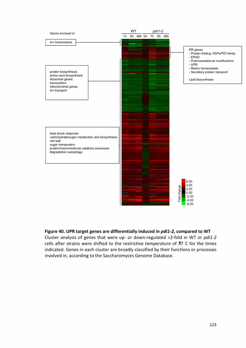
123
Figure 40. UPR target genes are differentially induced in pdi1-2, compared to WT Cluster analysis of genes that were up- or down-regulated >2-fold in WT or pdi1-2 cells after strains were shifted to the restrictive temperature of 37⁰ C for the times indicated. Genes in each cluster are broadly classified by their functions or processes involved in, according to the Saccharomyces Genome Database.

124
To further analyze the compensation by the UPR in pdi1-2, I looked at the UPR target
genes that were differentially upregulated in pdi1-2 compared with WT cells at each
time-point. Because some of the genes were also induced in WT cells due to heat
stress, only genes that were upregulated more than 1.5 fold in pdi1-2 over WT were
selected. This would indicate that these genes may be necessary for mitigating the
defects in pdi1-2. Since the response to heat stress was factored in, the list of UPR
genes induced in pdi1-2 due to its mutation was short. After 1 h shift to 37⁰C, two
distinct groups of genes were upregulated. The first group consists of genes involved
in redox homeostasis in the ER (MPD1, PDI1, EUG1, ERO1) while the other comprises
the HSPs (KAR2, SIL1, LHS1, JEM1, SCJ1). The roles of these genes were discussed in
Chapter 1. Interestingly, elevated expression of most of these genes (all except EUG1)
persisted even after 48 h shift to 37⁰C, indicating that they were specifically required
for viability of pdi1-2 (Fig. 41). In addition, three other genes, PMT3, DFR1 and SCS3
remained upregulated after 48h. PMT3 is an enzyme involved in O-mannosylation
(Girrbach and Strahl 2003); DFR1 is a dihydrofolate reductase which also has a role in
respiratory metabolism (Huang et al. 1992), while SCS3 encodes a protein required
for synthesis of inositol phospholipids (Hosaka et al. 1994) and is also required for ER
membrane synthesis during ER stress (Moir et al. 2012).
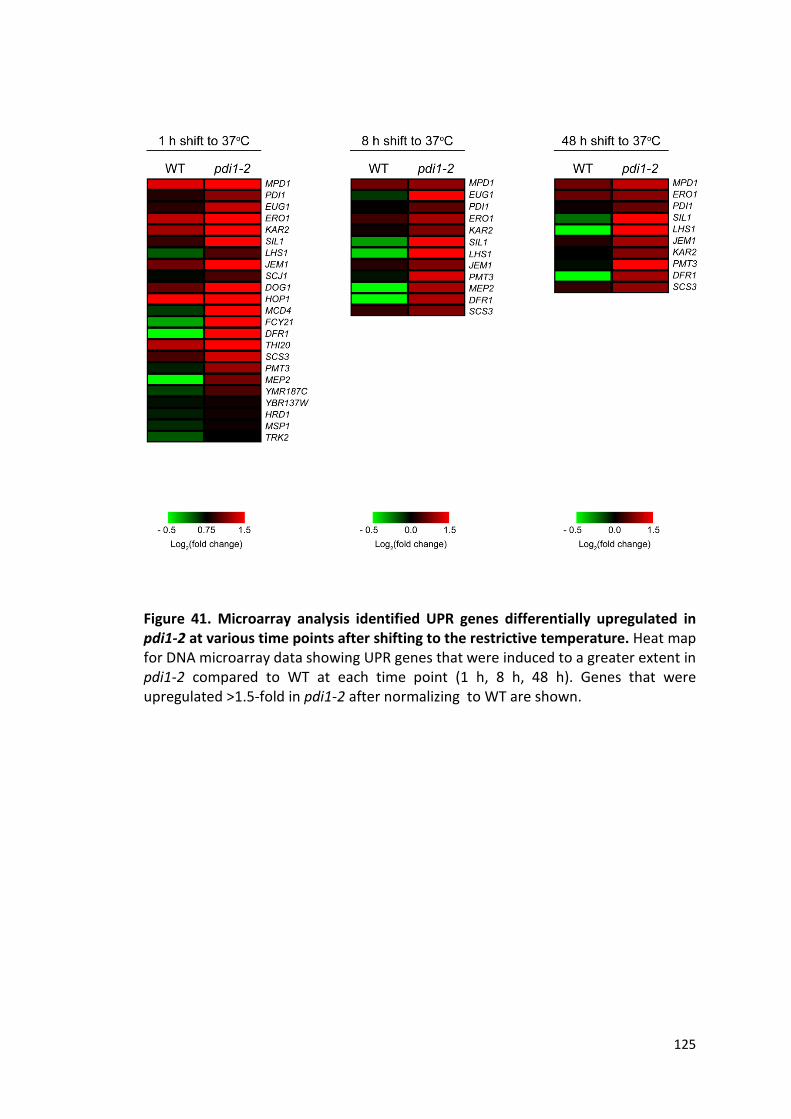
125
Figure 41. Microarray analysis identified UPR genes differentially upregulated in pdi1-2 at various time points after shifting to the restrictive temperature. Heat map for DNA microarray data showing UPR genes that were induced to a greater extent in pdi1-2 compared to WT at each time point (1 h, 8 h, 48 h). Genes that were upregulated >1.5-fold in pdi1-2 after normalizing to WT are shown.

126
4.9. Verification of microarray data by quantitative PCR
The microarray data was verified using quantitative real-time PCR (qPCR). Remaining
RNA from the microarray experiment was used for qPCR to ensure there were no
discrepancies arising from sample preparation. The expression level of eight genes
(KAR2, JEM1, LHS1, SIL1, PDI1, ERO1, MPD1, and PMT3) that remained upregulated
after 48 h was examined. This was expressed as fold change over the respective
expression level in WT or pdi1-2 strains before temperature shift. Fold change
measured using qPCR was generally higher than that obtained by microarray analysis,
likely due to the differences in sensitivity of the assays used. Nonetheless, there is
good correlation between the microarray and qPCR data, and the same general
trend of decreasing expression levels with time was observed (Fig. 42A). Some
differences were observed in WT cells between the two sets of data. Transcript
levels of LHS1, SIL1 and PMT3 after 8 h and 48 h temperature shifts were almost at
basal levels when measured using qPCR, but showed slight decreases in the
microarray analysis (Fig. 42A).
The expression levels of these genes after adaptation to stress (48 h shift to 37⁰C)
are shown in Fig. 42B. Expression of KAR2, PDI1, and MPD1 was markedly increased
in pdi1-2 compared to WT, as shown by both microarray and qPCR analysis.
Involvement of the other genes was less clear, and would require biochemical
analysis for verification.

127
Figure 42. Correlation between qPCR data with microarray data (A) Expression levels of key genes that were upregulated in pdi1-2 were examined using qPCR. These were expressed as fold changes over their respective levels at 0 h, and plotted together with fold changes obtained from the microarray data for WT and pdi1-2. (B) Fold changes of the key upregulated genes in pdi1-2 after 48 h shift to 37⁰C, expressed on a log2 scale.

128
4.10. Expression of KAR2 suppresses defects in pdi1-2Δire1
Interestingly, the Hsp70 chaperone KAR2 and its cofactors (SIL1 and LHS1) were
upregulated in pdi1-2Δire1 after it was shifted to the restrictive temperature, as
demonstrated by both microarray analysis (Fig. 41) and qPCR (Fig. 42B). Kar2p is a
major protein chaperone and is abundantly expressed in the ER. To test if Kar2p is
sufficient for cell viability, I expressed KAR2 in pdi1-2Δire1 using a centromeric
plasmid containing either its endogenous promoter (KAR2 level is increased two-
fold), or the TDH3 promoter (KAR2 is overexpressed). Surprisingly, KAR2 alone was
sufficient to suppress cell lethality in the absence of the UPR at both 30⁰C and 37⁰C.
However, overexpression of KAR2 did not improve cell viability over that of a modest
two-fold increase, suggesting that KAR2 did not need to be highly upregulated by the
UPR (Fig. 43)
Since PDI1 was one of the UPR targets that remained upregulated after a 48h shift to
37⁰C, the ts allele of PDI1 was overexpressed in pdi1-2Δire1 to test whether
increased levels of the mutant protein is sufficient to suppress its dysfunction.
Expression of Pdi1pts did not suppress cell lethality (Fig. 43), suggesting that the
defect in pdi1-2 could not be overcome by increasing dosage of the mutant protein.
One possible way that increased amount of Kar2p help the cells survive is by binding
accumulated unfolded proteins and preventing them from aggregating (Nishikawa et
al. 2001). A more intriguing alternative is that Kar2p functionally compensates for
the defect in pdi1-2. To elucidate the role of Kar2p, CPY maturation in pdi1-2Δire1
was analyzed when KAR2 was expressed. KAR2, in the absence of the UPR,
dramatically improved the folding of CPY, as seen from the increased amount of
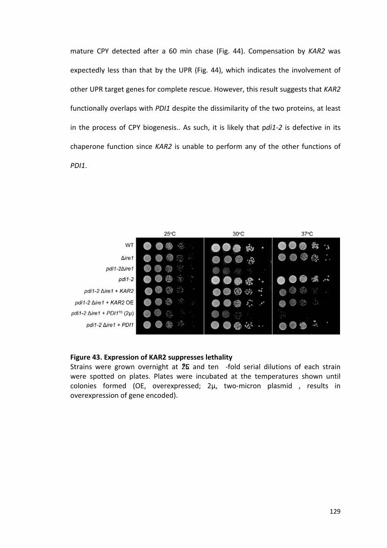
129
mature CPY detected after a 60 min chase (Fig. 44). Compensation by KAR2 was
expectedly less than that by the UPR (Fig. 44), which indicates the involvement of
other UPR target genes for complete rescue. However, this result suggests that KAR2
functionally overlaps with PDI1 despite the dissimilarity of the two proteins, at least
in the process of CPY biogenesis.. As such, it is likely that pdi1-2 is defective in its
chaperone function since KAR2 is unable to perform any of the other functions of
PDI1.
Figure 43. Expression of KAR2 suppresses lethality Strains were grown overnight at 25⁰C and ten -fold serial dilutions of each strain were spotted on plates. Plates were incubated at the temperatures shown until colonies formed (OE, overexpressed; 2µ, two-micron plasmid , results in overexpression of gene encoded).
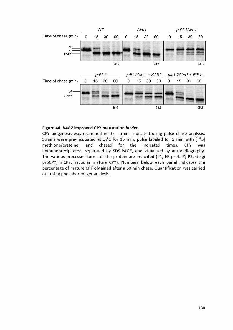
130
Figure 44. KAR2 improved CPY maturation in vivo CPY biogenesis was examined in the strains indicated using pulse chase analysis. Strains were pre-incubated at 37⁰C for 15 min, pulse labeled for 5 min with [ 35S] methione/cysteine, and chased for the indicated times. CPY was immunoprecipitated, separated by SDS-PAGE, and visualized by autoradiography. The various processed forms of the protein are indicated (P1, ER proCPY; P2, Golgi proCPY; mCPY, vacuolar mature CPY). Numbers below each panel indicates the percentage of mature CPY obtained after a 60 min chase. Quantification was carried out using phosphorimager analysis.

131
4.11. Co-expression of KAR2 interacting factors has no additive effect over KAR2 expression alone
The binding of Kar2p to its substrates is regulated by its interactions with other
Hsp40s (Kampinga and Craig 2010) and nucleotide exchange factors (NEFs). In the ER,
KAR2 shows genetic interactions with several of its cofactors - SCJ1, JEM1, SIL1 and
LHS1. To test whether these interacting partners were necessary to augment KAR2's
compensatory action in pdi1-2Δire1, I co-expressed these factors with KAR2, either
alone, or in combination. Cell growth was then assessed by spotting growth assay,
while CPY folding was monitored using pulse chase analysis.
Individual expression of SCJ1, JEM1, SIL1 and LHS1 in pdi1-2Δire1 was insufficient for
suppression of cell lethality and the defect in CPY folding at 37⁰C (Fig. 45). When co-
expressed with KAR2 alone or in combination with other cofactors, there was little
improvement in cell viability (Fig. 45) compared to KAR2 expression alone.
Expression of KAR2 resulted in ~60% mature CPY formed after a 60 min chase, while
co-expression of the cofactors gave rise to slightly lower amounts of mature CPY
(~50-60%). Therefore, CPY folding was similarly not better than what was achieved
when expressing KAR2 alone (Fig. 46). These data suggest that these genes play an
accessory role when induced by the UPR and serve to support KAR2, which appears
to be responsible for much of the compensation.
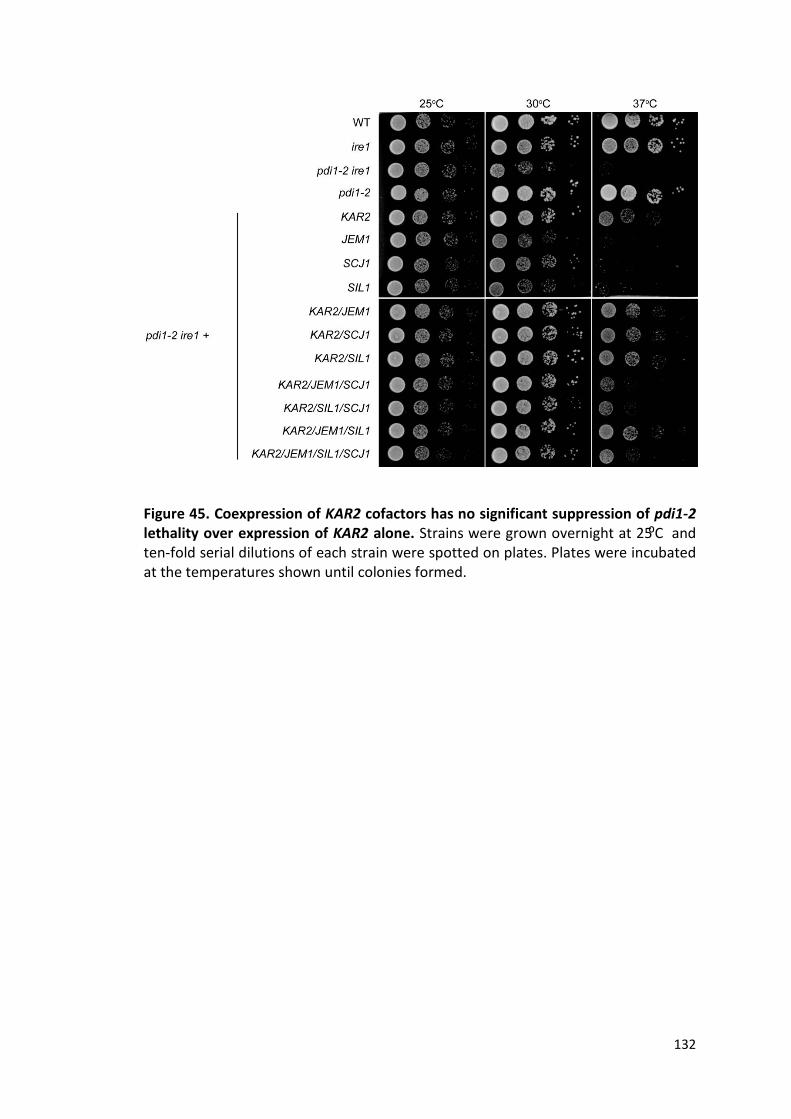
132
Figure 45. Coexpression of KAR2 cofactors has no significant suppression of pdi1-2 lethality over expression of KAR2 alone. Strains were grown overnight at 25⁰C and ten-fold serial dilutions of each strain were spotted on plates. Plates were incubated at the temperatures shown until colonies formed.

133

134
Figure 46. Coexpression of KAR2 with its cofactors does not improve CPY maturation over expression of KAR2 alone. (A) CPY biogenesis was examined in the strains indicated using pulse chase analysis. Strains were pre-incubated at 37⁰C for 15 min, pulse labeled for 5 min with [35S] methione/cysteine, and chased for the indicated times. CPY was immunoprecipitated, separated by SDS-PAGE, and visualized by autoradiography. The various processed forms of the protein are indicated (P1, ER proCPY; P2, Golgi proCPY; mCPY, vacuolar mature CPY). (B) Quantification of the percentage of mature CPY obtained after a 60 min chase for (A); quantification was carried out using phosphorimager analysis. Dotted line represents amount of mature CPY obtained in pdiΔire1 expressing KAR2; single represents coexpression of KAR2 with one other cofactor while combination indicates coexpression of KAR2 with various combinations of cofactors.

135
4.12. The Hsp70-like Lhs1p alone does not compensate for defects in pdi1-2Δire1
One of the genes upregulated by the UPR upon shifting pdi1-2Δire1 to the restrictive
temperature is LHS1, which encodes a Hsp70-like ER-resident chaperone. Lhs1p
shares 24% amino acid similarity to Kar2p (Baxter et al. 1996) and the two proteins
reciprocally regulate each other. Lhs1p acts as a specific nucleotide exchange factor
for Kar2p, while Kar2p activates the ATPase domain of Lhs1p in place of Hsp40s
(Steel et al. 2004). To test if Lhs1p plays a similar role as Kar2p in compensating for
the defects in pdi1-2, LHS1 was expressed in pdi1-2Δire1. Contrary to what was
observed for KAR2, pdi1-2Δire1 remained inviable at 37⁰C with LHS1 expression (FIg.
47A). Consequently, there was also no improvement in CPY biogenesis (Fig. 47C).
Next, to test if the coordinated regulation of Kar2p and Lhs1p was required for
complete compensation, the two genes were coexpressed. Coexpression of LHS1 did
not improve cell viability (Fig. 47A) or CPY maturation (Fig. 47C) beyond that of KAR2
alone, suggesting that the actions of LHS1 and KAR2 were not coupled in this case.
Since interaction between the two proteins are required for normal function in vivo
(Steel et al. 2004), this result showed that upregulation of both genes by the UPR
may serve to restore general ER homeostasis. However, only the increased
expression of KAR2, but not that of LHS1, is defect-specific.
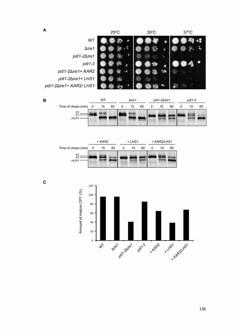
136
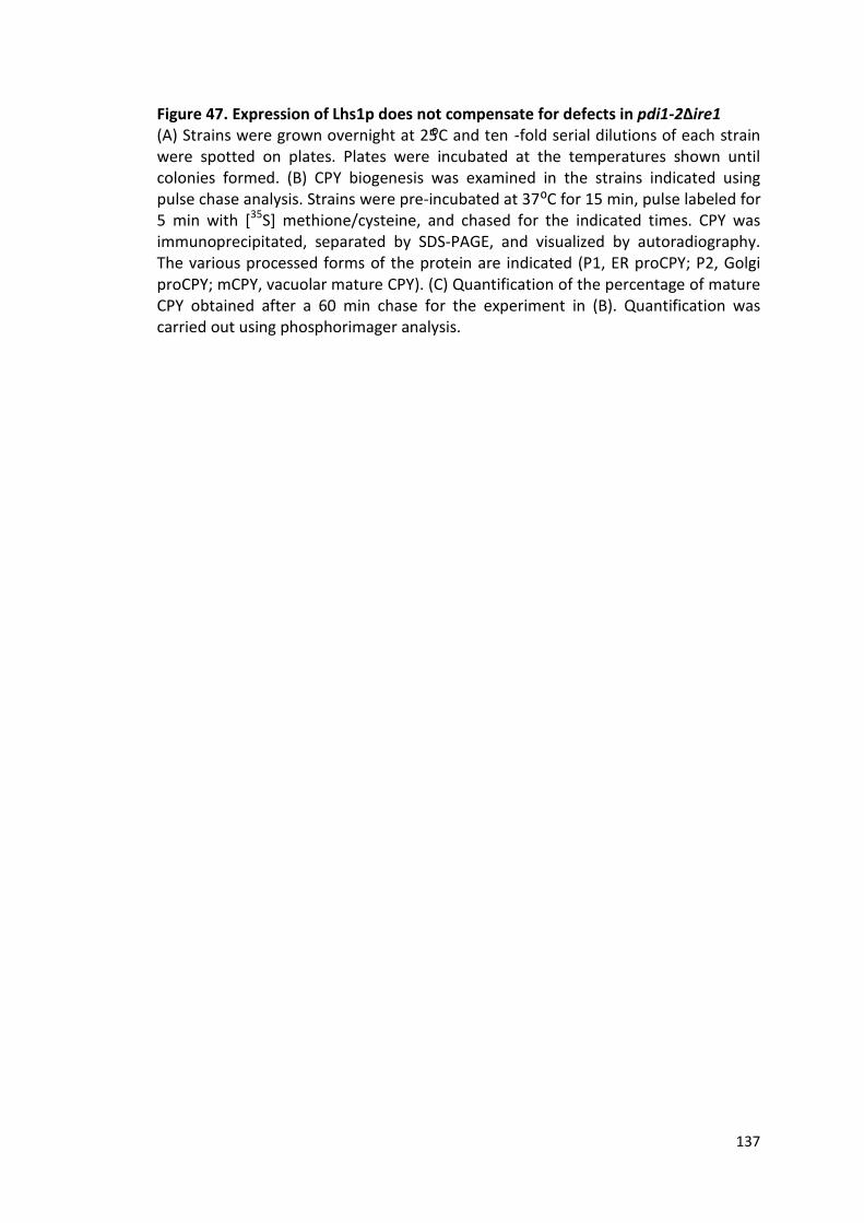
137
Figure 47. Expression of Lhs1p does not compensate for defects in pdi1-2Δire1 (A) Strains were grown overnight at 25⁰C and ten -fold serial dilutions of each strain were spotted on plates. Plates were incubated at the temperatures shown until colonies formed. (B) CPY biogenesis was examined in the strains indicated using pulse chase analysis. Strains were pre-incubated at 37⁰C for 15 min, pulse labeled for 5 min with [35S] methione/cysteine, and chased for the indicated times. CPY was immunoprecipitated, separated by SDS-PAGE, and visualized by autoradiography. The various processed forms of the protein are indicated (P1, ER proCPY; P2, Golgi proCPY; mCPY, vacuolar mature CPY). (C) Quantification of the percentage of mature CPY obtained after a 60 min chase for the experiment in (B). Quantification was carried out using phosphorimager analysis.

138
4.13. KAR2 and pdi1ts work synergistically in pdi1-2Δire1
PDI1 is an essential protein in the yeast ER lumen that is critical for protein folding
through its ability to catalyze disulfide bond breakage and formation. Unsurprisingly,
PDI1 is one of the genes upregulated by the UPR in pdi1-2Δire1 at the restrictive
temperature, as the cells respond to what is perceived as an insufficiency of PDI1
function. To study the effect of an increased expression of the ts PDI1 allele, I
overexpressed PDI1ts. On its own, it was insufficient to suppress the lethality of pdi1-
2Δire1 (Fig. 48A), but was able to improve CPY processing to a level comparable to
that of KAR2 (Fig. 48C). This could be because Pdi1pts is not entirely dysfunctional at
37⁰C, evident from the small percentage of mature CPY obtained after a 60 min
chase. Overexpression of Pdi1pts could, in the short term (e.g. during the short shift
during pulse chase analysis), improve cellular defects like CPY folding; it could not
however, restore ER homeostasis at steady state, as demonstrated by the inviability
of cells expressing PDI1ts in the growth assay.
If KAR2 is specifically compensating for the defect in the chaperone function of pdi1ts,
coexpression of KAR2 and pdi1ts may be expected to alleviate the defects observed
in pdi1-2Δire1. Indeed, pdi1-2Δire1 expressing both KAR2 and pdi1ts was viable at
37⁰C, and grow at a similar rate as Δire1 cells (Fig. 48A). Interestingly, coexpression
of both genes also fixed the defect in CPY maturation to the same extent as when
the UPR was induced, with 85% mature CPY obtained after a 60 min chase (Fig. 48C).
Taken together, this suggests that KAR2 and pdi1ts work synergistically in pdi1-2Δire1
and demonstrates an uncharacterized functional interaction between the two
proteins.
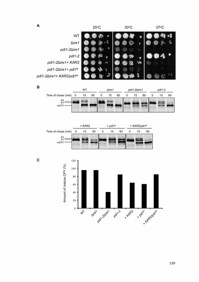
139

140
Figure 48. Synergy between KAR2 and pdi1ts (A) Strains were grown overnight at 25⁰C and ten -fold serial dilutions of each strain were spotted on plates. Plates were incubated at the temperatures shown until colonies formed. (B) CPY biogenesis was examined in the strains indicated using pulse chase analysis. Strains were pre-incubated at 37⁰C for 15 min, pulse labeled for 5 min with [35S] methione/cysteine, and chased for the indicated times. CPY was immunoprecipitated, separated by SDS-PAGE, and visualized by autoradiography. The various processed forms of the protein are indicated (P1, ER proCPY; P2, Golgi proCPY; mCPY, vacuolar mature CPY). (C) Quantification of the percentage of mature CPY obtained after a 60 min chase for the experiment in (B); quantification was carried out using phosphorimager analysis.

141
4.14. Characterizing the functional interaction between KAR2 and pdi1ts
To characterize the functional interaction between KAR2 and PDI1, I made point
mutants of KAR2 and expressed these in pdi1-2Δire1. KAR2(G417S) has a point
mutation in the ATPase domain while KAR2(T473F) and KAR2(P515L) each has a
point mutation in the substrate-binding domain (SBD). These mutations have been
characterized; ATPase domain mutants are defective in protein translocation into
the ER, while SBD mutants are defective in ERAD (Baxter et al. 1996, Holkeri et al.
1998, Kabani et al. 2003).
Expression of mutant forms of KAR2 did not suppress cell lethality, as demonstrated
by the lack of cell growth at 37⁰C and at a lower temperature of 34⁰ C (Fig. 49). In
addition, KAR2(G417S), an ATPase mutant, abolished cell growth even at 30⁰C ,
suggesting that it could have a dominant negative effect. In contrast, compensation
by KAR2 was more apparent at 34⁰C than at 37⁰C, presumably due to less
devastating defects in pdi1-2Δire1 at the lower temperature (Fig. 49). Together,
these suggest that a fully functional KAR2 is required to rescue pdi1-2Δire1.

142
Figure 49. A functional KAR2 is required for suppression of cell lethality in pdi1-2Δire1. Strains were grown overnight at 25⁰C and ten -fold serial dilutions of each strain were spotted on plates. Plates were incubated at the temperatures shown until colonies formed.
As KAR2 is an essential gene, known temperature-sensitive kar2 mutants were used
to create pdi1-2kar2 double mutants for further analysis of their genetic interaction.
kar2-113, kar2-203 and kar2-159 are ATPase mutants, of which kar2-203 and kar2-
159 are ts at temperatures of 30⁰C or higher and kar2-113 is ts at 34⁰C or higher (Fig.
50). kar2-1 and kar2-133 are SBD mutants and are ts at 37⁰C (Fig. 50).
Since all the kar2 mutants are ts at 37⁰C, the d ouble mutants were examined for
growth at lower temperatures. At 34⁰C, double mutants pdi1-2kar2-113, pdi1-2kar2-
1 and pdi1-2kar2-133 displayed synthetic lethality even in the presence of the UPR,
while their growth were significantly slower at 30⁰C (Fig. 50). The negative genetic
interaction observed in the double mutants suggests that KAR2 and pdi1ts work in
compensatory pathways. This result also indicates that UPR-induced functional KAR2
is required for suppression of defects in the pdi1 mutant.

143
Figure 50. Mutations in pdi1-2 and kar2 are synthetic lethal Strains were grown overnight at 25⁰C and ten -fold serial dilutions of each strain were spotted on plates. Plates were incubated at the temperatures shown until colonies formed.

144
4.15. Discussion
Protein folding is an essential and critical function of the cell, which explains the
extensive resources deployed to aid in functional protein maturation. Unproductive
protein folding can lead to protein aggregation and toxicity, which can result in
disease states like Parkinson's or Huntington's. Networks of chaperones and folding
factors cooperate in this process, while surveillance mechanisms ensure stringent
protein quality control and efficient degradation of rogue proteins.
In the ER, events in the secretory pathway are closely monitored and perturbations
leading to accumulation of unfolded/misfolded proteins activate the UPR. In this
study, an elegant genetic system was devised to attenuate the compensatory
mechanisms of the UPR in the face of physiologic stress induced by disruptions to
gene functions. This system allowed us to study the effect of a loss of gene function
without complications arising from the UPR response. In addition, it provided an
invaluable tool for examining the UPR under different physiologic stressors, UPR
kinetics, and the mechanisms through which the UPR compensates for cellular
defects. This is important because the physiologic UPR is more relevant to
understanding disease pathology, while traditionally, studies often used chemical-
induced stress (e.g. addition of DTT or Tm) which strongly activates the UPR.
For the first time, this study provided insight into how the UPR is modulated with
time after exposure to stress. Shifting pdi1-2 to the restrictive temperature led to
rapid activation of the UPR within the first hour, followed by subsequent damping of
the stress response as the cells adapt and reach a new homeostatic state (Fig. 40).
This is analogous to the well-studied heat shock response. This UPR kinetics suggests

145
that the initial stage of UPR activation is non-discerning, with massive upregulation
of many genes to maximize the chance of cell survival. With time, defect-specific
compensation occurs and redundant UPR targets are returned to basal levels. This
ensures that the cell is not overwhelmed by its own stress response and the
accompanying indirect effects after ER stress is alleviated, and corroborates the
observation that attenuation of the yeast UPR is essential for cell survival upon
recovery of ER function (Chawla et al. 2011).
In yeast, the IRE1/HAC1 signaling pathway upregulates close to 400 UPR target genes.
The mutant pdi1-2 is synthetic lethal with the UPR which means the UPR needs to be
constitutively activated for viability, yet only a small subset of UPR targets remains
upregulated at steady state (Fig. 41). Other studies in the laboratory on knockout
strains Δlhs1, Δscj1, and Δalg5 showed that the UPR output for each of these strains
differ dramatically. Many UPR target genes are induced in Δlhs1 but only a handful
are induced in Δalg5 (Thibault et al. 2011). How the cell regulates UPR output in
response to different stressors remains an open question, but it is clear that
additional regulation at the Ire1p/Hac1p levels and/or other signaling mechanisms
stemming from the ER are required to produce the diverse output shown in these
mutants. Nonetheless, the UPR demonstrates a surprising plasticity in dealing with
the specific needs of the cell during different ER stresses.
The amazing ability of the UPR to mask phenotypic expression was demonstrated in
this study using compensation-attenuated genetics. In pdi1-2, eliminating the UPR
rendered the cells inviable, revealing the catastrophic effect of the pdi1 mutation.
UPR activation completely masked this growth defect. This suggests that mild

146
phenotypes, or the lack thereof, in mutants routinely generated for research may be
a result of masking by the accompanying compensatory responses and are not
indicative of the contribution of these genes in the normal cell environment.
Disabling these compensatory mechanisms would therefore improve genetic analysis
and functional studies.
In addition, these compensatory pathways also provide an explanation for the
seemingly large number of non-essential genes in the yeast genome (~80%), despite
the fact that many curated genes have important known functions and do not show
functional overlap or redundancy with other genes. Indeed, a wide range of mutants
with non-essential genes deleted display synthetic lethality with the UPR (Costanzo
et al. 2010), underscoring the importance of the broad scope of UPR compensation
in maintaining cellular homeostasis.
Analysis of the pdi1-2 mutant provided direct evidence that the UPR is able to
alleviate ER stress by fixing specific defects. A dysfunction in the folding factor PDI1
led to upregulation of ER chaperones which restored CPY folding. On the other hand,
a recent study demonstrated that when lipid equilibrium is severely disrupted in an
Δopi3 mutant, the UPR did not restore or modify lipid composition but instead fixed
the deficiencies in protein biogenesis and ERAD, and maintained membrane
morphology (Thibault et al. 2012). In this case, the UPR compensates by remodeling
the entire protein homeostasis network to ensure cell survival rather than fixing lipid
disequilibrium. This demonstrates the versatility of the UPR, and its ability to provide
broad compensation to support cellular robustness.
147
In this study, the conditional synthetic lethal mutant, pdi1-2, was chosen for further
analysis. PDI1 is a relatively well-characterized gene but mutant alleles of PDI1 are
rarely identified from genetic screens possibly because it performs such important
functions in the cell. As such, research on PDI1 was mainly carried out in vitro, or
relied on the use of expressed truncated or mutated PDI1 constructs. The only other
known allele, pdi1-1, is specifically defective in its interaction with the mannosidase
Htm1p, and affects ERAD of glycosylated misfolded proteins (Gauss et al. 2011).
pdi1-2 provides a valuable tool for us to dissect the in vivo functions of PDI1 under
normal conditions and also to study the UPR response to a partial dysfunction of an
essential gene.
Based on the assays used in the present study, pdi1-2Δire1 exhibited folding defects
in endogenous proteins CPY and Gas1p (Fig. 23), an ERAD defect in CPY* (Fig. 27),
and cell lethality at the restrictive temperature of 37⁰C (Fig. 22). Activation of the
UPR in pdi1-2 restored cell growth and protein biogenesis, but not ERAD. It is
surprising that the ERAD defect was not fixed by the UPR, since many genes involved
in ERAD are targets of the UPR (Travers et al. 2000). Also, the UPR is known to have
the ability to fix defects in ERAD, as shown by restoration of CPY* degradation in
Δopi3 and Δcho2 strains upon UPR activation (Thibault et al. 2012). However these
ERAD genes were not specifically upregulated in pdi1-2, as shown by microarray
analysis. It is possible that the UPR prioritizes its compensation based on the needs
of the cell. Since protein folding is essential to the cell and is the more severe defect
in pdi1-2, it was imperative that protein biogenesis be restored, which would have
contributed greatly to cell survival. ERAD of CPY* was recently shown to require the

148
catalytic sites of Pdi1p (Grubb et al. 2012), which are intact in pdi1-2. However, there
could be an unknown function defective in pdi1-2 that may be required for ERAD,
and perhaps this function cannot be compensated for by the UPR.
A surprising finding from this study is that other PDI family members (MPD1, MPD2,
EUG1 and EPS1) were dispensable for suppression of cell lethality and defects in CPY
folding in pdi1-2 (Fig. 36). Even though MPD1, MPD2 and EUG1 are UPR targets and
MPD1 was upregulated in pdi1-2, activation of the UPR was sufficient for cell viability
and restoring CPY folding even when all other PDI family members were deleted.
This result provided insight into the PDI family of proteins. PDI family members are
classified according to the presence of the conserved thioredoxin-like domain.
However, the crystal structure of MPD1, which is the only member that can suppress
cell lethality in a pdi1-deleted strain devoid of other PDI family members, showed
that domain orientations and surface properties can vary greatly between PDI family
proteins (Vitu et al. 2008). These properties can affect substrate specificity and
interactions with other proteins in functional modules, suggesting that the
physiologic functions of PDI family members may show little overlap. Except for
MPD1 which seemed capable of carrying out the essential oxidative function of PDI1
when overexpressed, there appears to be little redundancy among the homologues
(Norgaard et al. 2001). Paradoxically, MPD1 is expressed at a very low level in the
cell -- 830 molecules per cell (Ghaemmaghami et al. 2003), which may explain why
despite its induction as shown by microarray analysis and qPCR, the deletion of
MPD1 had no bearing on compensation by the UPR.
149
The role of PDI1 as a chaperone in vivo is unclear, since studies implicating PDI1 as a
chaperone were all in vitro refolding assays involving mammalian PDI1 (Cai et al.
1994, Song and Wang 1995). It was intriguing to observe that the HSPs were some of
the key genes to be upregulated by the UPR in pdi1-2 (Fig. 41). These proteins are
neither similar in sequence and structure to Pdi1p, nor do they catalyze oxidative
protein folding. However, they are important chaperones involved in protein folding,
and their upregulation suggest that pdi1-2 may be defective in its chaperone
function. Expression of KAR2, but not pdi1ts, was sufficient to suppress inviability of
pdi1-2Δire1 and improve CPY maturation, albeit to a lesser extent than the UPR. (Fig.
43,44). Surprisingly, coexpression of KAR2 and pdi1ts restored cell viability and CPY
maturation rate to that comparable with the UPR, suggesting a synergistic functional
interaction between the two genes (Fig. 48). This also highlighted the fact that the
HSPs are fixing a specific defect in pdi1-2Δire1, and not just binding unfolded
proteins to keep them soluble to prevent aggregation and its associated toxicity.
Functional cooperation between PDI1 and KAR2 during protein folding has not been
characterized in vivo, though a study suggested the cooperation of KAR2 and PDI1 in
the refolding of denatured and reduced antibodies in vitro (Mayer et al. 2000). Here,
we established the genetic and functional interaction between PDI1 and KAR2 in vivo.
No physical interaction between Kar2p and Pdi1p has been reported. It is still unclear
whether this cooperation occurs under normal conditions, or only as part of the
compensation mechanism during stress. We postulate a model for CPY folding in the
cell. As the precursors of CPY get translocated into the ER, Kar2p binds to the
precursors and aids in the early stages of CPY folding. Kar2p subsequently releases

150
the partially folded CPY, which is then bound by Pdi1p. This helps to free Kar2p so
that it can then bind to newly-translocated polypeptides. Pdi1p helps CPY to fold into
its native conformation and catalyzes the formation of the correct disulfide bonds. In
pdi1-2 where the chaperone function of pdi1pts is defective, UPR induction further
increases the concentration of the abundantly-expressed Kar2p. There is now extra
Kar2p to bind and aid CPY folding till it reaches its native conformation. Pdi1pts is not
oxidation-deficient and continues to catalyze disulfide bond formation to produce
mature CPY. In the latter case, Pdi1pts acts as a passive "placeholder" (Kosuri et al.
2012) rather than an active folding chaperone. This means that the chaperone and
redox functions of Pdi1p can be uncoupled, and that the chaperone function is non-
essential only because the UPR is induced.
This notion is supported by two studies. It was shown that expression of the yeast
PDIa' domain from the endogenous PDI1 promoter was sufficient for near-WT
growth rate in a Δpdi1 strain (Xiao et al. 2004). A single catalytic domain is unlikely to
have any chaperone activity, but is capable of performing the essential oxidative
function. Though it was not shown in this particular study, it is very likely that the
UPR is actively compensating for the loss of chaperone function. A recent study
provided the mechanism for the above observation. Using single molecule atomic
force microscope, the authors demonstrated that the PDIa catalytic domain formed
mixed disulfides with the cysteines in a folding substrate without impeding protein
folding. When the substrate reached a near-native conformation, PDIa then
catalyzed native disulfide bond formation, thus acting as a passive "placeholder"
during protein folding (Kosuri et al. 2012). However, unlike the in vitro condition,

151
spontaneous protein folding is usually futile in the crowded environment of the ER
where the concentration of unfolded/folding proteins is high. Chaperones, like the
HSPs, thus come into play to aid in productive protein folding in vivo.
The ability of the HSP chaperone system to buffer against defects due to dysfunction
within the PDI family increases cellular robustness. Robustness is defined as "a
property that allows a system to maintain its functions against internal and external
perturbations" (Kitano 2004). As protein folding in the secretory pathway is a
fundamental process, buffering between two different networks that are part of this
process helps contribute to overall ER homeostasis. It is likely that when the cells
experience mild stress, buffering between networks of folding factors performing
different functions ensures protein folding function is maintained. When the severity
of ER stress requires the UPR to be activated, this form of buffering could act as the
initial defence before UPR target genes are fully expressed and able to carry out
their functions. Thus, the observation from this study that HSPs buffer against a
lethal pdi1 dysfunction alludes to multiple levels of regulation contributing to cellular
robustness.
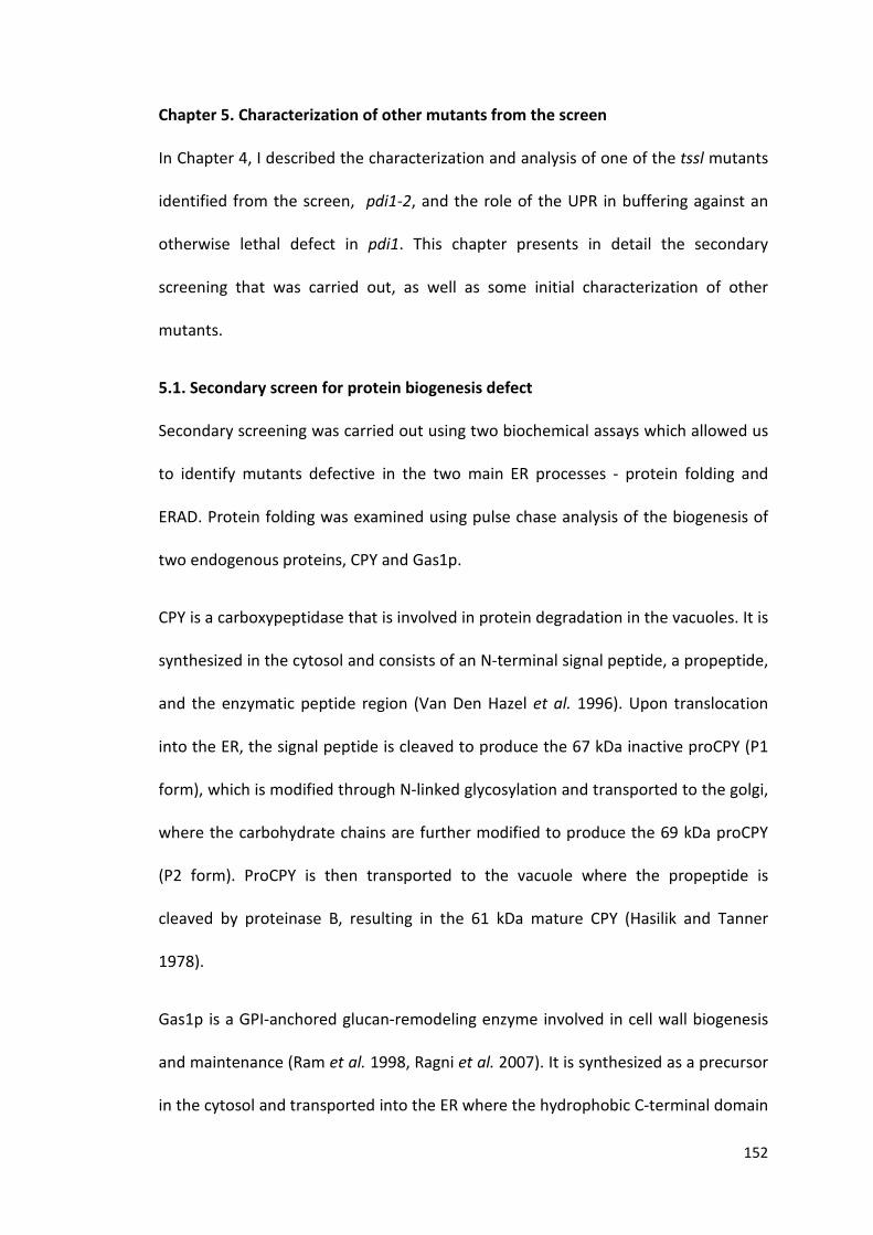
152
Chapter 5. Characterization of other mutants from the screen
In Chapter 4, I described the characterization and analysis of one of the tssl mutants
identified from the screen, pdi1-2, and the role of the UPR in buffering against an
otherwise lethal defect in pdi1. This chapter presents in detail the secondary
screening that was carried out, as well as some initial characterization of other
mutants.
5.1. Secondary screen for protein biogenesis defect
Secondary screening was carried out using two biochemical assays which allowed us
to identify mutants defective in the two main ER processes - protein folding and
ERAD. Protein folding was examined using pulse chase analysis of the biogenesis of
two endogenous proteins, CPY and Gas1p.
CPY is a carboxypeptidase that is involved in protein degradation in the vacuoles. It is
synthesized in the cytosol and consists of an N-terminal signal peptide, a propeptide,
and the enzymatic peptide region (Van Den Hazel et al. 1996). Upon translocation
into the ER, the signal peptide is cleaved to produce the 67 kDa inactive proCPY (P1
form), which is modified through N-linked glycosylation and transported to the golgi,
where the carbohydrate chains are further modified to produce the 69 kDa proCPY
(P2 form). ProCPY is then transported to the vacuole where the propeptide is
cleaved by proteinase B, resulting in the 61 kDa mature CPY (Hasilik and Tanner
1978).
Gas1p is a GPI-anchored glucan-remodeling enzyme involved in cell wall biogenesis
and maintenance (Ram et al. 1998, Ragni et al. 2007). It is synthesized as a precursor
in the cytosol and transported into the ER where the hydrophobic C-terminal domain

153
is removed and replaced by a GPI-anchor (Nuoffer et al. 1991). In addition, Gas1p
undergoes N-linked and O-linked glycosylation as it passes through the secretory
pathway (Doering and Schekman 1996).
The biogenesis of these two proteins can be tracked based on the gel mobility of
their precursors and mature forms. Defects along steps in the secretory pathway will
manifest as accumulation of the ER forms of the proteins, perhaps due to improper
folding or a block in the transport between organelles. Since both proteins are
modified post-translationally by glycosylation, defects in glycosylation can also be
detected by the presence of protein bands of lower molecular weight. This assay
served as a preliminary screen for mutants of interest that may have defects in the
secretory pathway.
To ensure that any phenotype observed was due to the mutation in the strain and
not due to secondary effects as a result of heat stress, the mutants were only shifted
to 37⁰C for 15 min before pulse chase analysis was carried o ut. These tssl
(temperature-sensitive synthetic lethal) mutants were isolated from the screen using
the colony colour assay (Section 3.2.1). Only tssl1 to tssl12 are shown in Figure 51 as
all the mutants did not show significant protein processing defects for both CPY and
Gas1p, with the exception of tssl3 (Fig. 51B). tssl3 was identified as pdi1-2Δire1 and
has been discussed in Chapter 4. All other mutants were able to process majority of
the precursors of CPY and Gas1p to the mature form after a 30 min chase, at a rate
similar to Δire1 cells (Fig. 51A,C). A slight translocation defect for CPY precursors was
observed in some of the mutants (tssl2, tssl6, tssl7, tssl9, tssl10). For some of the
mutants (tssl4 - tssl12), the transport of Wsc1p was examined concurrently. Wsc1p

154
was transported out of the ER in all of these strains, suggesting that general
trafficking was not defective (Fig. 51C).
The lack of phenotype could be because a 15 min preincubation at the restrictive
temperature was insufficient to disrupt the functions of the ts alleles in these mutant
strains. To test that, mutant strains were shifted to 37⁰C for 1 h before analysis.
Unfortunately, with the extended pre-incubation, ER forms of CPY and Gas1p
accumulated in all strains, including the parental strains. This prevented further
analysis since the control strains (WT and Δire1) appeared to be defective in protein
processing after a prolonged shift to 37⁰C. This was an unexpected result and
suggests that there could be inherent mutations in these strains which did not
manifest when the strains were cultured at ambient temperature (25⁰C) or at their
normal growth temperature (30⁰C).

155
Figure 51. Secondary screen for defects in biogenesis of CPY and Gas1p (A-C) Wild-type, Δire1 and tssl mutant strains were pre-incubated at 37°C for 15 min, pulse labeled for 5 min with [35S] methionine/cysteine, and chased for 30 min. CPY and Gas1p were immunoprecipitated, separated by SDS-PAGE and visualized by autoradiography. The various processed forms of the proteins are indicated (CPY: P1, ER proCPY; P2, Golgi proCPY; mCPY, vacuolar mature CPY. Gas1p: PM, plasma membrane).

156
As described in Chapter 3, the genetic screen was repeated using different strategies
-- counter selection using 5-FOA and direct screening for temperature sensitivity.
Mutants isolated from the 5-FOA and ts screens were shifted to the restrictive
temperature of 37⁰C for 1 h before pulse chase analysis was carried out. This is to
ensure sufficient time for inactivation of the ts allele, yet to minimize the stress
duration to avoid non-specific, secondary effects. Cells were then pulse-labeled for 5
min and cold-chased for 30 min. Out of the eight mutants analyzed, all displayed
normal processing for CPY (Fig. 52) and Gas1p (Fig. 53) except tssl36. There was
accumulation of the ER (P1) form of CPY after 30 min, even though a proportion of
proCPY was processed to the mature form (Fig. 52). Gas1p processing was more
severely affected, with almost no mature Gas1p detected after 30 min (Fig. 53). This
suggests that most of the mutants do not appear to have a defect in protein folding,
other than tssl36.

157
Figure 52 . Bioprocessing of CPY in mutant strains Wild-type, Δire1 and mutant strains were pre-incubated at 37°C for 1 h, pulse labeled for 5 min with [35S] methionine/cysteine, and chased for 30 min. CPY was immunoprecipitated, separated by SDS-PAGE and visualized by autoradiography. The various processed forms of the proteins are indicated (CPY: P1, ER proCPY; P2, Golgi proCPY; mCPY, vacuolar mature CPY).

158
Figure 53. Bioprocessing of Gas1p in mutant strains. Wild-type, Δire1 and mutant strains were pre-incubated at 37°C for 1 h, pulse labeled for 5 min with [35S] methionine/cysteine, and chased for 30 min. Gas1p was immunoprecipitated, separated by SDS-PAGE and visualized by autoradiography. The various processed forms of the proteins are indicated (PM, plasma membrane).

159
5.2. Secondary screen for ERAD defect
Since most of the mutants did not exhibit a defect in CPY and Gas1p biogenesis, I
screened the mutants for ERAD defects. This was carried out by examining the
degradation kinetics of a misfolded variant of CPY, CPY*. CPY* contains a point
mutation that results in an amino acid change from glycine to arginine at position
255 (G255R), located in a region highly conserved among serine proteases. It is
retained in the ER and gets degraded in the vacuole (Finger et al. 1993).
Mutants transformed with a plasmid expressing CPY* were shifted to 37⁰C for 1 h,
pulsed with radioisotope for 10 min and chased for 60 min. Out of the eight mutants
tested, four (tssl29, tssl31, tssl34, tssl35) did not have significant ERAD defects. The
degradation rate of CPY* in these mutants were similar to WT or Δire1 cells (Fig. 54).
Δire1 showed a slight stabilization of CPY* as expression of a misfolded protein
caused a degree of stress in the absence of the UPR. These four mutants were also
not impaired in protein folding, suggesting that their defects were either beyond
what could be detected by the assays used, or that they were not thermolabile and
required an extended shift to the restrictive temperature before a phenotype could
be observed.

160
Figure 54. Secondary screen for ERAD mutants using CPY* WT, Δire1, and tssl mutants expressing CPY* were pre-incuated at 37⁰C for 1h, pulse-labeled with [35S] methione/cysteine for 10 min, and chased for the times indicated. Anti-HA-precipitated CPY* was resolved by SDS-PAGE. Quantification was carried out using phosphorimager analysis.
Three of the mutants examined (tssl32, tssl33, tssl36) showed stabilization of CPY*,
indicating a defect in the clearance of misfolded proteins (Fig. 55). Interestingly, one
of the mutants (tssl30) showed enhanced clearance of CPY*compared to both WT
and Δire1 cells. In this mutant, only 10% of CPY* remained after a 30 min chase,
compared to ~40% in WT and Δire1 cells (Fig. 55). tssl30, tssl32, tssl33 appeared to
be ERAD-specific mutants since folding was not affected in these strains. tssl36 on
the other hand, had defects in both protein processing and ERAD, suggesting that
the mutation disrupts both critical functions of the ER.
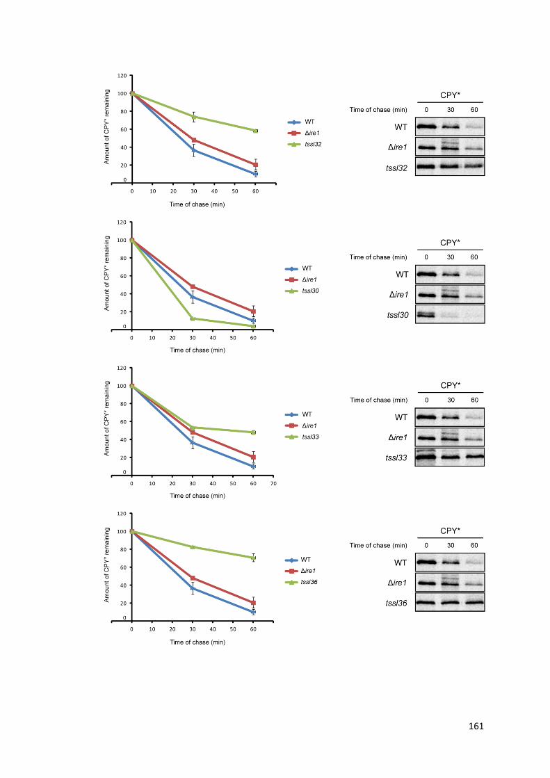
161

162
Figure 55. CPY* degradation is partially affected by putative ERAD mutants isolated from the screen. Wild-type, Δire1 and mutant strains were pre-incubated at 37°C for 1 h, pulse labeled for 10 min with [35S] methionine/cysteine, and chased for 60 min. Immunoprecipitation of CPY* was performed using anti-HA monoclonal antibody and normalized by total TCA precipitable counts. Proteins were separated by SDS-PAGE, visualized by autoradiography, and quantified by phosphorimager analysis. Results are depicted in the graph above.

163
5.3. tssl36 is a tip20 mutant
tssl36 was interesting as it showed a folding defect as well as a strong ERAD defect,
similar to pdi1-2. At the permissive temperature of 25⁰C, processing of both CPY and
Gas1p proceeded normally, and majority of the precursors formed the mature forms
after a 30 min chase (Fig. 56A). As shown previously, biogenesis of both proteins
were partially disrupted at 37⁰C. About half of proCPY and almost all of the Gas1p
precursor remained in the ER after 30 min (Fig. 56A). Biosynthesis of another
secreted protein, invertase, was similarly affected at 37⁰ C. The signal sequence of
the precursor of invertase is cleaved in the ER and it is then core-glycosylated. Upon
transport to the golgi, further elongation of the oligosaccharide chains occurs before
the mature protein is secreted out of the cell (Gillece et al. 1999). There was
increased accumulation of invertase precursors in the ER and/or golgi in tssl36 after
a 30 min chase, whereas only mature invertase was detected in WT and Δire1 cells
(Fig. 56B). This suggests that protein folding or transport along the secretory
pathway is compromised in this mutant.
164
Figure 56. Protein processing of CPY, Gas1p and invertase are affected in tssl36 (A) tssl36 was pre-incubated at 25⁰C or 37°C for 1 h, pulse labeled for 5 min with [35S] methionine/cysteine, and chased for 30 min. CPY and Gas1p were immunoprecipitated, separated by SDS-PAGE and visualized by autoradiography. The various processed forms of the proteins are indicated (CPY: P1, ER proCPY; P2, Golgi proCPY; mCPY, vacuolar mature CPY. Gas1p: PM, plasma membrane). (B) Wild-type, Δire1 and tssl36 were pre-incubated at 37°C for 1 h, pulse labeled for 5 min with [35S] methionine/cysteine, and chased for 30 min. Invertase was immunoprecipitated using anti-HA antibody, separated by SDS-PAGE and visualized by autoradiography. (inv, invertase).
While carrying out the biochemical analysis, tssl36 was also transformed with the
YCp50 yeast genomic library in order to clone the gene that is defective in this
mutant. A total of 70 000 transformants were obtained, of which six of them grew at
37⁰C. Plasmids from these colonies were extracted and re-transformed into the
mutant to verify complementation at 37⁰C. Out of these, only one complementing

165
clone was obtained. The complementing plasmid was sequenced and found to
encode the following genes (Table 8).
Table 8. List of genes encoded by the complementing plasmid
gDNA from the mutant was extracted and these genes were PCR-amplified and
sequenced. Sequencing confirmed a point mutation in TIP20, which resulted in an
amino acid change from glutamic acid to lysine at position 644 of the protein
sequence.
TIP20 encodes a membrane protein that is essential for trafficking from the golgi
complex to the ER (Lewis et al. 1997) and also prevents retrograde fusion of COPII
vesicles with the ER (Kamena and Spang 2004). It forms a vesicle tethering complex
with Dsl1p, and is involved in the fusion of COPI vesicles to the ER membranes
(Tripathi et al. 2009). How protein folding and/or transport is affected in this tip20

166
mutant is unclear, since the passage of maturing proteins is generally anterograde. It
is possible that ER-resident proteins, which tend to cycle between the ER and golgi
compartments, fail to be retrieved from the golgi back to the ER in this tip20 mutant.
This could result in decreased levels of folding enzymes and factors in the ER which
indirectly affects protein folding.

167
5.4. tssl30 causes enhanced ERAD of CPY*
An intriguing ERAD mutant identified from the screen was tssl30. It exhibited a
significantly enhanced rate of CPY* degradation compared to WT or Δire1 cells (Fig.
55) This type of ERAD mutant has not been observed before, as most ERAD mutants
resulted in reduced degradation of CPY*. As the secondary screen was carried out
after a 1h incubation at 37⁰C, I examined the degradation kinetics of CPY* in tssl30
after shifting to 37⁰C for different length of time. This was done to assess how
thermolabile the mutant allele is, and to confirm that the phenotype observed was
not due to indirect effects from heat stress. The three incubation times tested (15,
30, 60 min) all led to rapid CPY* degradation with the same kinetics (Fig. 57),
suggesting that the mutant allele is very thermolabile.
Figure 57. tssl30 is extremely thermolabile tssl30 was pre-incubated at 37°C for 15, 30 or 60 min, pulse labeled for 10 min with [35S] methionine/cysteine, and chased for 60 min. Immunoprecipitation of CPY* was performed using anti-HA monoclonal antibody and normalized by total TCA precipitable counts. Proteins were separated by SDS-PAGE, visualized by autoradiography, and quantified by phosphorimager analysis. Results are depicted in the graph above.

168
Figure 58. UPR activation inhibits enhanced degradation of CPY* WT, Δire1, tssl30Δire1, and tssl30 were pre-incubated at 37°C for 15 min, pulse labeled for 10 min with [35S] methionine/cysteine, and chased for 60 min. Immunoprecipitation of CPY* was performed using anti-HA monoclonal antibody and normalized by total TCA precipitable counts. Proteins were separated by SDS-PAGE, visualized by autoradiography, and quantified by phosphorimager analysis. Results are depicted in the graph above.
Preliminary analysis suggested that the enhanced degradation is corrected by the
UPR. When IRE1 was expressed, CPY* degradation proceeded at WT rate (Fig. 58).
Since the mutant is synthetic lethal with Δire1, it suggests that the mutation causing
enhanced degradation is detrimental to the cell. This could be because of
indiscriminate degradation of all proteins, not just misfolded ones, resulting in the
disruption of protein homeostasis. It would be interesting to identify the mutated
gene and study its role in regulation of protein degradation, but unfortunately I was
unable to clone this gene due to the extremely low transformation efficiency of this
mutant.

169
5.5. Discussion
Secondary screening for desired phenotypes is necessary for identifying mutants of
interest. In this study, I screened the tssl mutants for defects in bioprocessing of CPY
and Gas1p, as well as ERAD of CPY*. I identified two mutants that showed defects in
protein folding/trafficking. The first mutant, tssl3, was later identified as pdi1-2 and
described in detail in Chapter 4; the second mutant, tssl36, is a tip20 mutant and
also has defects in ERAD. In addition, three other ERAD mutants were identified, two
of which stabilized CPY*. Of interest is tssl30, which showed enhanced degradation
of CPY*.
It is unclear why the tip20 mutant (tssl36) has defects in both protein
folding/trafficking, and ERAD, since it is known to be involved in retrograde transport.
Perhaps it has an undiscovered function in anterograde transport, or the defects
could be indirect effects arising from the disruption to normal recycling of factors
that are involved in these processes. Initial characterization of tssl30 surprisingly
showed enhanced degradation of the model ERAD substrate, CPY*. This could be
due to the loss of quality control mechanisms governing degradation of misfolded
proteins, or just proteins in general. Such a defect could cause a loss of protein
homeostasis in the cell, which could lead to cell death. The role of the UPR in
ensuring survival of these mutants needs to be further characterized, which would
provide greater insights into the functions of these genes in protein trafficking and
protein degradation.
Unexpectedly, the number of colonies that needed to be screened to identify
mutants of interest was much greater than initially thought. This is due to the high

170
proportion of false positives as well as the rarity of temperature-sensitive synthetic
lethal mutants. As such, a more extensive screen is required if a broader range of
mutants is to be identified.

171
Chapter 6. Conclusion and future direction
The UPR provides an important buffer against dysregulation of protein homeostasis
in the secretory pathway. In S.cerevisiae, the UPR is non-essential during non-stress
conditions, but studies identifying the broad range of genes displaying synthetic
lethality with the UPR underscore the importance of this stress response when cells
undergo internal and external perturbations. In chapter 3 and chapter 5 of this thesis,
I described the genetic system that was set up to allow us to study loss of gene
functions in the absence of compensatory mechanisms of the UPR, as well as to
examine how the UPR buffers against such physiologic stress. Chapter 4 focuses on
one of the mutants identified from the screen, pdi1-2, which exhibited a mild ERAD
defect with the UPR on but was completely inviable in its absence. Analysis of this
mutant suggested that even though there is only one UPR transducer in the budding
yeast, the UPR displays plasticity and modifies its output according to the needs of
the cell. As far as we know, Hac1p is the sole transcription factor activating the UPR
target genes. How the cell regulates transcription of a specific subset of UPR genes
when Hac1p is expressed remains an open question.
One way we can study this is to examine the gene promoters that are bound by
Hac1p over time using chromatin immunoprecipitation coupled to high throughput
sequencing (ChIP-SEQ). This will give us an idea whether Hac1p remains bound to all
UPR target gene promoters over time, or whether its binding varies with time.
Constant binding would signal the action of other repressors and additional signaling
mechanisms that modifies the UPR output.

172
Interestingly, analysis of pdi1-2 revealed the cooperation between Kar2p and Pdi1p
in protein folding. It is not known if this synergistic action occurs under normal
condition, or is simply the result of buffering between different chaperone networks
carrying out the same function of protein folding. Further association studies need to
be carried out to understand the mechanism through which Kar2p fixes the folding
defect in pdi1-2. It would also be interesting to study if this relationship is reciprocal
and whether PDI family members can buffer against defects in the HSP chaperone
network.

173
7. References Araki, K., Nagata, K., 2011. Protein folding and quality control in the er. Cold Spring Harb
Perspect Biol 3 (11), a007526. Araki, K., Nagata, K., 2012. Protein folding and quality control in the er. Cold Spring Harb
Perspect Biol 4 (8), a015438. Baxter, B.K., James, P., Evans, T., Craig, E.A., 1996. Ssi1 encodes a novel hsp70 of the
saccharomyces cerevisiae endoplasmic reticulum. Mol Cell Biol 16 (11), 6444-56. Beh, C.T., Rose, M.D., 1995. Two redundant systems maintain levels of resident proteins
within the yeast endoplasmic reticulum. Proc Natl Acad Sci U S A 92 (21), 9820-3. Bertolotti, A., Zhang, Y., Hendershot, L.M., Harding, H.P., Ron, D., 2000. Dynamic interaction
of bip and er stress transducers in the unfolded-protein response. Nat Cell Biol 2 (6), 326-32.
Brizzio, V., Khalfan, W., Huddler, D., Beh, C.T., Andersen, S.S., Latterich, M., Rose, M.D., 1999. Genetic interactions between kar7/sec71, kar8/jem1, kar5, and kar2 during nuclear fusion in saccharomyces cerevisiae. Mol Biol Cell 10 (3), 609-26.
Brodsky, J.L., Goeckeler, J., Schekman, R., 1995. Bip and sec63p are required for both co- and posttranslational protein translocation into the yeast endoplasmic reticulum. Proc Natl Acad Sci U S A 92 (21), 9643-6.
Brodsky, J.L., Skach, W.R., 2011. Protein folding and quality control in the endoplasmic reticulum: Recent lessons from yeast and mammalian cell systems. Curr Opin Cell Biol 23 (4), 464-75.
Buck, T.M., Kolb, A.R., Boyd, C.R., Kleyman, T.R., Brodsky, J.L., 2010. The endoplasmic reticulum-associated degradation of the epithelial sodium channel requires a unique complement of molecular chaperones. Mol Biol Cell 21 (6), 1047-58.
Byrd, A.E., Aragon, I.V., Brewer, J.W., 2012. Microrna-30c-2* limits expression of proadaptive factor xbp1 in the unfolded protein response. J Cell Biol 196 (6), 689-98.
Cai, H., Wang, C.C., Tsou, C.L., 1994. Chaperone-like activity of protein disulfide isomerase in the refolding of a protein with no disulfide bonds. J Biol Chem 269 (40), 24550-2.
Carrara, M., Prischi, F., Ali, M.M., 2013. Upr signal activation by luminal sensor domains. Int J Mol Sci 14 (3), 6454-66.
Chakrabarti, A., Chen, A.W., Varner, J.D., 2011. A review of the mammalian unfolded protein response. Biotechnol Bioeng 108 (12), 2777-93.
Chang, S.C., Wooden, S.K., Nakaki, T., Kim, Y.K., Lin, A.Y., Kung, L., Attenello, J.W., Lee, A.S., 1987. Rat gene encoding the 78-kda glucose-regulated protein grp78: Its regulatory sequences and the effect of protein glycosylation on its expression. Proc Natl Acad Sci U S A 84 (3), 680-4.
Chawla, A., Chakrabarti, S., Ghosh, G., Niwa, M., 2011. Attenuation of yeast upr is essential for survival and is mediated by ire1 kinase. J Cell Biol 193 (1), 41-50.
Chen, X., Shen, J., Prywes, R., 2002. The luminal domain of atf6 senses endoplasmic reticulum (er) stress and causes translocation of atf6 from the er to the golgi. J Biol Chem 277 (15), 13045-52.
Corsi, A.K., Schekman, R., 1997. The lumenal domain of sec63p stimulates the atpase activity of bip and mediates bip recruitment to the translocon in saccharomyces cerevisiae. J Cell Biol 137 (7), 1483-93.
Costanzo, M., Baryshnikova, A., Bellay, J., Kim, Y., Spear, E.D., Sevier, C.S., Ding, H., Koh, J.L., Toufighi, K., Mostafavi, S., Prinz, J., St Onge, R.P., Vandersluis, B., Makhnevych, T., Vizeacoumar, F.J., Alizadeh, S., Bahr, S., Brost, R.L., Chen, Y., Cokol, M., Deshpande, R., Li, Z., Lin, Z.Y., Liang, W., Marback, M., Paw, J., San Luis, B.J., Shuteriqi, E., Tong, A.H., Van Dyk, N., Wallace, I.M., Whitney, J.A., Weirauch, M.T., Zhong, G., Zhu, H., Houry, W.A., Brudno, M., Ragibizadeh, S., Papp, B., Pal, C., Roth, F.P., Giaever, G., Nislow, C., Troyanskaya, O.G., Bussey, H., Bader, G.D., Gingras, A.C., Morris, Q.D.,

174
Kim, P.M., Kaiser, C.A., Myers, C.L., Andrews, B.J., Boone, C., 2010. The genetic landscape of a cell. Science 327 (5964), 425-31.
Cox, J.S., Shamu, C.E., Walter, P., 1993. Transcriptional induction of genes encoding endoplasmic reticulum resident proteins requires a transmembrane protein kinase. Cell 73 (6), 1197-206.
Cox, J.S., Walter, P., 1996. A novel mechanism for regulating activity of a transcription factor that controls the unfolded protein response. Cell 87 (3), 391-404.
Craven, R.A., Egerton, M., Stirling, C.J., 1996. A novel hsp70 of the yeast er lumen is required for the efficient translocation of a number of protein precursors. EMBO J 15 (11), 2640-50.
Credle, J.J., Finer-Moore, J.S., Papa, F.R., Stroud, R.M., Walter, P., 2005. On the mechanism of sensing unfolded protein in the endoplasmic reticulum. Proc Natl Acad Sci U S A 102 (52), 18773-84.
Cui, W., Li, J., Ron, D., Sha, B., 2011. The structure of the perk kinase domain suggests the mechanism for its activation. Acta Crystallogr D Biol Crystallogr 67 (Pt 5), 423-8.
De Hoon, M.J., Imoto, S., Nolan, J., Miyano, S., 2004. Open source clustering software. Bioinformatics 20 (9), 1453-4.
De Maio, A., 1999. Heat shock proteins: Facts, thoughts, and dreams. Shock 11 (1), 1-12. Deshaies, R.J., Schekman, R., 1987. A yeast mutant defective at an early stage in import of
secretory protein precursors into the endoplasmic reticulum. J Cell Biol 105 (2), 633-45.
Ding, W.X., Yin, X.M., 2008. Sorting, recognition and activation of the misfolded protein degradation pathways through macroautophagy and the proteasome. Autophagy 4 (2), 141-50.
Doering, T.L., Schekman, R., 1996. Gpi anchor attachment is required for gas1p transport from the endoplasmic reticulum in cop ii vesicles. EMBO J 15 (1), 182-91.
Ellgaard, L., Helenius, A., 2003. Quality control in the endoplasmic reticulum. Nat Rev Mol Cell Biol 4 (3), 181-91.
Ellgaard, L., Molinari, M., Helenius, A., 1999. Setting the standards: Quality control in the secretory pathway. Science 286 (5446), 1882-8.
Farhan, H., Rabouille, C., 2011. Signalling to and from the secretory pathway. J Cell Sci 124 (Pt 2), 171-80.
Farquhar, R., Honey, N., Murant, S.J., Bossier, P., Schultz, L., Montgomery, D., Ellis, R.W., Freedman, R.B., Tuite, M.F., 1991. Protein disulfide isomerase is essential for viability in saccharomyces cerevisiae. Gene 108 (1), 81-9.
Fawcett, T.W., Martindale, J.L., Guyton, K.Z., Hai, T., Holbrook, N.J., 1999. Complexes containing activating transcription factor (atf)/camp-responsive-element-binding protein (creb) interact with the ccaat/enhancer-binding protein (c/ebp)-atf composite site to regulate gadd153 expression during the stress response. Biochem J 339 ( Pt 1), 135-41.
Feldheim, D., Rothblatt, J., Schekman, R., 1992. Topology and functional domains of sec63p, an endoplasmic reticulum membrane protein required for secretory protein translocation. Mol Cell Biol 12 (7), 3288-96.
Ferro-Novick, S., Novick, P., Field, C., Schekman, R., 1984. Yeast secretory mutants that block the formation of active cell surface enzymes. J Cell Biol 98 (1), 35-43.
Finger, A., Knop, M., Wolf, D.H., 1993. Analysis of two mutated vacuolar proteins reveals a degradation pathway in the endoplasmic reticulum or a related compartment of yeast. Eur J Biochem 218 (2), 565-74.
Fonseca, S.G., Ishigaki, S., Oslowski, C.M., Lu, S., Lipson, K.L., Ghosh, R., Hayashi, E., Ishihara, H., Oka, Y., Permutt, M.A., Urano, F., 2010. Wolfram syndrome 1 gene negatively regulates er stress signaling in rodent and human cells. J Clin Invest 120 (3), 744-55.

175
Forsburg, S.L., 2001. The art and design of genetic screens: Yeast. Nat Rev Genet 2 (9), 659-68.
Frand, A.R., Kaiser, C.A., 1998. The ero1 gene of yeast is required for oxidation of protein dithiols in the endoplasmic reticulum. Mol Cell 1 (2), 161-70.
Gaddam, D., Stevens, N., Hollien, J., 2013. Comparison of mrna localization and regulation during endoplasmic reticulum stress in drosophila cells. Mol Biol Cell 24 (1), 14-20.
Gardner, B.M., Walter, P., 2011. Unfolded proteins are ire1-activating ligands that directly induce the unfolded protein response. Science 333 (6051), 1891-4.
Gauss, R., Kanehara, K., Carvalho, P., Ng, D.T., Aebi, M., 2011. A complex of pdi1p and the mannosidase htm1p initiates clearance of unfolded glycoproteins from the endoplasmic reticulum. Mol Cell 42 (6), 782-93.
Gautier, T., Berges, T., Tollervey, D., Hurt, E., 1997. Nucleolar kke/d repeat proteins nop56p and nop58p interact with nop1p and are required for ribosome biogenesis. Mol Cell Biol 17 (12), 7088-98.
Ghaemmaghami, S., Huh, W.K., Bower, K., Howson, R.W., Belle, A., Dephoure, N., O'shea, E.K., Weissman, J.S., 2003. Global analysis of protein expression in yeast. Nature 425 (6959), 737-41.
Gillece, P., Luz, J.M., Lennarz, W.J., De La Cruz, F.J., Romisch, K., 1999. Export of a cysteine-free misfolded secretory protein from the endoplasmic reticulum for degradation requires interaction with protein disulfide isomerase. J Cell Biol 147 (7), 1443-56.
Girrbach, V., Strahl, S., 2003. Members of the evolutionarily conserved pmt family of protein o-mannosyltransferases form distinct protein complexes among themselves. J Biol Chem 278 (14), 12554-62.
Grubb, S., Guo, L., Fisher, E.A., Brodsky, J.L., 2012. Protein disulfide isomerases contribute differentially to the endoplasmic reticulum-associated degradation of apolipoprotein b and other substrates. Mol Biol Cell 23 (4), 520-32.
Guo, J., Polymenis, M., 2006. Dcr2 targets ire1 and downregulates the unfolded protein response in saccharomyces cerevisiae. EMBO Rep 7 (11), 1124-7.
Gupta, S., Deepti, A., Deegan, S., Lisbona, F., Hetz, C., Samali, A., 2010. Hsp72 protects cells from er stress-induced apoptosis via enhancement of ire1alpha-xbp1 signaling through a physical interaction. PLoS Biol 8 (7), e1000410.
Guthrie, C., Fink, G.R. , 1991. Methods in enzymology: Guide to yeast genetics and molecular biology. pp. 933.
Hamilton, T.G., Flynn, G.C., 1996. Cer1p, a novel hsp70-related protein required for posttranslational endoplasmic reticulum translocation in yeast. J Biol Chem 271 (48), 30610-3.
Harding, H.P., Novoa, I., Zhang, Y., Zeng, H., Wek, R., Schapira, M., Ron, D., 2000. Regulated translation initiation controls stress-induced gene expression in mammalian cells. Mol Cell 6 (5), 1099-108.
Harding, H.P., Zhang, Y., Ron, D., 1999. Protein translation and folding are coupled by an endoplasmic-reticulum-resident kinase. Nature 397 (6716), 271-4.
Harding, H.P., Zhang, Y., Zeng, H., Novoa, I., Lu, P.D., Calfon, M., Sadri, N., Yun, C., Popko, B., Paules, R., Stojdl, D.F., Bell, J.C., Hettmann, T., Leiden, J.M., Ron, D., 2003. An integrated stress response regulates amino acid metabolism and resistance to oxidative stress. Mol Cell 11 (3), 619-33.
Hasilik, A., Tanner, W., 1978. Biosynthesis of the vacuolar yeast glycoprotein carboxypeptidase y. Conversion of precursor into the enzyme. Eur J Biochem 85 (2), 599-608.
Hayano, T., Yanagida, M., Yamauchi, Y., Shinkawa, T., Isobe, T., Takahashi, N., 2003. Proteomic analysis of human nop56p-associated pre-ribosomal ribonucleoprotein

176
complexes. Possible link between nop56p and the nucleolar protein treacle responsible for treacher collins syndrome. J Biol Chem 278 (36), 34309-19.
Haze, K., Yoshida, H., Yanagi, H., Yura, T., Mori, K., 1999. Mammalian transcription factor atf6 is synthesized as a transmembrane protein and activated by proteolysis in response to endoplasmic reticulum stress. Mol Biol Cell 10 (11), 3787-99.
Hetz, C., 2012. The unfolded protein response: Controlling cell fate decisions under er stress and beyond. Nat Rev Mol Cell Biol 13 (2), 89-102.
Hetz, C., Bernasconi, P., Fisher, J., Lee, A.H., Bassik, M.C., Antonsson, B., Brandt, G.S., Iwakoshi, N.N., Schinzel, A., Glimcher, L.H., Korsmeyer, S.J., 2006. Proapoptotic bax and bak modulate the unfolded protein response by a direct interaction with ire1alpha. Science 312 (5773), 572-6.
Hightower, L.E., 1980. Cultured animal cells exposed to amino acid analogues or puromycin rapidly synthesize several polypeptides. J Cell Physiol 102 (3), 407-27.
Holkeri, H., Paunola, E., Jamsa, E., Makarow, M., 1998. Dissection of the translocation and chaperoning functions of yeast bip/kar2p in vivo. J Cell Sci 111 ( Pt 6), 749-57.
Hollien, J., Lin, J.H., Li, H., Stevens, N., Walter, P., Weissman, J.S., 2009. Regulated ire1-dependent decay of messenger rnas in mammalian cells. J Cell Biol 186 (3), 323-31.
Hollien, J., Weissman, J.S., 2006. Decay of endoplasmic reticulum-localized mrnas during the unfolded protein response. Science 313 (5783), 104-7.
Hosaka, K., Nikawa, J., Kodaki, T., Ishizu, H., Yamashita, S., 1994. Cloning and sequence of the scs3 gene which is required for inositol prototrophy in saccharomyces cerevisiae. J Biochem 116 (6), 1317-21.
Huang, T., Barclay, B.J., Kalman, T.I., Von Borstel, R.C., Hastings, P.J., 1992. The phenotype of a dihydrofolate reductase mutant of saccharomyces cerevisiae. Gene 121 (1), 167-71.
Johnson, N., Powis, K., High, S., 2012. Post-translational translocation into the endoplasmic reticulum. Biochim Biophys Acta.
Joseph Sambrook, D.W.R., 2001. Molecular cloning : A laboratory manual, 3rd ed. Cold Spring Harbor, N.Y. : Cold Spring Harbor Laboratory, c2001.
Kabani, M., Beckerich, J.M., Gaillardin, C., 2000. Sls1p stimulates sec63p-mediated activation of kar2p in a conformation-dependent manner in the yeast endoplasmic reticulum. Mol Cell Biol 20 (18), 6923-34.
Kabani, M., Kelley, S.S., Morrow, M.W., Montgomery, D.L., Sivendran, R., Rose, M.D., Gierasch, L.M., Brodsky, J.L., 2003. Dependence of endoplasmic reticulum-associated degradation on the peptide binding domain and concentration of bip. Mol Biol Cell 14 (8), 3437-48.
Kabani, M., Mclellan, C., Raynes, D.A., Guerriero, V., Brodsky, J.L., 2002. Hspbp1, a homologue of the yeast fes1 and sls1 proteins, is an hsc70 nucleotide exchange factor. FEBS Lett 531 (2), 339-42.
Kaiser, C.A., Schekman, R., 1990. Distinct sets of sec genes govern transport vesicle formation and fusion early in the secretory pathway. Cell 61 (4), 723-33.
Kamena, F., Spang, A., 2004. Tip20p prohibits back-fusion of copii vesicles with the endoplasmic reticulum. Science 304 (5668), 286-9.
Kampinga, H.H., Craig, E.A., 2010. The hsp70 chaperone machinery: J proteins as drivers of functional specificity. Nat Rev Mol Cell Biol 11 (8), 579-92.
Katiyar, S., Till, E.A., Lennarz, W.J., 2001. Studies on the function of yeast protein disulfide isomerase in renaturation of proteins. Biochim Biophys Acta 1548 (1), 47-56.
Kawahara, T., Yanagi, H., Yura, T., Mori, K., 1997. Endoplasmic reticulum stress-induced mrna splicing permits synthesis of transcription factor hac1p/ern4p that activates the unfolded protein response. Mol Biol Cell 8 (10), 1845-62.
Kim, S., Sideris, D.P., Sevier, C.S., Kaiser, C.A., 2012. Balanced ero1 activation and inactivation establishes er redox homeostasis. J Cell Biol 196 (6), 713-25.
177
Kimata, Y., Kimata, Y.I., Shimizu, Y., Abe, H., Farcasanu, I.C., Takeuchi, M., Rose, M.D., Kohno, K., 2003. Genetic evidence for a role of bip/kar2 that regulates ire1 in response to accumulation of unfolded proteins. Mol Biol Cell 14 (6), 2559-69.
Kimata, Y., Oikawa, D., Shimizu, Y., Ishiwata-Kimata, Y., Kohno, K., 2004. A role for bip as an adjustor for the endoplasmic reticulum stress-sensing protein ire1. J Cell Biol 167 (3), 445-56.
Kimmig, P., Diaz, M., Zheng, J., Williams, C.C., Lang, A., Aragon, T., Li, H., Walter, P., 2012. The unfolded protein response in fission yeast modulates stability of select mrnas to maintain protein homeostasis. Elife 1, e00048.
Kimura, T., Hosoda, Y., Sato, Y., Kitamura, Y., Ikeda, T., Horibe, T., Kikuchi, M., 2005. Interactions among yeast protein-disulfide isomerase proteins and endoplasmic reticulum chaperone proteins influence their activities. J Biol Chem 280 (36), 31438-41.
Kitano, H., 2004. Biological robustness. Nat Rev Genet 5 (11), 826-37. Knop, M., Finger, A., Braun, T., Hellmuth, K., Wolf, D.H., 1996. Der1, a novel protein
specifically required for endoplasmic reticulum degradation in yeast. EMBO J 15 (4), 753-63.
Kojima, E., Takeuchi, A., Haneda, M., Yagi, A., Hasegawa, T., Yamaki, K., Takeda, K., Akira, S., Shimokata, K., Isobe, K., 2003. The function of gadd34 is a recovery from a shutoff of protein synthesis induced by er stress: Elucidation by gadd34-deficient mice. FASEB J 17 (11), 1573-5.
Komar, A.A., Hatzoglou, M., 2011. Cellular ires-mediated translation: The war of itafs in pathophysiological states. Cell Cycle 10 (2), 229-40.
Korennykh, A., Walter, P., 2012. Structural basis of the unfolded protein response. Annu Rev Cell Dev Biol 28, 251-77.
Korennykh, A.V., Egea, P.F., Korostelev, A.A., Finer-Moore, J., Zhang, C., Shokat, K.M., Stroud, R.M., Walter, P., 2009. The unfolded protein response signals through high-order assembly of ire1. Nature 457 (7230), 687-93.
Kosuri, P., Alegre-Cebollada, J., Feng, J., Kaplan, A., Ingles-Prieto, A., Badilla, C.L., Stockwell, B.R., Sanchez-Ruiz, J.M., Holmgren, A., Fernandez, J.M., 2012. Protein folding drives disulfide formation. Cell 151 (4), 794-806.
Kozutsumi, Y., Segal, M., Normington, K., Gething, M.J., Sambrook, J., 1988. The presence of malfolded proteins in the endoplasmic reticulum signals the induction of glucose-regulated proteins. Nature 332 (6163), 462-4.
Lafontaine, D.L., Tollervey, D., 2000. Synthesis and assembly of the box c+d small nucleolar rnps. Mol Cell Biol 20 (8), 2650-9.
Lee, A.H., Iwakoshi, N.N., Glimcher, L.H., 2003. Xbp-1 regulates a subset of endoplasmic reticulum resident chaperone genes in the unfolded protein response. Mol Cell Biol 23 (21), 7448-59.
Lee, K.P., Dey, M., Neculai, D., Cao, C., Dever, T.E., Sicheri, F., 2008. Structure of the dual enzyme ire1 reveals the basis for catalysis and regulation in nonconventional rna splicing. Cell 132 (1), 89-100.
Leidich, S.D., Drapp, D.A., Orlean, P., 1994. A conditionally lethal yeast mutant blocked at the first step in glycosyl phosphatidylinositol anchor synthesis. J Biol Chem 269 (14), 10193-6.
Leidich, S.D., Kostova, Z., Latek, R.R., Costello, L.C., Drapp, D.A., Gray, W., Fassler, J.S., Orlean, P., 1995. Temperature-sensitive yeast gpi anchoring mutants gpi2 and gpi3 are defective in the synthesis of n-acetylglucosaminyl phosphatidylinositol. Cloning of the gpi2 gene. J Biol Chem 270 (22), 13029-35.
Lewis, M.J., Rayner, J.C., Pelham, H.R., 1997. A novel snare complex implicated in vesicle fusion with the endoplasmic reticulum. EMBO J 16 (11), 3017-24.

178
Li, H., Korennykh, A.V., Behrman, S.L., Walter, P., 2010. Mammalian endoplasmic reticulum stress sensor ire1 signals by dynamic clustering. Proc Natl Acad Sci U S A 107 (37), 16113-8.
Liu, C.Y., Schroder, M., Kaufman, R.J., 2000. Ligand-independent dimerization activates the stress response kinases ire1 and perk in the lumen of the endoplasmic reticulum. J Biol Chem 275 (32), 24881-5.
Lodish H, B.A., Zipursky Sl, Et Al., 2000. Molecular cell biology section 17.3, overview of the secretory pathway. 4th edition ed. W. H. Freeman, New York.
Luo, D., He, Y., Zhang, H., Yu, L., Chen, H., Xu, Z., Tang, S., Urano, F., Min, W., 2008. Aip1 is critical in transducing ire1-mediated endoplasmic reticulum stress response. J Biol Chem 283 (18), 11905-12.
Lyman, S.K., Schekman, R., 1995. Interaction between bip and sec63p is required for the completion of protein translocation into the er of saccharomyces cerevisiae. J Cell Biol 131 (5), 1163-71.
Ma, Y., Hendershot, L.M., 2003. Delineation of a negative feedback regulatory loop that controls protein translation during endoplasmic reticulum stress. J Biol Chem 278 (37), 34864-73.
Makmura, L., Hamann, M., Areopagita, A., Furuta, S., Munoz, A., Momand, J., 2001. Development of a sensitive assay to detect reversibly oxidized protein cysteine sulfhydryl groups. Antioxid Redox Signal 3 (6), 1105-18.
Matlack, K.E., Misselwitz, B., Plath, K., Rapoport, T.A., 1999. Bip acts as a molecular ratchet during posttranslational transport of prepro-alpha factor across the er membrane. Cell 97 (5), 553-64.
Mayer, M., Kies, U., Kammermeier, R., Buchner, J., 2000. Bip and pdi cooperate in the oxidative folding of antibodies in vitro. J Biol Chem 275 (38), 29421-5.
Merksamer, P.I., Papa, F.R., 2010. The upr and cell fate at a glance. J Cell Sci 123 (Pt 7), 1003-6.
Meusser, B., Hirsch, C., Jarosch, E., Sommer, T., 2005. Erad: The long road to destruction. Nat Cell Biol 7 (8), 766-72.
Moir, R.D., Gross, D.A., Silver, D.L., Willis, I.M., 2012. Scs3 and yft2 link transcription of phospholipid biosynthetic genes to er stress and the upr. PLoS Genet 8 (8), e1002890.
Mori, K., 2009. Signalling pathways in the unfolded protein response: Development from yeast to mammals. J Biochem 146 (6), 743-50.
Neuhaus-Steinmetz, U., Rensing, L., 1997. Heat shock protein induction by certain chemical stressors is correlated with their cytotoxicity, lipophilicity and protein-denaturing capacity. Toxicology 123 (3), 185-95.
Ng, D.T., 2005. Screening for mutants defective in secretory protein maturation and er quality control. Methods 35 (4), 366-72.
Nguyen, D.T., Kebache, S., Fazel, A., Wong, H.N., Jenna, S., Emadali, A., Lee, E.H., Bergeron, J.J., Kaufman, R.J., Larose, L., Chevet, E., 2004. Nck-dependent activation of extracellular signal-regulated kinase-1 and regulation of cell survival during endoplasmic reticulum stress. Mol Biol Cell 15 (9), 4248-60.
Nishikawa, S., Endo, T., 1997. The yeast jem1p is a dnaj-like protein of the endoplasmic reticulum membrane required for nuclear fusion. J Biol Chem 272 (20), 12889-92.
Nishikawa, S.I., Fewell, S.W., Kato, Y., Brodsky, J.L., Endo, T., 2001. Molecular chaperones in the yeast endoplasmic reticulum maintain the solubility of proteins for retrotranslocation and degradation. J Cell Biol 153 (5), 1061-70.
Norgaard, P., Westphal, V., Tachibana, C., Alsoe, L., Holst, B., Winther, J.R., 2001. Functional differences in yeast protein disulfide isomerases. J Cell Biol 152 (3), 553-62.

179
Normington, K., Kohno, K., Kozutsumi, Y., Gething, M.J., Sambrook, J., 1989. S. Cerevisiae encodes an essential protein homologous in sequence and function to mammalian bip. Cell 57 (7), 1223-36.
Novick, P., Field, C., Schekman, R., 1980. Identification of 23 complementation groups required for post-translational events in the yeast secretory pathway. Cell 21 (1), 205-15.
Novick, P., Schekman, R., 1979. Secretion and cell-surface growth are blocked in a temperature-sensitive mutant of saccharomyces cerevisiae. Proc Natl Acad Sci U S A 76 (4), 1858-62.
Nuoffer, C., Jeno, P., Conzelmann, A., Riezman, H., 1991. Determinants for glycophospholipid anchoring of the saccharomyces cerevisiae gas1 protein to the plasma membrane. Mol Cell Biol 11 (1), 27-37.
Okada, T., Yoshida, H., Akazawa, R., Negishi, M., Mori, K., 2002. Distinct roles of activating transcription factor 6 (atf6) and double-stranded rna-activated protein kinase-like endoplasmic reticulum kinase (perk) in transcription during the mammalian unfolded protein response. Biochem J 366 (Pt 2), 585-94.
Oono, K., Yoneda, T., Manabe, T., Yamagishi, S., Matsuda, S., Hitomi, J., Miyata, S., Mizuno, T., Imaizumi, K., Katayama, T., Tohyama, M., 2004. Jab1 participates in unfolded protein responses by association and dissociation with ire1. Neurochem Int 45 (5), 765-72.
Oyadomari, S., Mori, M., 2004. Roles of chop/gadd153 in endoplasmic reticulum stress. Cell Death Differ 11 (4), 381-9.
Parlati, F., Dominguez, M., Bergeron, J.J., Thomas, D.Y., 1995. Saccharomyces cerevisiae cne1 encodes an endoplasmic reticulum (er) membrane protein with sequence similarity to calnexin and calreticulin and functions as a constituent of the er quality control apparatus. J Biol Chem 270 (1), 244-53.
Pincus, D., Chevalier, M.W., Aragon, T., Van Anken, E., Vidal, S.E., El-Samad, H., Walter, P., 2010. Bip binding to the er-stress sensor ire1 tunes the homeostatic behavior of the unfolded protein response. PLoS Biol 8 (7), e1000415.
Plemper, R.K., Bohmler, S., Bordallo, J., Sommer, T., Wolf, D.H., 1997. Mutant analysis links the translocon and bip to retrograde protein transport for er degradation. Nature 388 (6645), 891-5.
Ragni, E., Fontaine, T., Gissi, C., Latge, J.P., Popolo, L., 2007. The gas family of proteins of saccharomyces cerevisiae: Characterization and evolutionary analysis. Yeast 24 (4), 297-308.
Ram, A.F., Kapteyn, J.C., Montijn, R.C., Caro, L.H., Douwes, J.E., Baginsky, W., Mazur, P., Van Den Ende, H., Klis, F.M., 1998. Loss of the plasma membrane-bound protein gas1p in saccharomyces cerevisiae results in the release of beta1,3-glucan into the medium and induces a compensation mechanism to ensure cell wall integrity. J Bacteriol 180 (6), 1418-24.
Richardson, L.A., Reed, B.J., Charette, J.M., Freed, E.F., Fredrickson, E.K., Locke, M.N., Baserga, S.J., Gardner, R.G., 2012. A conserved deubiquitinating enzyme controls cell growth by regulating rna polymerase i stability. Cell Rep 2 (2), 372-85.
Rodriguez, D.A., Zamorano, S., Lisbona, F., Rojas-Rivera, D., Urra, H., Cubillos-Ruiz, J.R., Armisen, R., Henriquez, D.R., Cheng, E.H., Letek, M., Vaisar, T., Irrazabal, T., Gonzalez-Billault, C., Letai, A., Pimentel-Muinos, F.X., Kroemer, G., Hetz, C., 2012. Bh3-only proteins are part of a regulatory network that control the sustained signalling of the unfolded protein response sensor ire1alpha. EMBO J 31 (10), 2322-35.
Rose, M.D., Misra, L.M., Vogel, J.P., 1989. Kar2, a karyogamy gene, is the yeast homolog of the mammalian bip/grp78 gene. Cell 57 (7), 1211-21.

180
Rose, M.D., Novick, P., Thomas, J.H., Botstein, D., Fink, G.R., 1987. A saccharomyces cerevisiae genomic plasmid bank based on a centromere-containing shuttle vector. Gene 60 (2-3), 237-43.
Rubio, C., Pincus, D., Korennykh, A., Schuck, S., El-Samad, H., Walter, P., 2011. Homeostatic adaptation to endoplasmic reticulum stress depends on ire1 kinase activity. J Cell Biol 193 (1), 171-84.
Ruegsegger, U., Leber, J.H., Walter, P., 2001. Block of hac1 mrna translation by long-range base pairing is released by cytoplasmic splicing upon induction of the unfolded protein response. Cell 107 (1), 103-14.
Rutkowski, D.T., Kaufman, R.J., 2004. A trip to the er: Coping with stress. Trends Cell Biol 14 (1), 20-8.
Saldanha, A.J., 2004. Java treeview--extensible visualization of microarray data. Bioinformatics 20 (17), 3246-8.
Saris, N., Holkeri, H., Craven, R.A., Stirling, C.J., Makarow, M., 1997. The hsp70 homologue lhs1p is involved in a novel function of the yeast endoplasmic reticulum, refolding and stabilization of heat-denatured protein aggregates. J Cell Biol 137 (4), 813-24.
Sato, Y., Nadanaka, S., Okada, T., Okawa, K., Mori, K., 2011. Luminal domain of atf6 alone is sufficient for sensing endoplasmic reticulum stress and subsequent transport to the golgi apparatus. Cell Struct Funct 36 (1), 35-47.
Schindler, A.J., Schekman, R., 2009. In vitro reconstitution of er-stress induced atf6 transport in copii vesicles. Proc Natl Acad Sci U S A 106 (42), 17775-80.
Schlenstedt, G., Harris, S., Risse, B., Lill, R., Silver, P.A., 1995. A yeast dnaj homologue, scj1p, can function in the endoplasmic reticulum with bip/kar2p via a conserved domain that specifies interactions with hsp70s. J Cell Biol 129 (4), 979-88.
Shamaei-Tousi, A., Halcox, J.P., Henderson, B., 2007. Stressing the obvious? Cell stress and cell stress proteins in cardiovascular disease. Cardiovasc Res 74 (1), 19-28.
Shen, J., Snapp, E.L., Lippincott-Schwartz, J., Prywes, R., 2005a. Stable binding of atf6 to bip in the endoplasmic reticulum stress response. Mol Cell Biol 25 (3), 921-32.
Shen, X., Ellis, R.E., Sakaki, K., Kaufman, R.J., 2005b. Genetic interactions due to constitutive and inducible gene regulation mediated by the unfolded protein response in c. Elegans. PLoS Genet 1 (3), e37.
Sidrauski, C., Cox, J.S., Walter, P., 1996. Trna ligase is required for regulated mrna splicing in the unfolded protein response. Cell 87 (3), 405-13.
Sidrauski, C., Walter, P., 1997. The transmembrane kinase ire1p is a site-specific endonuclease that initiates mrna splicing in the unfolded protein response. Cell 90 (6), 1031-9.
Silberstein, S., Schlenstedt, G., Silver, P.A., Gilmore, R., 1998. A role for the dnaj homologue scj1p in protein folding in the yeast endoplasmic reticulum. J Cell Biol 143 (4), 921-33.
Simons, J.F., Ferro-Novick, S., Rose, M.D., Helenius, A., 1995. Bip/kar2p serves as a molecular chaperone during carboxypeptidase y folding in yeast. J Cell Biol 130 (1), 41-9.
Song, J.L., Wang, C.C., 1995. Chaperone-like activity of protein disulfide-isomerase in the refolding of rhodanese. Eur J Biochem 231 (2), 312-6.
Stearns, T., Ma, H., Botstein, D., 1990. Manipulating yeast genome using plasmid vectors. Methods Enzymol 185, 280-97.
Steel, G.J., Fullerton, D.M., Tyson, J.R., Stirling, C.J., 2004. Coordinated activation of hsp70 chaperones. Science 303 (5654), 98-101.
Szegezdi, E., Logue, S.E., Gorman, A.M., Samali, A., 2006. Mediators of endoplasmic reticulum stress-induced apoptosis. EMBO Rep 7 (9), 880-5.
Tachibana, C., Stevens, T.H., 1992. The yeast eug1 gene encodes an endoplasmic reticulum protein that is functionally related to protein disulfide isomerase. Mol Cell Biol 12 (10), 4601-11.

181
Tachikawa, H., Funahashi, W., Takeuchi, Y., Nakanishi, H., Nishihara, R., Katoh, S., Gao, X.D., Mizunaga, T., Fujimoto, D., 1997. Overproduction of mpd2p suppresses the lethality of protein disulfide isomerase depletion in a cxxc sequence dependent manner. Biochem Biophys Res Commun 239 (3), 710-4.
Tachikawa, H., Miura, T., Katakura, Y., Mizunaga, T., 1991. Molecular structure of a yeast gene, pdi1, encoding protein disulfide isomerase that is essential for cell growth. J Biochem 110 (2), 306-13.
Tachikawa, H., Takeuchi, Y., Funahashi, W., Miura, T., Gao, X.D., Fujimoto, D., Mizunaga, T., Onodera, K., 1995. Isolation and characterization of a yeast gene, mpd1, the overexpression of which suppresses inviability caused by protein disulfide isomerase depletion. FEBS Lett 369 (2-3), 212-6.
Teske, B.F., Wek, S.A., Bunpo, P., Cundiff, J.K., Mcclintick, J.N., Anthony, T.G., Wek, R.C., 2011. The eif2 kinase perk and the integrated stress response facilitate activation of atf6 during endoplasmic reticulum stress. Mol Biol Cell 22 (22), 4390-405.
Thibault, G., Ismail, N., Ng, D.T., 2011. The unfolded protein response supports cellular robustness as a broad-spectrum compensatory pathway. Proc Natl Acad Sci U S A 108 (51), 20597-602.
Thibault, G., Ng, D.T., 2012. The endoplasmic reticulum-associated degradation pathways of budding yeast. Cold Spring Harb Perspect Biol 4 (12).
Thibault, G., Shui, G., Kim, W., Mcalister, G.C., Ismail, N., Gygi, S.P., Wenk, M.R., Ng, D.T., 2012. The membrane stress response buffers lethal effects of lipid disequilibrium by reprogramming the protein homeostasis network. Mol Cell 48 (1), 16-27.
Tian, G., Xiang, S., Noiva, R., Lennarz, W.J., Schindelin, H., 2006. The crystal structure of yeast protein disulfide isomerase suggests cooperativity between its active sites. Cell 124 (1), 61-73.
Travers, K.J., Patil, C.K., Wodicka, L., Lockhart, D.J., Weissman, J.S., Walter, P., 2000. Functional and genomic analyses reveal an essential coordination between the unfolded protein response and er-associated degradation. Cell 101 (3), 249-58.
Tripathi, A., Ren, Y., Jeffrey, P.D., Hughson, F.M., 2009. Structural characterization of tip20p and dsl1p, subunits of the dsl1p vesicle tethering complex. Nat Struct Mol Biol 16 (2), 114-23.
Tsvetanova, N.G., Riordan, D.P., Brown, P.O., 2012. The yeast rab gtpase ypt1 modulates unfolded protein response dynamics by regulating the stability of hac1 rna. PLoS Genet 8 (7), e1002862.
Tyson, J.R., Stirling, C.J., 2000. Lhs1 and sil1 provide a lumenal function that is essential for protein translocation into the endoplasmic reticulum. EMBO J 19 (23), 6440-52.
Van Den Hazel, H.B., Kielland-Brandt, M.C., Winther, J.R., 1996. Review: Biosynthesis and function of yeast vacuolar proteases. Yeast 12 (1), 1-16.
Van Huizen, R., Martindale, J.L., Gorospe, M., Holbrook, N.J., 2003. P58ipk, a novel endoplasmic reticulum stress-inducible protein and potential negative regulator of eif2alpha signaling. J Biol Chem 278 (18), 15558-64.
Vembar, S.S., Jonikas, M.C., Hendershot, L.M., Weissman, J.S., Brodsky, J.L., 2010. J domain co-chaperone specificity defines the role of bip during protein translocation. J Biol Chem 285 (29), 22484-94.
Verghese, J., Abrams, J., Wang, Y., Morano, K.A., 2012. Biology of the heat shock response and protein chaperones: Budding yeast (saccharomyces cerevisiae) as a model system. Microbiol Mol Biol Rev 76 (2), 115-58.
Vitu, E., Gross, E., Greenblatt, H.M., Sevier, C.S., Kaiser, C.A., Fass, D., 2008. Yeast mpd1p reveals the structural diversity of the protein disulfide isomerase family. J Mol Biol 384 (3), 631-40.

182
Vogel, J.P., Misra, L.M., Rose, M.D., 1990. Loss of bip/grp78 function blocks translocation of secretory proteins in yeast. J Cell Biol 110 (6), 1885-95.
Volmer, R., Van Der Ploeg, K., Ron, D., 2013. Membrane lipid saturation activates endoplasmic reticulum unfolded protein response transducers through their transmembrane domains. Proc Natl Acad Sci U S A 110 (12), 4628-33.
Walter, P., Ron, D., 2011. The unfolded protein response: From stress pathway to homeostatic regulation. Science 334 (6059), 1081-6.
Wang, F.M., Chen, Y.J., Ouyang, H.J., 2011. Regulation of unfolded protein response modulator xbp1s by acetylation and deacetylation. Biochem J 433 (1), 245-52.
Wang, Q., Chang, A., 1999. Eps1, a novel pdi-related protein involved in er quality control in yeast. EMBO J 18 (21), 5972-82.
Wang, Q., Chang, A., 2003. Substrate recognition in er-associated degradation mediated by eps1, a member of the protein disulfide isomerase family. EMBO J 22 (15), 3792-802.
Wang, S., Ng, D.T., 2010. Evasion of endoplasmic reticulum surveillance makes wsc1p an obligate substrate of golgi quality control. Mol Biol Cell 21 (7), 1153-65.
Welihinda, A.A., Tirasophon, W., Green, S.R., Kaufman, R.J., 1997. Gene induction in response to unfolded protein in the endoplasmic reticulum is mediated through ire1p kinase interaction with a transcriptional coactivator complex containing ada5p. Proc Natl Acad Sci U S A 94 (9), 4289-94.
Welihinda, A.A., Tirasophon, W., Green, S.R., Kaufman, R.J., 1998. Protein serine/threonine phosphatase ptc2p negatively regulates the unfolded-protein response by dephosphorylating ire1p kinase. Mol Cell Biol 18 (4), 1967-77.
Welihinda, A.A., Tirasophon, W., Kaufman, R.J., 1999. The cellular response to protein misfolding in the endoplasmic reticulum. Gene Expr 7 (4-6), 293-300.
Welihinda, A.A., Tirasophon, W., Kaufman, R.J., 2000. The transcriptional co-activator ada5 is required for hac1 mrna processing in vivo. J Biol Chem 275 (5), 3377-81.
Xiao, R., Wilkinson, B., Solovyov, A., Winther, J.R., Holmgren, A., Lundstrom-Ljung, J., Gilbert, H.F., 2004. The contributions of protein disulfide isomerase and its homologues to oxidative protein folding in the yeast endoplasmic reticulum. J Biol Chem 279 (48), 49780-6.
Xie, W., Kanehara, K., Sayeed, A., Ng, D.T., 2009. Intrinsic conformational determinants signal protein misfolding to the hrd1/htm1 endoplasmic reticulum-associated degradation system. Mol Biol Cell 20 (14), 3317-29.
Xu, C., Wang, S., Thibault, G., Ng, D.T., 2013. Futile protein folding cycles in the er are terminated by the unfolded protein o-mannosylation pathway. Science 340 (6135), 978-81.
Xu, X., Azakami, H., Kato, A., 2004a. P-domain and lectin site are involved in the chaperone function of saccharomyces cerevisiae calnexin homologue. FEBS Lett 570 (1-3), 155-60.
Xu, X., Kanbara, K., Azakami, H., Kato, A., 2004b. Expression and characterization of saccharomyces cerevisiae cne1p, a calnexin homologue. J Biochem 135 (5), 615-8.
Yan, W., Frank, C.L., Korth, M.J., Sopher, B.L., Novoa, I., Ron, D., Katze, M.G., 2002. Control of perk eif2alpha kinase activity by the endoplasmic reticulum stress-induced molecular chaperone p58ipk. Proc Natl Acad Sci U S A 99 (25), 15920-5.
Yoshida, H., Matsui, T., Yamamoto, A., Okada, T., Mori, K., 2001. Xbp1 mrna is induced by atf6 and spliced by ire1 in response to er stress to produce a highly active transcription factor. Cell 107 (7), 881-91.
Yoshida, H., Uemura, A., Mori, K., 2009. Pxbp1(u), a negative regulator of the unfolded protein response activator pxbp1(s), targets atf6 but not atf4 in proteasome-mediated degradation. Cell Struct Funct 34 (1), 1-10.

183
Zhou, J., Liu, C.Y., Back, S.H., Clark, R.L., Peisach, D., Xu, Z., Kaufman, R.J., 2006. The crystal structure of human ire1 luminal domain reveals a conserved dimerization interface required for activation of the unfolded protein response. Proc Natl Acad Sci U S A 103 (39), 14343-8.