Science Against the People: the story of JASON. Copyright ...

Copyright 2014 Jason Michael Sobota

IDENTIFICATION AND CHARACTERIZATION OF A NOVEL CLASS OF
HYDROGEN PEROXIDE TARGETS IN ESCHERICHIA COLI
BY
JASON MICHAEL SOBOTA
DISSERTATION
Submitted in partial fulfillment of the requirements
for the degree of Doctor of Philosophy in Microbiology
in the Graduate College of the
University of Illinois at Urbana-Champaign, 2014
Urbana, Illinois
Doctoral Committee:
Professor James A. Imlay, Chair
Professor John E. Cronan, Jr.
Assistant Professor Thomas Kehl-Fie
Professor James M. Slauch

ii
ABSTRACT
In nature, microorganisms are constantly exposed to micromolar levels of
hydrogen peroxide (H2O2) from sources such as host defenses or byproducts of chemical
processes. This constant threat raises two major questions: what biomolecules are
damaged by H2O2, and how do cells defend themselves against it? Using Escherichia
coli strains that are devoid of peroxide scavengers (Hpx-), I was able to study the effects
of protracted, low-grade H2O2 stress.
In this study, a novel class of H2O2-sensitive enzymes is described. I discovered
that Hpx- cells were unable to metabolize gluconate via the pentose phosphate pathway.
By assaying activity of various enzymes in the pathway, one protein, ribulose 5-
phosphate 3-epimerase (Rpe), which requires a solvent-exposed divalent metal for
activity, was found to be sensitive to H2O2 both in vivo and in vitro. Although Zn, Mn,
Fe, and Co were found to activate Rpe to varying degrees, only Rpe loaded with Fe was
sensitive to H2O2, which suggests that Fe is the metal that activates Rpe under normal
conditions. In addition, Mn supplements were found to protect Rpe from damage by
H2O2 in vivo, probably by replacing Fe in the active site.
Growth studies also revealed that Hpx- cells are auxotrophic for aromatic amino
acids. Less than 0.3 µM H2O2 is necessary to cause this phenotype, making it the most
sensitive pathway known. This auxotrophy can be suppressed by adding shikimic acid,
an intermediate in the common pathway for aromatic amino acid synthesis (the shikimic
acid pathway), as well as Mn2+
. I have found that the first enzyme in the shikimic acid
pathway (DAHP synthase), which has been shown to have the ability to use Fe2+
as a
cofactor, loses activity in Hpx- cells grown in the presence of oxygen. This failure is due
to accumulation of damage to DAHP synthase by a Fenton reaction between H2O2 and
the active-site Fe atom. This damage results in pathway failure and an inability to
synthesize aromatic compounds. The cell attempts to compensate by inducing DAHP
synthase transcription, but this is not enough. Mn2+
can replace the Fe in the protein,
allowing growth, but still cannot fully activate DAHP synthase to wild-type levels.

iii
Thus, Fenton chemistry remains the sole mode of H2O2 toxicity currently known,
but the scope of targets of the Fenton reaction is increasing, as many proteins, such as
Rpe and DAHP synthase, may use Fe as a mononuclear metal cofactor. Certain aspects
of protection against damage by H2O2 are becoming clearer. While the OxyR response
increases production of H2O2 scavenging enzymes, it also plays a role in metal
homeostasis through regulation of MntH and Dps. These proteins work to increase Mn
concentrations, while sequestering Fe in the stressed cell, respectively. This process
allows Mn to metallate and thereby protect enzymes which would otherwise be
vulnerable to damage by H2O2. Therefore, adjusting metal availability is an important
defense mechanism against H2O2, given that enzymes that use divalent metals are
significant targets. Each mononuclear Fe-containing enzyme studied thus far could be
protected from oxidation by addition of Mn2+
, which may help to explain why cells
induce Mn import during H2O2 stress.

iv
To my family

v
ACKNOWLEDGEMENTS
First, I would like to my advisor, Dr. James Imlay, for his support and direction
regarding this work. Jim is a brilliant scientist from whom one can learn so much if one
is willing to. I would like to express my sincerest thanks to Jim for the opportunity to
work with and learn from him over the past several years.
Second, I would like to thank the members of my thesis committee, both past and
present. Dr. John Cronan, Dr. Jim Slauch, and Dr. Peter Orlean were always available
for advice, and I thank them all for their guidance throughout my graduate career. I
would also like to thank Dr. Thomas Kehl-Fie for serving in place of Dr. Orlean, who
was on sabbatical for the final stages of my doctoral work and thesis defense.
I would also like to thank the many members of the Imlay lab who have come and
gone throughout my time here: Dr. Lauren Costa-Seaver, Dr. Sunny Park, Dr. Ann Wu,
Dr. Radha Krishnakumar, Dr. Shery Varghese, Dr. Lee Macomber, Dr. Soojin Jang, Dr.
Adil Anjem, Dr. Surabhi Mishra, Sripriya Ravindrakumar, Dr. Yuanyuan Liu, Dr.
Mianzhi Gu, Kari Imlay, Maryam Khademian, Dr. Sergei Korshunov, Dr. Zheng Lu, Dr.
Stefano Mancini, and Dr. Julia Martin. The atmosphere they helped to create in the lab
made it enjoyable to come to work each day. Their thoughts and suggestions were
especially useful.
Last, but by no means least, I would like to thank my family. My parents have
been supportive of my work from the beginning, and for that I sincerely thank them.
Most of all, I must thank my wife, Kelli, and son, Jason Jr. Their patience through this
process has only been exceeded by their love and support, and for that I am eternally
grateful. Without them I would not have been able to become what I am today.

vi
TABLE OF CONTENTS
CHAPTER 1: INTRODUCTION .......................................................................................1
1.1 A BRIEF EVOLUTIONARY HISTORY OF LIFE ...................................................1
1.1.1 In the beginning .................................................................................................1
1.1.2 The rise of oxygen .............................................................................................1
1.1.3 Problems associated with oxygen ......................................................................3
1.2 MAJOR QUESTIONS IN THE FIELD OF OXIDATIVE STRESS .........................5
1.3 WHERE DO REACTIVE OXYGEN SPECIES COME FROM? ..............................7
1.3.1 External sources of ROS ....................................................................................7
1.3.2 Internal sources of ROS .....................................................................................8
1.4 WHAT ARE THE TARGETS OF ROS? .................................................................10
1.4.1 Targets of O2·- ..................................................................................................10
1.4.2 Targets of H2O2 ................................................................................................12
1.5 IRON IN PROTEINS ...............................................................................................16
1.5.1 Roles of iron .....................................................................................................17
1.5.2 Prevalence of iron in metabolism ....................................................................18
1.5.3 Susceptibility of iron proteins to ROS .............................................................19
1.5.4 Why is iron maintained in metabolic processes? .............................................19
1.6 HOW DO ORGANISMS COPE WITH ROS? ........................................................21
1.6.1 Responses to ROS ............................................................................................21
1.6.2 DNA repair systems .........................................................................................23
1.6.3 Alternative metabolism ...................................................................................24
1.7 WHAT ARE THE ECOLOGICAL IMPACTS OF ROS? .......................................26
1.7.1 Ongoing evolutionary battles ...........................................................................26
1.7.2 Habitation of niche environments ....................................................................28
1.7.3 Why are there obligate anaerobes? ..................................................................29
1.8 SCOPE OF THIS THESIS........................................................................................30
1.8.1 Damage to mononuclear iron enzymes by H2O2 .............................................30
1.8.2 Protection of mononuclear iron enzymes by manganese .................................31
1.9 REFERENCES .........................................................................................................32

vii
CHAPTER 2: THE IRON ENZYME RIBULOSE-5-PHOSPHATE
3-EPIMERASE IN ESCHERICHIA COLI IS RAPIDLY DAMAGED BY
HYDROGEN PEROXIDE BUT CAN BE PROTECTED BY MANGANESE ...............47
2.1 INTRODUCTION ....................................................................................................47
2.2 RESULTS .................................................................................................................49
2.2.1 Ribulose-5-phosphate 3-epimerase is vulnerable to hydrogen
peroxide in vivo ...............................................................................................49
2.2.2 Rpe can be metallated by various divalent metals which result in
variable properties ............................................................................................50
2.2.3 Fenton chemistry is responsible for the inactivation of Rpe ............................51
2.2.4 Repeated rounds of inactivation are required to permanently
damage Rpe ......................................................................................................52
2.2.5 Manganese protects Rpe from damage by hydrogen peroxide ........................53
2.2.6 Iron must be accessible to solvent in order for enzymes to be
vulnerable to hydrogen peroxide .....................................................................54
2.3 DISCUSSION ...........................................................................................................54
2.3.1 Fe enzymes are targets of oxidants ..................................................................55
2.3.2 The role of alternative metals in Fe enzymes ..................................................56
2.4 EXPERIMENTAL PROCEDURES .........................................................................58
2.4.1 Reagents ...........................................................................................................58
2.4.2 Bacterial growth ...............................................................................................58
2.4.3 Growth curves ..................................................................................................59
2.4.4 Enzyme assays .................................................................................................59
2.4.5 Purification of Rpe ...........................................................................................61
2.4.6 Determination of KM and kcat ...........................................................................62
2.4.7 Metal dissociation rates....................................................................................63
2.4.8 H2O2 challenge in vitro ....................................................................................63
2.4.9 H2O2 challenge in vivo.....................................................................................63
2.4.10 Ascorbate cycling in vitro ................................................................................63
2.4.11 Transketolase challenges .................................................................................64
2.4.12 Mn protection assays........................................................................................64

viii
2.4.13 H2O2 accumulation...........................................................................................65
2.4.14 Inactivation rate determination ........................................................................65
2.4.15 Substrate protection assay ................................................................................65
2.5 TABLES ...................................................................................................................66
2.6 FIGURES ..................................................................................................................68
2.7 REFERENCES .........................................................................................................78
CHAPTER 3: H2O2 IMPOSES AN AROMATIC AMINO ACID
AUXOTROPHY ON ESCHERICHIA COLI BY DAMAGING DAHP
SYNTHASE ......................................................................................................................82
3.1 INTRODUCTION ....................................................................................................82
3.2 RESULTS .................................................................................................................86
3.2.1 DAHP synthase is damaged by H2O2 in vivo ..................................................86
3.2.2 DAHP synthase is a mononuclear iron enzyme that is sensitive to
Fenton chemistry ..............................................................................................87
3.2.3 Damage to DAHP synthase in vivo is different than damage in vitro .............88
3.2.4 Manganese is able to protect DAHP synthase by replacing iron in
the active site ...................................................................................................89
3.2.5 E. coli attempts to compensate for diminished DAHP synthase
activity by increasing expression .....................................................................90
3.3 DISCUSSION ...........................................................................................................91
3.3.1 H2O2 and O2·- damage mononuclear iron proteins...........................................92
3.3.2 The role of manganese in protection against oxidative stress ..........................93
3.4 EXPERIMENTAL PROCEDURES .........................................................................95
3.4.1 Reagents ...........................................................................................................95
3.4.2 Bacterial growth ...............................................................................................95
3.4.3 Strains and strain construction .........................................................................96
3.4.4 Growth curves ..................................................................................................96
3.4.5 DAHP synthase assays .....................................................................................97
3.4.6 Purification of DAHP synthase ........................................................................99
3.4.7 Continuous assay of purified DAHP synthase ...............................................100

ix
3.4.8 H2O2 accumulation.........................................................................................100
3.5 TABLES .................................................................................................................102
3.6 FIGURES ................................................................................................................103
3.7 REFERENCES .......................................................................................................117
CHAPTER 4: CONCLUSIONS ....................................................................................123
4.1 SUMMARY OF CURRENT WORK .....................................................................123
4.1.1 Mononuclear iron enzymes are primary targets of H2O2 ...............................123
4.1.2 Mechanism of protection of mononuclear iron proteins by
manganese ......................................................................................................125
4.2 POSSIBLE FUTURE DIRECTIONS .....................................................................125
4.2.1 Why does E. coli require iron in a pathway needed to acquire it? .................125
4.2.2 Are mononuclear iron proteins mismetallated during H2O2 stress,
and if not, how does the cell prevent it? ........................................................126
4.2.3 Are there other mononuclear iron proteins that are sensitive to
H2O2? .............................................................................................................127
4.2.4 Are mononuclear iron proteins sensitive to H2O2 in organisms that
either do not use iron or produce H2O2? ........................................................127
4.2.5 Have eukaryotes evolved to use alternative metals in mononuclear
metalloproteins to protect against oxidative stress? .......................................128
4.3 FIGURES ................................................................................................................129
4.4 REFERENCES .......................................................................................................130
APPENDIX A: DETERMINATION OF FREE IRON LEVELS IN
RESPONSE TO RYHB IN ESCHERICHIA COLI .........................................................132
A.1 INTRODUCTION ..................................................................................................132
A.2 RESULTS ...............................................................................................................132
A.2.1 RyhB mutants display a growth phenotype as they reach stationary
phase ..............................................................................................................132

x
A.2.2 RyhB mutants do not accumulate high levels of intracellular free
iron .................................................................................................................133
A.3 DISCUSSION .........................................................................................................133
A.4 EXPERIMENTAL PROCEDURES .......................................................................134
A.4.1 Reagents .........................................................................................................134
A.4.2 Bacterial growth .............................................................................................134
A.4.3 EPR analysis ..................................................................................................135
A.5 FIGURES ................................................................................................................137
A.6 REFERENCES........................................................................................................139
APPENDIX B: ARE MONONUCLEAR IRON PROTEINS FROM OTHER
ORGANISMS SENSITIVE TO H2O2? ...........................................................................140
B.1 INTRODUCTION ...................................................................................................140
B.2 RESULTS................................................................................................................140
B.2.1 Are mononuclear iron proteins from the lactic acid bacterium
Lactococcus lactis sensitive to H2O2 in vitro? ...............................................140
B.2.2 Is Rpe from yeast sensitive to H2O2 in vitro?.................................................141
B.3 DISCUSSION .........................................................................................................141
B.4 EXPERIMENTAL PROCEDURES .......................................................................142
B.4.1 Reagents .........................................................................................................142
B.4.2 Growth medium..............................................................................................142
B.4.3 Strains and growth ..........................................................................................142
B.4.4 Preparation of yeast cell extracts ....................................................................143
B.4.5 Rpe assay ........................................................................................................143
B.4.6 Treatment of yeast Rpe with H2O2 .................................................................144
B.5 FIGURES ................................................................................................................145
B.6 REFERENCES ........................................................................................................146

1
CHAPTER 1: INTRODUCTION
A BRIEF EVOLUTIONARY HISTORY OF LIFE 1.1
In the beginning 1.1.1
When life first appeared on Earth some 3.8 billion years ago, the environment was
very different than exists today. With an atmosphere devoid of molecular oxygen, early
organisms would have encountered copious amounts of ferrous iron and plenty of sulfur
[3, 5, 83]. Other bio-relevant metals such as cobalt, manganese, nickel, and molybdenum
were most likely available, though at several orders of magnitude lower in concentration
than iron. Two other biologically relevant metals, copper and zinc, were extremely
scarce during the early evolution of life, as they probably existed as precipitates with
organic and inorganic sulfides [3, 196]. These differences in relative availability of
metals and profusion of sulfur led early metabolic pathways to be built using these
elements, both due to both their abundance and the wide array of chemical reactions that
iron and sulfur are able to facilitate [186, 187]. In fact, organisms today show evidence
of these early preferences, such that prokaryotes require more iron, manganese, and
cobalt and much less zinc relative to eukaryotes, which evolved much later and under
very different conditions [49].
The rise of oxygen 1.1.2
Life would continue to evolve under these anoxic, iron-rich conditions for the
next billion years. Over this time period basic metabolic processes were fine-tuned using
iron as an important cofactor, both as a stand-alone facilitator of difficult reactions, as
well as a partner of sulfur in iron-sulfur cluster proteins [83]. Thus, iron became deeply
entrenched in metabolism from the very beginning [164]. Should the earth have
remained anoxic, these metabolic processes would have continued to be adequate to

2
sustain life. However, the evolution of photosystem II would prove to challenge this
lifestyle.
The first oxygen-producing photosystems appeared between 2.4 to 2.7 billion
years ago [102, 110]. While it would take almost a billion years for oxygen to begin to
accumulate in the atmosphere, it most likely began oxidizing ferrous iron as it was
produced. This titration limited atmospheric oxygen level for some time, but made
reduced iron species harder for organisms to acquire from the environment. Because iron
had already established a firm foothold on various metabolic processes [164], it was
much easier for early life to evolve new ways to obtain iron, such as siderophores and
other iron import systems, rather than to rebuild metabolism from the bottom up. This
did not mean, however, that early organisms could not use oxygen as a tool in their
metabolic repertoire.
In fact, there is some evidence that early life was able to begin taking advantage
of oxygen before the appearance of photosystem II, over 2.9 billion years ago. LUCA, or
the Last Universal Common Ancestor of Eukaryotes, Archaea, and Bacteria, is thought to
have already evolved a cytochrome oxidase [28]. This early cytochrome oxidase likely
evolved from NO reductase, which was part of the denitrification pathway. Other
evidence of early aerobic metabolic processes includes the predicted presence of oxygen-
dependent pyridoxal synthetic enzymes predating the appearance of photosystem II [99].
These processes required molecular oxygen in order to function, so why would
they have appeared before photosystem II? Oxygen could have been generated by the
organisms themselves by enzymes such as methane monooxygenase found in the strict
anaerobe Methylomirabilis oxyfera [51]. It is also possible that some oxygen was
generated by the UV irradiation of glacial ice, causing local accumulation of hydrogen
peroxide (H2O2) in meltwaters, which was then converted into molecular oxygen and
water [100]. The presence of a mangano-catalase roughly 3 billion years ago both
suggests the presence of H2O2 and provides a mechanism for production of molecular
oxygen without photosystem II [99]. Some even suggest that catalase enzyme was the
precursor to manganese center of the photosynthetic oxygen-evolving complex [18].

3
However, there is still debate as to whether these processes really occurred before
photosystem II, as increasing evidence suggests replacement of alternative anaerobic
reactions with more kinetically favorable aerobic enzymes over evolutionary history
[148].
Problems associated with oxygen 1.1.3
The Great Oxygenation Event yielded a boon in aerobic metabolic processes. It
has been suggested that the presence of molecular oxygen in the atmosphere enabled
more than 1000 new and unique metabolic reactions [149]. These oxygen-dependent
enzymes were preferred over their anaerobic counterparts due to their thermodynamic
efficiency and irreversibility. New metabolites, including steroids, were also made
possible by the accumulation of oxygen [93]. These aerobic metabolites tend to be less
polar, making it easier for them to cross membranes. However, most good things come
with a price. While oxygen yielded a plethora of new routes to create an array of
metabolites, it brought with it new challenges that life would have to overcome in order
to benefit from this new tool.
1.1.3.1 Changes in metal availability
One major challenge to early life upon accumulation of molecular oxygen was a
change in the availability of biologically relevant metals. The geologic record suggests a
two-step change in metal availability [152]. The first major change, which occurred
between 2.4 and 1.8 billion years ago, resulted in significant drops in soluble levels of
iron and cobalt, with a slight drop in soluble manganese levels [196]. These drops were
accompanied by a significant increase in the availability of molybdenum [157]. These
were only the first set of changes that life would face with respect to metal availability.
The second major change in soluble metal concentrations occurred about 800
million years ago. This change saw levels of iron, manganese, and cobalt fall further,
while levels of molybdenum and nickel rose significantly [157, 196]. While there was no
appreciable change in levels of copper or zinc during the first major event, this round was
very different. The organic sulfides that had kept these metals as precipitates became

4
oxidized by the rise in molecular oxygen levels. This released huge amounts of copper
and zinc causing the soluble levels of these two metals to skyrocket.
Despite this sudden availability of copper and zinc, life had already established its
metabolic processes with metals such as iron, manganese, and cobalt. Early organisms
had a choice to make: completely overhaul metabolism, or develop new ways to acquire
the metals they had long been accustomed to. Because changing the entire suite of
metabolic processes would entail tweaking many different enzymes which carried out a
number of different types of reaction, it was much easier to solve the problem by
evolving ways of obtaining metals that had become scarce. These acquisition systems, if
effective, would allow all metabolic processes requiring a particular metal to function
without the need to redesign each enzyme individually.
1.1.3.2 Generation of reactive oxygen species
Coping with a decrease in the availability of necessary metals was not the only
challenge brought on by the accumulation of molecular oxygen. While molecular oxygen
itself is a rather weak oxidant, partially reduced oxygen species are much better electron
acceptors (Reaction 1.1). These reactive oxygen species (ROS), which include hydrogen
peroxide (H2O2), superoxide (O2·-), and the hydroxyl radical (HO·), have reduction
potentials well suited to oxidize various biomolecules, thereby damaging an array of
cellular targets [135].
O2 O2·- H2O2 HO· H2O (Reaction 1.1)
It was not immediately obvious, however, that these reactive oxygen species were
toxic. In fact, it was not until the discovery of dedicated scavenging systems that the
effects of these oxidants were appreciated. The first of these enzymes to be discovered
was by Oscar Loew in 1900. Loew observed a protein in a range of higher organisms
that catalyzed the conversion of two molecules of H2O2 to molecular oxygen and water,
e- e
-, 2H
+ e
-, H
+ e
-, H
+
-0.16 +0.94 +0.38 +2.33

5
which he named “catalase” [118]. While Loew recognized that metabolic processes must
generate H2O2 and that this might toxify the cell, he failed to develop any molecular
mechanisms for these processes.
It was then nearly 70 years before superoxide dismutases (SODs) were discovered
in the lab of Irwin Fridovich in 1969 [122]. As their name implies, SODs dismute two
O2·- molecules into molecular oxygen and H2O2. Others took steps to explore the effects
of O2·- on cells which lacked these scavengers, finding a number of processes that were
injured by this reactive oxygen species [26, 31, 181].
Since then, a wide range of catalases, SODs, superoxide reductases, as well as
peroxidases have been described in all domains of life, even obligate anaerobes [91, 118,
119, 129, 145]. Without these defenses, organisms would be unable to cope with the
resulting onslaught of oxidation they would otherwise experience. The requirement for
these scavenging enzymes during growth in oxygen has led to an entire field devoted to
the elucidation of the effects of their respective substrates. The field of oxidative stress
encompasses all aspects of reactive oxygen species, from how they are made, to what
they damage, and how cells cope with their effects. Each of these areas will be discussed
in turn as the major questions of the field of oxidative stress are explored.
MAJOR QUESTIONS IN THE FIELD OF OXIDATIVE STRESS 1.2
Much work has been done in the field of oxidative stress to understand the various
reactive oxygen species and how they affect cells. There are four major questions that
embody past, present, and future directions in oxidative stress research. While each of
these areas will be described in greater detail later in this chapter, an overview of each
will be given here as an introduction.
The first major question in oxidative stress is where do reactive oxygen species
come from? This question can be explored from two distinct angles – sources external to
the cell, and sources within the cell. For O2·-, the source is important, as its charged

6
nature makes it difficult for this oxidant to cross membranes [193]. Therefore, the
location of its generation will likely be the location of any targets. However, H2O2 can
easily cross biological membranes, so the targets of H2O2 will be less dependent on the
source of this oxidant [159].
This leads to the second major question in oxidative stress – what are the targets
of reactive oxygen species? In the case of superoxide, solvent-exposed Fe-S clusters are
major targets in a number of organisms [17, 65, 111, 116, 183, 188]. While other
phenotypes, such as auxotrophies for sulfur-containing, and aromatic amino acids have
been described [26], there is a lack of strong evidence in the literature with regard to the
cause of either. Suppression of these auxotrophies by mutations in dapD or fur imply a
role for iron, but the specific targets are yet to be identified [120]. In the case of H2O2,
the Fenton reaction, which will be discussed later, is the common theme. Oxidation of
sulfhydryls and lipid peroxidation have also been suggested as mechanisms of H2O2
damage, but these avenues are not likely factors in Escherichia coli [194]. Details of
specific phenotypes will be described later.
The third major question in the field of oxidative stress is how do organisms cope
with reactive oxygen species? This question follows directly from the first two, as an
understanding the sources and targets of reactive oxygen species is integral to
comprehending the strategies employed to overcome them. These strategies include
responding to the reactive oxygen species themselves, reacting to and repairing damage,
and changing metabolism to circumvent the damage altogether.
Finally, another point of consideration in oxidative stress, but by no means the
least important, is what are the evolutionary and ecological impacts of reactive oxygen
species? From ongoing evolutionary battles, to occupation of particular ecological
niches, to the very existence of obligate anaerobes, the roles of reactive oxygen species
remain important factors.
7
WHERE DO REACTIVE OXYGEN SPECIES COME FROM? 1.3
External sources of ROS 1.3.1
As was discussed in the previous section, there are two categories of major
sources of reactive oxygen species. The first category to be discussed includes those
sources external to the cell. In each of these cases, the reactive oxygen species is acting
on internal targets, but the primary source of the oxidant is extracellular.
One such source of reactive oxygen species is the immune response. In plants,
NADPH oxidases at wound sites produce vast amounts of O2·- and H2O2 in order to
defend them against invading microorganisms [11, 124]. NADPH oxidases are also
present in mammalian macrophages and neutrophils, where these systems operate in
concert with generation of reactive nitrogen species, release of antimicrobial peptides,
and sequestration of metals to kill invading microbes [63, 147]. It has been established
that organisms unable to defend themselves against O2·- generated by the immune
response are attenuated [108, 109], although the role of H2O2 in the oxidative burst
remains unsolved [10, 173].
Some organisms, either because they are unable to mount an immune response, or
as a secondary means of protection, employ redox-cycling warfare as a strategy to
compete with and defend against microorganisms in their vicinity. Plumbago, a
flowering shrub, generates the redox-cycling drug plumbagin, which is an organic
compound that generates reactive oxygen species by stealing electrons from metal centers
and flavins once the drug has been taken up by other organisms [73, 180]. Juglone, a
compound very similar in structure and function to plumbagin, is used by black walnut
trees to defend against microorganisms, as well as insects and other plants [88]. Bacteria
such as Streptomyces and Pseudomonas species, as well as Pantoea agglomerans, use
phenazine compounds such as pyocyanin to defend against microbial competitors [179].
All of these drugs work because they easily enter the cytoplasm and readily accept
electrons from various sources as promiscuity makes defending against them more
difficult.

8
Some organisms resort to producing H2O2 directly. One such organism, the
bombadier beetle, has a specialized gland which produces H2O2 [40]. In this chamber,
H2O2 is quickly combined with catalases, peroxidases, and hydroquinones to produce a
hot spray as a defense mechanism against larger predators. Other organisms, such as
lactic acid bacteria, are more subdued in their approach. These bacteria use pyruvate-,
lactate-, and NADH oxidases that produce as much as millimolar amounts of H2O2,
which is released into their surroundings as a means to compete with other organisms in
their environment [66, 162, 170].
Not all sources of reactive oxygen species are biological. Indeed, chemically-
generated species can be formed by a variety of reactions. For example, illumination of
riboflavin or other chromophores is a common source of reactive oxygen species in lab
medium [24]. Ascorbate is also commonly added to medium to generate reactive oxygen
species. In the environment, catechols from rotting plants can generate reactive oxygen
species, as can reduced sulfur species released from anaerobic sediments which can, in a
metal-dependent manner, produce reactive oxygen species when they rise to the
oxygenated layer.
Internal sources of ROS 1.3.2
While there are a number of external sources of reactive oxygen species that
organisms must contend with, it turns out that, in the absence of defense systems, cells
produce enough oxidants to damage themselves without help from their environment.
Indeed, when scavenging systems are removed from cells, a number of growth
phenotypes have been observed. These will be discussed later [26, 89, 140, 184].
However, these reactive oxygen species are accidental byproducts of aerobic metabolism,
not desired end products or useful intermediates.
It was initially suspected that H2O2 and O2·- were merely inadvertent products
stemming from partial reduction of oxygen by cytochrome oxidase in the respiratory
chain. However, this idea did not hold up to experimental scrutiny [127]. Another idea
was that flavoproteins or quinones in the respiratory chain were the primary contributors

9
to intracellular generation of reactive oxygen species. This theory was plausible, as in
vitro experiments had demonstrated the ability inverted membrane vesicles from bacteria
and mitochondria generated H2O2 and O2·- during in vitro respiration [30, 85, 125].
Indeed, other experiments showed reactive oxygen species generation in vitro by two
respiratory chain enzymes, NADH dehydrogenase II and succinate dehydrogenase [126].
However, these ideas were also shown to be incorrect, as E. coli NADH dehydrogenase
mutants did not decrease the rate of production of H2O2 or O2·-, and cells overproducing
this protein had only a slight increase in production of reactive oxygen species [160].
While the thought that the respiratory chain was the primary source of H2O2
proved to be false, the idea that flavoproteins were responsible was not. While the few
flavoproteins found in processes outside the respiratory chain and most flavoenzymes do
not contribute to H2O2 formation in a significant way, one such protein proved to be a
major player under certain growth conditions. NadB, which is a flavin-dependent
dehydrogenase, was found to be responsible for nearly 30% of the total H2O2 produced
endogenously during aerobic growth [104]. NadB typically uses fumarate as an
anaerobic electron acceptor during biosynthesis of nicotinamide. However, when cells
are growing aerobically, oxygen can serve as an alternative electron acceptor, resulting in
H2O2 production. This process seems to be tolerated by cells because the flux through
this particular pathway is usually very low. Additionally, NadB is rather unique, as most
other flavoproteins with significant flux, such as sulfite reductase, glutamate
dehydrogenase, and alkylhydroperoxide reductase, do not employ flavins as the terminal
redox moiety. This arrangement allows them to pass electrons to other carriers, such as
disulfides or heme, rather than to oxygen. This setup drastically decreases the lifetime of
the electrons on flavins, thereby preventing the adventitious reaction with oxygen that
would otherwise lead to H2O2 production. Fumarate reductase, another flavoprotein
similar to NadB, can also generate fair amounts of H2O2 [104]. This is another unique
case, however, as fumarate reductase is only expressed under anaerobic conditions, so
generation of H2O2 by this enzyme would be limited to a window of time immediately
following a shift from anaerobic to aerobic growth conditions.

10
Another source of H2O2 that has been discovered is autoxidation of menaquinone
[103]. This process can account for up to an additional 10% of the total intracellular
production of H2O2. However, the sources of more than half of the total H2O2 generated
during aerobic growth are yet to be determined. While it is possible that a single
predominant source will someday be found, it is far more likely that many processes
contribute small amounts of H2O2 toward the total, making those individual processes
harder to locate.
WHAT ARE THE TARGETS OF ROS? 1.4
Targets of O2·- 1.4.1
As a radical species, it might seem that O2·- might readily oxidize a host of
biological targets. However, due to electrostatic repulsion, electron-rich targets inhibit
abstraction of electrons by the anionic O2·- [16, 52, 82, 155]. It was reported in 1976 that
E. coli experienced a number of growth defects when exposed to hyperbaric oxygen [20].
These phenotypes, specifically deficiencies for aromatic, branched-chain, and sulfur-
containing amino acids, were explored further and found to be the result of O2·- stress by
using superoxide dismutase (SOD) mutants [26]. This finding shows that despite the
limits on its interactions with certain biomolecules, O2·- is still able to cause damage to
cells. Each of the three auxotrophies will be discussed in turn.
First, the branched-chain amino acid auxotrophy was found to result from damage
to a particular class of iron-sulfur (Fe-S) cluster enzymes, known as dehydratases [54,
111]. These dehydratases are characterized by a solvent-exposed [4Fe-FS] cluster in
which one iron atom is not fully coordinated but is left with one coordination site
available to bind to and activate substrate. While the dehydratase responsible for the
branched-chain amino acid auxotrophy was found to be dihydroxyacid dehydratase
(DHAD), another enzyme in the same pathway, isopropylmalate isomerase (IPMI) is also
a dehydratase sensitive to O2·- oxidation. In fact, there are at least six additional proteins

11
fitting this description, including aconitase A and B, fumarase A and B, 6-
phosphogluconate dehydratase (Edd), and serine dehydratase [55, 59, 60, 78, 116]. In
each of these cases, the exposed cluster reacts with O2·-, which univalently oxidizes the
cluster, causing the iron atom to fall out and the cluster to be degraded (Reactions 1.2 and
1.3). This occurs with second-order rate constants of between 106 and 10
7 M
-1 s
-1 [54].
[4Fe-4S]2+
+ O2·- + 2H
+ [4Fe-4S]
3+ + H2O2 (Reaction 1.2)
[4Fe-4S]3+
[3Fe-4S]1+
+ Fe2+
(Reaction 1.3)
The second phenotype, an auxotrophy for cysteine and methionine, is yet to be
explained. It has been observed that unidentified sulfur compounds are exported from the
cell, but it is unclear how this explains the deficiency [13]. Due to damage to Fe-S
clusters, the demand for cysteine, which is used to build the clusters, may be higher, but
this hypothesis has not been tested directly. The ability of a dapD mutations (which
causes cells to accumulate dipicolinic acid, a metal chelator), as well as deletion of fur
(ferric uptake repressor), to relieve this phenotype does propose a role for iron, but no
concrete answer has been determined [120].
The third phenotype, which can also be relieved by mutations in dapD and fur, is
the aromatic amino acid auxotrophy [120]. This phenotype has been suggested to result
from damage to the glycolate intermediate formed during the transketolase reaction [12].
The resulting oxidation product would cause the enzyme to become stalled in an inactive
state until the glycolate adduct was lost. It is unclear if, or how quickly, this resolving
reaction would occur. One important note is that this theory was tested using hyperbaric
oxygen in wild-type strains rather than by using SOD mutants. It is not clear that this is
the actual cause for the aromatic amino acid auxotrophy caused by O2·- in vivo. Further
work is necessary to fully explain this phenotype as well.

12
Targets of H2O2 1.4.2
1.4.2.1 The Fenton reaction
Unlike the charged O2·- radical, H2O2 can easily penetrate cellular membranes
[159]. In principle, H2O2 could act as both a univalent and divalent oxidant; however, the
high energy of activation associated with breaking the dioxygen bond makes H2O2 unable
to react with most biomolecules. Despite this, a bolus of H2O2 added to a wild-type E.
coli culture will immediately stop growth, and protracted, low-level exposure leads to a
loss in viability. This damage is not due to direct interaction of H2O2 with biomolecules
but is mediated by the Fenton reaction (Reaction 1.4).
Fe2+
+ H2O2 [FeO2+
] + OH- + H
+ Fe
3+ + OH
- + HO· (Reaction 1.4)
The Fenton reaction is the basis for all known modes of H2O2 toxicity in E. coli.
As shown in reaction 1.4, this reaction is dependent on the presence of reduced iron. The
initial reaction between the H2O2 and reduced iron produces a ferryl radical intermediate,
[FeO2+
]. The lifetime of this intermediate is unknown, but it is possible that this species
may dissociate away from the site of its formation decomposing to produce the highly-
reactive hydroxyl radical (HO·). A powerful oxidant, HO· can react at diffusion-limited
rates with virtually any biomolecule.
1.4.2.2 DNA damage
One of the most consequential phenotypes observed as a result of H2O2 stress is
DNA damage. H2O2 cannot itself react with DNA; however the negative charge of the
sugar-phosphate backbone attracts cations, such as iron, to its surface. Iron may also
interact with bases via π orbitals. Here, H2O2 participates in the Fenton reaction on the
face of DNA, producing HO· [75]. The HO· then immediately oxidizes the DNA,
resulting in formation of DNA radicals which are subsequently resolved into a variety of
different types of lesions [47, 81]. These lesions accumulate and eventually leave the cell

13
unable to replicate, resulting in a filamentation phenotype [140]. If the cells are unable to
carry out recombinational repair, these lesions ultimately lead to cell death [27, 41, 86].
One question regarding the extremity of this phenotype was the reaction rate
associated with the Fenton reaction. The published rate constant for ferrous iron with
H2O2 was 76 M-1
s-1
[189]. This rate is much too slow to explain the damage incurred by
cells, as the half-time of the reaction using the physiologically relevant steady-state
concentration value for H2O2, around 20 nM, is about five days. However, this
calculation was made at pH 3 using hexaqueous iron. When the rate was measured at a
physiological pH with iron liganded to DNA or ATP, the rate constant increased to 2000
– 6000 M-1
s-1
[140]. This rate is sufficient to explain the high levels of DNA damage
that result from H2O2 stress.
Evidence that DNA damage by H2O2 results from the Fenton reaction in vivo is
provided by the finding that cell-permeable chelators, such as desferrioxamine, are able
to rescue cells. This phenotype can also be rescued by overexpression of Dps, which is a
ferritin-like iron sequestration protein induced during H2O2 stress. Alternatively, when
either fur or dps is deleted, resulting higher intracellular levels of iron causes an increase
in DNA damage [140]. In addition, cells lacking the YaaA protein, which is involved in
controlling iron levels, also show DNA-damage phenotypes; although the actual
mechanism by which this protein acts is unknown [117]. Together, these findings make a
strong case that iron is integral to the toxicity of H2O2 with respect to DNA.
1.4.2.3 Protein and membrane damage
Iron serves an integral role in DNA damage by H2O2, but is the same true for
proteins and membranes? With this in mind, one obvious target of H2O2 to consider is
Fe-S cluster proteins, specifically those dehydratases with solvent-exposed clusters.
Here, the Fenton reaction would occur between H2O2 and the tri-coordinate iron atom of
the exposed cluster. When this class of enzymes was examined, they were indeed found
to be sensitive to H2O2, with inactivation rate constants in the range of 103 – 10
4 M
-1 s
-1
[89]. Some phenotypes related to damage to these dehydratases include an inability to
grow on TCA cycle intermediates, likely due to damage to clusters in aconitase and

14
fumarase, and if additional H2O2 is added, an auxotrophy for leucine, caused by damage
to two proteins in the biosynthetic pathway, dihydroxyacid dehydratase (DHAD) and
isopropylmalate isomerase (IPMI) [89].
The need for higher H2O2 concentrations to obtain a visible phenotype likely
derives from the ability of substrate to protect the enzyme. As damage occurs and
activity of the enzyme decreases, substrate accumulates. Eventually, substrate will be
able to compete with H2O2 for the active site, allowing the protein to turn over [89]. This
cannot be achieved without the additional ability of the cell to repair damaged clusters. If
cluster damage were irreversible, new protein would have to be synthesized de novo.
This task would become difficult in the case of the leucine auxotrophy, as there would be
no leucine available to synthesize new protein. In addition, de novo cluster assembly
comes under fire, as the Isc (Iron sulfur cluster) assembly system is also sensitive to H2O2
[90].
Aside from Fe-S proteins, another iron protein examined was Fur, the ferric
uptake repressor. The Fur regulon was found to be derepressed during H2O2 stress,
implying that the reduced iron needed to activate Fur was not available [184]. It is
unclear whether iron bound to Fur is oxidized, or if the phenotype is due to simply to
lower concentrations of available reduced iron. No matter what the actual mechanism
might be, derepression of the Fur regulon has dire implications for the cell. Some
proteins under the control of Fur include iron transporters, which become induced during
H2O2 stress. This leads to an increase in intracellular levels of iron, which as was
mentioned before, contribute to higher levels of DNA damage. Thus, it is important for
the cell to try to limit this response, which is why Dps in induced to sequester iron, and
Fur itself is induced in order to shut down the regulon.
Another proposed protein target is oxidation of sulfhydryls. While it is possible
for sulfhydryls to be oxidized by H2O2, the plausibility of this actually occurring in vivo
is very low, as there are only a few examples of proteins with highly reactive thiols. The
rate constant for H2O2 oxidation of a free thiolate anion does not exceed 20 M-1
s-1
[194]
and similar values for the oxidation of active-site cysteine residues have also been

15
reported [36, 42, 69, 165]. Thus even at 1 µM intracellular H2O2, the half-time of the
reaction would be nearly 10 hours – far too long to cause problems for the cell [82]. It is
possible that oxidation of sulfhydryls might become important if the sulfur atom is
involved in coordination of an iron atom. In this case, formation of the ferryl radical
intermediate of the Fenton reaction might be able to oxidize the sulfhydryl, thereby
quenching the radical, preventing HO· formation, and producing a sulfenic acid. Sulfenic
acid species are easily reduced by the cell, so these lesions would not cause irreversible
damage to the protein.
The final consideration for protein damage with respect to H2O2 stress is protein
carbonylation. It is known that hydroxyl radicals can lead to carbonylation, which is
elevated during exposure to redox-cycling compounds and H2O2 [48]. In addition, a
much higher incidence of protein carbonylation is observed in E. coli mutants that cannot
scavenge H2O2 [4]. In fact, when this is coupled with a mutation in Dps, resulting in
higher levels of iron along with elevated levels of H2O2, carbonylation levels are
extremely high. These facts imply that the Fenton reaction is also involved in generation
of protein carbonyls, but the mechanisms remain unknown.
Another injury commonly proposed to result from H2O2 stress is lipid
peroxidation [9, 64, 163]. While this mechanism of damage does seem to happen in
higher organisms and some bacteria, the process appears to require polyunsaturated fatty
acids. Since E. coli and most other bacterial lipids contain only saturated and
monounsaturated fatty acids in their membranes [136], this form of damage does not
seem to be relevant [15]. One notable exception is Borrelia burgdorferi, which is an
intracellular pathogen that acquires fatty acids from its host, including polyunsaturated
fatty acids. Indeed, B. burgdorferi did exhibit vulnerability to lipid peroxidation while E.
coli did not [21]. Thus, while an important consideration in other systems, lipid
peroxidation will not be addressed further with respect to E. coli.
1.4.2.4 The aromatic amino acid auxotrophy
The phenotypes discussed thus far occur in E. coli mutants in ahpCF, katE, and
katG, which are devoid of H2O2-scavenging activity, and given the name Hpx-, or

16
hydroperoxidase-deficient [158]. However, each of these phenotypes requires either rich
medium, additional mutations, or exogenous H2O2, in order to be clearly visible [89, 90,
117, 140, 184]. However, there is one phenotype that is present in minimal glucose
medium without addition of H2O2 or the need for additional mutations. This phenotype,
an auxotrophy for the three aromatic amino acids (phenylalanine, tryptophan, and
tyrosine), also occurs during O2·- stress [26, 89]. In the case of O2·
- stress, damage to
transketolase, as was described earlier, is prescribed to be the cause [12]. While the
chemistry required for O2·- to oxidize the transketolase reaction intermediate is plausible,
there is no involvement of iron in this mechanism, which is implied by the ability of a
ΔdapD mutation to suppress the auxotrophy. All known mechanisms of H2O2 toxicity
are mediated by the Fenton reaction and implicitly require iron, so it is unclear how
transketolase would also be damaged by H2O2 and responsible for the phenotype
observed. While it is possible to contrive a series of reactions for oxidation of the
transketolase intermediate by H2O2, the likelihood that both oxidants are acting on the
same protein by the same mechanism seems low because of difference in the mechanisms
of oxidation. The mechanism of damage by O2·- would require univalent oxidation of the
intermediate, while the mechanism of H2O2 damage would involve a divalent oxidation.
Elucidation of the cause of this auxotrophy is one of the goals of this work and will be
discussed later.
IRON IN PROTEINS 1.5
While numerous potential problems associated with the use of iron in aerobic
environments have been discussed in previous sections, iron remains an important and
integral part of aerobic life. In order to understand why life has continued to use this
potentially vulnerable metal, it is also important to appreciate its uses in biological
systems and prevalence in various processes. Exploration of these and other
considerations regarding the maintenance of iron in aerobic metabolism will help to
achieve this understanding.

17
Roles of iron 1.5.1
Iron is integrated into various aspects of metabolism, both aerobic and anaerobic.
It exists in various forms and carries out a variety of functions. The role of iron with
respect to metabolism, including structural elements it may be part of, fall into two major
classes: redox reactions and non-redox applications. The specific role iron plays may
differ within each category, but the type of reaction carried out is the distinguishing
feature of each of these classes.
1.5.1.1 Redox reactions
One major role for iron in biological systems is to serve as a redox moiety. Many
Fe-S cluster proteins serve this role. As the name implies, Fe-S cluster proteins have an
absolute requirement for iron. No other metal is known to be able to substitute in a
cluster. Typically, these clusters are buried within the polypeptide. The reduction
potential of Fe-S clusters can range from -0.6 V to +0.45 V [25], making them suitable
for a range of electron-transfer reactions, from ferredoxins to nitrogenases and
hydrogenases [112].
Not all redox enzymes employ Fe-S clusters, but some use iron itself to carry out
an array of functions. These proteins include heme-containing proteins like cytochrome
oxidases or catalases, mononuclear enzymes like iron-dependent superoxide dismutases,
binuclear ribonucleotide reductases, and 2-oxoglutarate-dependent oxidases. The ability
of iron to convert between oxidation states allows these enzymes to carry out their
respective redox reactions [35, 172, 182]. While other metals can also switch redox
states, the redox potential is different for different metals. In fact the redox potential of a
particular metal can be influenced by its ligands. While the particular function of each of
these enzymes is different, they all have one thing in common – each has been tuned to a
precise redox potential that requires a particular metal. Therefore, any changes in metal
composition in these proteins could potentially disrupt their redox state and prevent them
from carrying out the functions for which they were built [19, 121, 176].

18
1.5.1.2 Non-redox uses
The second role of iron in biological systems is in non-redox applications. These
proteins themselves fall into two main categories. The first category includes proteins
that require a metal for structure or proper positioning of cofactors. These proteins may
exercise some degree of selectivity, but in most cases, these proteins can employ various
metals with little to no change in activity. For example, methionine aminopeptidase [29],
transketolase [171], and isocitrate dehydrogenase [132] have been shown to be active
with a range of metals, such as cobalt, iron, manganese, nickel, and zinc.
Alternatively, many proteins require a metal for binding and activation of a
substrate. This is analogous to the role played by the Fe-S cluster in dehydratases but can
also be achieved using mononuclear metal centers. In this case, size, coordination state,
and geometry play a crucial role in the ability of the metal to carry out the function.
While some proteins may be able to use a number of metals, the activity of the protein is
often changed as a result of metal substitution, as was the case with ribulose 5-phosphate
epimerase (Rpe) [1]. Therefore, if the protein has evolved to use iron, substitution with
other metals may lead to decreased activity, if not inactivation of the protein.
Prevalence of iron in metabolism 1.5.2
While the importance of iron as a cofactor for various biological processes has
been illustrated, the number of known Fe-S cluster, heme-containing, and redox enzymes
is limited. Therefore, at face value, it may not seem as though iron is very prevalent in
metabolism. However, mononuclear metallo-proteins may represent a grossly
overlooked category of iron proteins, as was discussed with respect to non-redox uses of
iron. While searching enzyme databases may only reveal a handful of proteins in E. coli
that have been annotated as using iron as a cofactor, many studies may have missed the
pervasiveness of this metal simply due to experimental design.
There are many proteins in E. coli annotated as using a number of metals for
various applications, but one common theme is that when the proteins were studied, the
experiments were carried out under aerobic conditions. While researchers may have tried

19
to add iron in its reduced form to activate their protein of choice, it is extremely difficult
to ensure the iron remains reduced. Therefore, while studies attempted to metallate
divalent cation-requiring proteins with reduced iron, they were likely using iron in the
oxidized form, leading researcher to conclude that iron could not be used as a cofactor by
these enzymes [1]. If these experiments had been done under anaerobic conditions,
where iron is easily maintained in the reduced form, it is unclear how many of these
proteins may have been able to use iron. Thus, iron may actually be able to serve as a
cofactor to a much wider range of proteins than has been realized.
Susceptibility of iron proteins to ROS 1.5.3
While iron carries with it a number of benefits, including its ability to provide
cofactors with a wide range of reduction potentials and its ability to change between
multiple coordination geometries, it also poses potential problems for aerobic life. While
Fe-S proteins are extremely useful metabolic tools, they make cells vulnerable to
oxidative stress – an unavoidable occurrence in aerobic habitats.
Aside from Fe-S cluster proteins, any protein that uses iron as a cofactor or
substrate, such as Fur, is also susceptible to interference from reactive oxygen species. It
has been hypothesized that Fur might not only sense iron levels, but also serve as a sensor
for manganese as well [143]. This idea suggests that Fur might serve as a divalent metal
sensor, rather than one specific to iron. This also presents an interesting quandary: if
manganese will substitute for iron here, why has life not evolved an iron-independent
metabolism?
Why is iron maintained in metabolic processes? 1.5.4
The question of why life has not evolved iron-dependent metabolism is not a
simple one to answer. First, as described above, there are a number of reactions that
require Fe-S clusters, which have an absolute requirement for iron. Thus, if Fe-S clusters
are required for some enzymes, iron cannot be completely replaced without evolving
enzymes that can carry out the same reaction without the need for the cluster.

20
Interestingly, there is an example of this in E. coli. Fumarase, an enzyme in the TCA
cycle, has three separate isozymes. The two main isoforms use Fe-S clusters, one under
aerobic conditions and the other when cells are growing anaerobically. However, the
third fumarase isoform does not require a cluster and is induced during oxidative stress to
compensate for inactivation of the sensitive protein [144]. Even if this is not possible for
all processes that require Fe-S clusters, the number of these proteins is limited and would
only account for a small group of proteins that would be susceptible to damage. Why,
then, does life not evolve iron-independence in other processes?
The answer to this question is multi-faceted. First, reactions catalyzed by iron
are not so easily achieved by other metals. Despite some proteins having the ability to be
promiscuous in their metal preferences [29, 132, 171], other proteins are fine-tuned to the
redox potential of a particular metal [19, 121, 176]. This is also true for enzymes that
require their metal cofactor to switch between different coordination geometries. While
this is possible for metals other than iron, (for example, iron and manganese are both
especially facile at ligand exchange) one must remember the conditions under which life
evolved. When life first evolved, the environment was rich in reduced iron [3], and
metabolism was built around this metal [164]. Processes and pathways were constructed
relying on these rather unique properties of iron that could not be achieved, at least not
with the same kinetics, by using another metal. Over time, iron became so deeply
entrenched in metabolism that was much easier for life to evolve defense mechanisms to
address issues arising as a result of the use of iron in aerobic environments rather than
completely redesign metabolic processes that had existed for over a billion years.
Despite this fact, some organisms have developed iron-independent metabolism. Borellia
burgdorferi seems be free of any iron-containing enzymes [146] and Lactobacillus
acidophilus, a lactic acid bacterium, completely lacks Fe-S dehydratases [34].
Meanwhile, Streptococcus pneumoniae, another lactic acid bacterium, has evolved not to
use iron under conditions during which it is making H2O2 [170]. It is important to note
that this iron independence comes with a cost, as these organisms do not achieve the
same metabolism in an iron-free way; lacking the TCA cycle, the ability to respire, and
the ability to synthesize branched-chain amino acids.

21
HOW DO ORGANISMS COPE WITH ROS? 1.6
In order for organisms to survive the constant threat posed by reactive oxygen
species in aerobic habitats, life had to evolve defenses against the agents and the damage
they cause. If this did not occur, cells were either forced to develop new enzymes that
were resistant to oxidants but could achieve the same end as their sensitive counterparts,
or they lost the pathways altogether. The following section relates some of the strategies
E. coli and other organisms employ to survive in aerobic environments.
Responses to ROS 1.6.1
One way cells can tolerate the generation of reactive oxygen species is to induce a
suite of enzymes to cope with the related stress. There are three major responses
employed by model bacterial systems: the SoxRS response, the OxyR regulon, and the
PerR system.
1.6.1.1 The SoxRS response
The SoxRS system was discovered concurrently by the Demple and Weiss labs to
be responsible for the induction of manganese-superoxide dismutase (Mn-SOD) in
response to redox-cycling compounds [68, 144, 177]. Induction of Mn-SOD had been
observed by Hassan and Fridovich over a decade earlier [72]. The two-component
system uses the sensor (SoxR), which has an oxidizable [2Fe-2S] cluster, to induce
transcription of soxS [46, 61, 76]. The SoxS protein, in turn, activates transcription of a
number of genes encoding proteins involved in detoxification (Mn-SOD), Fe-S cluster
repair (YggX), flavodoxins, drug efflux and modification (AcrAB, MarAB, YgfZ, NfsA,
and NfnB), iron homeostasis (Fur), DNA repair (endonuclease IV), some oxidant-
resistant isozymes of fumarase (FumC) and aconitase (AcnA) [144]. This response
regulon was thought to be specific to O2·-, given the fact that Mn-SOD is a member of the
regulon. However, inclusion of Mn-SOD in the regulon is true in enterics, but not in
many other bacteria [43, 101, 138]. Therefore, some doubt was cast on the specificity of
the SoxRS regulon for O2·-.

22
Indeed, it was shown that the response to O2·- in particular by the SoxRS system
was not nearly as great as its response to redox-cycling compounds themselves [65, 115].
Nevertheless, these compounds do cause the production of intracellular reactive oxygen
species, including O2·-, making the SoxRS response important under these stress
conditions; however it is becoming increasingly apparent that this system is not specific
to O2·-, but to the redox-cycling compounds themselves [43, 70, 106, 166].
1.6.1.2 The OxyR regulon
During H2O2 stress, E. coli and many other bacterial species employ the OxyR
response to cope the problems that arise. OxyR itself is a transcriptional regulator that
has two sulfhydryls in its active site, one of which is tuned to be easily oxidized by H2O2.
In fact, the OxyR response can be activated by as little as 100 nM H2O2 and at a rate of
approximately 107 M
-1 s
-1. Upon oxidation and formation of a stabilizing disulfide, OxyR
activates transcription of genes involved in various processes [33, 113, 198]. H2O2-
scavenging enzymes, such as catalase and peroxidase, are induced to remove the stress
and keep H2O2 concentrations below damaging levels. Iron acquisition and metabolism
proteins, such as YaaA, Dps, and Fur, are induced as well [199]. YaaA is involved in
iron metabolism via a yet-to-be determined mechanism [117]; Dps, an iron storage
protein, is induced to sequester iron [197]; while Fur is likely induced in order to increase
repression of iron import [140]. Together, these scavenging and iron regulatory systems
work in concert to limit the damaging effects of the Fenton reaction.
Iron is not the only metal with regulatory ties to H2O2 stress. Manganese uptake
(MntH) proteins are also induced in an effort to protect the cell, the hypotheses and
models of which will be discussed later in detail [98]. Other proteins induced include
HemH (ferrochelatase), which is presumed to be necessary to help the cell meet the
increased demand for heme resulting from induction of catalase. Thiol homeostasis
proteins (glutaredoxins and thioredoxins) and Fe-S cluster synthesis and repair systems
(Suf) are also induced to help the cell cope with H2O2 stress [199].

23
1.6.1.3 The PerR system
The PerR system, found in Bacillus and other bacteria, is analogous to the OxyR
response in E. coli, but only with respect to its function as a response regulator during
H2O2 stress [23]. PerR itself is a homolog not of OxyR but of the Fur protein. Like Fur,
PerR acts as a repressor under normal conditions and is capable of binding either iron or
manganese. During H2O2 stress, PerR is only responsive when in the iron-bound form,
which is able to facilitate oxidation of a histidine residue in the active site. This histidine
then incorporates an oxygen atom, thereby displacing the iron, which leads to
dissociation of the PerR-DNA complex [114]. Members of the regulon are then able to
be transcribed, including perR itself as a feedback mechanism.
Members of the Bacillus PerR regulon resemble those under OxyR control in E.
coli [74]. These include peroxidase and catalase to scavenge H2O2, as well as a number
of genes necessary for heme synthesis. Fur and a Dps homolog, MrgA, are also
expressed, which work together with the scavenging enzymes to limit Fenton chemistry.
Another regulatory protein, Spx, is under PerR control [74]. Spx itself regulates genes
that appear to be important during thiol-specific oxidative stress, such as thioredoxins
[50, 134, 150, 200]. One unique aspect of the PerR regulon is that it also induces zinc
uptake, which does not occur in the E. coli response to H2O2 [58]. It has been suggested
that Bacillus might use high concentrations of zinc to protect sulfhydryls when high
concentrations of H2O2 are present.
DNA repair systems 1.6.2
Separate from the oxidative stress regulons, organisms also have inducible DNA
repair systems. As mentioned previously, DNA damage can have more impact than some
phenotypes associated with reactive oxygen species. While stress responses that
eliminate oxidants and limit iron will prevent further rounds of Fenton damage from
occurring, DNA lesions occurring before these responses can be implemented will persist
if not specifically addressed.

24
To counteract the damage to DNA, two different repair strategies are employed.
The first, base excision repair, uses formamidopyrimidine DNA glycosylase (MutM and
Fpg) in concert with endonucleases IV and VIII to initiate the removal of oxidized DNA
bases [92, 153, 174]. This system recognizes not specific lesions but general distortions
to the DNA helix. Thus, some lesions may go unrecognized and persist through DNA
replication, resulting in strand breaks. When this happens, the cell employs the second
repair system as a back-up – recombinational repair. The importance of these systems is
made obvious when mutations in both systems are constructed, as these strains are only
fully viable under anaerobic conditions [86, 131].
Alternative metabolism 1.6.3
While induction of defense systems to protect against and address damage that
results from oxidative stress is a useful and successful strategy, cells sometimes express
resistant isozymes to carry out processes that are otherwise sensitive to reactive oxygen
species. Eamples of these proteins include aconitase and fumarase in the TCA cycle and
the Suf pathway for iron sulfur cluster biosynthesis. In the case of fumarase and
aconitase, the normal aerobic isozymes of these proteins use Fe-S clusters, which are
sensitive to reactive oxygen species. However, under stress conditions, cells employ
non-sensitive counterparts [37, 116, 183]. It is unclear how the Suf systems protects the
clusters it builds from reactive oxygen species, but it is induced during oxidative stress
due to failure of the Isc iron sulfur cluster assembly system, which is sensitive to
oxidation [62, 90, 133, 137, 175, 195]. Some organisms take this one step further.
Rather than make proteins that are sensitive to oxidation or replace these with resistant
isozymes, some bacteria cope with the prevalence of oxidative stress with the complete
absence of sensitive processes [56, 146]. For some organisms, such as the lactic acid
bacteria mentioned previously, absence of these processes means they are auxotrophic for
metabolites like branched-chain amino acids and TCA cycle intermediates whose
biosynthetic pathways employ Fe-S cluster proteins.
A third strategy that is employed to counteract the deleterious effects of reactive
oxygen species is to shift from an iron-mediated metabolism to one that relies more

25
heavily on manganese [6, 7]. There are several strategies that the cell enlists to limit the
amount of intracellular free iron during oxidative stress, but the cell cannot survive
without an alternative. As a result, E. coli and other bacteria pair iron limitation with
manganese import [96, 141]. While this strategy does not protect cells from DNA
damage by reactive oxygen species, it is effective in protecting protein targets [4]. The
end result is a shift in the relative concentrations of manganese and iron, which may
influence the metallation state, and by extension, the vulnerability of metallo-proteins to
oxidation. This can be observed in the extreme, as some organisms have evolved
manganese dependent metabolic processes and achieved iron independence [34, 56, 146,
170].
Aside from direct replacement of iron in proteins, there have been several other
hypotheses proposed with respect to how manganese might protect the cell from reactive
oxygen species [2, 8, 32, 39, 79, 80, 87, 97, 151, 154, 161, 178]. Some believed that
manganese import protected the cell by activating the manganese-containing superoxide
dismutase, but this was proven to be incorrect [4]. Another popular idea was that
manganese was being imported to scavenge reactive oxygen species directly, as was
observed in vitro [14, 67, 80]. While this may be possible with O2·-, the relative amount
of O2·--scavenging activity is more than an order of magnitude lower than the SOD
activity in unstressed cells, making this scheme implausible as a means of protection.
With respect to H2O2, it was shown in E. coli that manganese does not influence
scavenging of H2O2 whatsoever [4]. Therefore, there must be a different mechanism by
which manganese is acting. The Daly lab has proposed a model for H2O2 protection that
uses small-molecule chelates of manganese to scavenge H2O2 in D. radiodurans [38].
While it is possible that this organism employs a strategy that is not present in E. coli,
more evidence is needed to substantiate this model.
Despite the finding that these particular models are incorrect or do not occur in E.
coli, it remains true that manganese does protect cells from oxidative stress. Therefore, it
is important to determine the possible mechanisms by which this occurs. One focus of
this study is to determine the avenue by which manganese protects cells from H2O2.

26
WHAT ARE THE ECOLOGICAL IMPACTS OF ROS? 1.7
Ultimately, the study of oxidative stress, from how oxidants are made, to what
biomolecules they damage and how cells defend against them leads to an investigation
into how these factors come together in an ecological sense. The ability to use or to
survive reactive oxygen species is an important aspect of the field of oxidative stress and
offers a number of interesting topics to consider, including evolutionary battles between
damage and defense, habitation of niche environments, and the existence of obligate
anaerobes. These wider views of the impact of oxidative stress will help to provide a
deeper understanding of the fundamental principles involved in the shaping of defenses
and metabolic pathways to withstand the ever-present threat posed by reactive oxygen
species.
Ongoing evolutionary battles 1.7.1
Ever since the world became aerobic, there has been an evolutionary battle to
survive reactive oxygen species. As cells began to cope with the existence of these
oxidants, other organisms developed ways to harness their power. While higher
organisms developed immune responses with an oxidative burst, pathogenic organisms
evolved ways to suppress them or to survive in spite of them. For example, Salmonella is
able to inhibit NADPH oxidase in macrophages to minimize the oxidative burst [185].
Borrellia burgdorferi, the causative agent of Lyme disease, has taken a different strategy
to evade the damage caused by reactive oxygen species. This bacterium has evolved a
completely iron-independent, manganese-dependent metabolism which allows it to
survive the immune response in its hosts [56, 146].
A close study of concurrent evolution of pathogens and hosts provides a number
of examples of one organism gaining the upper hand while the other evolves a way to
counteract it, be it by evolving new offensive weapons, such as release of oxidants or
limitation of metal in the host, to new defense mechanisms and metal acquisition systems
in the pathogen [95, 156, 168, 191]. An understanding of the responses of both
organisms with respect to components of oxidative stress may provide new insights into

27
particular targets of oxidation, as well as the importance of particular pieces of both host
and pathogen defenses.
Survival in the host is not the only evolutionary battle between organisms with
reactive oxygen species taking center stage. Production and excretion of redox-cycling
drugs is an important environmental source of reactive oxygen species. Pyocyanin, a
phenazine produced by Pseudomonas [179], plumbagin made by the Plumbago plant
[180], and juglone from the black walnut tree [88] are prime examples of nature’s taking
advantage of redox-cycling compounds to poison competitors. While the organisms that
produce these compounds are unaffected by them – or, in the case of phenazines, actually
benefit directly [44, 190] – other organisms that come into contact with the compounds
take them up, where they react with various components in the cell, such as flavins and
components of the respiratory chain, stealing electrons and producing reactive oxygen
species [73]. These reactive oxygen species made from the redox-cycling drugs prevent
other organisms from inhabiting the same area as the producer and serve as a natural
form of biochemical warfare. However, some organisms have responded by evolving
specific responses to these redox-cycling drugs. These responses include defenses
against oxidative stress, as well as drug modification and efflux systems that render the
compounds ineffective [68, 70, 105, 167, 169]. This is just one more example of
evolution to survive oxidative stress in response to organisms that use it as a weapon.
While this evolutionary battle with oxidative stress is more recognizable when
comparing hosts and pathogens, it is also important to consider how those organisms that
produce the reactive oxygen species or redox-cycling drugs avoid poisoning themselves.
For example, how do macrophages keep reactive oxygen species from damaging other
host cells? In this case, invading microorganisms are engulfed into a specific
compartment within the macrophage, the phagosome, where reactive oxygen species are
generated. The macrophage isolates the stress within itself, thus preventing other cells
from experiencing the negative effects of these oxidants. Another, more striking example
of this conundrum is the survival of lactic acid bacteria that produce H2O2 [66, 162, 170].
These bacteria can produce millimolar levels of H2O2 under some conditions but are still
able to survive. While excretion of H2O2 provides a competitive advantage to these

28
organisms, they must, at the same time, protect their own proteins and DNA from
damage.
It seems that lactic acid bacteria have adopted a two-pronged approach to
surviving these high levels of H2O2. First, lactic acid bacteria have cleared their
proteome of some of the iron-dependent processes that make other cells vulnerable to
H2O2 [34, 170]. This does create a need for lactic acid bacteria to acquire some
metabolites from their environment, but it also limits the number of processes that are
sensitive to oxidation. However, this alone would not be enough, as any amount of iron
in the cell at these concentrations of H2O2 would certainly lead to DNA damage, if not
damage to some proteins as well. Therefore, the second approach taken by lactic acid
bacteria has been to evolve a mangano-centric metabolism by importing millimolar levels
of manganese, allowing them to become nearly independent of iron [7]. This allows
these bacteria to survive the high levels of H2O2 they can produce while killing their
competition.
Habitation of niche environments 1.7.2
Another important aspect of the ecological importance of oxidative stress has
little to do with surviving the onslaught of reactive oxygen species imposed by other
organisms, but everything to do with surviving production of oxidants endogenously.
While strict anaerobes cannot survive even small amounts of oxygen and strict aerobes
cannot survive without it, organisms that have evolved the ability to survive in either
environment, or at least to tolerate low levels of oxygen, gain a competitive advantage by
expanding the scope of potential habitats. This is what allows some organisms to inhabit
the niche environments at the interface of aerobic and anaerobic environments. An
example of such an environment is the sediment in most bodies of water or deep in the
water column of exceptionally stagnant water bodies [22]. Here, organisms are forced to
cope with small amounts of oxygen and therefore the potential for exposure to reactive
oxygen species. Organisms that inhabit this interface have the unique ability to benefit
from the availability of oxygen for aerobic reactions while also having access to
metabolites that are only produced in anaerobic environments. Sulfide-oxidizing bacteria

29
provide an excellent example of this behavior [53, 94]. Sulfide is oxidized to sulfur in
the anoxic layer, since sulfur can be stored in large quantities in the cell. The bacteria
then migrate to the aerobic layer where they can use oxygen to further oxidize the sulfur
to sulfate. Sulfate-reducing bacteria, which were long thought to be strict anaerobes,
have also been observed to take advantage of life at the interface of the oxic/anoxic layers
[45, 57, 107, 128, 192]. These organisms have evolved to take full advantage of life at
the interface.
Why are there obligate anaerobes? 1.7.3
Throughout evolutionary history, life has sought to combat the deleterious effects
of reactive oxygen species associated with an aerobic lifestyle in order to take full
advantage of the benefits oxygen can provide. However, there are many examples of
organisms that have not made this evolutionary shift. Thus, the puzzle remains as to why
still obligate anaerobes remain when there are so many examples of defense and survival
mechanisms that can confer resistance to reactive oxygen species.
One early hypothesis as to why obligate anaerobes remain prevalent was that they
simply lacked scavenging systems and were unable to defend themselves against reactive
oxygen species [123]. This turns out to be completely false. In fact, obligate anaerobes
have been demonstrated to use a variety of combinations of scavenging systems including
catalases, peroxidases, superoxide dismutases, and superoxide reductases [91, 119, 129,
130, 145]. Another idea was that despite the presence of scavenging systems, anaerobes
did not have the ability to induce them. This also turns out to be false, as there are
numerous instances of PerR and OxyR homologs in obligate anaerobes, as well as
mutations in PerR that confer some degree of aerotolerance [77, 84, 142]. Therefore,
there must be some other reason that obligate anaerobes are unable to tolerate aerobic
environments.
Another possible explanation for the persistence of obligate anaerobes is that the
problem is not reactive oxygen species themselves, but oxygen itself. Indeed, a number
of anaerobic reactions involve low potential or free-radical enzymes that are easily

30
oxidized by molecular oxygen [71, 139]. These processes are therefore non-functional in
aerobic habitats and cannot be rescued by any amount of scavenging enzymes.
With these findings taken together, the implication is that abrupt and complete
oxygenation may immediately incapacitate obligate anaerobes, preventing them from
mounting any kind of response to reactive oxygen species. However, when they are
gradually exposed to low levels of oxygen, they may be capable enough of carrying out
metabolism to engage their defense mechanisms and persist through the stress. Thus,
even strict anaerobes may not be completely intolerant of oxygen but have also evolved
to tolerate it in moderation.
SCOPE OF THIS THESIS 1.8
Damage to mononuclear iron enzymes by H2O2 1.8.1
When Hpx- (catalase and peroxidase deficient) cells are stressed with H2O2 in
minimal glucose medium, only one phenotype is apparent: an auxotrophy for aromatic
amino acids. In this study I set out to determine the target of H2O2 responsible for this
phenotype. In this work I also identified an additional phenotype of H2O2 stress in the
Hpx- strain – sensitivity of the pentose phosphate pathway.
1.8.1.1 Ribulose 5-phosphate epimerase
The discovery of a sensitive target in the pentose phosphate pathway led to the
discovery that ribulose 5-phosphate epimerase (Rpe) was sensitive to H2O2. This finding
revealed a unique class of H2O2 target as the only proteins previously known to be
damaged directly by H2O2 were Fe-S cluster dehydratases. The novelty of this discovery
was compounded by the fact that Rpe was not thought to be activated by iron [1]. This
work showed that Rpe uses iron as its physiological cofactor and that mononuclear iron
proteins are a novel class of H2O2 sensitive protein.

31
1.8.1.2 DAHP synthase and the aromatic amino acid auxotrophy
With the finding that mononuclear iron proteins were sensitive to H2O2, I was
able to identify DAHP synthase, another mononuclear iron protein and the first enzyme
in the aromatic amino acid biosynthetic pathway, as the cause of the aromatic amino acid
auxotrophy. Work published on the same auxotrophy in SOD mutants identified
transketolase as the target of O2·- [12], but I showed that transketolase is not sensitive to
H2O2. I also showed that Hpx- cells attempt to compensate for a lack of DAHP synthase
activity by inducing expression of the aroF gene, which encodes the tyrosine-responsive
isozyme of DAHP synthase, but this does not sufficiently increase activity to allow
growth.
Protection of mononuclear iron enzymes by manganese 1.8.2
Manganese has been known to have protective effects during H2O2 stress. The
second goal of this study was to determine whether manganese could protect
mononuclear iron proteins from H2O2. In this work, I showed that manganese can protect
two mononuclear iron proteins, Rpe and DAHP synthase, during H2O2 stress. I also
showed that manganese was able to activate these proteins and provide resistance to H2O2
in vitro.

32
REFERENCES 1.9
1. Akana, J., et al. 2006. D-Ribulose 5-phosphate 3-epimerase: functional and
structural relationships to members of the ribulose-phosphate binding ( / )8-
barrel superfamily. Biochemistry 45:2493-503.
2. Al-Maghrebi, M., I. Fridovich, and L. Benov. 2002. Manganese
supplementation relieves the phenotypic deficits seen in superoxide-dismutase-
null Escherichia coli. Arch Biochem Biophys 402:104-9.
3. Anbar, A.D. 2008. Oceans. Elements and evolution. Science 322:1481-3.
4. Anjem, A., S. Varghese, and J.A. Imlay. 2009. Manganese import is a key
element of the OxyR response to hydrogen peroxide in Escherichia coli. Mol
Microbiol 72:844-58.
5. Archer, C. and D. Vance. 2006. Coupled Fe and S isotope evidence for Archean
microbial Fe(III) and sulfate reduction. Geology 34:153-156.
6. Archibald, F. 1983. Lactobacillus plantarum, an Organism Not Requiring Iron.
Fems Microbiology Letters 19:29-32.
7. Archibald, F. 1986. Manganese: its acquisition by and function in the lactic acid
bacteria. Crit Rev Microbiol 13:63-109.
8. Archibald, F.S. and I. Fridovich. 1981. Manganese and defenses against oxygen
toxicity in Lactobacillus plantarum. J Bacteriol 145:442-51.
9. Arenas, F.A., et al. 2011. The Escherichia coli BtuE protein functions as a
resistance determinant against reactive oxygen species. PLoS One 6:e15979.
10. Aussel, L., et al. 2011. Salmonella detoxifying enzymes are sufficient to cope
with the host oxidative burst. Mol Microbiol 80:628-40.
11. Bedard, K., B. Lardy, and K.H. Krause. 2007. NOX family NADPH oxidases:
not just in mammals. Biochimie 89:1107-12.
12. Benov, L. and I. Fridovich. 1999. Why superoxide imposes an aromatic amino
acid auxotrophy on Escherichia coli. The transketolase connection. J Biol Chem
274:4202-6.
13. Benov, L., N.M. Kredich, and I. Fridovich. 1996. The mechanism of the
auxotrophy for sulfur-containing amino acids imposed upon Escherichia coli by
superoxide. J Biol Chem 271:21037-40.

33
14. Berlett, B.S., et al. 1990. Manganese(II) catalyzes the bicarbonate-dependent
oxidation of amino acids by hydrogen peroxide and the amino acid-facilitated
dismutation of hydrogen peroxide. Proc Natl Acad Sci U S A 87:389-93.
15. Bielski, B.H., R.L. Arudi, and M.W. Sutherland. 1983. A study of the
reactivity of HO2/O2- with unsaturated fatty acids. J Biol Chem 258:4759-61.
16. Bielski, B.H.J. and H.W. Richter. 1977. Study of Superoxide Radical Chemistry
by Stopped-Flow Radiolysis and Radiation-Induced Oxygen-Consumption. J Am
Chem Soc 99:3019-3023.
17. Bilinski, T., et al. 1985. Is hydroxyl radical generated by the Fenton reaction in
vivo? Biochem Biophys Res Commun 130:533-9.
18. Blankenship, R.E. and H. Hartman. 1998. The origin and evolution of oxygenic
photosynthesis. Trends Biochem Sci 23:94-97.
19. Boal, A.K., et al. 2010. Structural basis for activation of class Ib ribonucleotide
reductase. Science 329:1526-30.
20. Boehme, D.E., K. Vincent, and O.R. Brown. 1976. Oxygen and Toxicity
Inhibition of Amino-Acid Biosynthesis. Nature 262:418-420.
21. Boylan, J.A., et al. 2008. Borrelia burgdorferi membranes are the primary targets
of reactive oxygen species. Mol Microbiol 68:786-99.
22. Brune, A., P. Frenzel, and H. Cypionka. 2000. Life at the oxic-anoxic interface:
microbial activities and adaptations. FEMS Microbiol Rev 24:691-710.
23. Bsat, N., L. Chen, and J.D. Helmann. 1996. Mutation of the Bacillus subtilis
alkyl hydroperoxide reductase (ahpCF) operon reveals compensatory interactions
among hydrogen peroxide stress genes. J Bacteriol 178:6579-86.
24. Buchko, G.W., et al. 1995. Photooxidation of d(TpG) by riboflavin and
methylene blue. Isolation and characterization of thymidylyl-(3',5')-2-amino-5-
[(2-deoxy-beta-D- erythro-pentofuranosyl)amino]-4H-imidazol-4-one and its
primary decomposition product thymidylyl-(3',5')-2,2-diamino-4-[(2-deoxy-beta-
D- erythro-pentofuranosyl)amino]-5(2H)-oxazolone. Nucleic Acids Res 23:3954-
61.
25. Capozzi, F., S. Ciurli, and C. Luchinat. 1998. Coordination sphere versus
protein environment as determinants of electronic and functional properties of
iron-sulfur proteins. Metal Sites in Proteins and Models 90:127-160.
26. Carlioz, A. and D. Touati. 1986. Isolation of Superoxide-Dismutase Mutants in
Escherichia coli - Is Superoxide-Dismutase Necessary for Aerobic Life. Embo
Journal 5:623-630.

34
27. Carlsson, J. and V.S. Carpenter. 1980. The recA+ gene product is more
important than catalase and superoxide dismutase in protecting Escherichia coli
against hydrogen peroxide toxicity. J Bacteriol 142:319-21.
28. Castresana, J. and M. Saraste. 1995. Evolution of energetic metabolism: the
respiration-early hypothesis. Trends Biochem Sci 20:443-8.
29. Chai, S.C., W.L. Wang, and Q.Z. Ye. 2008. Fe(II) is the native cofactor for
Escherichia coli methionine aminopeptidase. J Biol Chem 283:26879-85.
30. Chance, B., H. Sies, and A. Boveris. 1979. Hydroperoxide metabolism in
mammalian organs. Physiol Rev 59:527-605.
31. Chang, E.C., et al. 1991. Genetic and Biochemical-Characterization of Cu,Zn
Superoxide-Dismutase Mutants in Saccharomyces cerevisiae. J Biol Chem
266:4417-4424.
32. Chang, E.C. and D.J. Kosman. 1989. Intracellular Mn (II)-associated
superoxide scavenging activity protects Cu,Zn superoxide dismutase-deficient
Saccharomyces cerevisiae against dioxygen stress. J Biol Chem 264:12172-8.
33. Choi, H., et al. 2001. Structural basis of the redox switch in the OxyR
transcription factor. Cell 105:103-13.
34. Collins, E.B. and K. Aramaki. 1980. Production of Hydrogen peroxide by
Lactobacillus acidophilus. J Dairy Sci 63:353-7.
35. Coves, J., et al. 1995. Enzymic and chemical reduction of the iron center of the
Escherichia coli ribonucleotide reductase protein R2. The role of the C-terminus.
Eur J Biochem 233:357-63.
36. Crow, J.P., J.S. Beckman, and J.M. McCord. 1995. Sensitivity of the essential
zinc-thiolate moiety of yeast alcohol dehydrogenase to hypochlorite and
peroxynitrite. Biochemistry 34:3544-52.
37. Cunningham, L., M.J. Gruer, and J.R. Guest. 1997. Transcriptional regulation
of the aconitase genes (acnA and acnB) of Escherichia coli. Microbiology 143 (
Pt 12):3795-805.
38. Daly, M.J., et al. 2010. Small-molecule antioxidant proteome-shields in
Deinococcus radiodurans. PLoS One 5:e12570.
39. Daly, M.J., et al. 2004. Accumulation of Mn(II) in Deinococcus radiodurans
facilitates gamma-radiation resistance. Science 306:1025-8.
40. Dean, J., et al. 1990. Defensive spray of the bombardier beetle: a biological pulse
jet. Science 248:1219-21.

35
41. Demple, B., J. Halbrook, and S. Linn. 1983. Escherichia coli xth mutants are
hypersensitive to hydrogen peroxide. J Bacteriol 153:1079-82.
42. Denu, J.M. and K.G. Tanner. 1998. Specific and reversible inactivation of
protein tyrosine phosphatases by hydrogen peroxide: evidence for a sulfenic acid
intermediate and implications for redox regulation. Biochemistry 37:5633-42.
43. Dietrich, L.E., et al. 2006. The phenazine pyocyanin is a terminal signalling
factor in the quorum sensing network of Pseudomonas aeruginosa. Mol
Microbiol 61:1308-21.
44. Dietrich, L.E., et al. 2008. Redox-active antibiotics control gene expression and
community behavior in divergent bacteria. Science 321:1203-6.
45. Dilling, W. and H. Cypionka. 1990. Aerobic Respiration in Sulfate-Reducing
Bacteria. Fems Microbiology Letters 71:123-127.
46. Ding, H. and B. Demple. 1997. In vivo kinetics of a redox-regulated
transcriptional switch. Proc Natl Acad Sci U S A 94:8445-9.
47. Dizdaroglu, M., et al. 1991. Damage to the DNA bases in mammalian chromatin
by hydrogen peroxide in the presence of ferric and cupric ions. Arch Biochem
Biophys 285:317-24.
48. Dukan, S., et al. 2000. Protein oxidation in response to increased transcriptional
or translational errors. Proc Natl Acad Sci U S A 97:5746-9.
49. Dupont, C.L., et al. 2006. Modern proteomes contain putative imprints of ancient
shifts in trace metal geochemistry. Proc Natl Acad Sci U S A 103:17822-7.
50. Erwin, K.N., S. Nakano, and P. Zuber. 2005. Sulfate-dependent repression of
genes that function in organosulfur metabolism in Bacillus subtilis requires Spx. J
Bacteriol 187:4042-9.
51. Ettwig, K.F., et al. 2010. Nitrite-driven anaerobic methane oxidation by oxygenic
bacteria. Nature 464:543-+.
52. Fee, J.A. 1982. Is Superoxide Important in Oxygen Poisoning. Trends Biochem
Sci 7:84-86.
53. Fenchel, T. and R.N. Glud. 1998. Veil architecture in a sulphide-oxidizing
bacterium enhances countercurrent flux. Nature 394:367-369.
54. Flint, D.H., et al. 1993. The inactivation of dihydroxy-acid dehydratase in
Escherichia coli treated with hyperbaric oxygen occurs because of the destruction
of its Fe-S cluster, but the enzyme remains in the cell in a form that can be
reactivated. J Biol Chem 268:25547-52.

36
55. Flint, D.H., J.F. Tuminello, and M.H. Emptage. 1993. The inactivation of Fe-S
cluster containing hydro-lyases by superoxide. J Biol Chem 268:22369-76.
56. Fraser, C.M., et al. 1997. Genomic sequence of a Lyme disease spirochaete,
Borrelia burgdorferi. Nature 390:580-6.
57. Fukui, M. and S. Takii. 1990. Survival of Sulfate-Reducing Bacteria in Oxic
Surface Sediment of a Seawater Lake. Fems Microbiology Ecology 73:317-322.
58. Gaballa, A. and J.D. Helmann. 2002. A peroxide-induced zinc uptake system
plays an important role in protection against oxidative stress in Bacillus subtilis.
Mol Microbiol 45:997-1005.
59. Gardner, P.R. and I. Fridovich. 1991. Superoxide sensitivity of the Escherichia
coli 6-phosphogluconate dehydratase. J Biol Chem 266:1478-83.
60. Gardner, P.R. and I. Fridovich. 1991. Superoxide sensitivity of the Escherichia
coli aconitase. J Biol Chem 266:19328-33.
61. Gaudu, P., N. Moon, and B. Weiss. 1997. Regulation of the soxRS oxidative
stress regulon. Reversible oxidation of the Fe-S centers of SoxR in vivo. J Biol
Chem 272:5082-6.
62. Giel, J.L., et al. 2006. IscR-dependent gene expression links iron-sulphur cluster
assembly to the control of O2-regulated genes in Escherichia coli. Mol Microbiol
60:1058-75.
63. Glass, G.A., et al. 1986. The respiratory burst oxidase of human neutrophils.
Further studies of the purified enzyme. J Biol Chem 261:13247-51.
64. Gonzalez-Flecha, B. and B. Demple. 1997. Homeostatic regulation of
intracellular hydrogen peroxide concentration in aerobically growing Escherichia
coli. J Bacteriol 179:382-8.
65. Gort, A.S. and J.A. Imlay. 1998. Balance between endogenous superoxide stress
and antioxidant defenses. J Bacteriol 180:1402-10.
66. Gotz, F., B. Sedewitz, and E.F. Elstner. 1980. Oxygen utilization by
Lactobacillus plantarum. I. Oxygen consuming reactions. Arch Microbiol
125:209-14.
67. Gray, B. and A.J. Carmichael. 1992. Kinetics of superoxide scavenging by
dismutase enzymes and manganese mimics determined by electron spin
resonance. Biochem J 281 ( Pt 3):795-802.
68. Greenberg, J.T., et al. 1990. Positive control of a global antioxidant defense
regulon activated by superoxide-generating agents in Escherichia coli. Proc Natl
Acad Sci U S A 87:6181-5.

37
69. Griffiths, S.W., J. King, and C.L. Cooney. 2002. The reactivity and oxidation
pathway of cysteine 232 in recombinant human alpha 1-antitrypsin. J Biol Chem
277:25486-92.
70. Gu, M. and J.A. Imlay. 2011. The SoxRS response of Escherichia coli is
directly activated by redox-cycling drugs rather than by superoxide. Mol
Microbiol 79:1136-50.
71. Hans, M., et al. 1999. 2-hydroxyglutaryl-CoA dehydratase from Clostridium
symbiosum. Eur J Biochem 265:404-14.
72. Hassan, H.M. and I. Fridovich. 1977. Regulation of the synthesis of superoxide
dismutase in Escherichia coli. Induction by methyl viologen. J Biol Chem
252:7667-72.
73. Hassan, H.M. and I. Fridovich. 1979. Intracellular production of superoxide
radical and of hydrogen peroxide by redox active compounds. Arch Biochem
Biophys 196:385-95.
74. Helmann, J.D., et al. 2003. The global transcriptional response of Bacillus
subtilis to peroxide stress is coordinated by three transcription factors. J Bacteriol
185:243-53.
75. Henle, E.S., et al. 1999. Sequence-specific DNA cleavage by Fe2+
-mediated
fenton reactions has possible biological implications. J Biol Chem 274:962-71.
76. Hidalgo, E. and B. Demple. 1994. An iron-sulfur center essential for
transcriptional activation by the redox-sensing SoxR protein. EMBO J 13:138-46.
77. Hillmann, F., et al. 2008. PerR acts as a switch for oxygen tolerance in the strict
anaerobe Clostridium acetobutylicum. Mol Microbiol 68:848-60.
78. Hofmeister, A.E., S. Textor, and W. Buckel. 1997. Cloning and expression of
the two genes coding for L-serine dehydratase from Peptostreptococcus
asaccharolyticus: relationship of the iron-sulfur protein to both L-serine
dehydratases from Escherichia coli. J Bacteriol 179:4937-41.
79. Horsburgh, M.J., et al. 2002. MntR modulates expression of the PerR regulon
and superoxide resistance in Staphylococcus aureus through control of manganese
uptake. Mol Microbiol 44:1269-86.
80. Horsburgh, M.J., et al. 2002. Manganese: elemental defence for a life with
oxygen. Trends Microbiol 10:496-501.
81. Hutchinson, F. 1985. Chemical changes induced in DNA by ionizing radiation.
Prog Nucleic Acid Res Mol Biol 32:115-54.

38
82. Imlay, J.A. 2003. Pathways of oxidative damage. Annu Rev Microbiol 57:395-
418.
83. Imlay, J.A. 2006. Iron-sulphur clusters and the problem with oxygen. Mol
Microbiol 59:1073-82.
84. Imlay, J.A. 2008. How obligatory is anaerobiosis? Mol Microbiol 68:801-4.
85. Imlay, J.A. and I. Fridovich. 1991. Superoxide production by respiring
membranes of Escherichia coli. Free Radic Res Commun 12-13 Pt 1:59-66.
86. Imlay, J.A. and S. Linn. 1986. Bimodal pattern of killing of DNA-repair-
defective or anoxically grown Escherichia coli by hydrogen peroxide. J Bacteriol
166:519-27.
87. Inaoka, T., Y. Matsumura, and T. Tsuchido. 1999. SodA and manganese are
essential for resistance to oxidative stress in growing and sporulating cells of
Bacillus subtilis. J Bacteriol 181:1939-43.
88. Inbaraj, J.J. and C.F. Chignell. 2004. Cytotoxic action of juglone and
plumbagin: a mechanistic study using HaCaT keratinocytes. Chem Res Toxicol
17:55-62.
89. Jang, S. and J.A. Imlay. 2007. Micromolar intracellular hydrogen peroxide
disrupts metabolism by damaging iron-sulfur enzymes. J Biol Chem 282:929-37.
90. Jang, S. and J.A. Imlay. 2010. Hydrogen peroxide inactivates the Escherichia
coli Isc iron-sulphur assembly system, and OxyR induces the Suf system to
compensate. Mol Microbiol 78:1448-67.
91. Jenney, F.E., Jr., et al. 1999. Anaerobic microbes: oxygen detoxification without
superoxide dismutase. Science 286:306-9.
92. Jiang, D., et al. 1997. Escherichia coli endonuclease VIII: cloning, sequencing,
and overexpression of the nei structural gene and characterization of nei and nei
nth mutants. J Bacteriol 179:3773-82.
93. Jiang, Y.Y., et al. 2012. The impact of oxygen on metabolic evolution: a
chemoinformatic investigation. PLoS Comput Biol 8:e1002426.
94. Jorgensen, B.B. and V.A. Gallardo. 1999. Thioploca spp: filamentous sulfur
bacteria with nitrate vacuoles. Fems Microbiology Ecology 28:301-313.
95. Kehl-Fie, T.E. and E.P. Skaar. 2010. Nutritional immunity beyond iron: a role
for manganese and zinc. Curr Opin Chem Biol 14:218-24.

39
96. Kehres, D.G., et al. 2002. Regulation of Salmonella enterica serovar
Typhimurium mntH transcription by H2O2, Fe(2+), and Mn(2+). J Bacteriol
184:3151-8.
97. Kehres, D.G. and M.E. Maguire. 2003. Emerging themes in manganese
transport, biochemistry and pathogenesis in bacteria. FEMS Microbiol Rev
27:263-90.
98. Kehres, D.G., et al. 2000. The NRAMP proteins of Salmonella typhimurium and
Escherichia coli are selective manganese transporters involved in the response to
reactive oxygen. Mol Microbiol 36:1085-100.
99. Kim, K.M., et al. 2012. Protein Domain Structure Uncovers the Origin of
Aerobic Metabolism and the Rise of Planetary Oxygen. Structure 20:67-76.
100. Kirschvink, J.L. and R.E. Kopp. 2008. Palaeoproterozoic ice houses and the
evolution of oxygen-mediating enzymes: the case for a late origin of photosystem
II. Philosophical Transactions of the Royal Society B-Biological Sciences
363:2755-2765.
101. Kobayashi, K. and S. Tagawa. 2004. Activation of SoxR-dependent
transcription in Pseudomonas aeruginosa. J Biochem 136:607-15.
102. Kopp, R.E., et al. 2005. The paleoproterozoic snowball Earth: A climate disaster
triggered by the evolution of oxygenic photosynthesis. Proc Natl Acad Sci U S A
102:11131-11136.
103. Korshunov, S. and J.A. Imlay. 2006. Detection and quantification of superoxide
formed within the periplasm of Escherichia coli. J Bacteriol 188:6326-34.
104. Korshunov, S. and J.A. Imlay. 2010. Two sources of endogenous hydrogen
peroxide in Escherichia coli. Mol Microbiol 75:1389-401.
105. Koutsolioutsou, A., S. Pena-Llopis, and B. Demple. 2005. Constitutive soxR
mutations contribute to multiple-antibiotic resistance in clinical Escherichia coli
isolates. Antimicrob Agents Chemother 49:2746-52.
106. Krapp, A.R., M.V. Humbert, and N. Carrillo. 2011. The soxRS response of
Escherichia coli can be induced in the absence of oxidative stress and oxygen by
modulation of NADPH content. Microbiology 157:957-65.
107. Krekeler, D., et al. 1997. A sulfate-reducing bacterium from the oxic layer of a
microbial mat from Solar Lake (Sinai), Desulfovibrio oxyclinae sp. nov. Arch
Microbiol 167:369-375.
108. Krishnakumar, R., et al. 2004. Differences in enzymatic properties allow SodCI
but not SodCII to contribute to virulence in Salmonella enterica serovar
Typhimurium strain 14028. J Bacteriol 186:5230-8.

40
109. Krishnakumar, R., et al. 2007. Structural properties of periplasmic SodCI that
correlate with virulence in Salmonella enterica serovar Typhimurium. J Bacteriol
189:4343-52.
110. Kump, L.R. and M.E. Barley. 2007. Increased subaerial volcanism and the rise
of atmospheric oxygen 2.5 billion years ago. Nature 448:1033-1036.
111. Kuo, C.F., T. Mashino, and I. Fridovich. 1987. , -Dihydroxyisovalerate
dehydratase. A superoxide-sensitive enzyme. J Biol Chem 262:4724-7.
112. Lauble, H., et al. 1992. Crystal structures of aconitase with isocitrate and
nitroisocitrate bound. Biochemistry 31:2735-48.
113. Lee, C., et al. 2004. Redox regulation of OxyR requires specific disulfide bond
formation involving a rapid kinetic reaction path. Nat Struct Mol Biol 11:1179-
85.
114. Lee, J.W. and J.D. Helmann. 2006. The PerR transcription factor senses H2O2
by metal-catalysed histidine oxidation. Nature 440:363-7.
115. Liochev, S.I., et al. 1999. Induction of the soxRS regulon of Escherichia coli by
superoxide. J Biol Chem 274:9479-81.
116. Liochev, S.I. and I. Fridovich. 1992. Fumarase C, the stable fumarase of
Escherichia coli, is controlled by the soxRS regulon. Proc Natl Acad Sci U S A
89:5892-6.
117. Liu, Y., S.C. Bauer, and J.A. Imlay. 2011. The YaaA protein of the Escherichia
coli OxyR regulon lessens hydrogen peroxide toxicity by diminishing the amount
of intracellular unincorporated iron. J Bacteriol 193:2186-96.
118. Loew, O. 1900. A New Enzyme of General Occurrence in Organismis. Science
11:701-2.
119. Lombard, M., et al. 2000. Superoxide reductase as a unique defense system
against superoxide stress in the microaerophile Treponema pallidum. J Biol Chem
275:27021-6.
120. Maringanti, S. and J.A. Imlay. 1999. An intracellular iron chelator
pleiotropically suppresses enzymatic and growth defects of superoxide dismutase-
deficient Escherichia coli. J Bacteriol 181:3792-802.
121. Martin, J.E. and J.A. Imlay. 2011. The alternative aerobic ribonucleotide
reductase of Escherichia coli, NrdEF, is a manganese-dependent enzyme that
enables cell replication during periods of iron starvation. Mol Microbiol 80:319-
34.

41
122. McCord, J.M. and I. Fridovich. 1969. Superoxide Dismutase an Enzymic
Function for Erythrocuprein (Hemocuprein). J Biol Chem 244:6049-6055.
123. McCord, J.M., B.B. Keele, Jr., and I. Fridovich. 1971. An enzyme-based
theory of obligate anaerobiosis: the physiological function of superoxide
dismutase. Proc Natl Acad Sci U S A 68:1024-7.
124. Mehdy, M.C. 1994. Active Oxygen Species in Plant Defense against Pathogens.
Plant Physiol 105:467-472.
125. Messner, K.R. and J.A. Imlay. 1999. The identification of primary sites of
superoxide and hydrogen peroxide formation in the aerobic respiratory chain and
sulfite reductase complex of Escherichia coli. J Biol Chem 274:10119-28.
126. Messner, K.R. and J.A. Imlay. 2002. Mechanism of superoxide and hydrogen
peroxide formation by fumarate reductase, succinate dehydrogenase, and aspartate
oxidase. J Biol Chem 277:42563-71.
127. Minghetti, K.C. and R.B. Gennis. 1988. The two terminal oxidases of the
aerobic respiratory chain of Escherichia coli each yield water and not peroxide as
a final product. Biochem Biophys Res Commun 155:243-8.
128. Minz, D., et al. 1999. Diversity of sulfate-reducing bacteria in oxic and anoxic
regions of a microbial mat characterized by comparative analysis of dissimilatory
sulfite reductase genes. Applied and Environmental Microbiology 65:4666-4671.
129. Mishra, S. and J. Imlay. 2012. Why do bacteria use so many enzymes to
scavenge hydrogen peroxide? Arch Biochem Biophys 525:145-60.
130. Mishra, S. and J.A. Imlay. 2013. An anaerobic bacterium, Bacteroides
thetaiotaomicron, uses a consortium of enzymes to scavenge hydrogen peroxide.
Mol Microbiol 90:1356-71.
131. Morimyo, M. 1982. Anaerobic incubation enhances the colony formation of a
polA recB strain of Escherichia coli K-12. J Bacteriol 152:208-14.
132. Murakami, K., et al. 2006. Oxidative inactivation of reduced NADP-generating
enzymes in E. coli: iron-dependent inactivation with affinity cleavage of NADP-
isocitrate dehydrogenase. Arch Microbiol 186:385-92.
133. Nachin, L., et al. 2003. SufC: an unorthodox cytoplasmic ABC/ATPase required
for [Fe-S] biogenesis under oxidative stress. EMBO J 22:427-37.
134. Nakano, S., et al. 2003. Spx-dependent global transcriptional control is induced
by thiol-specific oxidative stress in Bacillus subtilis. Proc Natl Acad Sci U S A
100:13603-8.

42
135. Naqui, A., B. Chance, and E. Cadenas. 1986. Reactive Oxygen Intermediates in
Biochemistry. Annu Rev Biochem 55:137-166.
136. Nichols, D.S. and T.A. McMeekin. 2002. Biomarker techniques to screen for
bacteria that produce polyunsaturated fatty acids. J Microbiol Methods 48:161-70.
137. Outten, F.W., O. Djaman, and G. Storz. 2004. A suf operon requirement for Fe-
S cluster assembly during iron starvation in Escherichia coli. Mol Microbiol
52:861-72.
138. Palma, M., et al. 2005. Pseudomonas aeruginosa SoxR does not conform to the
archetypal paradigm for SoxR-dependent regulation of the bacterial oxidative
stress adaptive response. Infect Immun 73:2958-66.
139. Pan, N. and J.A. Imlay. 2001. How does oxygen inhibit central metabolism in
the obligate anaerobe Bacteroides thetaiotaomicron. Mol Microbiol 39:1562-71.
140. Park, S., X. You, and J.A. Imlay. 2005. Substantial DNA damage from
submicromolar intracellular hydrogen peroxide detected in Hpx- mutants of
Escherichia coli. Proc Natl Acad Sci U S A 102:9317-22.
141. Patzer, S.I. and K. Hantke. 2001. Dual repression by Fe(2+)-Fur and Mn(2+)-
MntR of the mntH gene, encoding an NRAMP-like Mn(2+) transporter in
Escherichia coli. J Bacteriol 183:4806-13.
142. Pieulle, L., et al. 1995. Isolation and characterization of the pyruvate-ferredoxin
oxidoreductase from the sulfate-reducing bacterium Desulfovibrio africanus.
Biochim Biophys Acta 1250:49-59.
143. Platero, R., et al. 2004. Fur is involved in manganese-dependent regulation of
mntA (sitA) expression in Sinorhizobium meliloti. Appl Environ Microbiol
70:4349-55.
144. Pomposiello, P.J., M.H. Bennik, and B. Demple. 2001. Genome-wide
transcriptional profiling of the Escherichia coli responses to superoxide stress and
sodium salicylate. J Bacteriol 183:3890-902.
145. Poole, L.B. 2005. Bacterial defenses against oxidants: mechanistic features of
cysteine-based peroxidases and their flavoprotein reductases. Arch Biochem
Biophys 433:240-54.
146. Posey, J.E. and F.C. Gherardini. 2000. Lack of a role for iron in the Lyme
disease pathogen. Science 288:1651-3.
147. Radtke, A.L. and M.X. O'Riordan. 2006. Intracellular innate resistance to
bacterial pathogens. Cell Microbiol 8:1720-9.

43
148. Raymond, J. and R.E. Blankenship. 2004. Biosynthetic pathways, gene
replacement and the antiquity of life. Geobiology 2:199-203.
149. Raymond, J. and D. Segre. 2006. The effect of oxygen on biochemical networks
and the evolution of complex life. Science 311:1764-1767.
150. Rochat, T., et al. 2012. Genome-wide identification of genes directly regulated
by the pleiotropic transcription factor Spx in Bacillus subtilis. Nucleic Acids Res
40:9571-83.
151. Runyen-Janecky, L., et al. 2006. Role and regulation of the Shigella flexneri Sit
and MntH systems. Infect Immun 74:4666-72.
152. Saito, M.A., D.M. Sigman, and F.M.M. Morel. 2003. The bioinorganic
chemistry of the ancient ocean: the co-evolution of cyanobacterial metal
requirements and biogeochemical cycles at the Archean-Proterozoic boundary?
Inorganica Chimica Acta 356:308-318.
153. Saito, Y., et al. 1997. Characterization of endonuclease III (nth) and
endonuclease VIII (nei) mutants of Escherichia coli K-12. J Bacteriol 179:3783-
5.
154. Sanchez, R.J., et al. 2005. Exogenous manganous ion at millimolar levels
rescues all known dioxygen-sensitive phenotypes of yeast lacking CuZnSOD. J
Biol Inorg Chem 10:913-23.
155. Sawyer, D.T. and J.S. Valentine. 1981. How Super Is Superoxide. Acc Chem
Res 14:393-400.
156. Schaible, U.E. and S.H. Kaufmann. 2004. Iron and microbial infection. Nat Rev
Microbiol 2:946-53.
157. Scott, C., et al. 2008. Tracing the stepwise oxygenation of the Proterozoic ocean.
Nature 452:456-U5.
158. Seaver, L.C. and J.A. Imlay. 2001. Alkyl hydroperoxide reductase is the
primary scavenger of endogenous hydrogen peroxide in Escherichia coli. J
Bacteriol 183:7173-81.
159. Seaver, L.C. and J.A. Imlay. 2001. Hydrogen peroxide fluxes and
compartmentalization inside growing Escherichia coli. J Bacteriol 183:7182-9.
160. Seaver, L.C. and J.A. Imlay. 2004. Are respiratory enzymes the primary sources
of intracellular hydrogen peroxide? J Biol Chem 279:48742-50.
161. Seib, K.L., et al. 2004. Defenses against oxidative stress in Neisseria
gonorrhoeae and Neisseria meningitidis: distinctive systems for different
lifestyles. J Infect Dis 190:136-47.

44
162. Seki, M., et al. 2004. Hydrogen peroxide production in Streptococcus pyogenes:
involvement of lactate oxidase and coupling with aerobic utilization of lactate. J
Bacteriol 186:2046-51.
163. Semchyshyn, H., et al. 2005. Hydrogen peroxide increases the activities of soxRS
regulon enzymes and the levels of oxidized proteins and lipids in Escherichia
coli. Cell Biol Int 29:898-902.
164. Semsey, S., et al. 2006. Genetic regulation of fluxes: iron homeostasis of
Escherichia coli. Nucleic Acids Research 34:4960-4967.
165. Shen, H., et al. 1993. Identification of cysteine residues involved in disulfide
formation in the inactivation of glutathione transferase P-form by hydrogen
peroxide. Arch Biochem Biophys 300:137-41.
166. Sheplock, R., et al. 2013. Species-specific residues calibrate SoxR sensitivity to
redox-active molecules. Mol Microbiol 87:368-81.
167. Shin, J.H., et al. 2011. Activation of the SoxR regulon in Streptomyces coelicolor
by the extracellular form of the pigmented antibiotic actinorhodin. J Bacteriol
193:75-81.
168. Sia, A.K., B.E. Allred, and K.N. Raymond. 2013. Siderocalins: Siderophore
binding proteins evolved for primary pathogen host defense. Curr Opin Chem
Biol 17:150-7.
169. Singh, A.K., et al. 2013. Comparative study of SoxR activation by redox-active
compounds. Mol Microbiol 90:983-96.
170. Spellerberg, B., et al. 1996. Pyruvate oxidase, as a determinant of virulence in
Streptococcus pneumoniae. Mol Microbiol 19:803-13.
171. Sprenger, G.A., et al. 1995. Transketolase A of Escherichia coli K12.
Purification and properties of the enzyme from recombinant strains. Eur J
Biochem 230:525-32.
172. Stubbe, J., et al. 2003. Radical initiation in the class I ribonucleotide reductase:
long-range proton-coupled electron transfer? Chem Rev 103:2167-201.
173. Taylor, P.D., C.J. Inchley, and M.P. Gallagher. 1998. The Salmonella
typhimurium AhpC polypeptide is not essential for virulence in BALB/c mice but
is recognized as an antigen during infection. Infect Immun 66:3208-17.
174. Tchou, J., et al. 1991. 8-oxoguanine (8-hydroxyguanine) DNA glycosylase and
its substrate specificity. Proc Natl Acad Sci U S A 88:4690-4.

45
175. Tokumoto, U., et al. 2004. Interchangeability and distinct properties of bacterial
Fe-S cluster assembly systems: functional replacement of the isc and suf operons
in Escherichia coli with the nifSU-like operon from Helicobacter pylori. J
Biochem 136:199-209.
176. Touati, D. 1988. Transcriptional and posttranscriptional regulation of manganese
superoxide dismutase biosynthesis in Escherichia coli, studied with operon and
protein fusions. J Bacteriol 170:2511-20.
177. Tsaneva, I.R. and B. Weiss. 1990. soxR, a locus governing a superoxide
response regulon in Escherichia coli K-12. J Bacteriol 172:4197-205.
178. Tseng, H.J., et al. 2001. Accumulation of manganese in Neisseria gonorrhoeae
correlates with resistance to oxidative killing by superoxide anion and is
independent of superoxide dismutase activity. Mol Microbiol 40:1175-86.
179. Turner, J.M. and A.J. Messenger. 1986. Occurrence, biochemistry and
physiology of phenazine pigment production. Adv Microb Physiol 27:211-75.
180. van der Vijver, L.M. 1972. Distribution of Plumbagin in Plumbaginaceae.
Phytochemistry 11:3247-3248.
181. van Loon, A.P.G.M., B. Pesoldhurt, and G. Schatz. 1986. A Yeast Mutant
Lacking Mitochondrial Manganese-Superoxide Dismutase Is Hypersensitive to
Oxygen. Proc Natl Acad Sci U S A 83:3820-3824.
182. Vance, C.K. and A.F. Miller. 2001. Novel insights into the basis for Escherichia
coli superoxide dismutase's metal ion specificity from Mn-substituted FeSOD and
its very high E(m). Biochemistry 40:13079-87.
183. Varghese, S., Y. Tang, and J.A. Imlay. 2003. Contrasting sensitivities of
Escherichia coli aconitases A and B to oxidation and iron depletion. J Bacteriol
185:221-30.
184. Varghese, S., et al. 2007. Submicromolar hydrogen peroxide disrupts the ability
of Fur protein to control free-iron levels in Escherichia coli. Mol Microbiol
64:822-30.
185. Vazquez-Torres, A., et al. 2000. Salmonella pathogenicity island 2-dependent
evasion of the phagocyte NADPH oxidase. Science 287:1655-1658.
186. Wachtershauser, G. 1992. Groundworks for an Evolutionary Biochemistry - the
Iron Sulfur World. Progress in Biophysics & Molecular Biology 58:85-201.
187. Wachtershauser, G. 2006. Evolution of life into a volcanic iron-nickle-sulphur-
world. Evolution und Menschwerdung 93:51-67.

46
188. Wallace, M.A., et al. 2004. Superoxide inhibits 4Fe-4S cluster enzymes involved
in amino acid biosynthesis. Cross-compartment protection by CuZn-superoxide
dismutase. J Biol Chem 279:32055-62.
189. Walling, C. 1975. Fentons Reagent Revisited. Acc Chem Res 8:125-131.
190. Wang, Y., et al. 2011. Phenazine-1-carboxylic acid promotes bacterial biofilm
development via ferrous iron acquisition. J Bacteriol 193:3606-17.
191. Weinberg, E.D. 2009. Iron availability and infection. Biochim Biophys Acta
1790:600-5.
192. Wieringa, E.B.A., J. Overmann, and H. Cypionka. 2000. Detection of
abundant sulphate-reducing bacteria in marine oxic sediment layers by a
combined cultivation and molecular approach. Environmental Microbiology
2:417-427.
193. Winterbourn, C.C., et al. 2006. Modeling the reactions of superoxide and
myeloperoxidase in the neutrophil phagosome: implications for microbial killing.
J Biol Chem 281:39860-9.
194. Winterbourn, C.C. and D. Metodiewa. 1999. Reactivity of biologically
important thiol compounds with superoxide and hydrogen peroxide. Free Radic
Biol Med 27:322-8.
195. Yeo, W.S., et al. 2006. IscR acts as an activator in response to oxidative stress for
the suf operon encoding Fe-S assembly proteins. Mol Microbiol 61:206-18.
196. Zerkle, A.L., C.H. House, and S.L. Brantley. 2005. Biogeochemical signatures
through time as inferred from whole microbial genomes. American Journal of
Science 305:467-502.
197. Zhao, G., et al. 2002. Iron and hydrogen peroxide detoxification properties of
DNA-binding protein from starved cells. A ferritin-like DNA-binding protein of
Escherichia coli. J Biol Chem 277:27689-96.
198. Zheng, M., F. Aslund, and G. Storz. 1998. Activation of the OxyR transcription
factor by reversible disulfide bond formation. Science 279:1718-21.
199. Zheng, M., et al. 2001. DNA microarray-mediated transcriptional profiling of the
Escherichia coli response to hydrogen peroxide. J Bacteriol 183:4562-70.
200. Zuber, P. 2004. Spx-RNA polymerase interaction and global transcriptional
control during oxidative stress. J Bacteriol 186:1911-8.

47
CHAPTER 2: THE IRON ENZYME RIBULOSE-5-PHOSPHATE 3-
EPIMERASE IN ESCHERICHIA COLI IS RAPIDLY DAMAGED BY
HYDROGEN PEROXIDE BUT CAN BE PROTECTED BY
MANGANESE*
INTRODUCTION 2.1
Catalases and peroxidases were among the first enzymes discovered, and their
ubiquity among the biota prompted biologists to speculate that hydrogen peroxide (H2O2)
must be a common by-product of the aerobic metabolism, and that if not scavenged it
must critically harm cells [26]. Since then workers have recognized that H2O2 is also
generated by extracellular processes, including photochemical processes in natural waters
and the autoxidation of sulfur and metal species at interfaces between anoxic and oxic
sediments. Perhaps more critically, H2O2 is generated by the antimicrobial responses of
both plant and mammalian higher organisms as a strategy to stave off infection [12, 28].
The local H2O2 concentrations rise to micromolar levels, which is sufficient to suppress
microbial growth.
Virtually all microbes engage specialized stress responses to fend off exogenous
H2O2. Among the bacteria, the OxyR and PerR systems are widespread [4, 24]. OxyR,
which has been closely studied in enterics, is a transcription factor that is activated when
H2O2 oxidizes a key cysteine residue; the subsequent formation of a disulfide bond locks
the protein into an active form that stimulates the transcription of ca. two dozen genes [5,
43]. Mutants that lack OxyR grow poorly or not at all in environments that contain H2O2.
Still, fundamental aspects of H2O2 stress remain poorly understood. Perhaps most
importantly, the primary intracellular targets of H2O2 have not been fully identified. A
* This chapter is a modified version of original work published previously in the following journal article:
Jason M. Sobota and James A. Imlay. Iron enzyme ribulose-5-phosphate 3-epimerase in Escherichia coli is
rapidly damaged by hydrogen peroxide but can be protected by manganese PNAS 2011 108 (13) 5402-
5407; published ahead of print March 14, 2011, doi:10.1073/pnas.1100410108

48
technical difficulty has hindered work on this problem: When laboratory cultures are
stressed with micromolar H2O2, growth briefly stops, but the H2O2 is quickly scavenged
and growth resumes. Under these circumstances it has been difficult to identify the
specific cellular processes and biomolecules that are affected. One way around this
problem has been the use of strains that lack scavenging enzymes [34]. E. coli
catalase/peroxidase mutants (katG katE ahpCF, denoted Hpx-) grow at wild-type rates in
anaerobic cultures. When inoculated into aerobic media, H2O2 accumulates to ca. 1
micromolar, which equilibrates between the cytoplasm and the extracellular environment.
This concentration exceeds the threshold that activates OxyR, and indeed Hpx- ΔoxyR
mutants die quickly in aerobic media [30]. Thus the Hpx- strains offer the opportunity to
identify the primary biomolecules that H2O2 damages, as well as the key defensive
strategies that OxyR triggers.
Thus far it has become clear that H2O2 damages DNA through standard Fenton
chemistry. The iron that is involved belongs to the cellular pool of unincorporated iron,
some of which presumably associates with nucleic acids [31]. A ferryl radical (FeO2+
) is
the immediate product, but ultimately a hydroxyl radical is released that can directly
attack both the sugar and base moieties of DNA [17]. Intracellular H2O2 also disables a
family of dehydratases that contain [4Fe-4S] clusters; the chemistry is similar, in that
H2O2 directly oxidizes a catalytic iron atom that is exposed to solvent [18]. The oxidized
cluster is unstable, the key iron atom dissociates, and enzyme activity is lost. These
enzymes were first shown to be oxidative targets in studies of superoxide [9-11, 23, 25].
Exacerbating this situation, H2O2 also inhibits the cellular iron-sulfur-cluster assembly
machinery, likely by oxidizing nascent clusters as they are assembled on the IscU
scaffold protein [19]. Finally, H2O2 has been shown to disrupt the Fur iron-sensing
system [40]. In this case the proximate cause has not been demonstrated, but it seems
likely that H2O2 oxidizes the Fe2+
iron atom that normally binds to the Fur transcription
factor.
The OxyR regulon stimulates the synthesis of several proteins whose roles are
well-suited to addressing these effects. The HPI catalase and AhpCF peroxidase are
induced to drive back down the intracellular H2O2 level [43]. The Dps iron-storage

49
protein sequesters unincorporated iron and thereby diminishes the amount of DNA
damage [13, 15, 30, 41]. An alternative iron-sulfur assembly machine, the Suf system, is
resistant to H2O2 and is induced to compensate for the failure of the Isc system [19]. The
Fur regulator is induced to higher levels as a hedge against its lower efficiency [40, 42].
Recently it was discovered that the induction of a manganese import system was
also critical, as Hpx- ΔmntH mutants stop growing when they are aerated [2]. E. coli
normally does not import significant amount of manganese. I speculated that E. coli
might employ ferrous iron as a cofactor in non-redox enzymes, which would place such
enzymes at risk during periods of H2O2 stress. Induction of the MntH transporter might
enable manganese to replace iron in these enzymes; since manganese does not react with
H2O2, the enzymes might be protected.
In the present study I sought to identify cellular processes that are inhibited by
micromolar H2O2. I identified the pentose-phosphate pathway as one such target and
determined that the bottleneck arises because of damage to a mononuclear enzyme that
contains ferrous iron. When manganese is provided to these cells, the enzyme is
protected and the pathway function is restored.
RESULTS 2.2
Ribulose-5-phosphate 3-epimerase is vulnerable to hydrogen peroxide 2.2.1
damage in vivo
Strains of E. coli that cannot scavenge H2O2 gradually accumulate ca. 1
micromolar H2O2 [34]. This level moderately exceeds the dose that activates that OxyR
response, and so it is likely that these strains confront protracted H2O2 stress of a
magnitude similar to that found in some natural habitats. Growth studies of this strain
have shown that most catabolic and biosynthetic pathways continue to function, which
conforms with the observation that this level of H2O2 is innocuous to the vast majority of
enzymes. However, in the process of testing the H2O2 sensitivity of central metabolic

50
pathways, I noted that an Hpx- ∆edd mutant is unable to grow in aerobic gluconate
medium (Figure 2.1A). This strain is designed to require flux through the pentose-
phosphate pathway (Figure 2.2). The cells began to lag when the H2O2 concentration had
reached approximately 0.8 µM (Figure 2.3), indicating that very low levels of H2O2 can
inactivate this pathway.
The fact that Hpx- edd
+ cells can grow on gluconate via the alternative Entner-
Doudoroff pathway implied that the block must occur downstream of gluconate import.
The five pentose-phosphate-pathway enzymes were assayed in extracts that had been
prepared from aerobic cultures of wild-type and Hpx- strains. No differences were found
in the levels of 6-phosphogluconate dehydrogenase (Gnd), ribose-5-phosphate isomerase
(Rpi), transketolase (Tkt), and transaldolase (Tal) (Figure 2.1B). However, ribulose 5-
phosphate 3-epimerase (Rpe) showed low activity. In order to determine whether H2O2
can inactivate Rpe directly, Hpx- cells were grown anaerobically, cell extracts were
prepared, and the extracts were then briefly challenged with H2O2 in vitro. Rpe rapidly
lost activity (Figure 2.1C). Even at 0o C, 5 µM H2O2 inactivated most of the enzyme
within 1 minute (Figure 2.1D); the measured rate constant for inactivation (4000 M-1
s-1
)
approximated those of H2O2-sensitive Fe/S dehydratases [18].
Rpe can be metallated by various divalent metals which result in variable 2.2.2
properties
In order to study Rpe in vitro, Rpe from E. coli was purified using a His tag,
which was removed after purification. Rpe is known to require a divalent metal for
activity, but the identity of the metal used in vivo remained uncertain [1]. A previous
study of Rpe from S. pyogenes reported that it co-purified with substantial Zn2+
, leading
the authors to speculate that this is its physiological metal. Akana et al. reported that
Mn2+
and Co2+
would also activate Rpe in vitro, but that Mg2+
and Fe2+
would not.
However, these experiments were conducted under aerobic conditions, and the Fe2+
likely
oxidized to Fe3+
. I used chelators to remove any associated metals from the purified Rpe
from E. coli, and I then tested various metals for their ability to activate the enzyme in
vitro under anaerobic conditions. Of the nine metals used, Ca2+
, Cd2+
, Cu2+
, Mg2+
, and

51
Ni2+
did not activate Rpe above background levels. However, Co2+
, Fe2+
, Mn2+
, and Zn2+
were able to activate Rpe in vitro.
The kinetic constants of the four metalloforms of Rpe were determined (Table
2.1). Strikingly, the Zn2+
metalloform had a much lower kcat and higher KM than the other
metalloforms; thus the catalytic efficiency (kcat/ KM) of the Zn2+
metalloform was much
worse than those of the Mn2+
, Fe2+
, and Co2+
forms, which were roughly comparable.
The dissociation rates for each of the activating metals were also measured (Table
2.1). Despite providing the most catalytically efficient metalloform, Mn2+
rapidly
dissociated from Rpe, while the Fe2+
metalloform was moderately more stable.
Interestingly, Zn2+
bound tightly to the enzyme.
Of these four metals, only Fe2+
and Zn2+
are found in E. coli in substantial
concentrations under normal growth conditions. E. coli lacks a Co2+
importer, and Mn2+
is not actively imported during routine growth [2]. It seemed likely that Fe2+
is the metal
that provides activity to Rpe under normal physiological conditions.
Fenton chemistry is responsible for the inactivation of Rpe 2.2.3
The proposed mechanism of Rpe implies that the metal will be exposed to solutes,
including H2O2 (Figure 2.4); therefore, I tested whether Fenton chemistry was responsible
for loss of Rpe activity. The sensitivity to H2O2 of each of the metalloforms of Rpe was
tested. When purified Rpe was loaded with Mn2+
, Co2+
, or Zn2+
, there was no loss in
activity when challenged with H2O2 (Figure 2.5). However, when Rpe was metallated
with Fe2+
, the activity was quickly lost. Since the enzyme that is found in cell extracts is
acutely sensitive to H2O2, I concluded that Fe2+
is indeed the native cofactor that provides
activity in lab cultures.
Reactions between H2O2 and Fe2+
initially generate ferryl radicals, which have a
substantial lifetime before decomposing into Fe3+
and a hydroxyl radical [16, 33].
Hydroxyl radicals react at near-diffusion-limited rates with most biomolecules, including
proteins, raising the prospect that H2O2 exposure might lead to covalent Rpe damage. I

52
found, however, that when iron-loaded Rpe was inactivated with H2O2, about 80% of the
initial Rpe activity could be recovered by adding Fe2+
back to the enzyme (Figure 2.6A).
Reductants alone were insufficient, confirming that the oxidized iron atom had
dissociated from the enzyme. Similar results were obtained by the addition of Co2+
or
Mn2+
. Full activity was never recovered. Thus I concluded that at most only a very
minor fraction of the protein was critically and irreversibly damaged; instead, most of the
activity loss was due to the dissociation of iron.
In vivo experiments showed a similar outcome. When Hpx- ∆edd cells were
exposed to a bolus of H2O2, activity quickly declined. After the H2O2 was removed from
the culture, activity rebounded to about 70% of the original activity, even though new
protein synthesis was blocked (Figure 2.6B). The recovery of activity inside the cell
reached the same level as when activating metals were added back to the lysate of the
H2O2-treated cells.
Repeated rounds of inactivation are required to permanently damage Rpe 2.2.4
The data suggested that when iron-loaded Rpe is oxidized by H2O2 the catalytic
iron cofactor is oxidized and lost; however, despite the presumptive formation of a
hydroxyl radical, only a small fraction of those events caused irreversible protein
damage. Inactivation was predominantly due to metal loss. In vivo, however, continual
enzyme re-metallation should occur. When Rpe activity was tracked during extended
Hpx- growth in aerobic medium, activity quickly fell. At early time points most of the
enzyme was in a non-metallated but reactivatable form, as activity could be recovered by
metal addition in vitro. However, the amount of recoverable activity progressively
declined, suggesting that Rpe gradually accrued irreversible damage from multiple
rounds of Fenton reactions (Figure 2.6C).
In order to replicate cycles of damage and re-metallation in vitro, purified Rpe
was exposed to iron, ascorbate, and oxygen in vitro. The oxidation of Fe2+
by oxygen
ensured the constant presence of a low level of H2O2 [29], while the reduction of iron by
ascorbate regenerated Fe2+
. At time points the iron and H2O2 were removed by DTPA

53
and catalase, and the amount of functional Rpe polypeptide was determined by the
addition of Co2+
prior to assay. When ascorbate was omitted, activity diminished only at
first, and it thereafter remained constant at about 65% of the starting activity, consistent
with a single cycle of enzyme damage. However, when ascorbate was added to enable
the cyclical activation and oxidation of the enzyme, activity dropped steadily, down to
15% after one hour and about 3% after 2 hours (Figure 2.6D). Taken together, these
results indicate that a single bolus of Fenton chemistry irreversibly damages only a
fraction of the Rpe molecules. Complete inactivation of the population results from
repeated cycles of the reaction.
Manganese protects Rpe from damage by hydrogen peroxide 2.2.5
The induction of a manganese importer, MntH, has been shown to be a critical
aspect of the response of E. coli to H2O2 [2, 20]. It was proposed recently that the role of
Mn in the protection against H2O2 stress might be to replace Fe2+
in the mononuclear
metal sites of enzymes, thereby diminishing the frequency of Fenton chemistry in those
sites. However, no specific examples of this hypothesis had been presented. To test this
idea with respect to Rpe, Hpx- ∆edd cells were grown aerobically on gluconate with or
without Mn supplementation. As shown in Figure 2.7A, Mn addition fully suppressed
the aerobic growth phenotype in Hpx- ∆edd cells. To verify that the protective effect was
due to protection of Rpe, its activity was measured in samples harvested from cultures
grown both with and without Mn supplements. Figure 2.7B shows that the supplements
boosted Rpe activity by several-fold at the ten-hour time point, when unsupplemented
cells began to lag. Because the dissociation rate of Mn in vitro is so fast, these assays
almost certainly underrepresented the activity of Rpe containing Mn. Therefore, I
sought to quantify the amount of functional Rpe polypeptide by providing Co2+
to the
extracts. The samples from Mn-supplemented cultures showed nearly 100% activity,
compared to less than 25% activity from untreated cultures (Figure 2.7B). These data
support the idea that Mn protects Rpe from H2O2 by replacing Fe2+
in the active site
during H2O2 stress.

54
Iron must be accessible to solvent in order for enzymes to be vulnerable to 2.2.6
hydrogen peroxide
The full range of enzymes that use Fe2+
as a cofactor in vivo—and therefore that
might be vulnerable to H2O2—is unknown. Presumably many enzymes that require
divalent metal cofactors might be able to use Fe2+
. However, Fenton chemistry requires
the direct binding of iron to H2O2, and so I suspected that only those enzymes that might
have under-coordinated iron in their active sites are potentially vulnerable. To test this
idea, transketolase was examined. Transketolase requires a divalent metal to form a
bridge between the polypeptide and a thiamine pyrophosphate (TPP) cofactor, but the
metal does not participate in the reaction chemistry. The metal itself is buried beneath
the bound cofactor and is not exposed to solvent [Isupov et. al, PDB code 1QGD]. I
determined that Fe2+
was able to activate transketolase as efficiently as did Mg2+
.
However, in contrast to Rpe, the addition of H2O2 did not affect the activity of the
Fe2+
/TPP-loaded enzyme (Figure 2.8). These results demonstrate that enzymes that use
Fe2+
as a metal cofactor are sensitive to H2O2 only if the metal site is exposed to solvent;
if the Fe2+
is fully coordinated, Fenton chemistry is blocked and the enzyme is not
vulnerable to H2O2. This feature presumably will circumvent damage to many enzymes,
like transketolase, that might occasionally or frequently bind Fe2+
as a divalent cofactor.
DISCUSSION 2.3
In addition to their facility at redox reactions, transition metals excel at surface
chemistry, in which they directly coordinate substrates and thereby activate them for a
wide variety of reaction types. Rpe is an example of the latter: its mononuclear metal
atom binds the carbonyl and carboxylate moieties of ribulose-5-phosphate, facilitating
epimerization by stabilizing the deprotonated anionic intermediate. In addition, over the
course of the reaction the metal alternatively coordinates and releases the aspartyl
residues that act as proton acceptors and donors to the substrate. Although a non-
transition metal such as magnesium can suffice in enzymes where its sole role is to

55
neutralize charge, transition metals are often requisite when the metal environment must
change during the reaction cycle, as they can tolerate changes in their coordination sphere
with minimal activation energy.
This study indicates that iron is the transition metal that activates Rpe. This
makes both ecological and evolutionary sense, as iron is typically abundant in anaerobic
habitats—including both the mammalian gut in which E. coli dwells and the anaerobic
world in which Rpe originally evolved. Indeed, the service of ferrous iron in non-redox
enzymes is probably systematically under-recognized, because iron is quickly oxidized
and released when these enzymes are studied in aerobic buffers. Thus, whereas in vitro
experiments suggest that manganese can activate scores of enzymes in E. coli
[http://www.Ecocyc.org/], in reality manganese is scarcely imported under routine
growth conditions [2]. More likely, iron is the cognate metal of many of these enzymes.
Fe enzymes are targets of oxidants 2.3.1
Catalase and peroxidase activities are sufficient to cope with the H2O2 that is
formed as a by-product of aerobic metabolism, but the diffusion of H2O2 into the cell
from environmental sources can nevertheless drive its steady-state levels above the
threshold of toxicity [35]. Rpe, and probably mononuclear iron enzymes in general, now
join iron-sulfur dehydratases as primary classes of enzymes that this H2O2 damages. In
both cases H2O2 directly oxidizes a solvent-exposed iron atom that normally serves to
bind substrate; the oxidized iron atom then dissociates from the enzyme. Interestingly,
the hydroxyl-like oxidants that are formed need not fatally damage the active sites of
either type of enzyme—the enzymes can be repeatedly reactivated, both in vivo and in
vitro. When H2O2 oxidizes iron-sulfur clusters, hydroxyl radical release is averted when
the nascent ferryl radical abstracts a second electron from the cluster [18]. I speculate
that Rpe might similarly be spared covalent damage because the ferryl radical usually
dissociates from the active site and does not release a hydroxyl radical until it enters the
bulk solution. Upon reflection, it is probably an essential feature of iron-containing
enzymes that they are not irreversibly damaged upon each encounter with H2O2. Even in
innocuous habitats wild-type E. coli contains about 20 nM steady-state H2O2 from

56
endogenous sources [35]; because the inactivation rates of these enzymes are likely 104-
105 M
-1 s
-1 at physiological temperatures (Figure 2.1D) [12], they are likely to be
oxidized by H2O2 on the order of every 10 min. This situation would be intolerable were
the polypeptide irreparably damaged each time.
The role of alternative metals in Fe enzymes 2.3.2
The reliance of metabolism upon iron-cofactored enzymes presents problems
during periods of either iron starvation or oxidative stress. One apparent solution is to
replace iron with manganese. When the problem is iron scarcity, the deactivation of the
Fur repressor stimulates synthesis of MntH, the manganese importer [20]. Presumably
the resultant manganese influx allows mononuclear enzymes such as Rpe to continue to
work. Because manganese does not share the reduction potential of iron, the solution is
slightly more complicated for redox enzymes: key iron-dependent enzymes, such as
superoxide dismutase and ribonucleotide reductase, are replaced by distinct isozymes that
bind and poise manganese at an appropriate potential [6, 27].
The induction of the manganese transporter is not sufficient to protect these
enzymes from H2O2, however, if the H2O2-stressed cell still contains ample iron that
might yet out-compete manganese for enzyme binding. Thus the induction of MntH by
OxyR is supplemented by its induction of the Fur repressor [42], which inhibits
expression of iron importers, and of Dps, which sequesters intracellular iron in a ferritin-
like storage protein [13, 15, 30, 41]. In collaboration these systems ensure that the
iron/manganese ratio tips towards the latter.
I was unable to directly demonstrate that the Rpe proteins recovered from H2O2-
stressed cells contain manganese; by the point of assay, manganese was not bound. This
is the expected outcome of the rapid dissociation of this weakly bound metal during
extract preparation. However, it is clear that manganese import blocked enzyme damage
and sustained activity. An alternative model for the anti-oxidant effects of manganese [7,
8, 22, 37-39] has been that it acts as a chemical scavenger of reactive oxygen species,
including H2O2 [14, 21, 22, 32, 37]. That model seems not to apply to E. coli, as direct

57
measurements showed that manganese import did not affect H2O2 levels [2]. Further, I
found that excess cobalt, which can also activate Rpe but does not chemically degrade
H2O2, was able to substitute for manganese in preserving the function of the pentose-
phosphate pathway during H2O2 stress (Figure 2.9). It seems possible that this
manganese protection scheme operates constitutively in those lactic acid bacteria that
produce H2O2 in high quantities, as these bacteria characteristically require millimolar
levels of intracellular manganese for good growth [3]. Work in the Daly lab has
suggested that additional mechanisms of manganese-based protection might also occur in
these organisms [39]. It will be interesting to see whether these organisms have evolved
enzymes that are better at binding manganese than is E. coli Rpe, or if the requirement for
such high intracellular manganese is a reflection of weak binding in these cells as well.
It has long been of interest to identify the mechanisms that ensure that enzymes
bind their cognate metals. The example of E. coli experiencing H2O2 stress, during
which cellular Mn2+
rises from 15 to 180 µM [2], indicates that metallation is
substantially controlled by the relative concentrations of the competing metals. But this
example also points out that this level of control is not enough, since zinc, which binds
more tightly to the Rpe active site than does either manganese or iron, is abundant inside
the cell. Indeed, when manganese-loaded Rpe was co-incubated with zinc in vitro,
activity steadily declined as the association/dissociation equilibrium of manganese was
interrupted by the permanent binding of zinc (Figure 2.10). The quick solvation of
manganese and iron from mononuclear enzymes may be the counterpoint to their facility
at the exchange reactions which allow them to be good surface catalysts. Inside the cell
competition from zinc is presumably suppressed by other intracellular ligands, such as
the glutathione pool, that substantially sequester it. Whether the cell also employs
systems that either actively load iron/manganese into mononuclear enzymes like Rpe, or
that facilitate the dissociation of inappropriately bound metals, remains to be determined.

58
EXPERIMENTAL PROCEDURES 2.4
Reagents 2.4.1
Amino acids, antibiotics, ascorbic acid, catalase (from bovine liver), cobalt (II)
chloride hexahydrate, diethylenetriaminepentaacetic acid (DTPA),
ethylenediaminetetraacetic acid(EDTA), D-erythrose 4-phosphate sodium salt, ferrous
ammonium sulfate hexahydrate, fructose 6-phosphate dipotassium salt, a-
glycerophosphate-triosephosphate isomerase from rabbit muscle, imidazole, manganese
(II) chloride tetrahydrate, β-nicotinamide adenine dinucleotide reduced disodium salt
(NADH), β-nicotinamide adenine dinucleotide phosphate sodium salt (NADP), 6-
phosphogluconic acid tri(cyclohexylammonium) salt, D-ribose 5-phosphate disodium
salt, D-ribulose 5-phosphate disodium salt, transketolase (from yeast), D-xylulose 5-
phosphate sodium salt, zinc chloride, and 30% H2O2 were from Sigma. Glycylglycine
was from Acros Organics, and Tris-HCl was from Fisher.
Bacterial growth 2.4.2
Luria-Bertani (LB) medium contained 10 g tryptone, 10 g NaCl, and 5 g yeast
extract per liter. Minimal medium was composed of minimal A salts supplemented with
0.5 µg/mL thiamin and 0.5 mM of the following amino acids: histidine, phenylalanine,
tryptophan, and tyrosine. Carbon sources glucose or gluconate were added at 0.2%,
where indicated.
Anoxic growth was carried out in a Coy anaerobic chamber which contained an
atmosphere consisting of 85% nitrogen, 10% hydrogen, and 5% carbon dioxide. Cultures
were maintained at 37°C. Aerobic cultures were grown by shaking cultures vigorously at
37°C.
59
Growth curves 2.4.3
Cells were first grown overnight to stationary phase in anaerobic minimal glucose
medium. Cultures were then inoculated to ~ 0.01 OD and grown anaerobically to 0.2
OD. Cells were then washed twice with minimal A salts, and used to inoculate fresh
aerobic minimal gluconate medium to 0.005 OD. Where indicated, 50 µM MnCl2 was
added both to anaerobic precultures and to aerobic growth medium.
Enzyme assays 2.4.4
2.4.4.1 In vivo exposure
Cultures of JI418 and JI422 cells used for in vivo exposure assays were grown
anaerobically in minimal glucose medium at least four generations to an OD of 0.4. A 25
mL aliquot was removed to determine initial activity, while 50 mL of the remaining
culture was washed twice in 25 mL minimal A salts. The washed cells were used to
inoculate 100 mL minimal A gluconate medium to 0.05 OD. Cultures were grown
shaking at 37°C to an OD of 0.1. Next, 50 mL cultures were moved into the anaerobic
chamber and washed twice in ice-cold 25 mL 50 mM glycylglycine buffer, pH 8.5, and
resuspended in 1 mL (2 mL for anaerobic sample) 50 mM glycylglycine buffer, pH 8.5
containing 1 mM DTPA. Samples were then sonicated for 2 minutes (3 seconds on/3
seconds off) and lysates were cleared by centrifugation at 14000 x g for 3 minutes.
Samples were then assayed immediately as described below.
2.4.4.2 In vitro exposure
Cultures of LC106 cells used for in vitro H2O2 exposure assays were grown
anaerobically in minimal glucose medium at least four generations to an OD of 0.4. All
extract preparation was done in an anaerobic chamber. The 25 mL cultures were washed
twice in ice-cold 25 mL 50 mM glycylglycine buffer, pH 8.5, and resuspended in 1 mL
50 mM glycylglycine buffer, pH 8.5 containing 1 mM DTPA. Samples were then
sonicated for 2 minutes (3 seconds on/3 seconds off) and lysates were cleared by
centrifugation at 14000 x g for 3 minutes. Samples were then assayed immediately as

60
described below. For H2O2 treatment, an aliquot was diluted 1:10 in buffer containing 10
µM H2O2 and incubated for 2 minutes. This mixture was then diluted in half with buffer
containing a 1:10000 dilution of catalase. Samples were then assayed as described
below.
2.4.4.3 6-phosphogluconate dehydrogenase
6-phosphogluconate dehydrogenase was assayed by adding extract at a final
dilution factor of 1:50 to a reaction mixture containing 20 mM Tris-HCl buffer, pH 7.8,
6.9 mM MgCl2, 4.2 mM 6-phosphogluconate, and 0.2 mM NADP in a final reaction
volume of 500 µL in a sealed, anaerobic cuvette. Absorbance was measured at 340 nm.
2.4.4.4 Transaldolase
Transaldolase was assayed by adding extract at a final dilution factor of 1:50 to a
reaction mixture containing 50 mM glycylglycine buffer, pH 8.5, 5 mM DTPA, 1 unit of
α-glycerophosphate dehydrogenase and 10 units of triosephosphate isomerase, (obtained
as a pre-mixed suspension) 2.8 mM fructose 6-phosphate, 0.2 mM erythrose 4-phosphate,
and 0.2 mM NADH in a final reaction volume of 500 µL in a sealed, anaerobic cuvette.
Absorbance was measured at 340 nm.
2.4.4.5 Transketolase
Assay for transketolase was assayed by adding extract at a final dilution factor of
1:50 to a reaction mixture containing 50 mM glycylglycine buffer, pH 8.5, 5 mM DTPA,
1 unit of a-glycerophosphate dehydrogenase and 10 units of triosephosphate isomerase,
(obtained as a pre-mixed suspension) 1 mM xylulose 5-phosphate, 1 mM ribose 5-
phosphate, and 0.2 mM NADH in a final reaction volume of 500 µL in a sealed,
anaerobic cuvette. Absorbance was measured at 340 nm.
2.4.4.6 Ribose 5-phosphate isomerase
Assay for ribose 5-phosphate isomerase was identical to the assay for
transketolase, except that 1 mM ribulose 5-phosphate was added instead of ribose 5-

61
phosphate. The mixture also contained 1 unit of transketolase from a stock of 5 units/mL
that was pre-treated with 0.2 mM MgCl2 and 2 mM thiamin pyrophosphate.
2.4.4.7 Ribulose 5-phosphate epimerase
Assay for ribulose 5-phosphate epimerase was identical to the assay for
transketolse, except that 1 mM ribulose 5-phosphate was added instead of xylulose 5-
phosphate. The mixture also contained 1 unit of transketolase from a stock of 5 units/mL
that was pre-treated with 0.2 mM MgCl2 and 2 mM thiamin pyrophosphate.
Purification of Rpe 2.4.5
Rpe was purified by cloning of the E. coli Rpe gene into the pET21b vector using
the forward primer 5’-
GCAATGCATATGAAACAGTATTTGATTGCCCCCTCAATTCTGTCG-3’, which
included an NdeI restriction site, and the reverse primer 5’-
GCAATGCTCGAGTTATTCATGACTTACCTTTGCCAGTTCACTGCG-3’, which
included an XhoI restriction site. After ligation, plasmids were transformed into BL21
and selected on LB plates containing 50 µg/mL ampicillin. One transformant found to
have the correct Rpe sequence was chosen for Rpe purification. E. coli strain BL21
containing plasmid pRpe-His10 was grown in LB glucose medium containing 50 µg/mL
ampicillin to stationary phase, diluted into fresh LB glucose containing 50 µg/mL
ampicillin to 0.006 OD, and grown at 37°C to 0.2 OD. This preculture was then used to
inoculate 1 L fresh LB glucose containing 50 µg/mL ampicillin to 0.005 OD and cells
were grown at 37°C to 0.6 OD. IPTG was then added to a final concentration of 0.4 mM,
the culture was shifted to 15°C, and incubated for 18 hours.
Purification was performed as described by Akana et al. with some modifications.
Buffers used were: binding buffer (20 mM Tris-HCl, pH 7.9, 0.5 M NaCl, and 5 mM
imidazole), elution buffer (20 mM Tris-HCl, pH 7.9, 0.5 M NaCl, and 1 M imidazole),
wash buffer (20 mM Tris-HCl, pH 7.9, 0.5 M NaCl, and 60 mM imidazole), and strip
buffer (20 mM Tris-HCl, pH 7.9, 0.5 M NaCl, and 100 mM EDTA). Cells were
centrifuged and washed with 30 mL binding buffer, resuspended in 20 mL binding

62
buffer, and sonicated. Cell extracts were cleared by centrifugation for 15 minutes at 4000
x g at 4°C. Two His Gravitrap columns (GE Healthcare) were equilibrated at 4°C using
10 mL binding buffer. Each column was loaded with 10 mL cleared extract, washed with
a 10 mL mixture of 15% elution buffer and 85% wash buffer. A second wash using 3 mL
of a 50% wash buffer and 50% strip buffer mixture was run before elution was conducted
by adding 3 mL elution buffer to each column. Eluates were pooled and dialyzed in 3.5
L 50 mM Tris-HCl, pH 8.0 containing 100 mM NaCl and 5 mM CaCl2 at 4°C for 6
hours. Buffer was changed and dialysis was run an additional 6 hours.
To remove the 10-His tag, dialysate was cleaved using a Factor Xa
Cleavage/Capture Kit (Novagen). The cleavage mixture contained 800 µL 10X
Cleavage/Capture buffer, 100 µL Factor Xa (2U/µL), 6 mL dialysate, and 1.1 mL dH2O.
This mixture was incubated at room temperature for 36 hours, then moved to ice. Factor
Xa was then removed according to kit instructions. Flowthrough from multiple columns
were pooled together. Next, to remove uncleaved Rpe-His10, a His Gravitrap column
was equilibrated with 10 mL 50mM Tris-HCl, pH 8.0 containing 500 mM NaCl at 4°C.
Flowthrough from Factor Xa removal was then loaded onto the column and washed with
6 mL 50mM Tris-HCl, pH 8.0 containing 500 mM NaCl. Rpe was mostly retained on the
column, so 6 mL binding buffer was used to wash the column, followed by elution of
cleaved Rpe using 6 mL of a mixture containing 50% strip buffer and 50% wash buffer.
The resulting purified Rpe had a protein concentration of 1.52 mg/mL.
Determination of KM and kcat 2.4.6
Purified Rpe was diluted 1:1000 into 50 mM glycylglycine buffer, pH 8.5, for Co,
Fe, and Mn, or 1:100 for Zn in the anaerobic chamber. Rpe was further diluted 100-fold
into 50 mM glycylglycine buffer (pH 8.5), with a final concentration of 500 µM metal
and incubated for 10 minutes at 23°C. Rpe was then assayed as above using various
RuMP concentrations. KM and kcat values were determined by using a double reciprocal
plot.

63
Metal dissociation rates 2.4.7
In the anaerobic chamber, purified Rpe was diluted 1:100 into 50 mM
glycylglycine buffer, pH 8.5 containing 500 µM of each metal for 5 minutes before being
diluted 10-fold into 50 mM glycylglycine buffer, pH 8.5 containing 1 mM DTPA and
incubated at 0°C or 23°C. Aliquots were taken at regular intervals and Rpe activity was
measured as described above.
H2O2 challenge in vitro 2.4.8
Pure Rpe was diluted 1:10000 (1:1000 for Zn) into 50 mM glycylglycine buffer,
pH 8.5 containing 100 µM of each metal for 5 minutes anaerobically before being diluted
10-fold into the Rpe assay mixture. The reaction was run for 6 minutes to determine
initial activity before being treated with 5 µL 50 mM H2O2 (final concentration of 500
µM). Rates were compared before and after H2O2 treatment.
H2O2 challenge in vivo 2.4.9
Cultures of LC106 cells used for in vitro H2O2 exposure assays were grown
anaerobically in minimal glucose medium at least four generations to an OD of 0.4.
Chloramphenicol was added to a final concentration of 150 µg/mL and cultures were
incubated at 37°C for an additional 5 minutes. A 25 mL aliquot was removed to
determine initial activity, while 75 mL of the remaining culture was treated with 100 µM
H2O2 and shifter to aerobic conditions where the culture was shaken at 37°C for 10
minutes. Catalase was then added and cultures were shifted back to the anaerobic
chamber. All extract preparation was done in an anaerobic chamber as described earlier.
Ascorbate cycling in vitro 2.4.10
Pure Rpe was diluted 1:50000 into 50 mM glycylglycine buffer, pH 8.5,
containing 20 µM DTPA. CoCl2 was added last to a final concentration of 50 µM,
samples were incubated for 10 minutes anaerobically, then assayed for Rpe activity to

64
determine maximum activity level. For cycling samples, 10 µL pure Rpe was added to
305 µL 50 mM glycylglycine buffer, pH 8.5, and 10 µL of 1 mM DTPA in each of 3
tubes. Next, 25 µL of 1 mM CoCl2 was added to tube 3 and incubated for 10 minutes.
Buffer was added to bring the volume of each of the three tubes to 425 µL. To tube 1, 50
µL more buffer was added, while 50 µL 100 mM ascorbate was added to tubes 2 and 3.
Finally, 25 µL of 1 mM Fe was added to each of the three tubes. (Final concentrations
are: 1:50000 dilution of Rpe, 20 µM DTPA, 50 µM Fe or Co, and 10 mM ascorbate.)
Samples were then moved to aerobic conditions and incubated at room temperature.
Aliquots of 100 µL were removed periodically and diluted into a final volume of 200 µL
containing, in order of addition, 100 µM DTPA, 1:10000 dilution of catalase, and 200
µM CoCl2. Samples were moved back into the anaerobic chamber and assayed for Rpe
activity after 10 minutes.
Transketolase challenges 2.4.11
Pure transketolase (Sigma) was metallated by adding 200 µM Fe or MgCl2 to a 50
ug/mL transketolase solution in 50 mM glycylglycine buffer, pH 8.5. After 2 minutes,
TPP was added to a concentration of 2 mM either 2 minutes before or after addition of
either H2O2 or DTPA (final concentration of 500 µM). Samples were then incubated for
an additional 2 minutes before being assayed as described above.
Mn protection assays 2.4.12
JI422 cells were grown anaerobically at least four generations to 0.3 OD in
minimal glucose medium with or without 50 µM MnCl2. A 25 mL aliquot was removed
to measure initial Rpe activity, while remaining 75 mL cultures were washed twice with
minimal A salts. Washed cells were then used to inoculate 250 mL aerobic cultures in
minimal gluconate medium with or without 50 µM MnCl2 to 0.04 OD. Cultures were
grown shaking at 37°C and 50 mL aliquots were removed every 2.5 hours. Extracts were
prepared anaerobically as described above. For reactivation, 100 µL of a 1:10 extract
dilution was added to 100 µL buffer containing 500 µM CoCl2 to reactivate apo-Rpe.
After 10 minutes, Rpe activity was measured as described previously.

65
H2O2 accumulation 2.4.13
Cells were first grown overnight to stationary phase in anaerobic minimal glucose
medium. Cultures were then inoculated to ~ 0.01 OD and grown anaerobically to 0.2
OD. Cells were then washed twice with minimal A salts, and used to inoculate fresh
aerobic minimal gluconate medium to 0.005 OD. Aliquots were removed periodically
and filter sterilized before being frozen on dry ice. After all samples were collected,
H2O2 concentrations were measured by the Amplex Red/horseradish peroxidase method.
Fluorescence was measured using a Shimadzu RF Mini-150 fluorometer.
Inactivation rate determination 2.4.14
Cultures of LC106 cells used for in vitro H2O2 exposure assays were grown
anaerobically in minimal glucose medium at least four generations to an OD of 0.4. All
extract preparation was done in an anaerobic chamber. The 25 mL cultures were washed
twice in ice-cold 25 mL 50 mM glycylglycine buffer, pH 8.5, and resuspended in 1 mL
50 mM glycylglycine buffer, pH 8.5 containing 1 mM DTPA. Samples were then
sonicated for 2 minutes (3 seconds on/3 seconds off) and lysates were cleared by
centrifugation at 14000 x g for 3 minutes. Samples were then diluted 1:10 in buffer
containing 1, 5, or 10 µM H2O2 and incubated on ice. Aliquots of 90 µL were taken at
regular intervals and added to 10 µL of a 1:100 dilution of catalase. Rpe activity was
then assayed as described previously.
Substrate protection assay 2.4.15
Purified Rpe was diluted 1:10000 into 50 mM glycylglycine buffer, pH 8.5
containing 100 µM Fe. After 5 minutes, metallated Rpe was then assayed after 1:10
dilution into assay mixture containing either 1 mM or 2 mM ribulose After 5 minutes,
metallated Rpe was then assayed after 1:10 dilution into assay mixture containing either 1
mM or 2 mM ribulose. The reaction was run for 3 minutes to determine initial activity
before being treated with 5 µL 50 mM H2O2 (final concentration of 500 µM). Rates were
compared before and after H2O2 treatment.

66
TABLES 2.5
Table 2.1: Properties of various Rpe metalloforms. kcat and KM values were determined as described in
Materials and Methods. Error represents standard deviation from the mean of three independent
measurements. Kcat/KM values were derived using the average values for kcat and KM. Dissociation rates
were determined by measuring Rpe activity of aliquots taken over time from a mixture containing
metallated Rpe diluted into an excess of DTPA and incubated at the temperature specified. Measurements
represent the averages of three independent experiments. ND is not determined.

67
Strain Genotype Reference
MG1655 F- wild type E. coli Genetic Stock
Center
LC106 ΔahpF::kan Δ(katG::Tn10)1 Δ(katE12::Tn10) [36]
JI418 Δ(edd-zwf)22~zeb-1::Tn10 Laboratory Stock
JI422 Δ(edd-zwf)22~zeb-1::Tn10 ΔahpF::kan
Δ(katG::Tn10)1 Δ(katE12::Tn10)
Laboratory Stock
BL21 (DE3) F- ompT hsdSB(rB
- mB
-) gal dcm (DE3) Novagen
Plasmids Relevant characteristics Reference
pET16b overexpression vector Novagen
pRpe-His10 pET16b::rpe This study
Table 2.2: Strain list. These strains and plasmids were used in the Rpe study.

68
FIGURES 2.6
Figure 2.1: Hpx− Δedd cells cannot grow on gluconate because of damage to Rpe. (A) After anaerobic
preculture, cells were diluted into aerobic minimal gluconate medium at time 0. Data presented are
representative of multiple experiments. (B) Activities measured in extracts prepared from Hpx− Δedd cells
grown aerobically. Values were normalized to those of anaerobic cells. Gnd, 6-phosphogluconate
dehydrogenase; Rpi, ribose-5-phosphate isomerase; Tal, transaldolase; Tkt, transketolase. (C) Activities
measured before (gray bars) and after (black bars) 10 μM H2O2 was added to cell extracts prepared from
anaerobically grown Hpx− cells. Error bars represent SD from the mean of three independent experiments.
(D) Time course of the inactivation at 0 °C of Rpe in cell extracts treated with the indicated concentrations
of H2O2. Extracts were prepared from anaerobically grown Hpx− cells.

69
Figure 2.2: The pentose–phosphate pathway. Relevant intermediates are listed, with key enzymes
denoted in brackets. Arrows indicate direction of each reaction.

70
Figure 2.3: H2O2 accumulation during aerobic growth of the Hpx- strain. After anaerobic preculture,
cells were diluted into aerobic minimal gluconate medium at time 0. Growth was monitored (circles), and
aliquots were removed for determination of H2O2 concentration (squares). H2O2 equilibrates across the cell
membranes so that extracellular and intracellular concentrations are essentially equivalent. Data presented
are representative of multiple experiments.

71
Figure 2.4: Rpe active site structure and mechanism. Rpe interconverts ribulose-5-phosphate and
xylulose-5-phosphate by abstracting a proton from one face of the C-3 carbon and then adding a proton to
the opposite face. The metal coordinates substrate and stabilizes the intermediate oxyanion [25].

72
Figure 2.5: Sensitivity of Rpe metalloforms to H2O2 in vitro. Pure Rpe was metallated with the
indicated metals. Activity then was measured before (gray bars) and after (black bars) treatment with 500
μM H2O2 for 2 min. Error bars represent SD from the mean of three independent measurements.

73
Figure 2.6: Reversibility of Rpe inactivation. (A) A single cycle of in vitro damage is mostly reversible.
Pure Rpe was metallated with 100 μM Fe2+
before being treated with 200 μM H2O2 for 5 min. Catalase and
DTPA were added to remove Fe and H2O2, and Rpe was assayed. Damaged enzyme then was reactivated
by anaerobic incubation with 100 μM Fe2+
and 500 μM ascorbate. (B) Brief damage in vivo is mostly
reversible. Hpx− cultures were grown in anaerobic medium, and chloramphenicol then was added to block
further protein synthesis. Cells then were aerated and exposed to H2O2 (100 μM) for 10 min. Cultures were
returned to the anaerobic chamber, treated with catalase, and assayed before (+H2O2) or after (+Fe2+
) in
vitro reactivation. In vivo reactivation was measured by incubating cells for an additional 20 min after the
termination of H2O2 stress. (C) Protracted stress irreversibly damages Rpe in vivo. Anaerobic cells were
diluted into aerobic minimal gluconate medium at time 0. At indicated time points Rpe activity was
measured before and after in vitro reactivation with Co2+
. (D) Repetitive Fenton chemistry irreversibly
damages Rpe in vitro. Pure Rpe was metallated with Fe2+
or Co2+
. After 10 min, samples were diluted into
aerobic buffer containing Fe2+
and ascorbate to trigger cycles of H2O2 damage and remetallation by Fe2+
. At
indicated time points, aliquots were removed, damage was terminated by the addition of catalase and
DTPA, and Co2+
was added to activate the remaining functional apoenzyme. Error bars represent the SD
from the mean of three experiments. The enzyme is not inactivated if the active site is occupied by Co.

74
Figure 2.7: Mn supplements protect Rpe against irreversible damage in vivo. (A) After anaerobic
preculture, cells were diluted at time 0 into aerobic minimal gluconate medium with or without 50 μM
MnCl2. Data are representative of multiple growth experiments. (B) Cells were handled as in A except that
aerobic inoculum was to 0.05 OD. At various times, aliquots were taken, and Rpe activity was measured
before and after reactivation with Co. Time 0 is an anaerobic time point. Error bars represent the SD from
the mean of three independent experiments.

75
Figure 2.8: Transketolase and necessity of solvent exposure for metal sensitivity to chelation and to
H2O2. Metals were added to pure transketolase (Sigma) before the addition of TPP and 500 μM H2O2.
After mixing, samples were incubated for 5 min and then assayed.

76
Figure 2.9: Co supplements allow Hpx- edd cells to grow on gluconate. After anaerobic preculture,
cells were diluted into aerobic minimal gluconate medium with or without various concentrations of CoCl2
at time zero. Concentrations of CoCl2 used were: 0 μM (circles) and 50 μM (squares).

77
Figure 2.10: Zn2+
outcompetes Mn2+
for the Rpe active site. The activity of pure Rpe premetallated with
Mn was monitored with (squares) or without (circles) the subsequent addition of Zn. Two hundred μM
Mn2+
and 200 μM Zn2+
were present during the incubation period. Mn equilibrates between the active site
and the bulk solution; when present, Zn deactivates the enzyme by binding irreversibly to the active site of
the transient apoenzyme.

78
REFERENCES 2.7
1. Akana, J., et al. 2006. D-Ribulose 5-phosphate 3-epimerase: functional and
structural relationships to members of the ribulose-phosphate binding ( / )8-
barrel superfamily. Biochemistry 45:2493-503.
2. Anjem, A., S. Varghese, and J.A. Imlay. 2009. Manganese import is a key
element of the OxyR response to hydrogen peroxide in Escherichia coli. Mol
Microbiol 72:844-58.
3. Archibald, F. 1986. Manganese: its acquisition by and function in the lactic acid
bacteria. Crit Rev Microbiol 13:63-109.
4. Carmel-Harel, O. and G. Storz. 2000. Roles of the glutathione- and thioredoxin-
dependent reduction systems in the Escherichia coli and Saccharomyces
cerevisiae responses to oxidative stress. Annu Rev Microbiol 54:439-61.
5. Choi, H., et al. 2001. Structural basis of the redox switch in the OxyR
transcription factor. Cell 105:103-13.
6. Compan, I. and D. Touati. 1993. Interaction of six global transcription
regulators in expression of manganese superoxide dismutase in Escherichia coli
K-12. J Bacteriol 175:1687-96.
7. Daly, M.J., et al. 2010. Small-molecule antioxidant proteome-shields in
Deinococcus radiodurans. PLoS One 5:e12570.
8. Daly, M.J., et al. 2004. Accumulation of Mn(II) in Deinococcus radiodurans
facilitates gamma-radiation resistance. Science 306:1025-8.
9. Flint, D.H., J.F. Tuminello, and M.H. Emptage. 1993. The inactivation of Fe-S
cluster containing hydro-lyases by superoxide. J Biol Chem 268:22369-76.
10. Gardner, P.R. and I. Fridovich. 1991. Superoxide sensitivity of the Escherichia
coli 6-phosphogluconate dehydratase. J Biol Chem 266:1478-83.
11. Gardner, P.R. and I. Fridovich. 1991. Superoxide sensitivity of the Escherichia
coli aconitase. J Biol Chem 266:19328-33.
12. Glass, G.A., et al. 1986. The respiratory burst oxidase of human neutrophils.
Further studies of the purified enzyme. J Biol Chem 261:13247-51.
13. Grant, R.A., et al. 1998. The crystal structure of Dps, a ferritin homolog that
binds and protects DNA. Nat Struct Biol 5:294-303.
14. Horsburgh, M.J., et al. 2002. Manganese: elemental defence for a life with
oxygen. Trends Microbiol 10:496-501.

79
15. Ilari, A., et al. 2002. Iron incorporation into Escherichia coli Dps gives rise to a
ferritin-like microcrystalline core. J Biol Chem 277:37619-23.
16. Imlay, J.A., S.M. Chin, and S. Linn. 1988. Toxic DNA damage by hydrogen
peroxide through the Fenton reaction in vivo and in vitro. Science 240:640-2.
17. Imlay, J.A. and S. Linn. 1988. DNA damage and oxygen radical toxicity.
Science 240:1302-9.
18. Jang, S. and J.A. Imlay. 2007. Micromolar intracellular hydrogen peroxide
disrupts metabolism by damaging iron-sulfur enzymes. J Biol Chem 282:929-37.
19. Jang, S. and J.A. Imlay. 2010. Hydrogen peroxide inactivates the Escherichia
coli Isc iron-sulphur assembly system, and OxyR induces the Suf system to
compensate. Mol Microbiol 78:1448-67.
20. Kehres, D.G., et al. 2002. Regulation of Salmonella enterica serovar
Typhimurium mntH transcription by H2O2, Fe(2+), and Mn(2+). J Bacteriol
184:3151-8.
21. Kehres, D.G. and M.E. Maguire. 2003. Emerging themes in manganese
transport, biochemistry and pathogenesis in bacteria. FEMS Microbiol Rev
27:263-90.
22. Kehres, D.G., et al. 2000. The NRAMP proteins of Salmonella typhimurium and
Escherichia coli are selective manganese transporters involved in the response to
reactive oxygen. Mol Microbiol 36:1085-100.
23. Kuo, C.F., T. Mashino, and I. Fridovich. 1987. , -Dihydroxyisovalerate
dehydratase. A superoxide-sensitive enzyme. J Biol Chem 262:4724-7.
24. Lee, J.W. and J.D. Helmann. 2006. The PerR transcription factor senses H2O2
by metal-catalysed histidine oxidation. Nature 440:363-7.
25. Liochev, S.I. and I. Fridovich. 1992. Fumarase C, the stable fumarase of
Escherichia coli, is controlled by the soxRS regulon. Proc Natl Acad Sci U S A
89:5892-6.
26. Loew, O. 1900. A New Enzyme of General Occurrence in Organismis. Science
11:701-2.
27. Martin, J.E. and J.A. Imlay. 2011. The alternative aerobic ribonucleotide
reductase of Escherichia coli, NrdEF, is a manganese-dependent enzyme that
enables cell replication during periods of iron starvation. Mol Microbiol 80:319-
34.
28. Mehdy, M.C. 1994. Active Oxygen Species in Plant Defense against Pathogens.
Plant Physiol 105:467-472.

80
29. Murakami, K., et al. 2006. Oxidative inactivation of reduced NADP-generating
enzymes in E. coli: iron-dependent inactivation with affinity cleavage of NADP-
isocitrate dehydrogenase. Arch Microbiol 186:385-92.
30. Park, S., X. You, and J.A. Imlay. 2005. Substantial DNA damage from
submicromolar intracellular hydrogen peroxide detected in Hpx- mutants of
Escherichia coli. Proc Natl Acad Sci U S A 102:9317-22.
31. Rai, P., et al. 2001. Localization of Fe(2+) at an RTGR sequence within a DNA
duplex explains preferential cleavage by Fe(2+) and H2O2. J Mol Biol 312:1089-
101.
32. Runyen-Janecky, L., et al. 2006. Role and regulation of the Shigella flexneri Sit
and MntH systems. Infect Immun 74:4666-72.
33. Rush, J.D., Z. Maskos, and W.H. Koppenol. 1990. Distinction between
hydroxyl radical and ferryl species. Methods Enzymol 186:148-56.
34. Seaver, L.C. and J.A. Imlay. 2001. Alkyl hydroperoxide reductase is the
primary scavenger of endogenous hydrogen peroxide in Escherichia coli. J
Bacteriol 183:7173-81.
35. Seaver, L.C. and J.A. Imlay. 2001. Hydrogen peroxide fluxes and
compartmentalization inside growing Escherichia coli. J Bacteriol 183:7182-9.
36. Seaver, L.C. and J.A. Imlay. 2004. Are respiratory enzymes the primary sources
of intracellular hydrogen peroxide? J Biol Chem 279:48742-50.
37. Seib, K.L., et al. 2004. Defenses against oxidative stress in Neisseria
gonorrhoeae and Neisseria meningitidis: distinctive systems for different
lifestyles. J Infect Dis 190:136-47.
38. Tseng, H.J., et al. 2002. Virulence of Streptococcus pneumoniae: PsaA mutants
are hypersensitive to oxidative stress. Infect Immun 70:1635-9.
39. Tseng, H.J., et al. 2001. Accumulation of manganese in Neisseria gonorrhoeae
correlates with resistance to oxidative killing by superoxide anion and is
independent of superoxide dismutase activity. Mol Microbiol 40:1175-86.
40. Varghese, S., et al. 2007. Submicromolar hydrogen peroxide disrupts the ability
of Fur protein to control free-iron levels in Escherichia coli. Mol Microbiol
64:822-30.
41. Zhao, G., et al. 2002. Iron and hydrogen peroxide detoxification properties of
DNA-binding protein from starved cells. A ferritin-like DNA-binding protein of
Escherichia coli. J Biol Chem 277:27689-96.

81
42. Zheng, M., et al. 1999. OxyR and SoxRS regulation of fur. J Bacteriol 181:4639-
43.
43. Zheng, M., et al. 2001. DNA microarray-mediated transcriptional profiling of the
Escherichia coli response to hydrogen peroxide. J Bacteriol 183:4562-70.

82
CHAPTER 3: H2O2 IMPOSES AN AROMATIC AMINO ACID
AUXOTROPHY ON ESCHERICHIA COLI BY DAMAGING DAHP
SYNTHASE
INTRODUCTION 3.1
In 1900, when Loew first discovered catalase, an enzyme that could scavenge
H2O2, the field of oxidative stress was born [43]. Although researchers recognized that
there must be a reason why cells would want to rid themselves of this oxidant, years went
by before real insights into its toxicity were realized. It would take more than 70 years
for another reactive oxygen species scavenger, superoxide dismutase (SOD), to be
discovered [46]. When McCord took an inventory of what organisms possessed these
two scavenging systems, he found that in his sample set only aerobes possessed catalases
and SODs [47]. This led some workers to conclude that the reason for the existence of
obligate anaerobes was their lack of enzymes capable of detoxification of reactive oxygen
species. At the time, this conclusion seemed valid, and this idea subsequently found its
way into a number of textbooks. However, with the coming of the genomic age and
further studies on enzymes capable of scavenging H2O2 and O2·-, it has become apparent
that anaerobes in fact do possess a number of scavenging enzymes, including peroxidases
and superoxide reductases [37, 44, 51]. Thus, it remains a mystery as to why anaerobes
cannot tolerate oxygenated environments, although it may be that oxygen itself poses the
real threat.
The ubiquitous existence of scavenging systems for reactive oxygen species
(ROS) implies a universal requirement to eliminate these toxic compounds. Indeed, there
are a number of sources from which an organism may be exposed to these compounds.
For example, the immune responses of plants and animals, including the oxidative burst
of macrophages and neutrophils, bombard invading microorganisms with a bolus of
reactive oxygen species, including H2O2 and O2·- [8, 21, 48, 55]. Some bacteria (such as
lactic acid bacteria) are even capable of producing these oxidants to poison their

83
competitors [23, 60, 62]. Other organisms, including plants and some bacteria, secrete
redox-cycling drugs like juglone and phenazines that, when taken up by organisms in the
same environment, produce O2·- and H2O2 which are toxic to the competitors [34, 66].
Competing organisms are not the only sources of reactive oxygen species. The
environment itself can be a source of H2O2, generated from the oxidation of organic
molecules. Perhaps the most pertinent source of reactive oxygen species is the organism
itself. O2·- can be generated within the cell as an inadvertent byproduct of aerobic
metabolism when molecular oxygen picks up an electron from flavins [33, 49].
Subsequent dismutation forms H2O2 [10, 49]. In E. coli, H2O2 generated by this
mechanism can accumulate to ca. 1 µM when H2O2 scavenging systems have been
deleted [57].
Because there are so many avenues by which cells can come into contact with
reactive oxygen species, it is necessary for organisms to have a way to respond to these
toxins. In the case of H2O2, this response is mediated by OxyR. OxyR is a
transcriptional regulator with an active-site cysteine residue that is poised to react with
H2O2 [12, 41]. When this occurs, OxyR undergoes a conformational change that allows
it to activate a number of genes, including ones encoding scavenging systems, Fe-S
cluster assembly and repair systems, and manganese import, to name a few [69]. There is
a similar response to redox-cycling drugs which produce O2·-, via the SoxRS system [22,
42]. SoxRS was once thought to respond to O2·- directly, but recent work has shown that
this response may not be so specific to O2·-, but more likely a response to the redox
cycling drugs [26, 39]. SoxRS is a two- component system where the sensor, SoxR,
contains an oxidizable Fe-S cluster. When the cluster is oxidized, SoxRS activates
transcription of members if the regulon [53]. These member proteins include Mn-SOD
and a number of drug efflux and detoxification systems. These responses help cells to
tolerate these unavoidable reactive oxygen species.
In order to better understand why scavenging systems play such a crucial role in
the ability of organisms to survive reactive oxygen species, it is important to understand
what the targets of these oxidants are. To do this, studies have taken advantage of
mutants that either lack cytosolic SODs (SOD mutants) or lack both catalases and

84
peroxidase (Hpx-) and are unable to scavenge these reactive oxygen species. Hpx
- cells,
as mentioned previously, accumulate up to 1 µM H2O2, which is enough to poison
themselves [58]. Several phenotypes result from H2O2 exposure. First is DNA damage,
which is probably the most consequential of all the phenotypes. This occurs when the
Fenton reaction takes place on the face of DNA, resulting in hydroxyl radical formation
and oxidation of the DNA itself. If unchecked, the accumulation of DNA lesions can
lead to loss in cell viability [52]. A second class of H2O2 target includes solvent-exposed
Fe-S cluster dehydratases. These proteins have a [4Fe-4S] cluster where the fourth Fe
atom is not fully coordinated but is free to bind and activate substrate. Damage to these
proteins, though they require addition of H2O2 beyond 1 µM to become apparent, causes
growth phenotypes such as a leucine auxotrophy, failure of the Isc Fe-S cluster assembly
system, and an inability to grow on TCA cycle intermediates [35, 36]. In each case, the
Fenton reaction occurs at the site of the under-coordinated Fe atom in the Fe-S clusters of
the dehydratases. Finally, the newest targets of H2O2 to be discovered are mononuclear
Fe proteins. Examples of these proteins include ribulose-5-phosphate 3-epimerase (Rpe),
peptide deformylase (Pdf), cytosine deaminase (Cda), and threonine dehydrogenase
(Tdh) [3]. Each of these proteins uses a solvent-exposed Fe atom to bind and activate
substrate in a way analogous to the Fe-S cluster in dehydratases. This solvent exposure
also makes them vulnerable to oxidation by H2O2. Again, the damage is due to Fenton
chemistry at the active-site Fe atom.
SOD mutants have similar phenotypes due to damage to a number of the same
proteins. While O2·- does not directly damage DNA, SOD mutants do experience a
higher incidence of DNA damage than wild-type cells. This higher incidence of DNA
damage is likely facilitated by increased levels of Fe, which has been released from
damaged Fe-S proteins, available to participate in the Fenton reaction on the face of the
DNA [30]. SOD mutants are also branched-chain amino acid auxotrophs, which is due to
damage to the Fe-S clusters of the same dehydratases responsible for failure of the
pathway in Hpx- cells [18, 40]. Mononuclear Fe enzymes are not immune to damage by
superoxide either. However, while Fenton chemistry in Hpx- cells leads to irreversible
enzyme damage, the failure of these proteins to function properly in SOD mutants

85
appears to be caused by mismetallation with Zn. This was observed with Rpe and Tdh
[27].
While scavenging reactive oxygen species will remove the stress, it seems that the
stress can be tolerated by import of Mn [1, 11, 31, 38, 59, 65]. In fact, almost all
phenotypes, in both Hpx- and SOD mutants, can be relieved by addition of Mn to the
growth medium (Anjem & Imlay, unpublished data). While damage to DNA cannot be
prevented by Mn, damage to Rpe, Pdf, Cda, and Tdh can be avoided if these proteins are
metallated with Mn [3]. When Mn is in the active site, most of these proteins retain a
large percentage of their Fe-loaded activity. While Fe is readily oxidized by either O2·- or
H2O2, Mn is not. This allows the enzyme and by extension, the pathway, to function
during oxidative stress, albeit at a slightly lesser rate. Mn has also been observed to
prevent H2O2, and to a lesser extent O2·- from causing high levels of Fe-S cluster damage,
although the mechanism by which Fe-S clusters are protected by Mn remains unclear
(Anjem & Imlay, unpublished data).
One additional phenotype that has been described in both SOD and Hpx- mutants
is an auxotrophy for aromatic amino acids [9, 35]. While this phenotype has been known
for years, there have been no compelling explanations for the cause of this auxotrophy.
Although one study has suggested that the phenotype in SOD mutants is due to damage
to the reaction intermediate of transketolase, these results may not be the real explanation
[9]. Until a few years ago, there were no obvious candidates other than transketolase.
However, recent studies identifying mononuclear Fe proteins as novel targets of these
reactive oxygen species shed new light on this problem. In this study, I report that DAHP
synthase, the first enzyme in the aromatic amino acid biosynthetic pathway (shikimic
acid pathway), is a mononuclear Fe enzyme that is damaged by H2O2 and O2·- and is the
target responsible for the aromatic amino acid auxotrophy. Furthermore, Mn can protect
DAHP synthase from both oxidants by replacing Fe in the active site of the protein.
86
RESULTS 3.2
DAHP synthase is damaged by H2O2 in vivo 3.2.1
More than a decade ago, it was determined that E. coli strains that cannot
scavenge H2O2 gradually accumulate about 1 µM H2O2 [58]. This Hpx- strain has been
shown to have a number of growth deficiencies, but the cause of the most sensitive
phenotype, an auxotrophy for the aromatic amino acids (Figure 3.1), has remained
elusive. In fact, less than 0.3 µM H2O2 is necessary to elicit this growth defect (Figure
3.2), which requires that all three aromatic amino acids (phenylalanine, tryptophan, and
tyrosine) be added to the medium in order to fully restore growth to wild-type levels.
This requirement implied failure of the common pathway for aromatic biosynthesis,
rather than damage to the branch pathways for the individual amino acids.
The common aromatic biosynthetic pathway, the shikimic acid pathway, begins
with phosphoenolpyruvate (PEP) and erythrose 4-phosphate (E4P) as substrates and
progresses through a series of seven reactions, leading to the production of chorismate
(Figure 3.3). Chorismate is the end product that branches not only to the individual
biosynthetic pathways for the three aromatic amino acids but also to those for folate,
ubiquinone, menaquinone, and siderophores. In order to narrow down where the point of
damage to the pathway was occurring, I added the central intermediate, shikimate, to
growth medium to determine whether the block was upstream or downstream of
shikimate. When shikimate was added, Hpx- cells were able to grow as quickly as when
all three aromatic amino acids were provided (Figure 3.4). This indicated that the block
in the pathway was upstream of shikimate. Recently I identified a new class of proteins
that are targets of H2O2 – mononuclear iron enzymes. These proteins use a single
solvent-exposed iron atom to bind and activate substrate. The first enzyme of the
common biosynthetic pathway, 3-deoxy-D-arabinoheptulosonate 7-phosphate (DAHP)
synthase, is one such protein. This activity is supplied by 3 isozymes, and each is
believed to be cofactored by iron in in vivo based on atomic absorption spectra of
purified DAHP synthases [45].

87
When I exposed extracts to H2O2 in vitro, I found that the protein was acutely
sensitive to H2O2. The DAHP synthase activity fell below 10% of that of the untreated
activity (Figure 3.5). Control experiments were performed to determine which of the
three isozymes of DAHP synthase were contributing to the activity, as well as the
sensitivity of each to H2O2. This was done by feedback inhibition of two of the three
isozymes of DAHP synthase by addition of combinations of two aromatic amino acids.
In vitro measurements indicated that all three isozymes were sensitive to H2O2 treatment
(data not shown) and that the phenylalanine-responsive isozyme, AroG, accounted for
most of the activity, as has been described previously [15, 64]. This demonstrated the
ability of H2O2 to directly damage DAHP synthase, but does this actually occur in the cell
during H2O2 stress? To investigate this I measured the activity of DAHP synthase from
aerobic cultures of Hpx- cells over time. While wild-type cells maintained a constant
level of activity, Hpx- cells experienced a progressive decrease in DAHP synthase
activity, to a final level of only 20 to 25% of the wild-type activity (Figure 3.6). This
result confirmed that DAHP synthase is sensitive to H2O2 in vivo.
DAHP synthase is a mononuclear iron enzyme that is sensitive to Fenton 3.2.2
chemistry
Other mononuclear iron proteins have been shown to be activated by a number of
different metals. To determine what metals are able to activate DAHP synthase, as well
as which metals might sensitize it to damage by H2O2, DAHP synthase was purified
using a His tag, which was cleaved off after purification. Because the AroG isozyme of
DAHP synthase comprises the largest percentage of total activity under normal growth
conditions [15] [64], I chose to use this isozyme in our in vitro studies. Published assays
have shown that cobalt is able to activate DAHP synthase [63]. However, E. coli does
not import cobalt; therefore it is unlikely that cobalt is physiologically relevant with
respect to in vivo metallation of DAHP synthase. Three physiologically relevant metals,
iron, manganese, and zinc, were added to apo-DAHP synthase and assayed for their
ability to activate the protein. Each of the three metals tested was able to activate DAHP
synthase, to varying degrees. Iron yielded protein with the highest activity, whereas
88
manganese was able to activate DAHP synthase to about 70% of the iron-activated
sample (Figure 3.7). Zinc appeared to be a less suitable metal for activation. While
DAHP synthase did become activated after zinc addition, the maximum level of activity
was only 20% of the levels obtained using iron (Figure 3.7). These three metalloforms
were also assayed for their sensitivity to H2O2. When pure protein was challenged with
100 µM H2O2, only the iron-metallated isoform was sensitive to H2O2, losing almost 90%
of its activity (Figure 3.7). This result suggests that iron metallates DAHP synthase in
vivo.
While the use of pure DAHP synthase to determine the kinetics of inactivation as
well as the mechanism of damage would have been the most desirable method, the pure
protein was not stable enough to facilitate such experiments. Therefore, cell extracts
from anaerobically-grown Hpx- cells were used to determine the rate of inactivation of
DAHP synthase by H2O2. The apparent inactivation rate constant was found to be about
500 – 1000 M-1
s-1
at 0°C, which is roughly 5-fold lower than the inactivation rate
constants (4000 – 5000 M-1
s-1
at 0°C) determined for other mononuclear iron proteins,
such as Rpe.
Damage to DAHP synthase in vivo is different than damage in vitro 3.2.3
In order to determine the mechanism by which H2O2 damages DAHP synthase,
enzyme that was damaged in vivo was reactivated in vitro by addition of iron and the
reducing agent tris(2-carboxyethyl)phosphine (TCEP). TCEP was included because one
of the ligands responsible for coordination of the metal ion in the active site is a cysteine
residue (Figure 3.8). It seemed plausible that if the Fenton reaction were occurring while
iron was bound to the cysteine, oxidation of the sulfhydryl might occur, requiring
reduction of the resultant sulfenic acid before reactivation could occur. When DAHP
synthase activity was diminished through the aeration of Hpx- cells, the activity could be
recovered completely by the addition of iron in vitro without the need for reductant
(Figure 3.8). It has been reported that mononuclear Fe proteins accumulate Zn in their
active sites during O2·- stress [27]. In order to confirm determine whether this was also
occurring during H2O2 stress, penicillamine, a metal chelator with preference for zinc,

89
was also added to remove any metals that may have remained in the active site. When
this treatment was followed by activation with iron or zinc, lower activity was recovered
(Figure 3.8). While it is unclear why full reactivation was not achieved after this
treatment, it is clear that DAHP synthase is not poisoned due to mismetallation by zinc
during the H2O2 stress.
In contrast, when DAHP synthase was inactivated in vitro using 100 µM H2O2 in
the presence of chelator to prevent remetallation, only 13% of the activity could be
recovered with iron addition (Figure 3.9). When TCEP was included, up to 24% of the
activity was recovered, implying that the cysteine residue was partially oxidized in vitro
during the H2O2 challenge. In order to determine whether the cysteine residue itself was
sensitive to oxidation, apo-DAHP synthase was prepared by treating Hpx- cell extracts
with chelator for 30 minutes. After the apo-protein had been generated, the extracts were
treated with different amounts of H2O2 to determine the inactivation rate constant for the
cysteine residue. The apparent rate constant measured for the cysteine residue itself was
only ca. 5 M-1
s-1
, which approximates the values obtained for other thiols [13, 16, 25, 61,
68]. Thus, it appears that there may be some protective component in the cell, such as
reducing systems or manganese, which are either too dilute or otherwise inactivated in
the extract, thereby preventing protection of the thiol.
Manganese is able to protect DAHP synthase by replacing iron in the active 3.2.4
site
The ability of manganese to protect cells from oxidants has been well established,
including with respect to protection of mononuclear iron enzymes from H2O2. For
example, Rpe activity was protected by addition of manganese, as was described earlier,
and the activities of other mononuclear iron proteins have been protected by manganese
addition as well [3]. Therefore, I tested whether manganese supplements would allow
Hpx- mutants to grow aerobically without aromatic amino acids. As seen in Figure 3.10,
addition of 50 µM manganese was able to completely suppress the growth phenotype. In
fact, as little as 0.5 µM manganese was able to restore growth to the Hpx- strain (Figure
3.11). Evidence that this suppression is the result of protection of DAHP synthase by
90
metallation by manganese was obtained by assaying DAHP synthase activity taken from
manganese-fed Hpx- cells. The DAHP synthase activity in these cells was between 60
and 70% that of wild-type cells (Figure 3.6), which matched almost perfectly with the
amount of activity obtained when pure DAHP synthase was metallated with manganese
versus iron (Figure 3.7). In addition, when the manganese importer, MntH, was deleted
from the Hpx- strain, DAHP synthase activity was even lower than in the Hpx
- strain
itself, falling to less than 5% of the wild-type activity, near the limit of detection (Figure
3.6). Thus, it appears that manganese can protect DAHP synthase from damage by H2O2.
Other evidence the supports the idea that manganese protection of DAHP
synthase is by protecting the active site of the protein is provided by additional media
supplementation experiments. Manganese itself was not the only metal supplement that
was able to protect DAHP synthase. When cobalt was added to growth medium, there
was some relief of the aromatic amino acid auxotrophy as seen by an improvement in
growth (Figure 3.12). While cobalt is toxic itself at higher concentrations, there was
improvement in growth rate at concentrations up to 50 μM cobalt. Cobalt can activate
DAHP synthase, but it does not scavenge H2O2, thus protection by cobalt must be by
replacement of iron in the active site of DAHP synthase. Furthermore, addition of an
iron chelator, deferoxamine, also restores growth to aerobically-grown Hpx- cells (Figure
3.13). Because iron chelation starves the cell for iron, enzymes must be metallated by
alternative metals, such as manganese. These other metals will not be sensitive to
oxidation by H2O2 and will thereby protect DAHP synthase from damage. Taken
together, these data support the idea that the mechanism of manganese protection of
DAHP synthase from H2O2 is by replacing iron in the enzyme active site.
E. coli attempts to compensate for diminished DAHP synthase activity by 3.2.5
increasing expression
It seemed likely that E. coli would make an attempt to compensate for decreased
output of the aromatic amino acid biosynthetic pathway by inducing transcription of
genes in the pathway. DAHP synthases are the first proteins in the biosynthetic pathway,
and expression of the proteins is known to be controlled by TrpR or TyrR, transcriptional

91
regulators in response to levels of tryptophan and tyrosine, respectively. I hypothesized
that expression of one or more isozymes of DAHP synthase would be induced during
H2O2 stress. To determine whether the levels of DAHP synthase expression were being
affected under these conditions, I constructed single-copy transcriptional fusions of the
aroG or aroF promoters to the lacZ gene and inserted these into the λ attachment site of
the E. coli chromosome. While the transcription of aroG was not affected by H2O2
stress, likely due to a lesser degree of regulation by TyrR, transcription of the aroF gene,
which encodes the tyrosine-responsive isozyme of DAHP synthase, increased by 7.5-fold
over wild-type levels (Figure 3.14). The aroF gene is regulated by TyrR, a tyrosine-
sensitive repressor, so the levels of transcription were measured in a ΔtyrR mutant as
well. In the tyrR null strain, aroF expression increased about 28-fold (Figure 3.14).
Thus, induction in response to H2O2 is not complete, but it is apparent that expression
increases as a way to compensate for diminished activity of the pathway. I also
investigated whether the addition of manganese would return expression of aroF to wild-
type levels, since the growth phenotype is suppressed by manganese supplements. While
the expression did diminish some (from 7.5 to 5-fold above wild-type), there must still be
enough injury to warrant an increase in expression to maintain adequate flux through the
aromatic biosynthetic pathway (Figure 3.14).
DISCUSSION 3.3
Early life forms arose and developed in an anoxic, Fe-rich environment [2, 5, 32].
These early predecessors of modern-day organisms built their metabolism around what
was readily available to them. This led enzymes catalyzing reactions that could be more
easily achieved by incorporating Fe, either as a redox moiety, or as a way to facilitate
substrate binding and activation, to be built with Fe as an integral cofactor. While Fe is
very good at surface chemistry and would have achieved these goals for early life in the
absence of oxygen, photosystem II brought with it a need for organisms to adapt. The Fe
that made the lives of early organisms possible was now a source of vulnerability. In

92
some enzymes the necessity of solvent exposure of Fe for interaction with substrate also
meant exposure to oxidants.
As more enzymes are studied, I find more frequently that Fe remains an integral
part of aerobic life. Despite the vulnerability to reactive oxygen species posed by this
metal, life finds a way to make it work. DAHP synthase represents the fifth mononuclear
protein I have studied that uses Fe under normal physiological conditions despite being
sensitive to oxidation by both H2O2 and O2·- (Figures 3.5 & 3.6) [3, 27]. It is not difficult
to comprehend why these enzymes would remain despite their inactivation by reactive
oxygen species. The metabolic processes employed by early organisms evolved over
billions of years, and while the rise of atmospheric oxygen levels occurred gradually over
time, it was much easier for organisms to develop new strategies to counteract the effects
of these new reactive oxygen species than to completely rewrite the book on central
metabolism.
H2O2 and O2·- damage mononuclear iron proteins 3.3.1
To combat the onslaught of reactive oxygen species, organisms evolved
scavenging systems to help cope with their effects. As a result, normal physiological
levels of reactive oxygen species are kept at bay by the activities of SODs, catalases, and
peroxidases. However, under conditions of ROS stress, when organisms come into
contact with high levels of O2·-, H2O2, or redox-cycling compounds in the environment,
these scavenging systems may not be enough. Increases in the steady-state levels of O2·-
and H2O2 can still damage cells. Together with Rpe, Pdf, Cda, and Tdh, DAHP synthase
represents another example of the vulnerability of mononuclear Fe proteins to reactive
oxygen species. These mononuclear Fe proteins join Fe-S cluster proteins as the primary
protein targets of both O2·- and H2O2. While O2·
- and H2O2 appear to target the same
classes of proteins, the mechanisms by which the damage occurs does not seem to be
identical.
To date, all solvent-exposed Fe proteins studies have been damaged by H2O2 via
the Fenton reaction. This leads to production of the ferryl radical intermediate, and

93
subsequently, the highly reactive hydroxyl radical. If multiple damage cycles occur, the
hydroxyl radical can eventually irreversibly inactivate these proteins by oxidizing the
polypeptide. In the case of DAHP synthase, it is possible that oxidation of the cysteine
residue that coordinates the metal atom prevents repeated oxidation cycles from
occurring. There is evidence that this may occur, as DAHP synthase damaged in vitro at
lower concentrations of H2O2 can be partially reactivated by Fe with the use of a
reductant. However, pure DAHP synthase loses the ability to be reactivated at high
levels of H2O2 exposure, even with the use of reductants, implying that the cysteine
residue can be oxidized beyond the sulfenic acid form. While this strategy leaves the
protein in a temporarily inactive state at low levels of H2O2, when the stress is removed,
the cell may be able to reduce the sulfenic acid back to the sulfhydryl and allow the
protein to regain function.
On the other hand, O2·- does not merely inactivate DAHP synthase by oxidation of
the Fe atom, but further complicates the problem by allowing mismetallation by zinc (Gu
and Imlay, unpublished data). This appears to be the mechanism by which O2·-
inactivates other mononuclear Fe proteins as well [27]. While this mechanism of damage
is well supported and it is conceivable that another metal would be able to bind the metal
site in these proteins, it remains unclear why this does not happen during H2O2 stress. In
the case of DAHP synthase, oxidation of the cysteine residue may prevent other metals
from making their way into the active site. However, not all mononuclear Fe proteins
employ a cysteine residue to bind metal. Are these proteins also mismetallated? Further
investigation is necessary to determine what the metallation status of other mononuclear
Fe proteins is during H2O2 stress, but it is clear that this phenomenon does not occur in
DAHP synthase.
The role of manganese in protection against oxidative stress 3.3.2
One mechanism by which cells attempt to protect themselves from reactive
oxygen species is to induce scavenging systems to remove them. Another strategy is to
limit Fe availability. Since Fe is responsible for DNA damage observed during both O2·-
and H2O2 stress, this strategy makes sense. However, how are proteins that require Fe

94
going to function if this metal is sequestered? Most extant enzymes will have no problem
under these circumstances, but enzymes that lose Fe will be affected. Relief of some
SOD mutant phenotypes has been observed when Mn was added to the growth medium.
While Mn cannot protect against DNA damage and has only a limited effect on SOD
mutant phenotypes associated with Fe-S cluster damage, it clearly protects mononuclear
Fe proteins from being mismetallated (Gu & Imlay, unpublished data). Despite this
protective effect of Mn on SOD mutants, Mn import is not induced in response to O2·-.
However, MntH, the sole Mn transporter in E. coli, is a member of the OxyR regulon
which is induced during H2O2 stress [69]. Under H2O2 stress conditions, Mn can protect
against both Fe-S cluster phenotypes (Anjem and Imlay, unpublished data) as well as
damage to mononuclear Fe proteins [3]. Some believed that Mn could scavenge reactive
oxygen species itself, but this was shown not to be true in E. coli [4]. While it is unclear
how Mn might protect Fe-S clusters, I suspect that it may be mediated by Fe sparing.
However, the effect is direct in the case of mononuclear Fe proteins, as these enzymes
can use Mn in place of Fe during stress conditions (Figure 3.6) [3].
So why do these proteins not use Mn all the time? Mn is able to activate many
mononuclear Fe proteins, but provide less activity than does Fe. Additionally, these
proteins seem to have a slightly higher affinity for Fe, probably because iron is generally
better at binding ligands, according to the Irving-Williams series. The key in determining
whether Mn or Fe will be used in these proteins seems to be based on their relative
abundance and availability. Under normal circumstances, E. coli does not import Mn, so
levels remain low, and proteins are metallated with Fe. However, under H2O2 stress
conditions, Fe is oxidized and sequestered, while Mn is imported via MntH [24]. This
causes levels of available Fe to fall, while the relative concentration of Mn increases,
allowing Mn to serve as the activating metal in the absence of Fe. This may be why
mismetallation by Zn is not a problem during H2O2 stress, but is during O2·- stress, since
Mn is not imported under those conditions.
Some organisms have found ways around the problems posed by using Fe. One
example, B. burgdorferi, which is the causative agent for Lyme Disease, does not require
Fe at all [19, 54]. Lactic acid bacteria import extremely high amounts of Mn, allowing

95
them to produce H2O2 and withstand concentrations that rise to millimolar levels [6, 7].
Lactic acid bacteria can also employ isozymes of proteins that do not require Fe, or
dispense with pathways requiring such enzymes altogether. The cost, in some cases, is
that these organisms become auxotrophic for certain metabolites, but if their habitat
provides these needs, those pathways become dispensable. One interesting question is
whether eukaryotes have evolved in a similar fashion to low-Fe organisms with respect to
metal specificity or use of alternative, metal-free isozymes. These questions – how do
low-Fe organisms carry out reactions that are vulnerable to reactive oxygen species in E.
coli, and have eukaryotes evolved to no longer require Fe for these processes – are foci of
current work in this field. Answering these questions may help us to understand how
these organisms can withstand oxidative stress so much more effectively than E. coli.
EXPERIMENTAL PROCEDURES 3.4
Reagents 3.4.1
Amino acids, antibiotics, catalase (from bovine liver),
diethylenetriaminepentaacetic acid (DTPA), ethylenediaminetetraacetic acid (EDTA), D-
erythrose 4-phosphate sodium salt, phospho(enol)pyruvic acid tri(cyclohexylammonium)
salt, ferrous ammonium sulfate hexahydrate, imidazole, manganese (II) chloride
tetrahydrate, zinc chloride, trichloroacetic acid, sodium (meta) periodate, sodium arsenite,
thiobarbituric acid, cyclohexanone, and 30% H2O2 were from Sigma. Tris-HCl was from
Fisher.
Bacterial growth 3.4.2
Luria-Bertani (LB) medium contained 10 g tryptone, 10 g NaCl, and 5 g yeast
extract per liter. Minimal medium was composed of minimal A salts [50] supplemented
with 0.2% glucose, 0.5 µg/mL thiamin and 0.5 mM histidine. Other amino acids were
added at 0.5 mM where indicated.

96
Anoxic growth was carried out in a Coy anaerobic chamber which contained an
atmosphere consisting of 85% nitrogen, 10% hydrogen, and 5% carbon dioxide. Cultures
were maintained at 37°C. Aerobic cultures were grown by shaking cultures vigorously at
37°C.
Strains and strain construction 3.4.3
A comprehensive strain list provided as table 3.5.1. Constructions in the Hpx-
background were carried out in an anaerobic chamber to ensure that no suppressor
mutations were selected. Null mutations were constructed using the λ Red recombinase
method [14]. All mutations were moved into the desired strain backgrounds by P1
transduction [50] and confirmed by PCR. Antibiotic cassettes were removed by
transformation of pCP20 followed by removal of the plasmid [14].
Single-copy lacZ fusions to the aroG promoter were made by integration of the
construct into the l attachment site. The native aroG gene was maintained on the
chromosome [28, 29]. The promoter region was amplified using the primers: 5’-
GCATCGCTGCAGTTCTTTCTCCTTTTTCAAAG-3’ and 5’-
GCATCGGAATTCCATGATGGCGATCCTGTTTA- 3’ with PstI and EcoRI restriction
sites, respectively. The modified CRIM plasmid, pSJ501, which contains a
chloramphenicol resistance cassette, was used to allow anaerobic selections. The aroG
promoter region was inserted into pSJ501 and confirmed by sequencing. The construct
was then integrated into the chromosome and transduced into the desired recipient
strains.
Growth curves 3.4.4
Cells were first grown overnight to stationary phase in anaerobic minimal glucose
medium. Cultures were then inoculated to ~ 0.01 OD and grown anaerobically to 0.2
OD. Cells were then used to inoculate fresh aerobic minimal medium to 0.005 OD.
Where indicated, the specified concentrations of MnCl2, CoCl, or deferroxamine were
added to aerobic growth medium.

97
DAHP synthase assays 3.4.5
3.4.5.1 In vivo H2O2 exposure
Cells for assays were grown in minimal medium without aromatic amino acids to
ensure expression of DAHP synthase. Samples were collected by centrifugation of 50
mL culture for 10 min at 6,000 x g at 4°C. Supernatants were discarded and pellets were
resuspended in 25 mL cold 0.1 M KPi, pH 6.4. Samples were spun again for 10 min at
6,000 x g at 4°C. Supernatants were again discarded and pellets were moved into an
anaerobic chamber and resuspend in 1 mL cold anaerobic 0.1 M KPi, pH 6.4 + 9 mM
PEP + 100 µM DTPA. Samples were then lysed by sonication for 1 minute using 3
second pulses. Extracts were then spun for 1 min at 20,000 x g at 4°C. Supernatants
were then assayed immediately.
The assay used to determine DAHP synthase activity generates a cleavage
product of DAHP by treatment with periodate which can be visualized by formation of a
chromophore upon addition of thiobarbituric acid. The method described below is a
variation of that described by Weissbach and Hurwitz [67] and modified by Doy and
Brown [17].
Part I: Tubes were prepared anaerobically, containing 0.1 M KPi, pH 6.4, 0.75
mM PEP and 0.6 mM E4P, unless otherwise indicated, and incubated at 37°C for 10
minutes (Total volume is 500 µL – extract volume). Treated extracts (100 µL extract +
volume from additions, see below) were added to start the reaction. Reactions were
incubated at 37°C for 1 min, then an aliquot of 150 µL was taken from the reaction and
pipetted directly into a 1.5 mL tube containing 50 µL ice-cold 10% TCA. Samples were
kept on ice while remaining samples were harvested until the final sample had been on
ice for 5 minutes. Samples were then spun 10 min at 20,000 x g at 4°C to remove debris
and removed from the anaerobic chamber.
Part II: 125 µL of each sample in TCA was pipetted into a glass test tube
containing 125 µL 25 mM periodate in 0.075 M H2SO4, and the mixture was then
incubated at 37°C for 30 min. Next, 250 µL 2% arsenite in 0.5 M HCl was added to each

98
sample. Samples were then shaken well for a few seconds by hand until the yellow color
disappeared. Next, 1 mL 0.6% thiobarbituric acid in 0.5 M Na2SO4 was added to each
sample and mixed well by shaking by hand for a few seconds. Samples were then closed
by capping the tubes and heated vigorously (slowly boiling in a 110°C heating block) for
15 min, then placed in a water bath at room temperature for 5 minutes. 1 mL
cyclohexanone was added to a 15 mL glass centrifuge tube for each sample before 1 mL
of each reaction mixture was added to the centrifuge tubes. Samples were mixed well by
shaking gently back and forth for a few seconds, then spun at 1000 x g at room
temperature to separate layers. Absorbance of 500 µL of the top organic layer was
measured at 549 nm using a Perkin-Elmer Lambda 25 spectrophotometer.
3.4.5.2 In vitro H2O2 exposure
Cultures were grown anaerobically overnight in minimal A glucose medium +
thiamin, subcultured anaerobically for Hpx- cells or aerobically for Kat
- cells [56] in the
same medium, and grown to 0.2 OD. Cells were harvested as above, and extracts were
prepared anaerobically in 9 mM PEP + 1 mM DTPA as above. Samples were challenged
anaerobically +/- 100 µM H2O2 for 5 minutes before being added to the reaction mixture,
as was done for in vivo exposure. The remainder of the assay was identical to in vivo
exposure determinations of DAHP synthase activity.
3.4.5.3 In vitro reactivation of DAHP synthase
Reactivation of DAHP synthase damaged in vivo or in vitro was carried out
identically to the description above, except that 100 µL extract was treated with 11 µL of
1 mM H2O2 for 5 minutes or 11 µL 50 mM (NH4)2Fe(SO4)2 +/- 1.1 µL 50 mM TCEP for
15 minutes. In addition, for protein damaged in vivo, 20 µL of 30 mM Penicillamine for
20 minutes. Penicillamine-treated samples were then treated with either 13.4 µL 50 mM
(NH4)2Fe(SO4)2 or ZnCl2.
3.4.5.4 Inactivation rate determination
Rates of inactivation of DAHP synthase were measured as for in vitro
measurements, using Hpx- cells, except that inactivation was carried out using 5, 20, or

99
100 µM H2O2 for 2 min before 10 µL of a 1:1000 dilution of catalase was added and
activity was determined. These measurements were done in triplicate at 4, 10, and 20ºC.
Purification of DAHP synthase 3.4.6
DAHP synthase was purified by cloning of the E. coli aroG gene into the T7-
containing pET42a vector using the forward primer 5’-
CGACCTACCATGGGCAGCATCGAAGGTCGTATGAATTATCAGAACGACGATT
TACGCATC-3’, which included an NcoI restriction site, and the reverse primer 5’-
GCTGGATGGATCCTTACCCGCGACGCGCTTTTACTGCATTC -3’, which included
a BamHI restriction site. After ligation, plasmids were transformed into DH5α and
selected on LB plates containing 50 µg/mL ampicillin. One transformant found to have
the correct aroG sequence was chosen for DAHP synthase purification. This plasmid
was purified and transformed into E. coli strain BL21. This strain, containing plasmid
pAroG-His was grown in LB glucose medium containing 50 µg/mL ampicillin to
stationary phase, diluted into fresh LB glucose containing 50 µg/mL ampicillin to 0.006
OD, and grown at 37°C to 0.2 OD. This preculture was then used to inoculate 1 L fresh
LB glucose containing 50 µg/mL ampicillin to 0.005 OD and cells were grown at 37°C to
0.6 OD. IPTG was then added to a final concentration of 0.4 mM, the culture was shifted
to 15°C, and incubated for 18 hours.
Purification was performed as follows. Buffers used were: sample buffer (50 mM
KPi, pH 7.4, 0.5 M NaCl, 200 µM PEP, and 20 mM imidazole), elution buffer (50 mM
KPi, pH 7.4, 0.5 M NaCl, 200 µM PEP, and 0.5 M imidazole), cleavage buffer (50 mM
Tris-HCl, pH 8.0, 0.1 M NaCl, 200 µM PEP, and 5 mM CaCl2), and storage buffer (50
mM KPi, pH 6.4, 200 µM PEP, and 2 mM EDTA). Cells were centrifuged and washed
with 30 mL ice-cold sample buffer, resuspended in 20 mL ice-cold binding buffer, and
sonicated. Cell extracts were cleared by centrifugation for 15 minutes at 4000 x g at 4°C.
Two His Gravitrap pre-packed columns (GE Healthcare) were equilibrated aerobically at
4°C using 10 mL binding buffer. Each column was loaded with 5 mL cleared extract and
washed with 10 mL sample buffer. Elution was conducted by adding 3 mL elution
buffer to each column. This step was repeated to ensure all protein had been eluted from

100
the column. Eluates were pooled and 3 mL was dialyzed against 3 L cleavage buffer at
4°C for 8 hours.
To remove the 10-His tag, dialysate was cleaved using a Factor Xa
Cleavage/Capture Kit (Novagen). The cleavage mixture contained 10 µL Factor Xa
(2U/µL) and 990 µL dialysate per mL. This mixture was incubated at 4°C for 72 hours.
Factor Xa was then removed according to kit instructions. Flowthrough from multiple
columns were pooled together. Next, to remove uncleaved AroG-His10, a His Gravitrap
column was equilibrated with 10 mL sample buffer at 4°C. Flowthrough from Factor Xa
removal was then loaded onto the column and washed with 5 mL sample buffer. The
resulting purified DAHP synthase was dialyzed against a 1000-fold excess of storage
buffer for 8 hours at 4°C and stored on ice. Pure DAHP synthase obtained was 0.48
mg/mL and at least 90% pure as determined by SDS-PAGE.
Continuous assay of purified DAHP synthase 3.4.7
Purified DAHP synthase was assayed as previously described [20]. Pure DAHP
synthase was diluted 4-fold and treated with 50 µM TCEP and 500 µM excess over
chelator of each metal in 0.1 M KPi (pH 6.4) anaerobically for 10 minutes at ambient
temperature. This mixture was then diluted 1:20 into a 0.5 mL reaction mixture
containing saturating amounts of PEP and erythrose 4-phosphate, with or without 100
µM H2O2. This sample was assayed by measuring disappearance of the PEP signal at
232 nm over a period of at least 30 minutes. Specific activity obtained from Fe-
metallated DAHP synthase was approximately 2.4 ΔA/min/mg protein.
H2O2 accumulation 3.4.8
Cells were first grown overnight to stationary phase in anaerobic minimal glucose
medium. Cultures were then inoculated to ~ 0.01 OD and grown anaerobically to 0.2
OD. Cells were then used to inoculate fresh aerobic minimal glucose medium to 0.005
OD. Aliquots were removed periodically and filter sterilized before being frozen on dry
ice. After all samples were collected, H2O2 concentrations were measured by the Amplex

101
Red/horseradish peroxidase method. Fluorescence was measured using a Shimadzu RF
Mini-150 fluorometer.

102
TABLES 3.5
Table 3.1: Strain list. These strains and plasmids were used in the DAHP synthase study.
Strain Genotype Source
MG1655 F- wild-type E. coli Stock Center
LC106 ΔahpF ::kan Δ(katG17 ::Tn10 )1 Δ(katE12 ::Tn10 )1 [56]
JI367 Δ(katG17 ::Tn10 )1 Δ(katE12 ::Tn10 )1 [54]
AA30 LC106 ΔmntH2 ::cat [4]
BW25113 lacI rrnB ΔlacZ hsdK ΔaraBAD ΔrhaBAD [14]
JS279 BW25113 ΔtyrR1 ::cat This study
JS282 MG1655 ΔtyrR1 ::cat This study
SJ130 ΔlacZ1 with pINTTS Lab stock
JS285 SJ130 att λ ::[pSJ501::aroF' -lacZ+
] This study
JS289 MG1655 Δ(tyrR1 ::cat )1 This study
JS291 MG1655 att λ ::[pSJ501::aroF' -lacZ+
] This study
JS295 JS289 att λ ::[pSJ501::aroF' -lacZ+
] This study
JS299 LC106 att λ ::[pSJ501::aroF' -lacZ+
] This study
BL21 (DE3) F- ompT gal dcm lon hsdS B (rB
- mB
-) λ(DE3) Novagen
Plasmid Genotype Source
pINTTS temperature-sensitive plasmid carrying λ integrase [28]
pSJ501 pAH125 derivative with cat flanked by frt sites Lab stock
pET42a Expression vector with T7 promoter and N-terminal 8-His tag Novagen
pAroG-His pET42a with aroG inserted into the NcoI and BamHI sites This study

103
FIGURES 3.6
.
Figure 3.1: Growth of Hpx- cells in minimal A glucose medium. Cultures were grown anaerobically
overnight in minimal A glucose medium + thiamin and subcultured anaerobically in the same medium.
Cells were diluted into aerobic medium containing or lacking aromatic amino acids at time zero, and
growth was monitored aerobically.
WT
Hpx-
WT (FWY)
Hpx- (FWY)
0.001
0.01
0.1
1
0 2 4 6 8 10
OD
60
0n
m
Time (hr)

104
Figure 3.2: Accumulation of H2O2 during aerobic growth by Hpx- cells in minimal A glucose
medium. Cultures were grown anaerobically overnight in minimal A glucose medium + thiamin and
subcultured anaerobically in the same medium. Cells were diluted into aerobic medium containing or
lacking aromatic amino acids at time zero, and growth was monitored aerobically. At times shown,
aliquots were removed and the H2O2 concentration of the growth medium was determined by the Amplex
Red method. Error bars represent standard deviation from the mean of three independent replicate cultures.
Hpx-
Hpx- (FWY)
0
0.1
0.2
0.3
0.4
0.5
0 1 2 3 4 5 6
[H2O
2]
(µM
)
Time (hr)

105
Figure 3.3: Enzymes and intermediates of the shikimate pathway. Solid arrows represent reactions,
dashed arrows represent multiple reactions, chemical intermediates are named, and enzymes are denoted in
brackets next to the reaction they catalyze. Abbreviations are: phosphoenol pyruvate (PEP), erythrose 4-
phosphate (E4P), 3-deoxy-D-arabino-heptulosonate 7-phosphate (DAHP), dehydroquinate (DHQ),
dehydroshikimate (DHS), 5-Enolpyruvoylshikimate-3-phosphate (5-EPS-3-P), tryptophan (Trp), tyrosine
(Tyr), and phenylalanine (Phe).

106
Figure 3.4: Shikimate addition and aerobic growth of Hpx- cells. Cultures were grown anaerobically
overnight in Minimal A Glucose Medium + thiamin and subcultured anaerobically in the same medium.
Cells were diluted in the same aerobic medium +/- 50 µM shikimate at time zero, and growth was
monitored aerobically.
Hpx-
Hpx- (shik)
Hpx- (FWY)
0.001
0.01
0.1
1
0 2 4 6 8 10 12 14
OD
60
0n
m
Time (hr)

107
Figure 3.5: DAHP synthase enzyme activity is sensitive to H2O2 in vitro. Cultures were grown
anaerobically overnight in minimal A glucose medium + thiamin, subcultured (A) anaerobically (Hpx-cells)
or (B) aerobically (Kat- cells) in the same medium, and grown to 0.2 OD. Cells were harvested, and
extracts were prepared anaerobically in 9 mM PEP + 1 mM DTPA. Samples were challenged anaerobically
+/- 100 µM H2O2 for 5 minutes (dark bars are without H2O2; light bars contain H2O2). Reactions were run
for 1 min, then stopped by addition of TCA. Determinations of DAHP produced were carried out
aerobically. Error bars represent standard deviation from the mean of three independent cultures.
5%
0%
20%
40%
60%
80%
100%
120%
Per
cen
t D
AH
PS
Acti
vit
y
8%
0%
20%
40%
60%
80%
100%
120%P
erce
nt
DA
HP
S A
ctiv
ity
A B

108
Figure 3.6: DAHP synthase enzyme activity is lost in Hpx- cells. Cultures were grown anaerobically
overnight in minimal A glucose medium + thiamin, subcultured anaerobically in the same medium and
grown to 0.2 OD. An anaerobic sample was harvested, and cells were inoculated to 0.05 OD into aerobic
medium +/- 1 µM MnCl2, where indicated. Cells were harvested at times noted and extracts were prepared
anaerobically in 0.1 M KPi buffer (pH 6.4) containing 9 mM PEP + 1 mM DTPA. Reactions were run for 1
min then stopped by addition of TCA. Error bars represent standard deviation from the mean of three
independent cultures.
0%
20%
40%
60%
80%
100%
120%
140%
160%
-O2 1 hr 3 hr 6 hr
Per
cen
t A
ctiv
ity
Rel
ati
ve
to A
na
ero
bic
Sa
mp
le Wild type
Hpx-
Hpx- ΔmntH
Hpx- + MnHpx- + Mn
Hpx- ΔmntH
Hpx-
WT
-O2

109
Figure 3.7: Enzyme activity of pure DAHP synthase with various metals. Pure apo-DAHP synthase
was treated with 50 µM TCEP and a 500 µM excess of each metal anaerobically for 10 minutes. The
enzyme was then diluted 1:20 into a reaction mixture containing saturating amounts of PEP and erythrose
4-phosphate, with or without 100 µM H2O2. Activities are given relative to Fe-loaded DAHP synthase
without H2O2 challenge. Error bars represent standard deviation from the mean of three independent
samples.
100%
71%
21%
12%
62%
17%
0%
20%
40%
60%
80%
100%
120%
Fe Mn Zn
Per
cen
t A
ctiv
ity
rel
ate
d t
o F
e-l
oa
ded
No H2O2
+ H2O2+ H2O
2
No H2O
2

110
Figure 3.8: DAHP synthase enzyme activity is reactivatable in Hpx- cells. Cultures were grown
anaerobically overnight in minimal A glucose medium + thiamin, subcultured anaerobically in the same
medium, and grown to 0.2 OD. An anaerobic sample was harvested, and cells were inoculated to 0.05 OD
into aerobic medium. Cells were harvested at 0.15 OD and extracts were prepared anaerobically in 9 mM
PEP + 1 mM DTPA. Samples were treated with 100 µM H2O2 for 5 min, 500 µM Fe, Zn, or Fe/TCEP for
15 min, or 5 mM penicillamine (Pen) for 20 min followed by Zn, Fe, or Fe/TCEP treatments as noted
above. Reactions were run for 1 min, then stopped by addition of TCA. Error bars represent standard
deviation from the mean of three independent cultures.
0%
20%
40%
60%
80%
100%
120%
140%
Per
cen
t A
ctiv
ity
Rel
ati
ve
to F
e-l
oa
ded
WT
Hpx-
-O2 +O
2 + H
2O
2 + Fe + Fe/TCEP + Pen + Pen
+ Fe
+ Pen
+ Fe/TCEP
+ Pen
+ Zn
Hpx-
WT
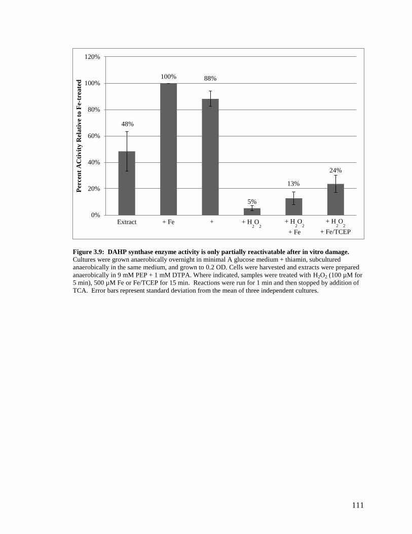
111
Figure 3.9: DAHP synthase enzyme activity is only partially reactivatable after in vitro damage.
Cultures were grown anaerobically overnight in minimal A glucose medium + thiamin, subcultured
anaerobically in the same medium, and grown to 0.2 OD. Cells were harvested and extracts were prepared
anaerobically in 9 mM PEP + 1 mM DTPA. Where indicated, samples were treated with H2O2 (100 µM for
5 min), 500 µM Fe or Fe/TCEP for 15 min. Reactions were run for 1 min and then stopped by addition of
TCA. Error bars represent standard deviation from the mean of three independent cultures.
48%
100% 88%
5%
13%
24%
0%
20%
40%
60%
80%
100%
120%
Per
cen
t A
Cti
vit
y R
ela
tiv
e to
Fe-t
rea
ted
Extract + H2O
2 + Fe +
Fe/TCEP
+ H2O
2
+ Fe
+ H2O
2
+ Fe/TCEP

112
Figure 3.10: Manganese addition and aerobic growth of Hpx-
cells. Cultures were grown anaerobically
overnight in Minimal A Glucose Medium + thiamin and subcultured anaerobically in the same medium.
Cells were diluted in the same aerobic medium +/- 50 µM Mn at time zero, and growth was monitored
aerobically.
WT
Hpx-
0.001
0.01
0.1
1
10
0 2 4 6 8 10 12
OD
60
0
Time (hr)
Hpx-
ΔmntH
Hpx-
+ Mn

113
A B
Figure 3.11: Manganese addition and aerobic growth of Hpx- cells. Cultures were grown anaerobically
overnight in Minimal A Glucose Medium + thiamin and subcultured anaerobically in the same medium.
Cells were diluted in the same aerobic medium with (A) or without (B) aromatic amino acids +/- Mn as
indicated at time zero, and growth was monitored aerobically.
0.001
0.010
0.100
1.000
10.000
0 5 10 15
OD
60
0
Time (hr)
No Mn 0.5 uM Mn
5 uM Mn 50 uM Mn
0.001
0.010
0.100
1.000
10.000
0 5 10 15
OD
60
0
Time (hr)
No Mn (+ Aros) 0.5 uM Mn (+ Aros)
5 uM Mn (+ Aros) 50 uM Mn (+ Aros)

114
Figure 3.12: Cobalt addition and aerobic growth of Hpx- cells. Cultures were grown anaerobically
overnight in Minimal A Glucose Medium + thiamin and subcultured anaerobically in the same medium.
Cells were diluted in the same aerobic medium +/- Co or 50 mM shikimate (shik) as indicated at time zero,
and growth was monitored aerobically. Points represent the OD measured after 10 hours of aeration.
Hpx-
Hpx- (+ shik)
0
0.02
0.04
0.06
0.08
0.1
0.12
0 50 100 150 200 250
OD
60
0
[Co] (μM)

115
Figure 3.13: Deferroxamine addition and aerobic growth of Hpx-
cells. Cultures were grown
anaerobically overnight in Minimal A Glucose Medium + thiamin and subcultured anaerobically in the
same medium. Cells were diluted in the same aerobic medium +/- 1 mM deferoxamine (DFO) at time zero,
and growth was monitored aerobically.
His
Aros DFO
0.001
0.01
0.1
1
0 2 4 6 8 10 12
OD
60
0n
m
Time (hr)

116
Figure 3.14: DAHP synthase transcription increases in response to H2O2 stress. Cultures were grown
anaerobically overnight in minimal A glucose medium + thiamin, subcultured anaerobically in the same
medium, and grown to 0.15 OD. Cells were then inoculated into fresh aerobic medium +/- 50 mM
aromatic amino acids (Aros) or +/- 50 mM Mn, where indicated, and grown aerobically for 4 hours. Cell
extracts were prepared, and β-galactosidase activity from a ParoF’-lacZ transcriptional fusion integrated
into the λ attachment site was determined [50]. Error bars represent standard deviation from the mean of
three independent cultures.
7.5
0.4
5.0
0.4
27.5 28.0
1.0 0.5
0.0
5.0
10.0
15.0
20.0
25.0
30.0
35.0
Fo
ld I
nd
uct
ion
co
mp
are
d t
o w
ild
-ty
pe
Hpx-
Hpx-
+ Aros Hpx
-
+ Mn
Hpx-
+ Mn
+ Aros
Δtyr
R
ΔtyrR
+ Aros
WT WT
+ Aros

117
REFERENCES 3.7
1. Al-Maghrebi, M., I. Fridovich, and L. Benov. 2002. Manganese
supplementation relieves the phenotypic deficits seen in superoxide-dismutase-
null Escherichia coli. Arch Biochem Biophys 402:104-9.
2. Anbar, A.D. 2008. Oceans. Elements and evolution. Science 322:1481-3.
3. Anjem, A. and J.A. Imlay. 2012. Mononuclear iron enzymes are primary targets
of hydrogen peroxide stress. J Biol Chem 287:15544-56.
4. Anjem, A., S. Varghese, and J.A. Imlay. 2009. Manganese import is a key
element of the OxyR response to hydrogen peroxide in Escherichia coli. Mol
Microbiol 72:844-58.
5. Archer, C. and D. Vance. 2006. Coupled Fe and S isotope evidence for Archean
microbial Fe(III) and sulfate reduction. Geology 34:153-156.
6. Archibald, F. 1983. Lactobacillus plantarum, an Organism Not Requiring Iron.
Fems Microbiology Letters 19:29-32.
7. Archibald, F. 1986. Manganese: its acquisition by and function in the lactic acid
bacteria. Crit Rev Microbiol 13:63-109.
8. Bedard, K., B. Lardy, and K.H. Krause. 2007. NOX family NADPH oxidases:
not just in mammals. Biochimie 89:1107-12.
9. Benov, L. and I. Fridovich. 1999. Why superoxide imposes an aromatic amino
acid auxotrophy on Escherichia coli. The transketolase connection. J Biol Chem
274:4202-6.
10. Chance, B., H. Sies, and A. Boveris. 1979. Hydroperoxide metabolism in
mammalian organs. Physiol Rev 59:527-605.
11. Chang, E.C. and D.J. Kosman. 1989. Intracellular Mn (II)-associated
superoxide scavenging activity protects Cu,Zn superoxide dismutase-deficient
Saccharomyces cerevisiae against dioxygen stress. J Biol Chem 264:12172-8.
12. Choi, H., et al. 2001. Structural basis of the redox switch in the OxyR
transcription factor. Cell 105:103-13.
13. Crow, J.P., J.S. Beckman, and J.M. McCord. 1995. Sensitivity of the essential
zinc-thiolate moiety of yeast alcohol dehydrogenase to hypochlorite and
peroxynitrite. Biochemistry 34:3544-52.

118
14. Datsenko, K.A. and B.L. Wanner. 2000. One-step inactivation of chromosomal
genes in Escherichia coli K-12 using PCR products. Proc Natl Acad Sci U S A
97:6640-5.
15. DeLeo, A.B., J. Dayan, and D.B. Sprinson. 1973. Purification and kinetics of
tyrosine-sensitive 3-deoxy-D-arabino-heptulosonic acid 7-phosphate synthetase
from Salmonella. J Biol Chem 248:2344-53.
16. Denu, J.M. and K.G. Tanner. 1998. Specific and reversible inactivation of
protein tyrosine phosphatases by hydrogen peroxide: evidence for a sulfenic acid
intermediate and implications for redox regulation. Biochemistry 37:5633-42.
17. Doy, C.H. and K.D. Brown. 1965. Control of aromatic biosynthesis: the
multiplicity of 7-phospho-2-oxo-3-deoxy-D-arabino-heptonate D-erythrose-4-
phosphate-lyase (pyruvate-phosphorylating) in Escherichia coli W. Biochim
Biophys Acta 104:377-89.
18. Flint, D.H., et al. 1993. The inactivation of dihydroxy-acid dehydratase in
Escherichia coli treated with hyperbaric oxygen occurs because of the destruction
of its Fe-S cluster, but the enzyme remains in the cell in a form that can be
reactivated. J Biol Chem 268:25547-52.
19. Fraser, C.M., et al. 1997. Genomic sequence of a Lyme disease spirochaete,
Borrelia burgdorferi. Nature 390:580-6.
20. Furdui, C., et al. 2004. Insights into the mechanism of 3-deoxy-D-arabino-
heptulosonate 7-phosphate synthase (Phe) from Escherichia coli using a transient
kinetic analysis. J Biol Chem 279:45618-25.
21. Glass, G.A., et al. 1986. The respiratory burst oxidase of human neutrophils.
Further studies of the purified enzyme. J Biol Chem 261:13247-51.
22. Gort, A.S. and J.A. Imlay. 1998. Balance between endogenous superoxide stress
and antioxidant defenses. J Bacteriol 180:1402-10.
23. Gotz, F., B. Sedewitz, and E.F. Elstner. 1980. Oxygen utilization by
Lactobacillus plantarum. I. Oxygen consuming reactions. Arch Microbiol
125:209-14.
24. Grass, G., et al. 2005. The metal permease ZupT from Escherichia coli is a
transporter with a broad substrate spectrum. J Bacteriol 187:1604-11.
25. Griffiths, S.W., J. King, and C.L. Cooney. 2002. The reactivity and oxidation
pathway of cysteine 232 in recombinant human alpha 1-antitrypsin. J Biol Chem
277:25486-92.

119
26. Gu, M. and J.A. Imlay. 2011. The SoxRS response of Escherichia coli is
directly activated by redox-cycling drugs rather than by superoxide. Mol
Microbiol 79:1136-50.
27. Gu, M. and J.A. Imlay. 2013. Superoxide poisons mononuclear iron enzymes by
causing mismetallation. Mol Microbiol 89:123-34.
28. Haldimann, A. and B.L. Wanner. 2001. Conditional-replication, integration,
excision, and retrieval plasmid-host systems for gene structure-function studies of
bacteria. J Bacteriol 183:6384-93.
29. Hasan, N., M. Koob, and W. Szybalski. 1994. Escherichia coli genome
targeting, I. Cre-lox-mediated in vitro generation of ori- plasmids and their in vivo
chromosomal integration and retrieval. Gene 150:51-6.
30. Henle, E.S., et al. 1999. Sequence-specific DNA cleavage by Fe2+
-mediated
fenton reactions has possible biological implications. J Biol Chem 274:962-71.
31. Horsburgh, M.J., et al. 2002. MntR modulates expression of the PerR regulon
and superoxide resistance in Staphylococcus aureus through control of manganese
uptake. Mol Microbiol 44:1269-86.
32. Imlay, J.A. 2006. Iron-sulphur clusters and the problem with oxygen. Mol
Microbiol 59:1073-82.
33. Imlay, J.A. and I. Fridovich. 1991. Superoxide production by respiring
membranes of Escherichia coli. Free Radic Res Commun 12-13 Pt 1:59-66.
34. Inbaraj, J.J. and C.F. Chignell. 2004. Cytotoxic action of juglone and
plumbagin: a mechanistic study using HaCaT keratinocytes. Chem Res Toxicol
17:55-62.
35. Jang, S. and J.A. Imlay. 2007. Micromolar intracellular hydrogen peroxide
disrupts metabolism by damaging iron-sulfur enzymes. J Biol Chem 282:929-37.
36. Jang, S. and J.A. Imlay. 2010. Hydrogen peroxide inactivates the Escherichia
coli Isc iron-sulphur assembly system, and OxyR induces the Suf system to
compensate. Mol Microbiol 78:1448-67.
37. Jenney, F.E., Jr., et al. 1999. Anaerobic microbes: oxygen detoxification without
superoxide dismutase. Science 286:306-9.
38. Kehres, D.G., et al. 2002. Regulation of Salmonella enterica serovar
Typhimurium mntH transcription by H2O2, Fe(2+), and Mn(2+). J Bacteriol
184:3151-8.

120
39. Krapp, A.R., M.V. Humbert, and N. Carrillo. 2011. The soxRS response of
Escherichia coli can be induced in the absence of oxidative stress and oxygen by
modulation of NADPH content. Microbiology 157:957-65.
40. Kuo, C.F., T. Mashino, and I. Fridovich. 1987. , -Dihydroxyisovalerate
dehydratase. A superoxide-sensitive enzyme. J Biol Chem 262:4724-7.
41. Lee, C., et al. 2004. Redox regulation of OxyR requires specific disulfide bond
formation involving a rapid kinetic reaction path. Nat Struct Mol Biol 11:1179-
85.
42. Liochev, S.I., et al. 1999. Induction of the soxRS regulon of Escherichia coli by
superoxide. J Biol Chem 274:9479-81.
43. Loew, O. 1900. A New Enzyme of General Occurrence in Organismis. Science
11:701-2.
44. Lombard, M., et al. 2000. Superoxide reductase as a unique defense system
against superoxide stress in the microaerophile Treponema pallidum. J Biol Chem
275:27021-6.
45. McCandliss, R.J. and K.M. Herrmann. 1978. Iron, an essential element for
biosynthesis of aromatic compounds. Proc Natl Acad Sci U S A 75:4810-3.
46. McCord, J.M. and I. Fridovich. 1969. Superoxide Dismutase an Enzymic
Function for Erythrocuprein (Hemocuprein). J Biol Chem 244:6049-6055.
47. McCord, J.M., B.B. Keele, Jr., and I. Fridovich. 1971. An enzyme-based
theory of obligate anaerobiosis: the physiological function of superoxide
dismutase. Proc Natl Acad Sci U S A 68:1024-7.
48. Mehdy, M.C. 1994. Active Oxygen Species in Plant Defense against Pathogens.
Plant Physiol 105:467-472.
49. Messner, K.R. and J.A. Imlay. 1999. The identification of primary sites of
superoxide and hydrogen peroxide formation in the aerobic respiratory chain and
sulfite reductase complex of Escherichia coli. J Biol Chem 274:10119-28.
50. Miller, J.H. 1972. Experiments in molecular geneticsCold Spring Harbor, N.Y.:
Cold Spring Harbor Laboratory. xvi, 466 p.
51. Mishra, S. and J. Imlay. 2012. Why do bacteria use so many enzymes to
scavenge hydrogen peroxide? Arch Biochem Biophys 525:145-60.
52. Park, S. and J.A. Imlay. 2003. High levels of intracellular cysteine promote
oxidative DNA damage by driving the fenton reaction. J Bacteriol 185:1942-50.

121
53. Pomposiello, P.J., M.H. Bennik, and B. Demple. 2001. Genome-wide
transcriptional profiling of the Escherichia coli responses to superoxide stress and
sodium salicylate. J Bacteriol 183:3890-902.
54. Posey, J.E. and F.C. Gherardini. 2000. Lack of a role for iron in the Lyme
disease pathogen. Science 288:1651-3.
55. Radtke, A.L. and M.X. O'Riordan. 2006. Intracellular innate resistance to
bacterial pathogens. Cell Microbiol 8:1720-9.
56. Seaver, L.C. and J.A. Imlay. 2001. Alkyl hydroperoxide reductase is the
primary scavenger of endogenous hydrogen peroxide in Escherichia coli. J
Bacteriol 183:7173-81.
57. Seaver, L.C. and J.A. Imlay. 2001. Hydrogen peroxide fluxes and
compartmentalization inside growing Escherichia coli. J Bacteriol 183:7182-9.
58. Seaver, L.C. and J.A. Imlay. 2004. Are respiratory enzymes the primary sources
of intracellular hydrogen peroxide? J Biol Chem 279:48742-50.
59. Seib, K.L., et al. 2004. Defenses against oxidative stress in Neisseria
gonorrhoeae and Neisseria meningitidis: distinctive systems for different
lifestyles. J Infect Dis 190:136-47.
60. Seki, M., et al. 2004. Hydrogen peroxide production in Streptococcus pyogenes:
involvement of lactate oxidase and coupling with aerobic utilization of lactate. J
Bacteriol 186:2046-51.
61. Shen, H., et al. 1993. Identification of cysteine residues involved in disulfide
formation in the inactivation of glutathione transferase P-form by hydrogen
peroxide. Arch Biochem Biophys 300:137-41.
62. Spellerberg, B., et al. 1996. Pyruvate oxidase, as a determinant of virulence in
Streptococcus pneumoniae. Mol Microbiol 19:803-13.
63. Staub, M. and G. Denes. 1969. Purification and properties of the 3-deoxy-D-
arabino-heptulosonate-7-phosphate synthase (phenylalanine sensitive) of
Escherichia coli K12. I. Purification of enzyme and some of its catalytic
properties. Biochim Biophys Acta 178:588-98.
64. Tribe, D.E., H. Camakaris, and J. Pittard. 1976. Constitutive and repressivle
enzymes of the common pathway of aromatic biosynthesis in Escherichia coli K-
12: regulation of enzyme synthesis at different growth rates. J Bacteriol
127:1085-97.
65. Tseng, H.J., et al. 2002. Virulence of Streptococcus pneumoniae: PsaA mutants
are hypersensitive to oxidative stress. Infect Immun 70:1635-9.

122
66. Turner, J.M. and A.J. Messenger. 1986. Occurrence, biochemistry and
physiology of phenazine pigment production. Adv Microb Physiol 27:211-75.
67. Weissbach, A. and J. Hurwitz. 1959. The formation of 2-keto-3-deoxyheptonic
acid in extracts of Escherichia coli B. I. Identification. J Biol Chem 234:705-9.
68. Winterbourn, C.C. and D. Metodiewa. 1999. Reactivity of biologically
important thiol compounds with superoxide and hydrogen peroxide. Free Radic
Biol Med 27:322-8.
69. Zheng, M., et al. 2001. DNA microarray-mediated transcriptional profiling of the
Escherichia coli response to hydrogen peroxide. J Bacteriol 183:4562-70.

123
CHAPTER 4: CONCLUSIONS
SUMMARY OF CURRENT WORK 4.1
Mononuclear iron enzymes are primary targets of H2O2 4.1.1
Under normal growth conditions, Escherichia coli produces low amounts of H2O2
as an inadvertent byproduct of aerobic metabolism. When its H2O2 scavengers, catalase
and peroxidase, are deleted from the cell, the resultant Hpx- strain exhibits a number of
phenotypes. The first phenotype discovered, damage to DNA, was found to be due to
hydroxyl radical oxidation of the DNA backbone as a result of Fenton chemistry
occurring on the DNA [18]. Other phenotypes, including an inability to grow on TCA
cycle intermediates, failure of the Isc system, and a leucine auxotrophy, were all due to
Fenton chemistry leading to oxidation of solvent-exposed [4Fe-4S] cluster dehydratases
[14, 15]. In this study, two other phenotypes were explored.
The first phenotype I examined was a block in the pentose phosphate pathway. I
found that when Hpx- E. coli cells were forced to metabolize gluconate through the
pentose phosphate pathway, they were unable to grow more than one or two doublings in
air. I measured the activity of each of these five enzymes downstream of gluconate in the
pentose phosphate pathway and found that only one, Rpe, lost activity upon H2O2
exposure. This result was interesting for a number of reasons. First, no one had ever
observed damage to proteins other than [4Fe-4S] cluster dehydratases by H2O2, making
Rpe a completely novel protein target. Second, Rpe was not known to use iron as a
cofactor [1]. This result meant either that Rpe must actually use iron, although other
researchers found it was unable to be activated by iron, or that damage to Rpe was not
Fenton-based. All other known forms of H2O2 damage are mediated by the Fenton
reaction [2, 14, 15, 17, 18, 21], so the idea that Rpe was somehow an exception did not
seem likely. When Rpe was purified and tested anaerobically, I found that not only was
iron able to activate it, but that iron activation yielded the highest amount of activity of

124
any metal. In addition, iron was the only metal that conferred H2O2 sensitivity, which
meant that iron was the physiological metal for Rpe. Thus, damage to Rpe mirrors that of
the [4Fe-4S] cluster dehydratases, as both use the metal to bind and activate substrate. It
is not a surprise, then, that these proteins have similar inactivation rates, about 4 – 5 x 10³
M-1
s-1
. Once again, Fenton chemistry is the reason for damage by H2O2.
There is another important aspect of this finding to note beyond the sensitivity of
Rpe to H2O2. By performing the metallation reaction in the anaerobic chamber, I was
able to maintain iron in the ferrous form, while researchers working with the protein
under aerobic conditions were unable to keep the iron reduced. It is conceivable that this
experimental artifact could have misled a number of researchers performing metallation
studies on mononuclear metalloproteins. Adil Anjem tested three other such proteins and
found that all were activated by iron and sensitive to H2O2, in vitro and in vivo, showing
that these proteins also used iron as their physiological cofactor [2]. Thus, the
mononuclear iron protein class may continue to grow as more work is done to determine
the metals used by various proteins.
The second phenotype I explored was the aromatic amino acid auxotrophy. When
work first began on this phenotype, it was not known that mononuclear iron proteins
were targets of H2O2. Since the aromatic amino acid biosynthetic pathway contained no
Fe-S cluster proteins, no protein seemed more likely to be damaged than any other. In
fact, there was reason to believe that transketolase was the cause, as this would also
explain the problem with the pentose phosphate pathway. However, when the sensitivity
of Rpe was found, two problems were solved. First, the two phenotypes were separated
from one another, meaning that another target existed. Second, identification of the
mononuclear iron proteins as a class of H2O2 targets shed new light on potential targets in
the shikimic acid pathway for aromatic biosynthesis. I identified the first enzyme in the
pathway, DAHP synthase, as a mononuclear iron protein and found that it, like the others
in this class, was sensitive to H2O2.

125
Mechanism of protection of mononuclear iron proteins by manganese 4.1.2
Manganese has long been known to have protective effects during oxidative
stress. Manganese was initially believed to protect cells by scavenging reactive oxygen
species; however, this mechanism of protection was shown to be incorrect in E. coli, as
scavenging rates did not change when manganese was added to cells [3]. In this study, I
sought to show that manganese protection, at least in part, is due to metallation of
mononuclear iron proteins. While metallation by iron causes these proteins to be
sensitive to H2O2, metallation by manganese provides a moderate amount of activity that
is not sensitive to oxidation by H2O2. I showed that manganese is able to protect two
mononuclear iron enzymes, Rpe and DAHP synthase, from oxidation by H2O2 in vivo
and in vitro. As mentioned previously, I believe that mononuclear iron proteins are much
more common than is currently known, so the protective effect of manganese on this
class of proteins is an important factor in combatting the negative effects of reactive
oxygen species. E. coli recognizes the importance of manganese during oxidative stress,
as evidenced by the upregulation of the manganese transporter, MntH, by OxyR.
Without MntH, Hpx- cells are unable to grow, even with amino acid supplements,
highlighting the importance of manganese during H2O2 stress.
POSSIBLE FUTURE DIRECTIONS 4.2
Why does E. coli require iron in a pathway needed to acquire it? 4.2.1
One additional problem that damage to DAHP synthase causes for E. coli is
limitation of its ability to import iron. In fact, there are at least two other proteins in the
pathway that might also use iron but are not annotated as such. While this might not be
such a problem during oxidative stress, as iron limitation is one of the desired outcomes
of induction of the stress responses, it is more difficult to comprehend why E. coli would
require iron for a pathway that produces the precursor for synthesis of enterochelin, a
siderophore used to acquire iron from the environment. Preliminary results of

126
enterochelin synthesis during H2O2 stress indicate that damage to DAHP synthase results
in an inability to make siderophores (Figure 4.1). It is inconceivable that iron levels
should fall so low that there is no DAHP synthase activity, but there are seven different
metabolites from five distinct pathways that require chorismate, the end product of the
shikimic acid pathway for their synthesis [10]. Perhaps the answer as to how E. coli
manages this problem comes from an investigation of the KM for chorismate of the
branch pathways. The pathway with the lowest KM, somewhat surprisingly, is for
tryptophan synthesis [7]. The pathways for folate and ubiquinone biosynthesis have the
next lowest KM values for chorismate [20, 22]. It is easy to see why these pathways
might be given preference over other metabolites, as they are key to keeping energy
levels high enough to keep metabolism running. Next in the chorismate hierarchy is
synthesis of enterochelin. This pathway has a KM for chorismate of 14 µM [16], more
than three times lower than the next processes, which are the pathways for phenylalanine
[9] and tyrosine [13] biosynthesis and at 195 µM, the pathway for menaquinone
biosynthesis [8]. This analysis of apparent KM values indicates the importance of the
ability of the cell to synthesize these iron importers, greater even than synthesis of two
amino acids. Further exploration of the pathway and its activity during iron starvation
may provide insight into how E. coli prioritizes iron, as well as flux through this pathway.
Are mononuclear iron proteins mismetallated during H2O2 stress, and if not, 4.2.2
how does the cell prevent it?
While I showed that DAHP synthase does not become mismetallated by zinc
during oxidative stress, other mononuclear iron proteins have not been so rigorously
tested for this phenotype. In fact, there is evidence that SOD mutants lose activity of
mononuclear iron proteins first by oxidation of the iron atom, but that activity remains
low because the iron is replaced by zinc [12]. It is possible that during H2O2 stress,
manganese import by induction of MntH shifts the metal balance in the cell to favor
metallation by manganese over iron or zinc, but in SOD mutants, there is no induction of
manganese import. Therefore, during O2·- stress, no metals are available to compete with

127
zinc for the active sites of mononuclear iron proteins, resulting in accumulation of
inactive zinc-metallated protein.
Are there other mononuclear iron proteins that are sensitive to H2O2? 4.2.3
As work continues to identify additional phenotypes of H2O2 stress, more
mononuclear iron proteins may be identified that cause them. Ongoing work in the lab is
using RNA-Seq to identify genes that are upregulated in response to oxidative stress.
While some genes – notably, the members of the OxyR regulon – are expected to appear,
other genes whose products are sensitive to H2O2 may also be induced. DAHP synthase
is an example of this, as E. coli attempts to produce more protein in response to the
decreased activity of the pathway. Additionally, proteins that use metal cofactors for
non-redox reactions may be additional targets. While some of the novelty has been lost,
it remains important to identify additional targets of H2O2 in order to fully appreciate the
scope of the effects reactive oxygen species have on cells.
Are mononuclear iron proteins sensitive to H2O2 in organisms that either do 4.2.4
not use iron or produce H2O2?
Some bacteria, such as Borrelia Burgdorferi and Lactococcus plantarum, are
considered to be low-iron or even iron-free organisms that import little to no iron [4, 5,
11, 19]. These organisms, presumably as a way to compensate for a lack of iron, import
high levels of manganese. It is possible that these, and organisms like them, have
evolved to use manganese in the place of iron in mononuclear metalloproteins. Use of
manganese in these proteins may be one explanation for the ability of lactic acid bacteria
to survive under conditions where they are producing up to millimolar levels of H2O2.
One way to examine this possibility is to examine Lactococcus counterparts of the
proteins found to be mononuclear iron enzymes in E. coli. If these proteins are found to
be sensitive to H2O2 as well, it will be interesting to determine what metal is being used
in these proteins, since the organism is not known to import iron. Alternatively, if the
proteins are resistant and found to bind manganese, it would be beneficial to determine if

128
there are differences that might allow the protein to differentiate between iron and
manganese. This could be tested both by expressing the Lactococcus proteins in E. coli
to see if they become sensitive, and conversely by expressing the E. coli proteins in
Lactococcus to see if they are now resistant to H2O2.
Have eukaryotes evolved to use alternative metals in mononuclear 4.2.5
metalloproteins to protect against oxidative stress?
It is possible that eukaryotes also suffer from the same damage to mononuclear
iron enzymes experienced by E. coli cells as a result of oxidative stress. It would be
interesting to determine whether damage to any of these proteins in humans is
responsible for any known diseases. It is also likely, however, that eukaryotic organisms
have taken evolutionary steps to rid themselves of this vulnerability. Preliminary results
using Baker’s yeast suggest that this may be the case, as Rpe assayed from aerobically
grown Saccharomyces cerevisiae showed no inactivation by 1 mM H2O2 and is described
fully in Appendix B. This result suggests either that yeast have evolved to use a metal
other than iron in the active site of Rpe, or that yeast Rpe somehow shields its metal from
H2O2. Further work must be carried out to determine the mechanism of resistance of
yeast Rpe to H2O2; such work might include experiments such as expression of yeast Rpe
in E. coli and vice versa. Other mononuclear iron proteins should also be examined to
determine whether this resistance in yeast is specific to Rpe or extends to mononuclear
iron proteins in general.

129
FIGURES 4.3
Figure 4.1: Enterochelin synthesis of Hpx- cells. Cultures were grown anaerobically overnight in
Minimal A Glucose Medium + thiamin and subcultured anaerobically in the same medium. Cells were
diluted in the same aerobic medium +/- aromatic or 19 amino acids as indicated at time zero and cells were
grown overnight. Cells were then spun out and the supernate was used to assay for catechol by the method
of Arnow [6]. (BDL = below detectable limit.)

130
REFERENCES 4.4
1. Akana, J., et al. 2006. D-Ribulose 5-phosphate 3-epimerase: functional and
structural relationships to members of the ribulose-phosphate binding ( / )8-
barrel superfamily. Biochemistry 45:2493-503.
2. Anjem, A. and J.A. Imlay. 2012. Mononuclear iron enzymes are primary targets
of hydrogen peroxide stress. J Biol Chem 287:15544-56.
3. Anjem, A., S. Varghese, and J.A. Imlay. 2009. Manganese import is a key
element of the OxyR response to hydrogen peroxide in Escherichia coli. Mol
Microbiol 72:844-58.
4. Archibald, F. 1983. Lactobacillus plantarum, an Organism Not Requiring Iron.
Fems Microbiology Letters 19:29-32.
5. Archibald, F. 1986. Manganese: its acquisition by and function in the lactic acid
bacteria. Crit Rev Microbiol 13:63-109.
6. Arnow, L.E. 1937. Proposed Chemical Mechanisms for the Production of Skin
Erythema and Pigmentation by Radiant Energy. Science 86:176.
7. Baker, T.I. and I.P. Crawford. 1966. Anthranilate synthetase. Partial
purification and some kinetic studies on the enzyme from Escherichia coli. J Biol
Chem 241:5577-84.
8. Daruwala, R., et al. 1997. Menaquinone (vitamin K2) biosynthesis:
overexpression, purification, and characterization of a new isochorismate synthase
from Escherichia coli. J Bacteriol 179:3133-8.
9. Dopheide, T.A., P. Crewther, and B.E. Davidson. 1972. Chorismate mutase-
prephenate dehydratase from Escherichia coli K-12. II. Kinetic properties. J Biol
Chem 247:4447-52.
10. Dosselaere, F. and J. Vanderleyden. 2001. A metabolic node in action:
chorismate-utilizing enzymes in microorganisms. Crit Rev Microbiol 27:75-131.
11. Fraser, C.M., et al. 1997. Genomic sequence of a Lyme disease spirochaete,
Borrelia burgdorferi. Nature 390:580-6.
12. Gu, M. and J.A. Imlay. 2013. Superoxide poisons mononuclear iron enzymes by
causing mismetallation. Mol Microbiol 89:123-34.
13. Hudson, G.S., V. Wong, and B.E. Davidson. 1984. Chorismate
mutase/prephenate dehydrogenase from Escherichia coli K12: purification,
characterization, and identification of a reactive cysteine. Biochemistry 23:6240-
9.

131
14. Jang, S. and J.A. Imlay. 2007. Micromolar intracellular hydrogen peroxide
disrupts metabolism by damaging iron-sulfur enzymes. J Biol Chem 282:929-37.
15. Jang, S. and J.A. Imlay. 2010. Hydrogen peroxide inactivates the Escherichia
coli Isc iron-sulphur assembly system, and OxyR induces the Suf system to
compensate. Mol Microbiol 78:1448-67.
16. Liu, J., et al. 1990. Overexpression, purification, and characterization of
isochorismate synthase (EntC), the first enzyme involved in the biosynthesis of
enterobactin from chorismate. Biochemistry 29:1417-25.
17. Park, S. and J.A. Imlay. 2003. High levels of intracellular cysteine promote
oxidative DNA damage by driving the fenton reaction. J Bacteriol 185:1942-50.
18. Park, S., X. You, and J.A. Imlay. 2005. Substantial DNA damage from
submicromolar intracellular hydrogen peroxide detected in Hpx- mutants of
Escherichia coli. Proc Natl Acad Sci U S A 102:9317-22.
19. Posey, J.E. and F.C. Gherardini. 2000. Lack of a role for iron in the Lyme
disease pathogen. Science 288:1651-3.
20. Siebert, M., K. Severin, and L. Heide. 1994. Formation of 4-hydroxybenzoate
in Escherichia coli: characterization of the ubiC gene and its encoded enzyme
chorismate pyruvate-lyase. Microbiology 140 ( Pt 4):897-904.
21. Varghese, S., et al. 2007. Submicromolar hydrogen peroxide disrupts the ability
of Fur protein to control free-iron levels in Escherichia coli. Mol Microbiol
64:822-30.
22. Viswanathan, V.K., J.M. Green, and B.P. Nichols. 1995. Kinetic
characterization of 4-amino 4-deoxychorismate synthase from Escherichia coli. J
Bacteriol 177:5918-23.

132
APPENDIX A: DETERMINATION OF FREE IRON LEVELS IN
RESPONSE TO RYHB IN ESCHERICHIA COLI
INTRODUCTION A.1
RyhB is a non-coding small RNA in Escherichia coli that was discovered by
Massé and Gottesman in 2002 [2]. RyhB is repressed by metallated Fur protein, but
when ferrous iron levels are low and the Fur regulon is derepressed, RyhB is expressed.
A global transcription microarray analysis using a plasmid expressing RyhB showed it
was able to directly repress at least 18 transcripts, including many genes that require iron
for activity [3]. It has been suggested that the purpose of RyhB is to spare iron from
dispensable iron proteins in order to allow metallation of indispensable proteins that
require it [1]. Because previous work was performed in complex medium, which has a
high level of iron, it was difficult to see distinct phenotypes of RyhB deletion. This study
provides evidence that RyhB spares iron in media where iron levels are very low.
RESULTS A.2
RyhB mutants display a growth phenotype as they reach stationary phase A.2.1
While rich media, such as LB, are replete with iron, minimal media contain low
levels of iron unless it is supplemented. Therefore, when looking for growth phenotypes
related to iron homeostasis, minimal defined media allow for the elucidation of growth
phenotypes that would not be apparent in rich medium. For this reason, strains obtained
from the Eric Masse lab – EM1055 (wild-type), EM1238 (ΔryhB), EM1256 (Δfur), and
EM1257 (ΔryhB, Δfur) – were grown in M63 glucose medium. While there was no
marked difference in the growth of these strains during logarithmic growth, as the cells
approached stationary phase, strains containing the ΔryhB allele were unable to reach as

133
high an optical density as strains that were wild-type for RyhB (Figure A.1A). However,
when 1 µM FeSO4 was added to the growth medium, this growth phenotype was
abolished (Figure A.1B). Thus, RyhB either is required to provide iron for some process
that becomes important during stationary phase, or is needed to allow proper distribution
of iron when it becomes scarce.
RyhB mutants do not accumulate high levels of intracellular free iron A.2.2
In order to examine whether RyhB was able to affect intracellular levels of iron in
minimal medium, electron paramagnetic resonance (EPR) spectroscopy was performed.
EPR provides a quantitative measure of chelatable iron levels inside the cell, which, by
extension, is a measure of the free and available iron pools in the cell. If RyhB were
sparing iron for indispensable processes that require it, then a RhyB mutant should have
lower levels of free iron than a wild-type cell under conditions where iron is scarce. Bars
1 and 3 of Figure A.2 show that this is indeed true. Even when iron is added to the
medium, the RyhB mutant strain does not accumulate high levels of free iron (Figure
A.2, bar 4). This is not the case for the fur mutant, which accumulates 4.5-fold more iron
than the wild-type strain when no iron was added (Figure A.2, bar 5) or more than 16-
fold more when iron was added to the growth medium (Figure A.2, bar 6). This is not
surprising, as deletion of fur will lead to constitutive expression of iron import systems,
as well as iron-sparing by RyhB. Interestingly, when the ryhB and fur deletions were
combined, the increase of intracellular free iron seen in the fur mutant was completely
abolished (Figure A.2, bars 7 and 8). This suggests that iron sparing by RyhB is what
allows the fur mutants to accumulate so much free iron.
DISCUSSION A.3
Determination of the role of RyhB in iron homeostasis is integral to acquiring a
full understanding of how iron pools are regulated during changing environmental
conditions. RyhB has been shown to be involved in iron sparing [1] in rich medium.

134
This report both verifies that this is true of minimal medium, but also demonstrates a
growth phenotype as cells approach stationary phase. It is important to note that this
medium was not treated to remove trace amounts of iron that might contaminate the
various components of the medium. Therefore, if iron levels in the medium were more
limited, the growth lag observed as cells reach stationary phase may occur earlier as iron
pools are exhausted.
Taken together, the growth phenotype and the free iron levels seem to agree with
the idea that RyhB spares the cell iron, which is detected as free iron. Failure to spare the
iron (under iron-limited conditions) causes the cells to stop growing, however the specific
defect that was responsible for the growth defect was not identified. Thus, RhyB is an
integral member of the Fur regulon whose importance to the cell during conditions that
limit the availability of iron warrants further study.
EXPERIMENTAL PROCEDURES A.4
Reagents A.4.1
Diethylenetriaminepentaacetic acid (DTPA), desferrioxamine mesylate, and FeCl3
hexahydrate were from Sigma, while FeSO4 was from Bioshop. All other chemicals used
were from Fisher.
Bacterial growth A.4.2
Cells were grown overnight in M63 medium containing 0.2% glucose in the
absence or in the presence of 1 µM FeSO4. Overnight cultures were then used to
inoculate 15 mL same medium to 0.14 OD and growth was monitored at 37°C.

135
EPR analysis A.4.3
A.4.3.1 Growth
Cells were grown overnight at 37°C in M63 glucose medium, diluted 1:10 into
250 mL freshly prepared M63 glucose +/- 1 µM FeSO4, and then grown at 37°C in 1L
baffled flasks with vigorous shaking to a cell density of approximately 0.9 OD at 600
nm. Cells were then harvested and prepared for EPR analysis.
A.4.3.2 Sample preparation
Cultures were placed on ice for 10 minutes, and then spun for 10 minutes at 6,000
rpm at 4°C in 250 mL bottles using the Beckman JA-14 rotor. Pellets were resuspended
in 8 mL M63 glucose + 1 mL 0.1 M DTPA (pH 7.0) that had been pre-warmed to 37°C.
Next, 1 mL 0.2 M DFO (pH 8.0) was added and the culture was shaken for 15 minutes at
37°C.
Samples were then put on ice for 15 minutes before being spun for 5 minutes at
8,000 rpm at 4°C. Supernatants were discarded and pellets were washed twice with 5 mL
ice-cold 20 mM Tris-HCl (pH 7.5), spinning for 5 minutes at 8,000 rpm at 4°C after each
wash. Finally, pellets were resuspended in 300 µL ice-cold 20 mM Tris-HCl (pH 7.4)
containing 30% glycerol. About 200 µL of each of the final suspensions was added to
separate EPR tubes, shaken to the bottom of the tube, and frozen immediately on dry ice.
Optical densities of the samples were measured for use in determination of
intracellular volumes. To do this, 10 µL of each sample was diluted into 990 µL 20 mM
Tris-HCl (pH 7.4) containing 30% glycerol and mixed well. Each of these was then
diluted 1:10 into the same buffer and the OD was measured. Three individual OD
replicates were measured for each sample.
A.4.3.3 Preparation of Fe standards
A fresh stock of 10 mM FeCl3·6H2O was prepared by adding 0.27g FeCl3·6H2O to
10 mL of 20 mM Tris-Cl (pH 7.4) + 10% glycerol. Dilutions were then prepared in the
same buffer to theoretical concentrations from 0 to 100 µM Fe. Concentrations of the

136
standards were then determined by measuring the A420 with the extinction coefficient of
2865 M-1
cm-1
. About 200 µL of each standard was added to separate EPR tubes for
generation of a standard curve.
A.4.3.4 EPR spectrometer parameters
Field controller:
Field set: 01573 (g = 4.3)
Scan source: Ext
Scan range (field sweep): 4 x 100
Function selector:
High frequency
Cavity Modulation: I: High II: Low
Gain: 3.2 x 10³ Norm
Power: 10 mW
Norm: 180
Modulation: 1.25 x 10
Function: 100 kHz
Time Constant: 0.032
Field Sweep: Off
Computer:
Scans: 7 scans; 30 sec/scan; 3 sec between scans

137
FIGURES A.5
Figure A.1: Growth of ryhB mutants is impaired near stationary phase in minimal medium. Cells
were grown overnight in M63 medium containing 0.2% glucose in the absence (A) or in the presence (B) of
1 µM FeSO4. Overnight cultures were then used to inoculate 15 mL same medium to 0.14 OD and growth
was monitored at 37°C. Strains used were EM1055 (WT), EM1238 (ΔryhB), EM1256(Δfur), and
EM1257(ΔryhB, Δfur).
WT
ΔryhB Δfur
ΔryhB, Δfur
0.100
1.000
0.00 2.00 4.00 6.00 8.00 10.00 12.00
OD
60
0n
m
Time (hr)
No Fe
WT ΔryhB
Δfur ΔryhB, Δfur
0.100
1.000
0 2 4 6 8 10 12
OD
60
0n
m
Time (hr)
1 µM Fe
A
B

138
Figure A.2: Concentration of intracellular free iron. Cells were grown in the absence or presence of 1
μM of FeSO4 until an OD600 of 0.9, at which point they were assayed for free intracellular Fe by EPR.
Strains used were EM1055 (WT), EM1238 (ΔryhB), EM1256(Δfur), and EM1257(ΔryhB, Δfur). This
figure has been published in collaboration with Dr. Eric Massé [4].

139
REFERENCES A.6
1. Jacques, J.F., et al. 2006. RyhB small RNA modulates the free intracellular iron
pool and is essential for normal growth during iron limitation in Escherichia coli.
Mol Microbiol 62:1181-90.
2. Masse, E. and S. Gottesman. 2002. A small RNA regulates the expression of
genes involved in iron metabolism in Escherichia coli. Proc Natl Acad Sci U S A
99:4620-5.
3. Masse, E., C.K. Vanderpool, and S. Gottesman. 2005. Effect of RyhB small
RNA on global iron use in Escherichia coli. J Bacteriol 187:6962-71.
4. Salvail, H., et al. 2010. A small RNA promotes siderophore production through
transcriptional and metabolic remodeling. Proc Natl Acad Sci U S A 107:15223-
8.

140
APPENDIX B: ARE MONONUCLEAR IRON PROTEINS FROM
OTHER ORGANISMS SENSITIVE TO H2O2?
INTRODUCTION B.1
There are a number of lactic acid bacteria that can continue to grow despite their
production of high levels of H2O2 [1, 3, 4]. It is of interest, both evolutionarily and
physiologically, to learn how these organisms can maintain the activity of enzymes, such
as the mononuclear iron proteins, in the presence of oxidants that would be damaging to
the homologous proteins in E. coli. Another evolutionary question regarding
mononuclear iron proteins is whether eukaryotes have maintained these proteins as Fe-
containing enzymes, or if these organisms have evolved to use different metals. These
questions were the focus of studies described in this appendix.
RESULTS B.2
Are mononuclear iron proteins from the lactic acid bacterium Lactococcus B.2.1
lactis sensitive to H2O2 in vitro?
In an effort to determine the sensitivity of mononuclear iron proteins from
bacteria that produce H2O2 during growth, I tried to determine growth conditions that
would allow Lactococcus lactis to produce H2O2 while still synthesizing proteins of
interest, such as Rpe and DAHP synthase. In order for DAHP synthase to be expressed,
the aromatic amino acids should be absent from the medium. I therefore attempted to
grow L. lactis on a minimal medium that has been described by others as a means to
study this organism’s physiology [2]. Despite making the medium as described, the L.
lactis strain did not well grow on a plate, even after three days at 30°C. When these cells
were inoculated into minimal liquid medium, no growth was ever observed. Thus, work

141
must still be done to determine the composition of a minimal medium that will support
the growth of L. lactis.
Is Rpe from yeast sensitive to H2O2 in vitro? B.2.2
To test the sensitivity to H2O2 of a eukaryotic homolog of a mononuclear iron
protein, Baker’s yeast was used as a model. Wild-type yeast cells were grown
aerobically at 30°C on yeast nitrogen base (YNB), a minimal medium for growing yeast,
supplemented with 2% glucose. Cells were harvested aerobically by centrifugation and
the pellets were transferred to the anaerobic chamber for extract preparation and assay.
Extracts were prepared by bead-beating the resuspended pellets and cleared by
centrifugation immediately before being assayed for Rpe activity. When these extracts
were treated with 1 mM H2O2 for 2 min, Rpe activity was no different than without H2O2
treatment (Figure B.1), both having a specific activity of about 0.45 U/mg protein (by
comparison, E.coli has a specific activity of 4.5 U/mg protein). Addition of a chelator
during H2O2 treatment had no effect on the activity of yeast Rpe. Thus, it appears that
the yeast homolog of Rpe is resistant to H2O2 in vitro. The mechanism of this resistance
will require further investigation.
DISCUSSION B.3
While there is little to discuss regarding the lactic acid bacteria, it remains an
interesting area of research that should be pursued. However, the preliminary finding
that Rpe from Baker’s yeast is resistant to H2O2 implies that yeast either use an
alternative metal in this protein, or that the metal is somehow shielded from H2O2.
Because the role of the metal in Rpe should be the same in yeast as it is in E. coli, solvent
exposure of the metal atom should be required in order for it to bind to and activate the
substrate. Therefore, it seems unlikely that yeast employs some shielding mechanism,
but this idea remains a formal possibility. More work is necessary to determine the
mechanism by which yeast Rpe remains active in the presence of H2O2.

142
EXPERIMENTAL PROCEDURES B.4
Reagents B.4.1
Amino acids, catalase (from bovine liver), diethylenetriaminepentaacetic
acid (DTPA), α-glycerophosphate-triosephosphate isomerase from rabbit muscle, MOPS
and associated medium components, β-nicotinamide adenine dinucleotide reduced
disodium salt (NADH), D-ribose 5-phosphate disodium salt, D-ribulose 5-phosphate
disodium salt, transketolase (from yeast), and 30% H2O2 were from Sigma.
Glycylglycine was from Acros Organics. Yeast Nitrogen Base, dextrose, and Tris-HCl
were from Fisher.
Growth medium B.4.2
Luria-Bertani (LB) medium contained 10 g tryptone, 10 g NaCl, and 5 g yeast
extract per liter. Minimal SA medium was a MOPS medium prepared according to [2]
supplemented with 0.2% glucose, 0.5 µg/mL thiamin and 0.5 mM histidine. Other amino
acids were added at 0.5 mM where indicated.
Strains and growth B.4.3
Lactococcus lactis strain IL1403 was a gift from Dr. John Cronan. L. lactis was
streaked on LB glucose solid medium and incubated overnight at 30°C. Single colonies
were picked from the LB plate and onto Minimal SA plates, which were then placed at
30°C.
The Baker’s yeast, Saccharomyces cerevisiae, wild-type strain X-2180-1a was a
gift from Dr. Peter Orlean. The yeast was streaked onto solid Difco YNB medium
containing 2% glucose. For Rpe activity determination, an overnight culture of X-2180-
1a was inoculated using a single colony into 5 mL liquid YNB + 2% glucose at 30°C.
The overnight was then diluted into 15 mL of the same medium to 0.005 OD and grown

143
to 0.1 OD at 30°C. This pre-culture was then diluted to 0.0015 OD in 50 mL YNB +
0.2% glucose and grown at 30°C to 0.4 OD.
Preparation of yeast cell extracts B.4.4
When yeast cells reached 0.4 OD as described above, the culture was chilled on
ice for 20 minutes and then spun for 8 minutes at 4,000 rpm at 4°C. After removing the
supernatant, the cell pellet was resuspended in 50 mL ice-cold 50 mM glycylglycine (pH
8.5) and spun again for 8 minutes at 4°C. The supernatant was discarded and the pellet
was moved into an anaerobic chamber. The pellet was then resuspended in 2 mL
anaerobic glycylglycine and spun 3 minutes at 14,000 x g at 4°C. The supernatant was
again discarded, and the pellet was resuspended in 2 mL glycylglycine containing 1 mM
DTPA and the suspension was transferred to a glass tube. Next, 0.5 mm glass beads
(acid-washed, rinsed, and thoroughly dried before use) were added to about 1 cm below
the level of the liquid and the sample was vortexed for 30 seconds, then placed on ice for
30 seconds. The vortexing and icing combinations was repeated four additional times.
The liquid was then poured off, leaving the beads behind, and spun for 3 minutes at
14,000 x g at 4°C. The supernatant was then assayed for Rpe activity immediately.
Rpe assay B.4.5
Rpe was assayed by diluting the yeast extract 1:10 in 50 mM glycylglycine, then
dilution again to a final dilution factor of 1:200 to a reaction mixture containing 50 mM
glycylglycine buffer, pH 8.5, 5 mM DTPA, 1 unit of α-glycerophosphate dehydrogenase
and 10 units of triosephosphate isomerase (obtained as a pre-mixed suspension), 1 mM
ribulose 5-phosphate, 1 mM ribose 5-phosphate, and 0.2 mM NADH in a final reaction
volume of 500 µL in a sealed, anaerobic cuvette. The mixture also contained 1 unit of
transketolase from a stock of 5 units/mL that was pre-treated with 0.2 mM MgCl2 and 2
mM thiamin pyrophosphate. Absorbance was measured at 340 nm.

144
Treatment of yeast Rpe with H2O2 B.4.6
Undiluted extracts prepared above were diluted 1:5 in buffer containing 1 mM
H2O2 and incubated at room temperature for 2 minutes. (100 µL extract into 350 µL 50
mM glycylglycine containing 50 µL 10 mM H2O2). Aliquots of 90 µL were taken at
regular intervals and added to 10 µL of a 1:100 dilution of catalase. Rpe activity was
then assayed as described previously.

145
FIGURES B.5
Figure B.1: Rpe from yeast is resistant to H2O2. Cultures were grown overnight at 30°C in YNB
medium with 2% glucose. Overnight cultures were used to inoculate 15 mL culture in the same medium to
0.01 OD and grown at 30°C to about 0.1 OD. These cultures were then used to inoculate 50 mL of the
same medium to 0.0015 OD and cells were grown at 30°C to about 0.4 OD. Cells were then harvested by
centrifugation of remaining culture for 8 min at 6,000 x g at 4°C. Pellets were washed with 50 mL ice-cold
50 mM glycylglycine and spun again for 8 min. Pellets were then moved to the anaerobic chamber and
assayed for Rpe activity before and after treatment with 1 mM H2O2. 1 mM DTPA was added during H2O2
treatment to block remetallation. Error bars represent standard deviation from the mean of three
independent measurements. Specific activity of untreated sample was 0.448 U/mg protein.
0.0%
20.0%
40.0%
60.0%
80.0%
100.0%
120.0%
140.0%
No H2O2 + H2O2 + DTPA
Per
cen
t A
ctiv
ity
Untreated + 1 mM H2O
2

146
REFERENCES B.6
1. Gotz, F., B. Sedewitz, and E.F. Elstner. 1980. Oxygen utilization by
Lactobacillus plantarum. I. Oxygen consuming reactions. Arch Microbiol
125:209-14.
2. Jensen, P.R. and K. Hammer. 1993. Minimal Requirements for Exponential
Growth of Lactococcus lactis. Appl Environ Microbiol 59:4363-6.
3. Seki, M., et al. 2004. Hydrogen peroxide production in Streptococcus pyogenes:
involvement of lactate oxidase and coupling with aerobic utilization of lactate. J
Bacteriol 186:2046-51.
4. Spellerberg, B., et al. 1996. Pyruvate oxidase, as a determinant of virulence in
Streptococcus pneumoniae. Mol Microbiol 19:803-13.