Copyright © 2010 Pearson Education, Inc. THE SPECIAL SENSES CHAPTER # 15(b)
34
pyright © 2010 Pearson Education, Inc. THE SPECIAL SENSES CHAPTER # 15(b)
-
Upload
jared-blake -
Category
Documents
-
view
217 -
download
1
Transcript of Copyright © 2010 Pearson Education, Inc. THE SPECIAL SENSES CHAPTER # 15(b)

Copyright © 2010 Pearson Education, Inc.
THE SPECIAL SENSES
CHAPTER # 15(b)
Copyright © 2010 Pearson Education, Inc.
Light
• Our eyes respond to visible light, a small portion of the electromagnetic spectrum
• Light: packets of energy called photons (quanta) that travel in a wavelike fashion
• Rods and cones respond to different wavelengths of the visible spectrum
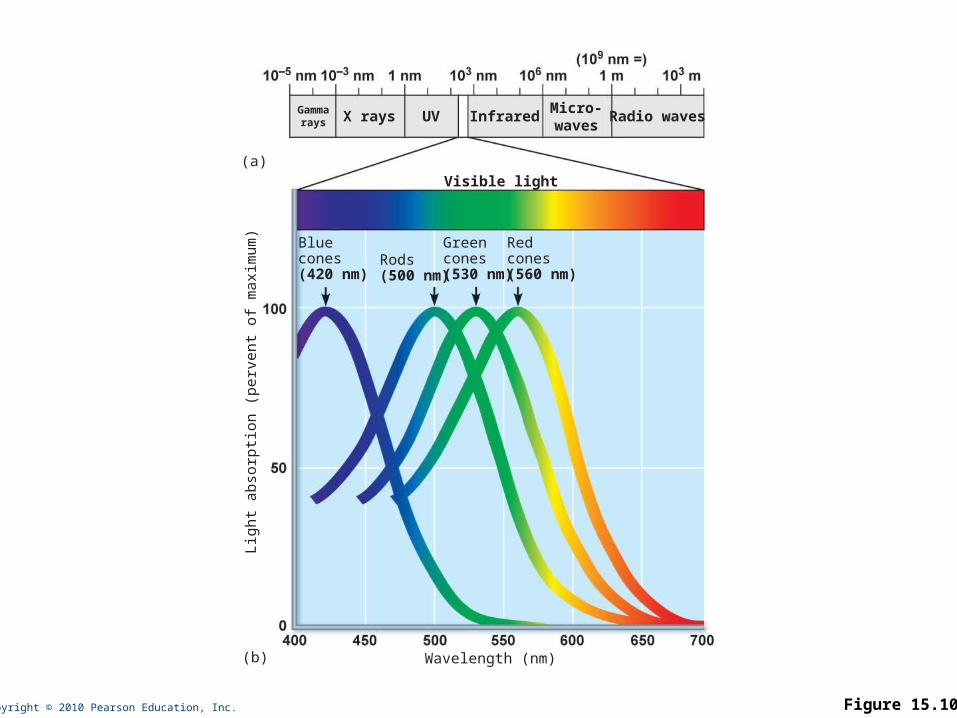
Copyright © 2010 Pearson Education, Inc. Figure 15.10
Wavelength (nm)
Visible light
(b)
(a)
Bluecones(420 nm)
Rods(500 nm)
Greencones(530 nm)
Redcones(560 nm)
X rays UV InfraredMicro-waves
Radio wavesGammarays
Lig
ht
abso
rpti
on (
perv
ent
of
maxim
um
)

Copyright © 2010 Pearson Education, Inc.
Refraction and Lenses
• Refraction
• Bending of a light ray due to change in speed when light passes from one transparent medium to another
• Occurs when light meets the surface of a different medium at an oblique angle

Copyright © 2010 Pearson Education, Inc.
Refraction and Lenses
• Light passing through a convex lens (as in the eye) is bent so that the rays converge at a focal point
• The image formed at the focal point is upside-down and reversed right to left
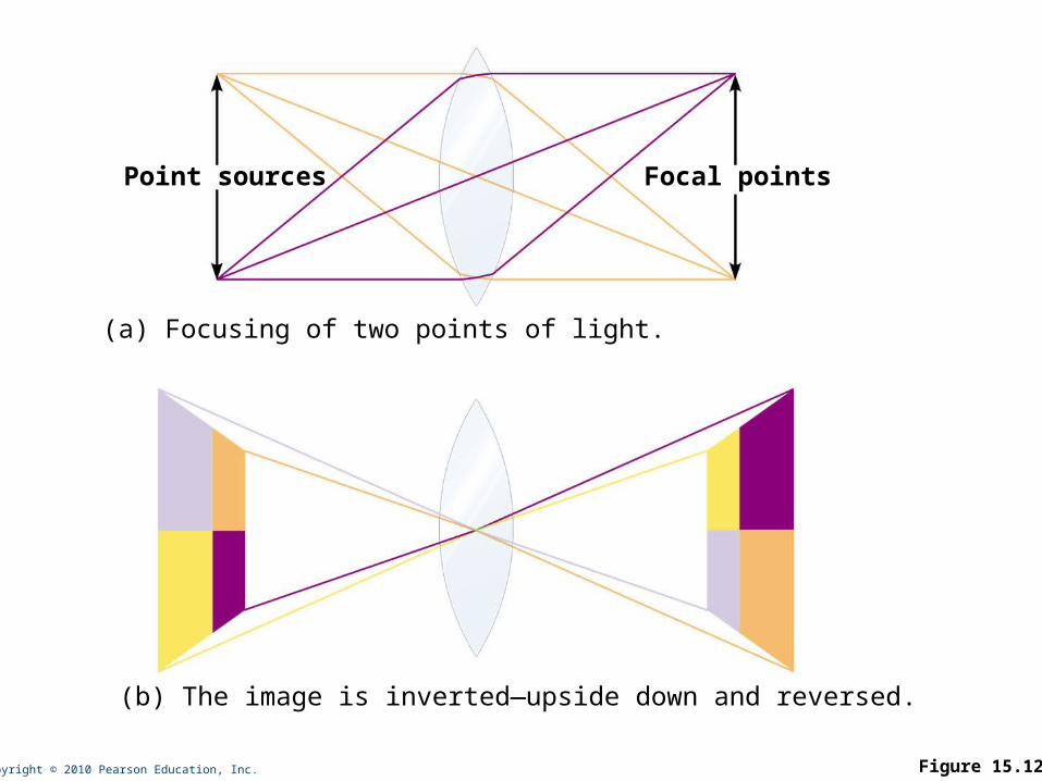
Copyright © 2010 Pearson Education, Inc. Figure 15.12
Point sources
(a) Focusing of two points of light.
(b) The image is inverted—upside down and reversed.
Focal points

Copyright © 2010 Pearson Education, Inc.
Focusing Light on the Retina
• Pathway of light entering the eye: cornea, aqueous humor, lens, vitreous humor, neural layer of retina, photoreceptors
• Light is refracted
• At the cornea
• Entering the lens
• Leaving the lens
• Change in lens curvature allows for fine focusing of an image

Copyright © 2010 Pearson Education, Inc.
Focusing for Distant Vision
• Light rays from distant objects are nearly parallel at the eye and need little refraction beyond what occurs in the at-rest eye
• Far point of vision: the distance beyond which no change in lens shape is needed for focusing; 20 feet for emmetropic (normal) eye
• Ciliary muscles are relaxed
• Lens is stretched flat by tension in the ciliary zonule

Copyright © 2010 Pearson Education, Inc. Figure 15.13a
Lens
Invertedimage
Ciliary zonule
Ciliary muscle
Nearly parallel raysfrom distant object
(a) Lens is flattened for distant vision. Sympatheticinput relaxes the ciliary muscle, tightening the ciliary zonule, and flattening the lens.
Sympathetic activation
Copyright © 2010 Pearson Education, Inc.
Focusing for Close Vision
• Light from a close object diverges as it approaches the eye; requires that the eye make active adjustments

Copyright © 2010 Pearson Education, Inc.
Focusing for Close Vision
• Close vision requires
• Accommodation—changing the lens shape by ciliary muscles to increase refractory power
• Near point of vision is determined by the maximum bulge the lens can achieve
• Presbyopia—loss of accommodation over age 50
• Constriction—the accommodation pupillary reflex constricts the pupils to prevent the most divergent light rays from entering the eye
• Convergence—medial rotation of the eyeballs toward the object being viewed
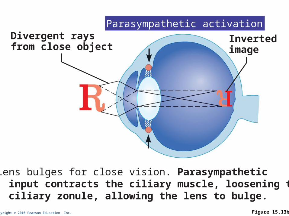
Copyright © 2010 Pearson Education, Inc. Figure 15.13b
Divergent raysfrom close object
(b) Lens bulges for close vision. Parasympathetic input contracts the ciliary muscle, loosening the ciliary zonule, allowing the lens to bulge.
Invertedimage
Parasympathetic activation

Copyright © 2010 Pearson Education, Inc.
Problems of Refraction
• Myopia (nearsightedness)—focal point is in front of the retina, e.g. in a longer than normal eyeball
• Corrected with a concave lens
• Hyperopia (farsightedness)—focal point is behind the retina, e.g. in a shorter than normal eyeball
• Corrected with a convex lens
• Astigmatism—caused by unequal curvatures in different parts of the cornea or lens
• Corrected with cylindrically ground lenses, corneal implants, or laser procedures
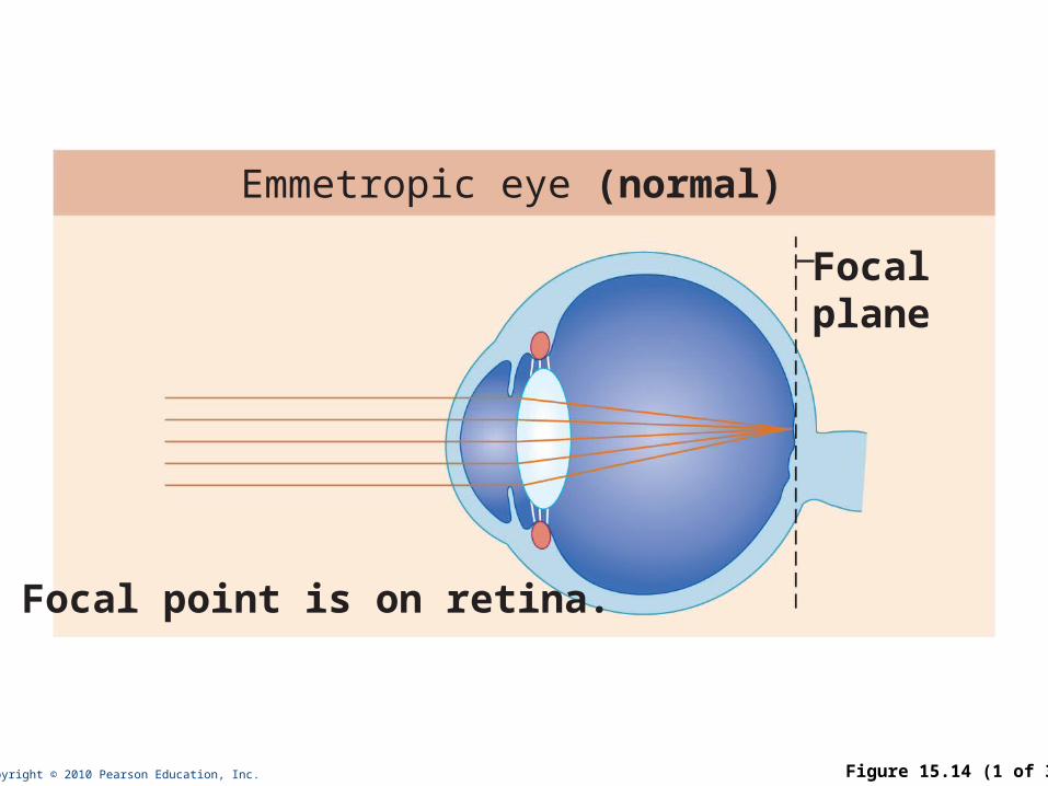
Copyright © 2010 Pearson Education, Inc. Figure 15.14 (1 of 3)
Focalplane
Focal point is on retina.
Emmetropic eye (normal)
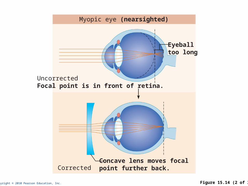
Copyright © 2010 Pearson Education, Inc. Figure 15.14 (2 of 3)
Concave lens moves focalpoint further back.
Eyeballtoo long
UncorrectedFocal point is in front of retina.
Corrected
Myopic eye (nearsighted)

Copyright © 2010 Pearson Education, Inc. Figure 15.14 (3 of 3)
Eyeballtoo short
UncorrectedFocal point is behind retina.
CorrectedConvex lens moves focalpoint forward.
Hyperopic eye (farsighted)

Copyright © 2010 Pearson Education, Inc.
Functional Anatomy of Photoreceptors
• Rods and cones
• Outer segment of each contains visual pigments (photopigments)—molecules that change shape as they absorb light
• Inner segment of each joins the cell body

Copyright © 2010 Pearson Education, Inc. Figure 15.15a
Process ofbipolar cell
Outer fiber
Apical microvillus
Discs containingvisual pigments
Melaningranules
Discs beingphagocytized Pigment cell nucleus
Inner fibers
Rod cell body
Cone cell body
Synaptic terminals
Rod cell body
Nuclei
Mitochondria
Connectingcilia
Basal lamina (borderwith choroid)
The outer segments of rods and cones are embedded in the pigmented layer of the retina.
Pig
men
ted
layer
Ou
ter
seg
men
tIn
ner
seg
men
t

Copyright © 2010 Pearson Education, Inc.
Rods
• Functional characteristics
• Very sensitive to dim light
• Best suited for night vision and peripheral vision
• Perceived input is in gray tones only
• Pathways converge, resulting in fuzzy and indistinct images

Copyright © 2010 Pearson Education, Inc.
Cones
• Functional characteristics
• Need bright light for activation (have low sensitivity)
• Have one of three pigments that furnish a vividly colored view
• Nonconverging pathways result in detailed, high-resolution vision

Copyright © 2010 Pearson Education, Inc.
Chemistry of Visual Pigments
• Retinal
• Light-absorbing molecule that combines with one of four proteins (opsin) to form visual pigments
• Synthesized from vitamin A
• Two isomers: 11-cis-retinal (bent form) and all-trans-retinal (straight form)
• Conversion of 11-cis-retinal to all-trans-retinal initiates a chain of reactions leading to transmission of electrical impulses in the optic nerve

Copyright © 2010 Pearson Education, Inc. Figure 15.15b
Rod discs
Visualpigmentconsists of• Retinal• Opsin
(b) Rhodopsin, the visual pigment in rods, is embedded in the membrane that forms discs in the outer segment.

Copyright © 2010 Pearson Education, Inc.
Excitation of Rods
• The visual pigment of rods is rhodopsin (opsin + 11-cis-retinal)
• In the dark, rhodopsin forms and accumulates
• Regenerated from all-trans-retinal
• Formed from vitamin A
• When light is absorbed, rhodopsin breaks down
• 11-cis isomer is converted into the all-trans isomer
• Retinal and opsin separate (bleaching of the pigment)
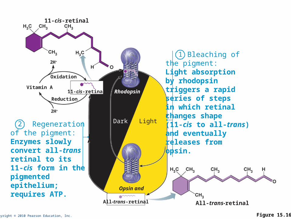
Copyright © 2010 Pearson Education, Inc. Figure 15.16
11-cis-retinal
Bleaching ofthe pigment:Light absorptionby rhodopsintriggers a rapidseries of stepsin which retinalchanges shape(11-cis to all-trans)and eventuallyreleases fromopsin.
1
Rhodopsin
Opsin and
Regenerationof the pigment:Enzymes slowlyconvert all-transretinal to its11-cis form in thepigmentedepithelium;requires ATP.
Dark Light
All-trans-retinal
Oxidation
2H+
2H+
Reduction
Vitamin A
2
11-cis-retinal
All-trans-retinal

Copyright © 2010 Pearson Education, Inc.
Excitation of Cones
• Method of excitation is similar to that of rods
• There are three types of cones, named for the colors of light absorbed: blue, green, and red
• Intermediate hues are perceived by activation of more than one type of cone at the same time
• Color blindness is due to a congenital lack of one or more of the cone types

Copyright © 2010 Pearson Education, Inc.
Light Adaptation
• Occurs when moving from darkness into bright light
• Large amounts of pigments are broken down instantaneously, producing glare
• Pupils constrict
• Dramatic changes in retinal sensitivity: rod function ceases
• Cones and neurons rapidly adapt
• Visual acuity improves over 5–10 minutes

Copyright © 2010 Pearson Education, Inc.
Dark Adaptation
• Occurs when moving from bright light into darkness
• The reverse of light adaptation
• Cones stop functioning in low-intensity light
• Pupils dilate
• Rhodopsin accumulates in the dark and retinal sensitivity increases within 20–30 minutes
Copyright © 2010 Pearson Education, Inc.
Visual Pathway
• Axons of retinal ganglion cells form the optic nerve
• Medial fibers of the optic nerve decussate at the optic chiasma
• Most fibers of the optic tracts continue to the lateral geniculate body of the thalamus
Copyright © 2010 Pearson Education, Inc.
Visual Pathway
• The optic radiation fibers connect to the primary visual cortex in the occipital lobes
• Other optic tract fibers send branches to the midbrain, ending in superior colliculi (initiating visual reflexes)
Copyright © 2010 Pearson Education, Inc.
Visual Pathway
• A small subset of ganglion cells in the retina contain melanopsin (circadian pigment), which projects to:
• Pretectal nuclei (involved with pupillary reflexes)
• Suprachiasmatic nucleus of the hypothalamus, the timer for daily biorhythms
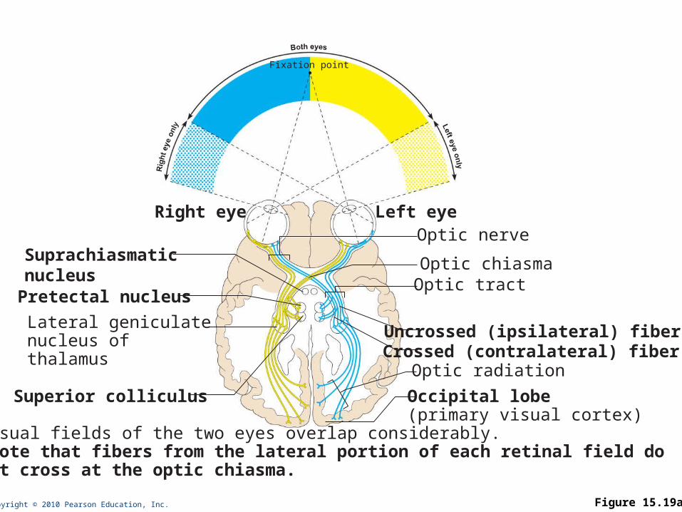
Copyright © 2010 Pearson Education, Inc. Figure 15.19a
Pretectal nucleus
Right eye Left eye
Fixation point
Optic radiation
Optic tractOptic chiasma
Uncrossed (ipsilateral) fiberCrossed (contralateral) fiber
Optic nerve
Lateral geniculatenucleus ofthalamus
Superior colliculus Occipital lobe (primary visual cortex)
The visual fields of the two eyes overlap considerably. Note that fibers from the lateral portion of each retinal field do not cross at the optic chiasma.
Suprachiasmaticnucleus

Copyright © 2010 Pearson Education, Inc.
Depth Perception
• Both eyes view the same image from slightly different angles
• Depth perception (three-dimensional vision) results from cortical fusion of the slightly different images
Copyright © 2010 Pearson Education, Inc.
Thalamic Processing
• Lateral geniculate nuclei of the thalamus
• Relay information on movement
• Segregate the retinal axons in preparation for depth perception
• Emphasize visual inputs from regions of high cone density
• Sharpen contrast information

Copyright © 2010 Pearson Education, Inc.
Cortical Processing
• Two areas in the visual cortex
1. Striate cortex (primary visual cortex)
• Processes contrast information and object orientation
2. Prestriate cortices (visual association areas)
• Processes form, color, and motion input from striate cortex
• Complex visual processing extends into other regions
• Temporal lobe—processes identification of objects
• Parietal cortex and postcentral gyrus—process spatial location