Copyright © 2010 Pearson Education, Inc. Bell Ringer!!!! (All Bell Ringers Due on Friday) What...
43
pyright © 2010 Pearson Education, Inc. Bell Ringer!!!! (All Bell Ringers Due on Friday) • What bones articulate with the manubrium? • How would a complete fracture of the dens affect the mobility of the vertebral column? • What bones contain the paranasal sinuses?
-
Upload
lisandro-wolden -
Category
Documents
-
view
216 -
download
0
Transcript of Copyright © 2010 Pearson Education, Inc. Bell Ringer!!!! (All Bell Ringers Due on Friday) What...
Copyright © 2010 Pearson Education, Inc.
Bell Ringer!!!!(All Bell Ringers Due on Friday)
• What bones articulate with the manubrium?
• How would a complete fracture of the dens affect the mobility of the vertebral column?
• What bones contain the paranasal sinuses?
Copyright © 2010 Pearson Education, Inc.
The Appendicular Skeleton
http://www.cteonline.org/portal/default/Resources/Viewer/ResourceViewer?action=2&resid=12835
Copyright © 2010 Pearson Education, Inc.
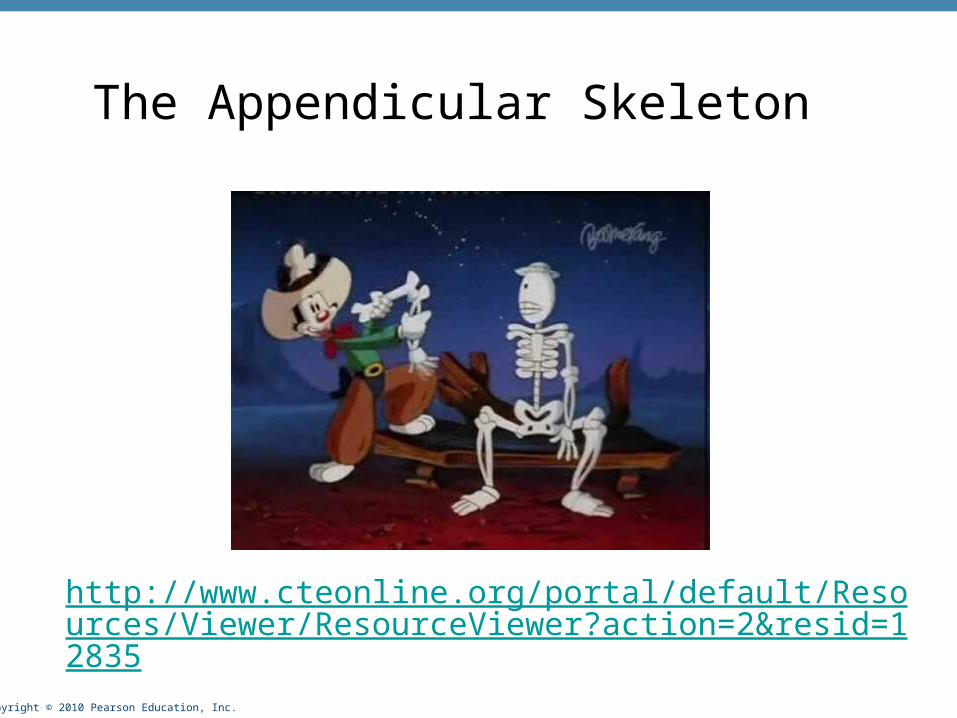
Bones of the limbs and their girdles
• Pectoral girdle attaches the upper limbs to the body trunk
• Pelvic girdle secures the lower limbs
Copyright © 2010 Pearson Education, Inc.
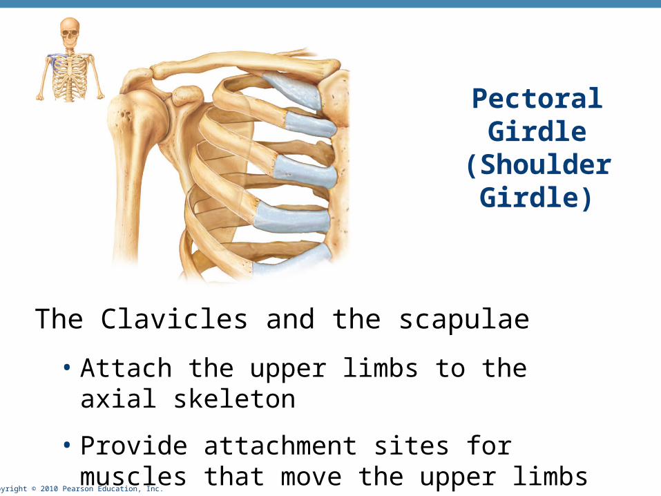
Pectoral Girdle (Shoulder Girdle)
The Clavicles and the scapulae
• Attach the upper limbs to the axial skeleton
• Provide attachment sites for muscles that move the upper limbs
Copyright © 2010 Pearson Education, Inc.
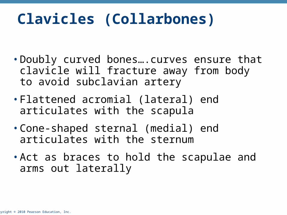
Clavicles (Collarbones)
• Doubly curved bones….curves ensure that clavicle will fracture away from body to avoid subclavian artery
• Flattened acromial (lateral) end articulates with the scapula
• Cone-shaped sternal (medial) end articulates with the sternum
• Act as braces to hold the scapulae and arms out laterally
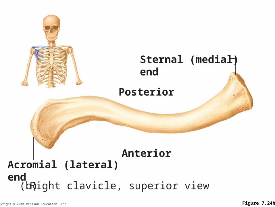
Copyright © 2010 Pearson Education, Inc. Figure 7.24b
Acromial (lateral)end(b) Right clavicle, superior view
Posterior
Sternal (medial)end
Anterior
Copyright © 2010 Pearson Education, Inc.
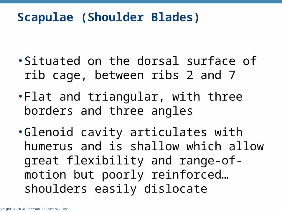
Scapulae (Shoulder Blades)
• Situated on the dorsal surface of rib cage, between ribs 2 and 7
• Flat and triangular, with three borders and three angles
• Glenoid cavity articulates with humerus and is shallow which allow great flexibility and range-of-motion but poorly reinforced…shoulders easily dislocate
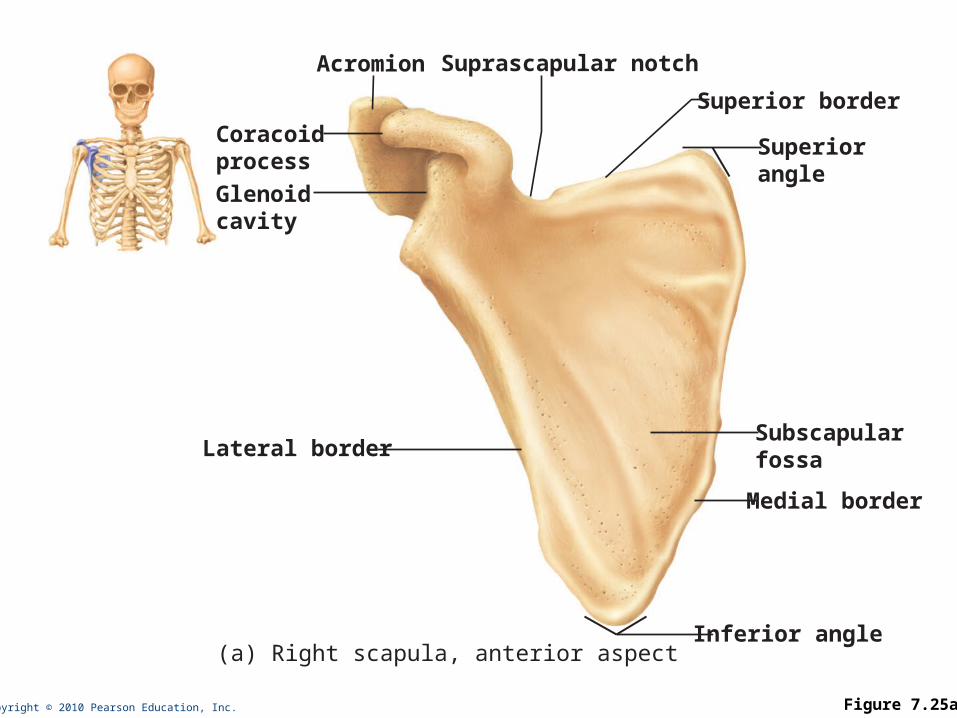
Copyright © 2010 Pearson Education, Inc. Figure 7.25a
Acromion
Coracoidprocess
Suprascapular notch
Superior border
Superiorangle
Subscapularfossa
Medial border
Inferior angle
Glenoidcavity
Lateral border
(a) Right scapula, anterior aspect
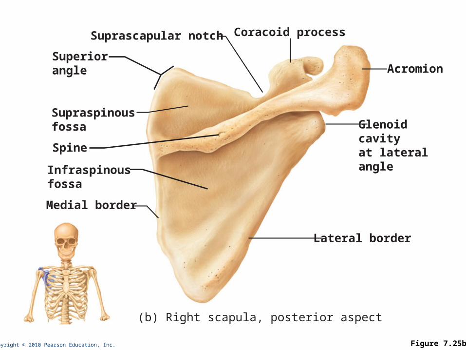
Copyright © 2010 Pearson Education, Inc. Figure 7.25b
Superiorangle
Medial border
Coracoid processSuprascapular notch
Acromion
Glenoidcavityat lateralangle
Lateral border
Infraspinousfossa
Spine
(b) Right scapula, posterior aspect
Supraspinousfossa
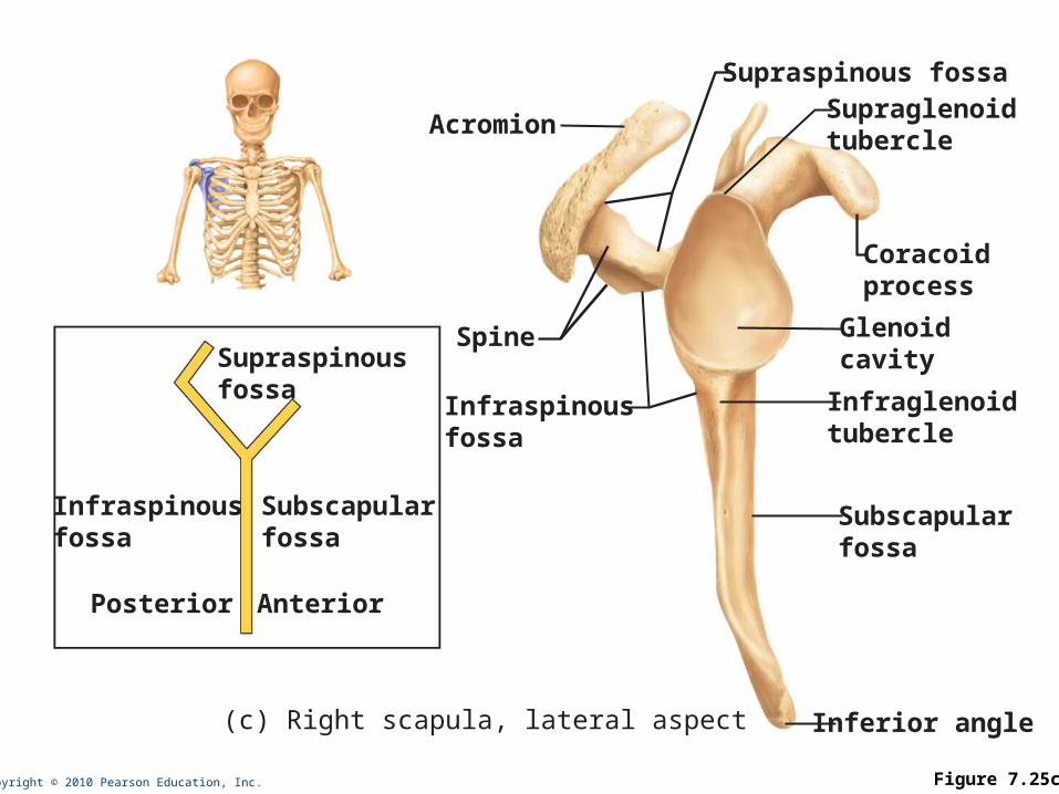
Copyright © 2010 Pearson Education, Inc. Figure 7.25c
Coracoidprocess
Glenoidcavity
Acromion
Infraspinousfossa
Spine
(c) Right scapula, lateral aspect
Infraglenoidtubercle
Supraglenoidtubercle
Supraspinous fossa
Subscapularfossa
Inferior angle
Supraspinousfossa
Infraspinousfossa
Subscapularfossa
Posterior Anterior
Copyright © 2010 Pearson Education, Inc.
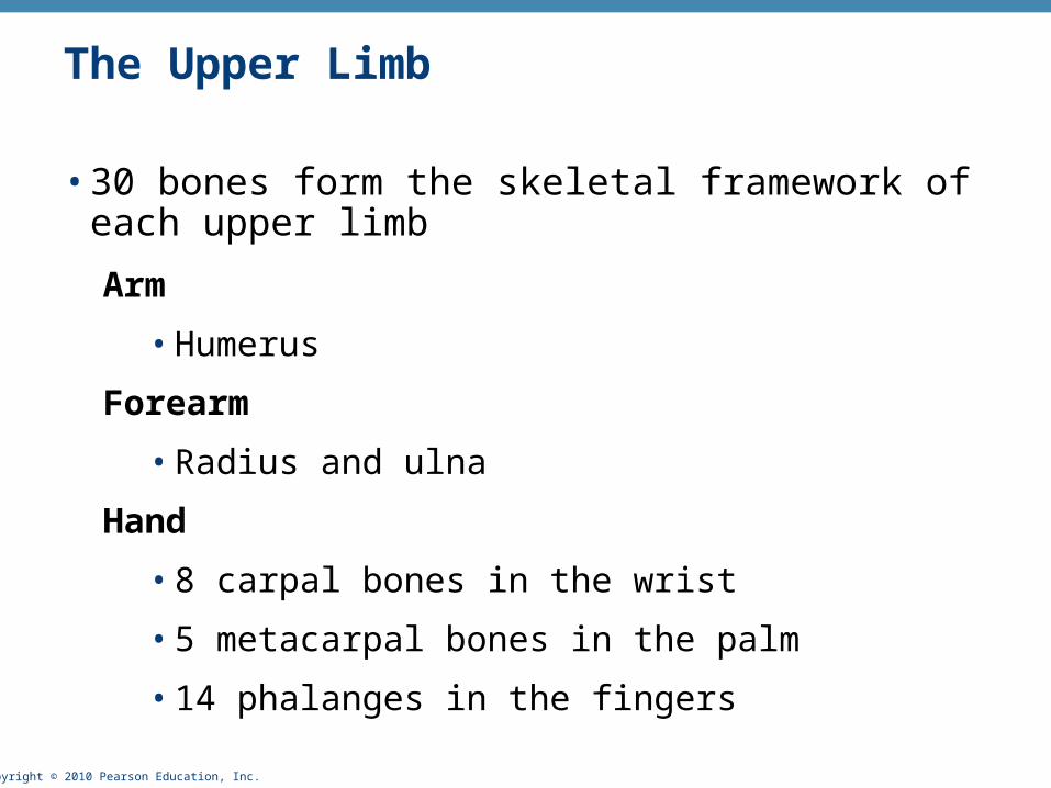
The Upper Limb
• 30 bones form the skeletal framework of each upper limb
Arm
• Humerus
Forearm
• Radius and ulna
Hand
• 8 carpal bones in the wrist
• 5 metacarpal bones in the palm
• 14 phalanges in the fingers
Copyright © 2010 Pearson Education, Inc.
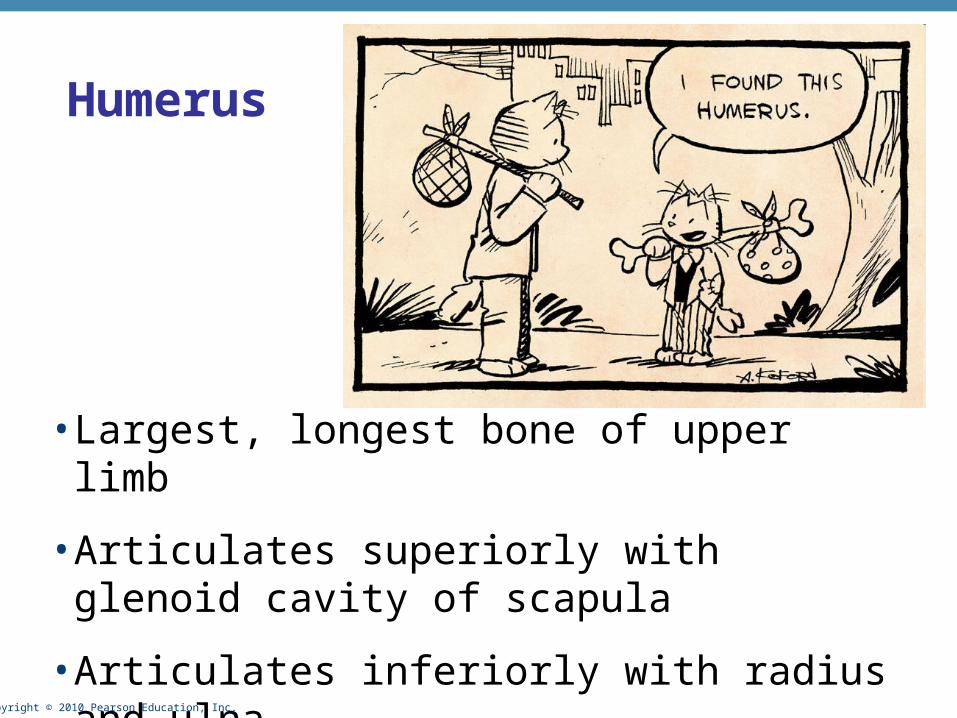
• Largest, longest bone of upper limb
• Articulates superiorly with glenoid cavity of scapula
• Articulates inferiorly with radius and ulna
Humerus
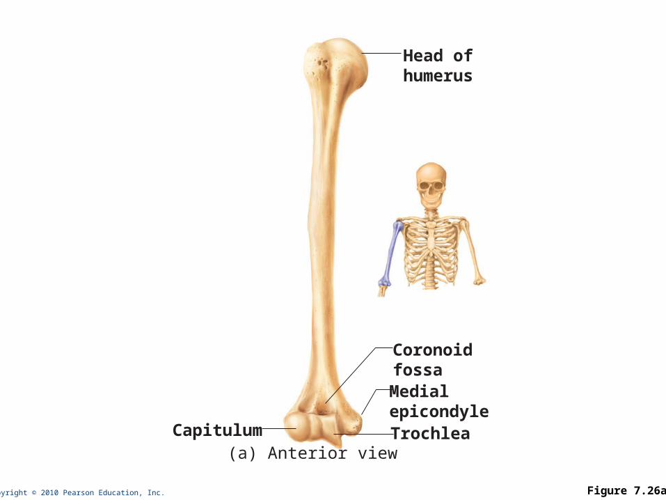
Copyright © 2010 Pearson Education, Inc. Figure 7.26a
Capitulum
Head ofhumerus
CoronoidfossaMedialepicondyleTrochlea
(a) Anterior view
Copyright © 2010 Pearson Education, Inc.
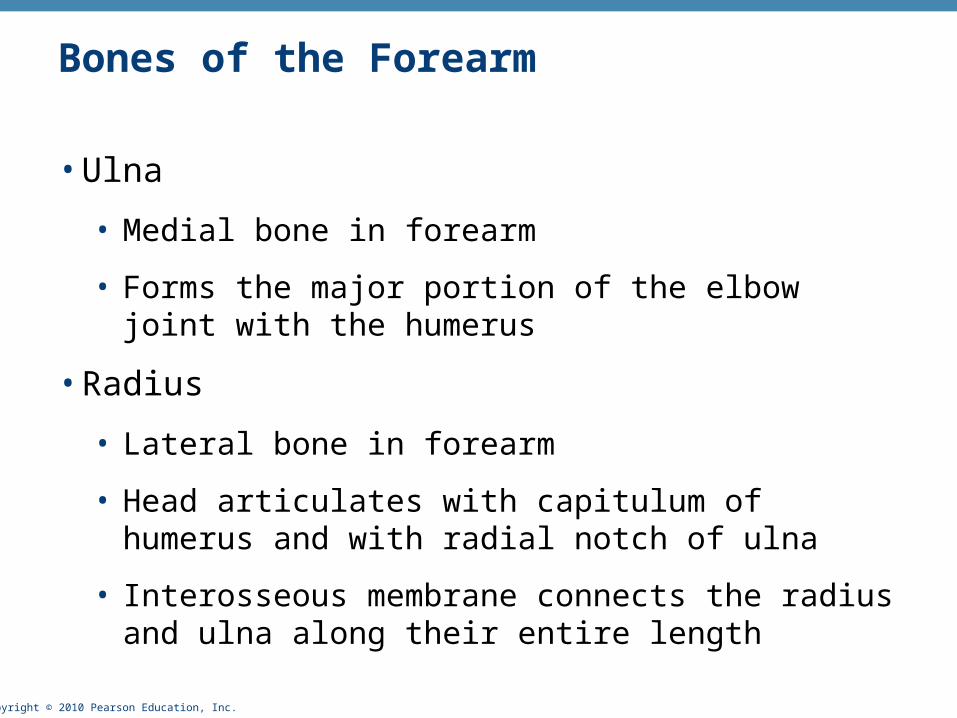
Bones of the Forearm
• Ulna
• Medial bone in forearm
• Forms the major portion of the elbow joint with the humerus
• Radius
• Lateral bone in forearm
• Head articulates with capitulum of humerus and with radial notch of ulna
• Interosseous membrane connects the radius and ulna along their entire length
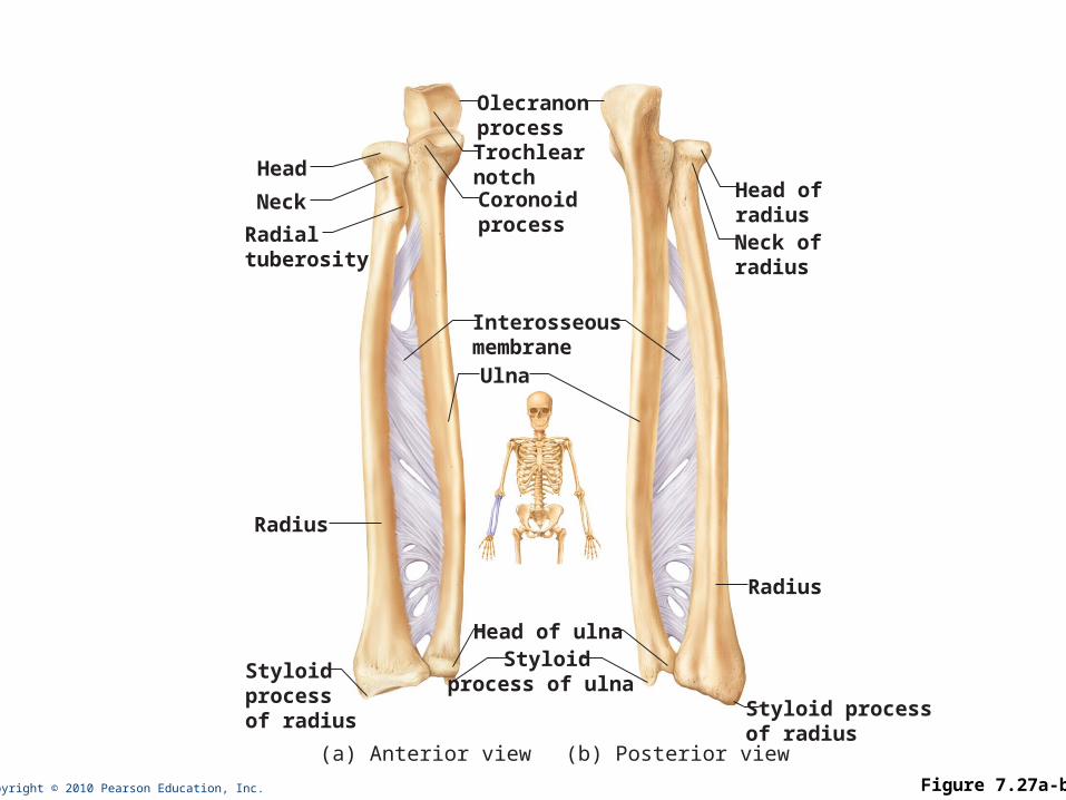
Copyright © 2010 Pearson Education, Inc. Figure 7.27a-b
OlecranonprocessTrochlearnotchCoronoidprocess
Styloid processof radius
Radius
Neck ofradius
Head ofradius
Head of ulnaStyloid
process of ulna
InterosseousmembraneUlna
Head
Neck
Radialtuberosity
Radius
Styloidprocessof radius
(a) Anterior view (b) Posterior view
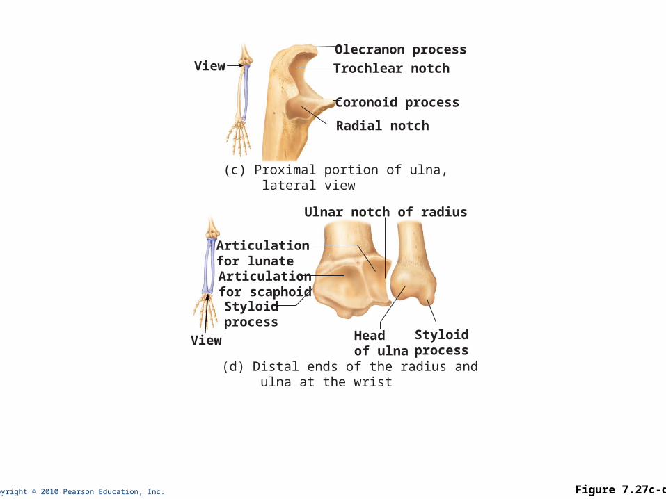
Copyright © 2010 Pearson Education, Inc. Figure 7.27c-d
(c) Proximal portion of ulna, lateral view
Olecranon process
Trochlear notch
Coronoid process
Radial notch
View
(d) Distal ends of the radius and ulna at the wrist
Ulnar notch of radius
Headof ulna
Styloidprocess
Articulationfor scaphoid
Articulationfor lunate
Styloidprocess
View
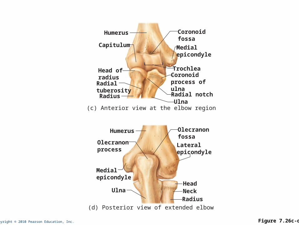
Copyright © 2010 Pearson Education, Inc. Figure 7.26c-d
Coronoidfossa
Radius
Radialtuberosity
Head ofradius
Capitulum
Trochlea
(c) Anterior view at the elbow region
Humerus
Medialepicondyle
Coronoidprocess of ulna
UlnaRadial notch
Olecranonfossa
Ulna
Olecranonprocess
Medialepicondyle
(d) Posterior view of extended elbow
Humerus
Lateralepicondyle
Head
RadiusNeck
Copyright © 2010 Pearson Education, Inc.
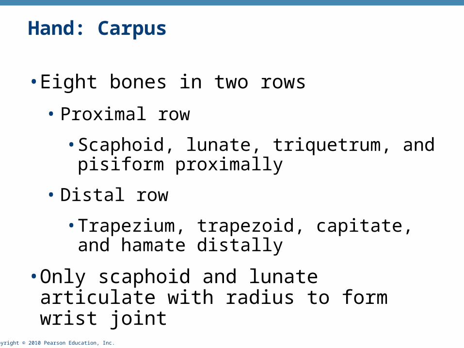
Hand: Carpus
• Eight bones in two rows
• Proximal row
• Scaphoid, lunate, triquetrum, and pisiform proximally
• Distal row
• Trapezium, trapezoid, capitate, and hamate distally
• Only scaphoid and lunate articulate with radius to form wrist joint
Copyright © 2010 Pearson Education, Inc.
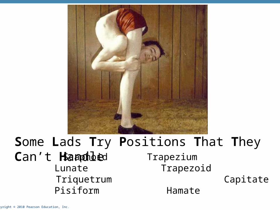
Some Lads Try Positions That They Can’t Handle Scaphoid Trapezium
Lunate Trapezoid Triquetrum Capitate
Pisiform Hamate
Copyright © 2010 Pearson Education, Inc.
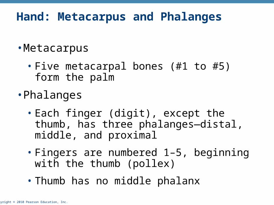
Hand: Metacarpus and Phalanges
• Metacarpus
• Five metacarpal bones (#1 to #5) form the palm
• Phalanges
• Each finger (digit), except the thumb, has three phalanges—distal, middle, and proximal
• Fingers are numbered 1–5, beginning with the thumb (pollex)
• Thumb has no middle phalanx
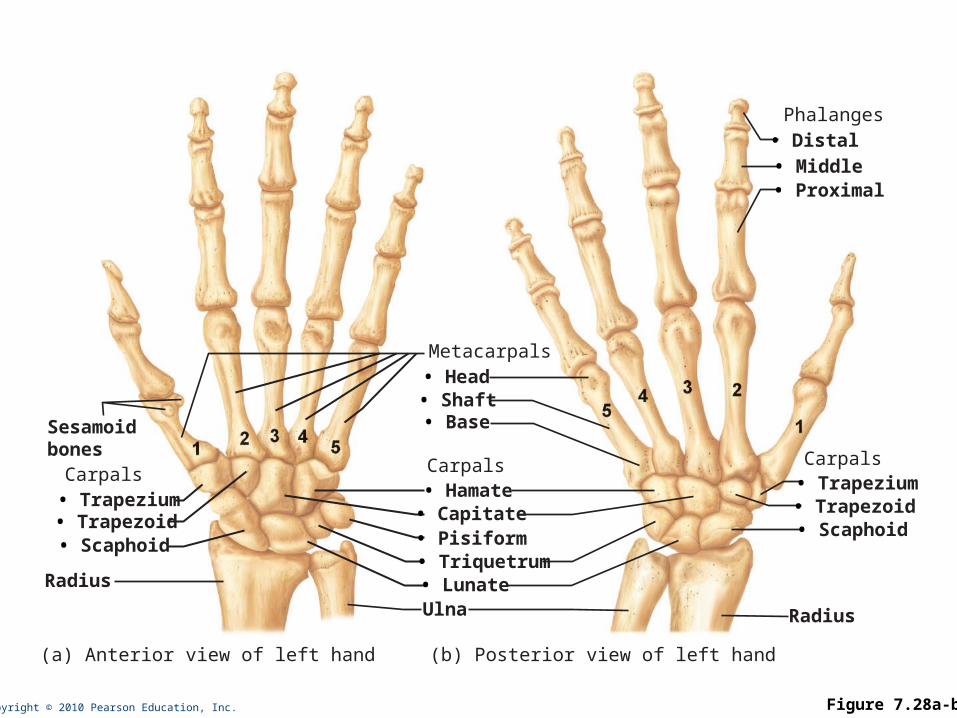
Copyright © 2010 Pearson Education, Inc. Figure 7.28a-b
• Trapezoid• Trapezium
• Scaphoid
Phalanges
Carpals
Radius
• Proximal• Middle• Distal
• Triquetrum• Lunate
• Capitate• Hamate
• Pisiform
Metacarpals
Carpals
(b) Posterior view of left hand
Ulna
• Base• Shaft• Head
• Trapezoid• Trapezium
• Scaphoid
Carpals
(a) Anterior view of left hand
Radius
Sesamoidbones
Copyright © 2010 Pearson Education, Inc.
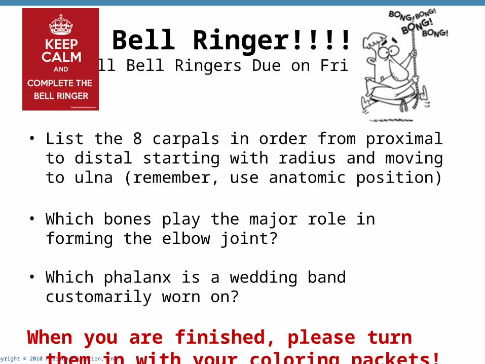
Bell Ringer!!!!(All Bell Ringers Due on Friday)
• List the 8 carpals in order from proximal to distal starting with radius and moving to ulna (remember, use anatomic position)
• Which bones play the major role in forming the elbow joint?
• Which phalanx is a wedding band customarily worn on?
When you are finished, please turn them in with your coloring packets!
Copyright © 2010 Pearson Education, Inc.
Pelvic (Hip) Girdle
• Two hip bones (each also called coxal bone or os coxae)
• Attach the lower limbs to the axial skeleton with strong ligaments
• Transmit weight of upper body to lower limbs
• Support pelvic organs
• Each hip bone consists of three fused bones: ilium, ischium, and pubis
• Together with the sacrum and the coccyx, these bones form the bony pelvis
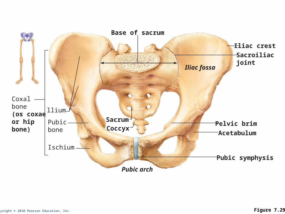
Copyright © 2010 Pearson Education, Inc. Figure 7.29
Coxalbone(os coxaeor hip bone)
llium
Sacroiliacjoint
Iliac fossa
Pubicbone
Ischium
Sacrum
Base of sacrum
Pelvic brim
Acetabulum
Pubic symphysis
Iliac crest
Coccyx
Pubic arch
Copyright © 2010 Pearson Education, Inc.
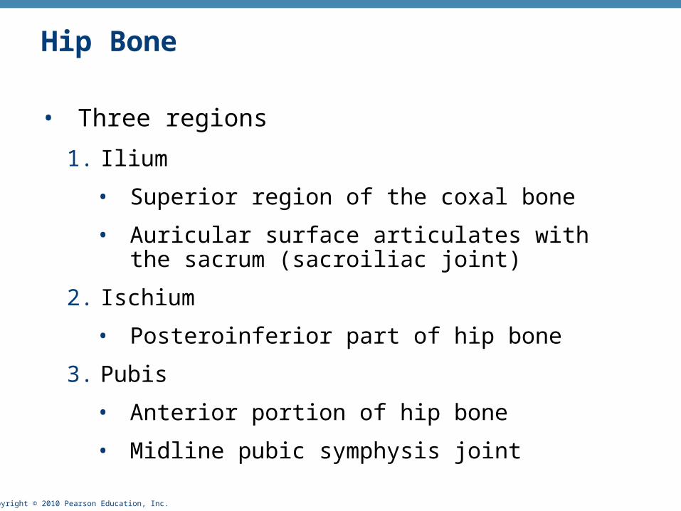
Hip Bone
• Three regions
1. Ilium
• Superior region of the coxal bone
• Auricular surface articulates with the sacrum (sacroiliac joint)
2. Ischium
• Posteroinferior part of hip bone
3. Pubis
• Anterior portion of hip bone
• Midline pubic symphysis joint
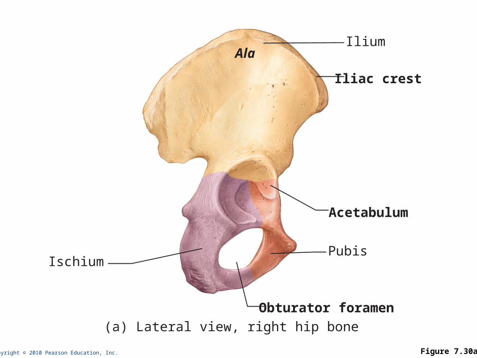
Copyright © 2010 Pearson Education, Inc. Figure 7.30a
IliumAla
Ischium
Obturator foramen
Acetabulum
Iliac crest
Pubis
(a) Lateral view, right hip bone
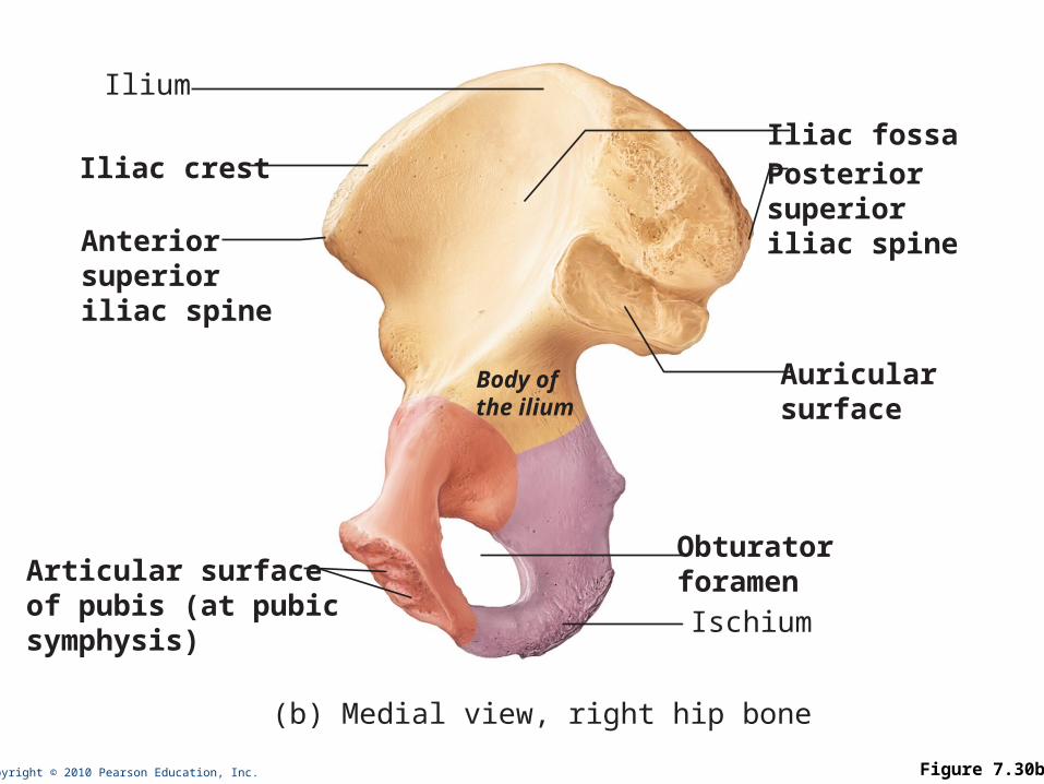
Copyright © 2010 Pearson Education, Inc. Figure 7.30b
Iliac fossa
Ilium
Iliac crest
Anteriorsuperioriliac spine
Posteriorsuperioriliac spine
Obturatorforamen
Body ofthe ilium
Ischium
(b) Medial view, right hip bone
Auricularsurface
Articular surfaceof pubis (at pubic symphysis)
Copyright © 2010 Pearson Education, Inc.
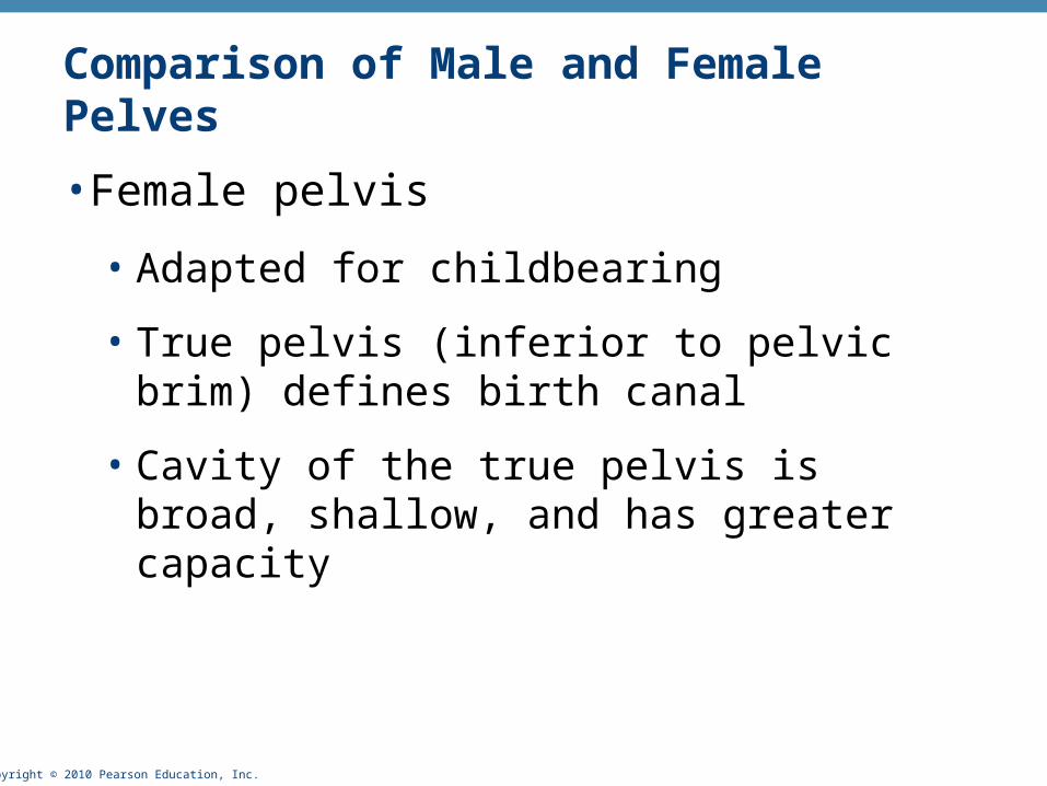
Comparison of Male and Female Pelves
• Female pelvis
• Adapted for childbearing
• True pelvis (inferior to pelvic brim) defines birth canal
• Cavity of the true pelvis is broad, shallow, and has greater capacity
Copyright © 2010 Pearson Education, Inc.
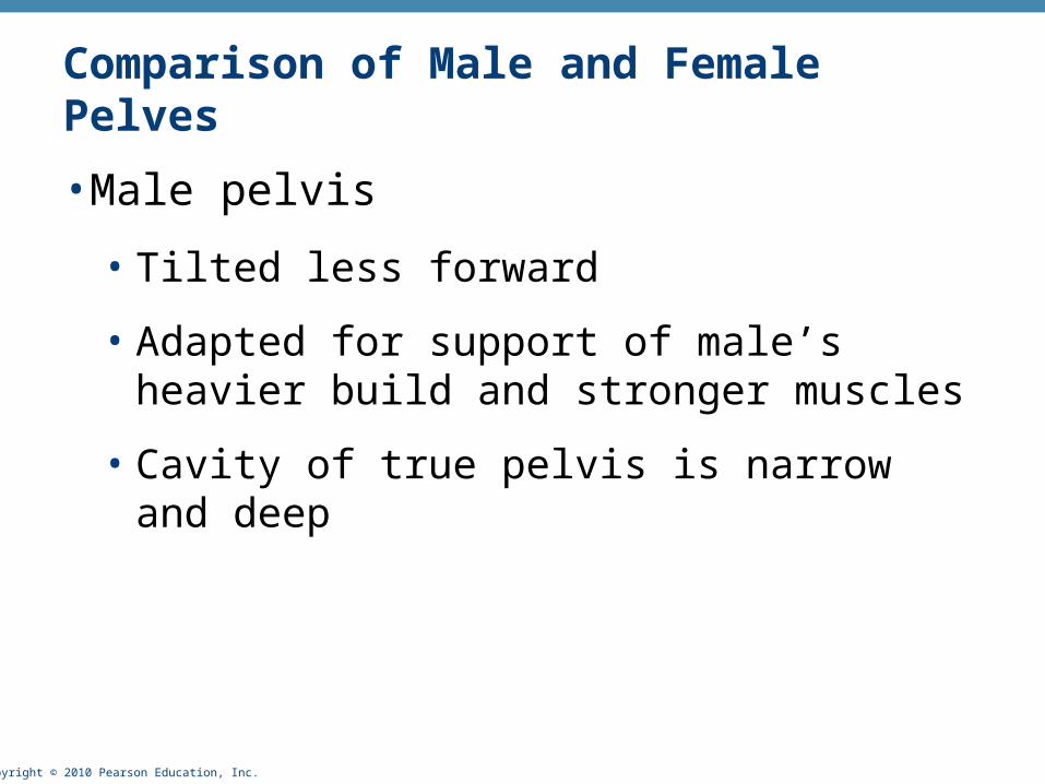
Comparison of Male and Female Pelves
• Male pelvis
• Tilted less forward
• Adapted for support of male’s heavier build and stronger muscles
• Cavity of true pelvis is narrow and deep
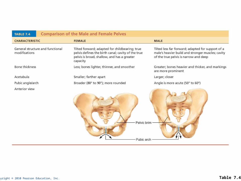
Copyright © 2010 Pearson Education, Inc. Table 7.4
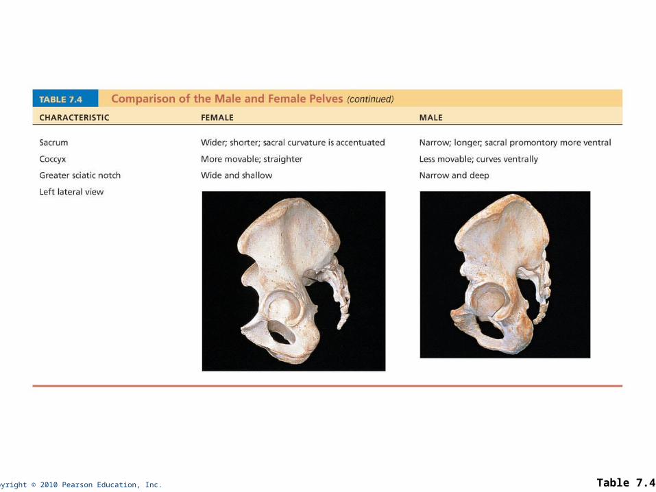
Copyright © 2010 Pearson Education, Inc. Table 7.4
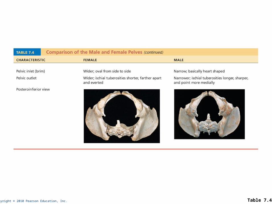
Copyright © 2010 Pearson Education, Inc. Table 7.4
Copyright © 2010 Pearson Education, Inc.
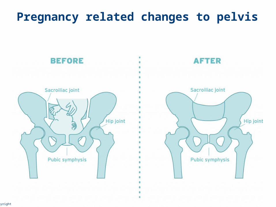
Pregnancy related changes to pelvis
Copyright © 2010 Pearson Education, Inc.
The Lower Limb
• Carries the weight of the body
• Subjected to exceptional forces
• Three segments of the lower limb
• Thigh: femur
• Leg: tibia and fibula
• Foot: 7 tarsal bones in the ankle, 5 metatarsal bones in the metatarsus, and 14 phalanges in the toes
Copyright © 2010 Pearson Education, Inc.
Femur
• Largest and strongest bone in the body
• Articulates proximally with the acetabulum of the hip and distally with the tibia and patella
• Hip socket (acetabulum) firmly secures head of femur into place and provides greater stability but less range-of-motion than pectoral girdle
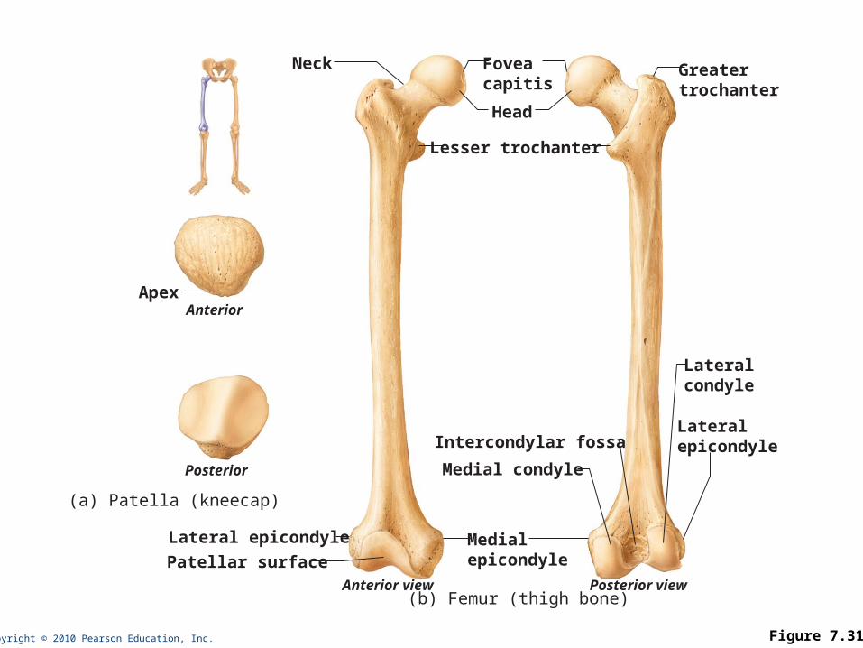
Copyright © 2010 Pearson Education, Inc. Figure 7.31
Neck Foveacapitis
Greatertrochanter
Head
Lesser trochanter
Lateralcondyle
LateralepicondyleIntercondylar fossa
Medial condyle
Medialepicondyle
Anterior view Posterior view(b) Femur (thigh bone)
Lateral epicondyle
Patellar surface
Posterior
ApexAnterior
(a) Patella (kneecap)
Copyright © 2010 Pearson Education, Inc.
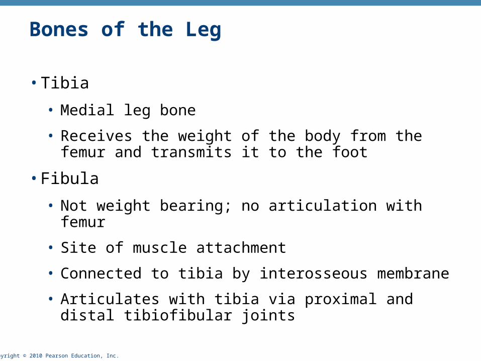
Bones of the Leg
• Tibia
• Medial leg bone
• Receives the weight of the body from the femur and transmits it to the foot
• Fibula
• Not weight bearing; no articulation with femur
• Site of muscle attachment
• Connected to tibia by interosseous membrane
• Articulates with tibia via proximal and distal tibiofibular joints
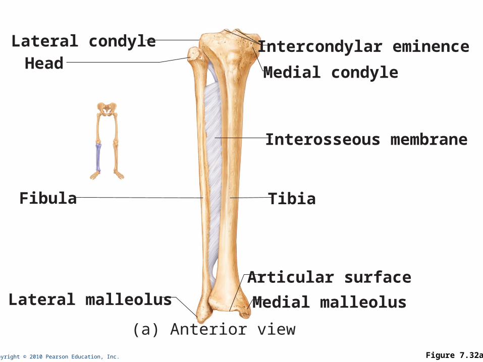
Copyright © 2010 Pearson Education, Inc. Figure 7.32a
Medial condyle
Articular surface
Interosseous membrane
Tibia
Medial malleolus
Intercondylar eminence
Lateral malleolus
Lateral condyle
Fibula
Head
(a) Anterior view
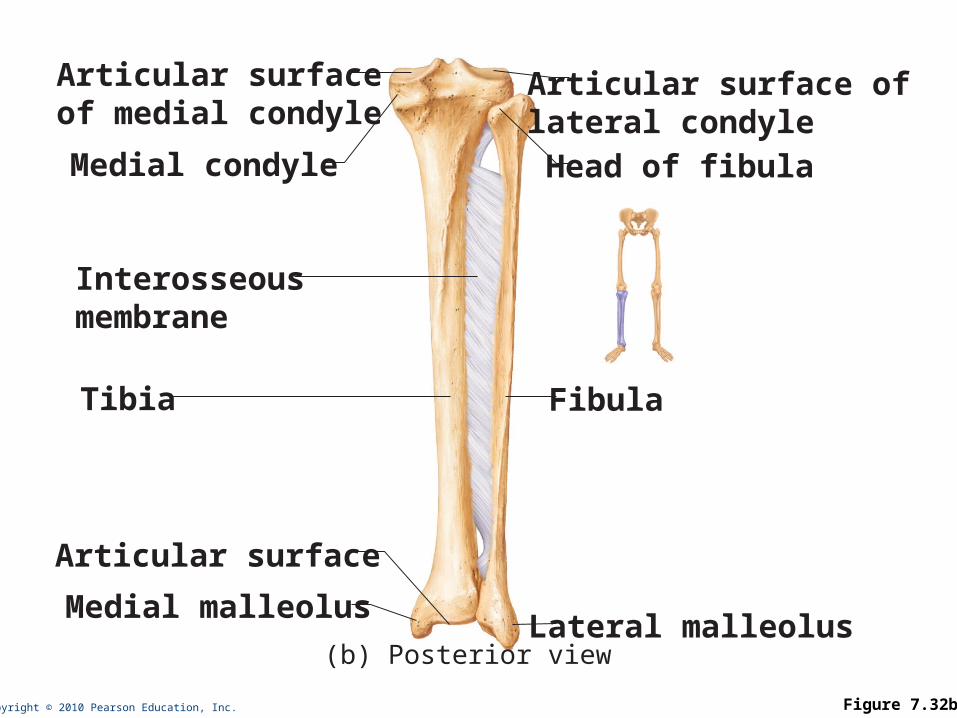
Copyright © 2010 Pearson Education, Inc. Figure 7.32b
Medial condyle
Articular surface oflateral condyle
Articular surfaceof medial condyle
Articular surface
Interosseousmembrane
Tibia Fibula
Head of fibula
Medial malleolusLateral malleolus
(b) Posterior view
Copyright © 2010 Pearson Education, Inc.
Foot: Tarsals
• Seven tarsal bones form the posterior half of the foot
• Talus transfers most of the weight from the tibia to the calcaneus
• Other tarsal bones: cuboid, navicular, and the medial, intermediate, and lateral cuneiforms
Copyright © 2010 Pearson Education, Inc.
Foot: Metatarsals and Phalanges
• Metatarsals:
• Five metatarsal bones (#1 to #5)
• Enlarged head of metatarsal 1 forms the “ball of the foot”
• Phalanges
• The 14 bones of the toes
• Each digit (except the hallux) has three phalanges
• Hallux has no middle phalanx
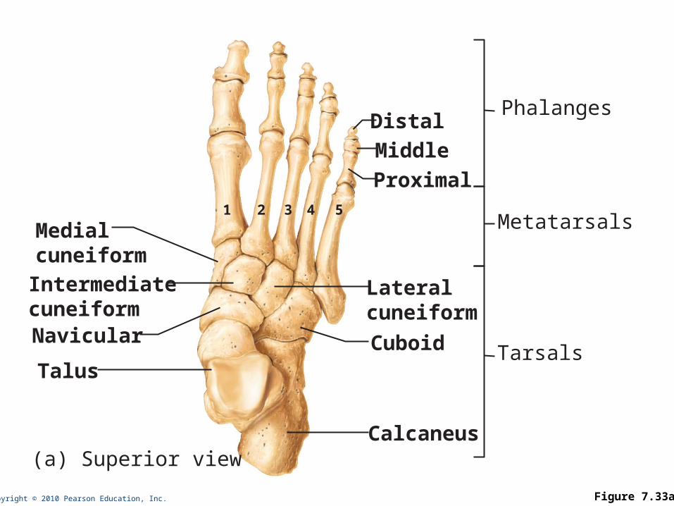
Copyright © 2010 Pearson Education, Inc. Figure 7.33a
Medialcuneiform
Phalanges
Metatarsals
TarsalsNavicular
Intermediatecuneiform
Talus
Calcaneus(a) Superior view
Cuboid
Lateralcuneiform
Proximal54321
Middle
Distal
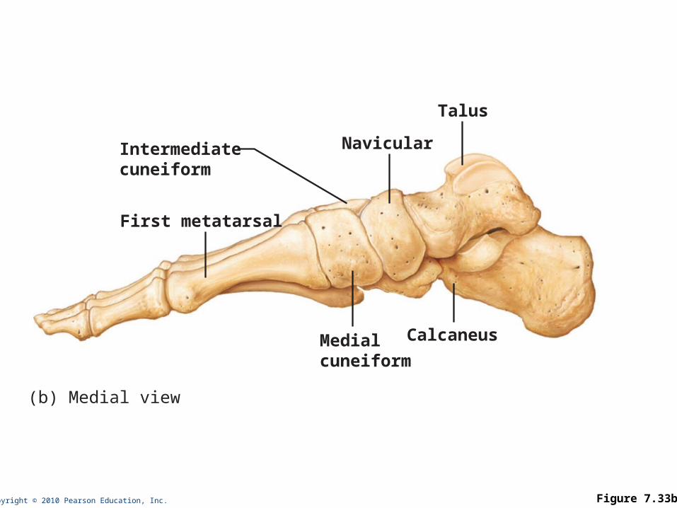
Copyright © 2010 Pearson Education, Inc. Figure 7.33b
(b) Medial view
Intermediatecuneiform
Talus
Navicular
First metatarsal
Medialcuneiform
Calcaneus