Copyright 2009, John Wiley & Sons, Inc. Chapter 18: The Endocrine System.
89
Copyright 2009, John Wiley & Sons, Inc. Chapter 18: The Endocrine System
-
Upload
sophia-riley -
Category
Documents
-
view
215 -
download
0
Transcript of Copyright 2009, John Wiley & Sons, Inc. Chapter 18: The Endocrine System.

Copyright 2009, John Wiley & Sons, Inc.
Chapter 18: The Endocrine System
Copyright 2009, John Wiley & Sons, Inc.
Nervous and Endocrine Systems Act together to coordinate functions of all body
systems Nervous system
Nerve impulses/ Neurotransmitters Faster responses, briefer effects, acts on specific target
Endocrine system Hormone – mediator molecule released in 1 part of the
body but regulates activity of cells in other parts Slower responses, effects last longer, broader influence

18_table_01

Copyright 2009, John Wiley & Sons, Inc.
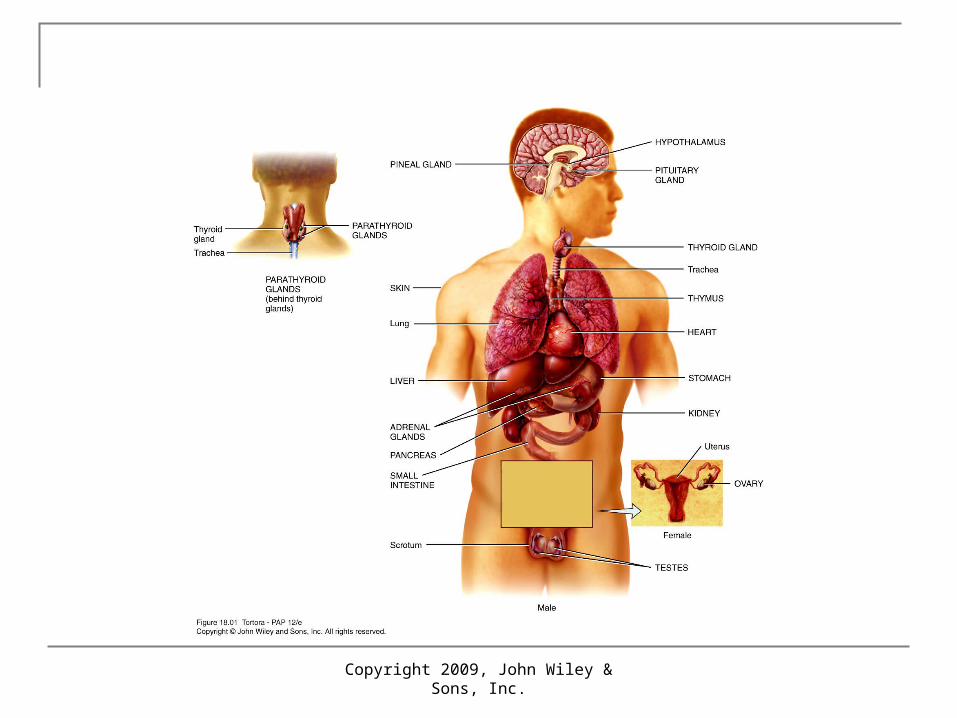
Endocrine Glands
2 kinds of glands Exocrine – ducted Endocrine – ductless
Secrete products into interstitial fluid, diffuse into blood
Endocrine glands include Pituitary, thyroid, parathyroid, adrenal and pineal glands Hypothalamus, thymus, pancreas, ovaries, testes, kidneys,
stomach, liver, small intestine, skin, heart, adipose tissue, and placenta not exclusively endocrine glands
Copyright 2009, John Wiley & Sons, Inc.

Functions of Hormones
1. help regulate chemical composition and volume of internal
environment (interstitial fluid) metabolism and energy balance contraction of smooth and cardiac muscle glandular secretions some immune system activities
2. control growth and development3. regulate operation of reproductive systems4. help establish circadian rhythms
Copyright 2009, John Wiley & Sons, Inc.

Copyright 2009, John Wiley & Sons, Inc.
Hormone Activity
Hormones affect only specific target tissues with specific receptors
Receptors constantly synthesized and broken down Down-regulation – less sensitive Up-regulation – more sensitive

Copyright 2009, John Wiley & Sons, Inc.
Circulating and Local Hormones Hormone types
Circulating – circulate in blood throughout body
Local hormones – act locally Paracrine – act on
neighboring cells Autocrine – act on
the same cell that secreted them

Copyright 2009, John Wiley & Sons, Inc.
Chemical Classes of Hormones
Lipid-soluble – use transport proteins Steroid Thyroid Nitric oxide (NO)
Water-soluble – circulate in “free” form Amine Peptide/ protein Eicosanoid: arachidonic acid
Prostaglandins Leukotrienes

Hormone Transport in the Blood water-soluble hormones circulate “dissolved”
in plasma as “free” form Lipid-soluble hormones require transport
proteins synthesized in liver three functions
water solubility retard filtration in kidney ready reserve
free fraction (0.1-10%) provide active hormone
Copyright 2009, John Wiley & Sons, Inc.
Copyright 2009, John Wiley & Sons, Inc.
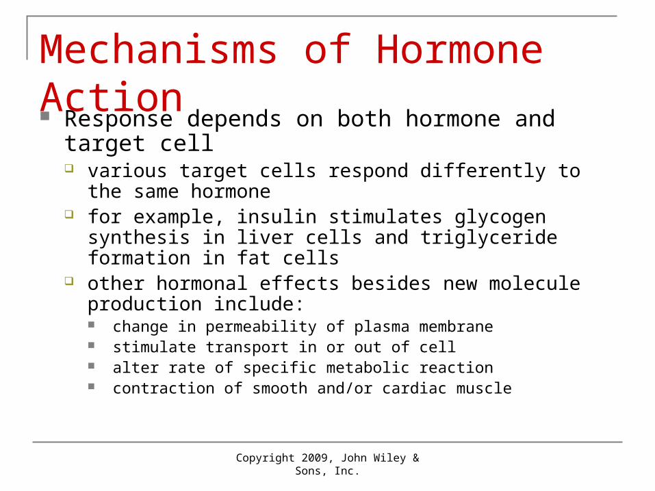
Mechanisms of Hormone Action Response depends on both hormone and target cell
various target cells respond differently to the same hormone for example, insulin stimulates glycogen synthesis in liver cells
and triglyceride formation in fat cells other hormonal effects besides new molecule production
include: change in permeability of plasma membrane stimulate transport in or out of cell alter rate of specific metabolic reaction contraction of smooth and/or cardiac muscle
Copyright 2009, John Wiley & Sons, Inc.
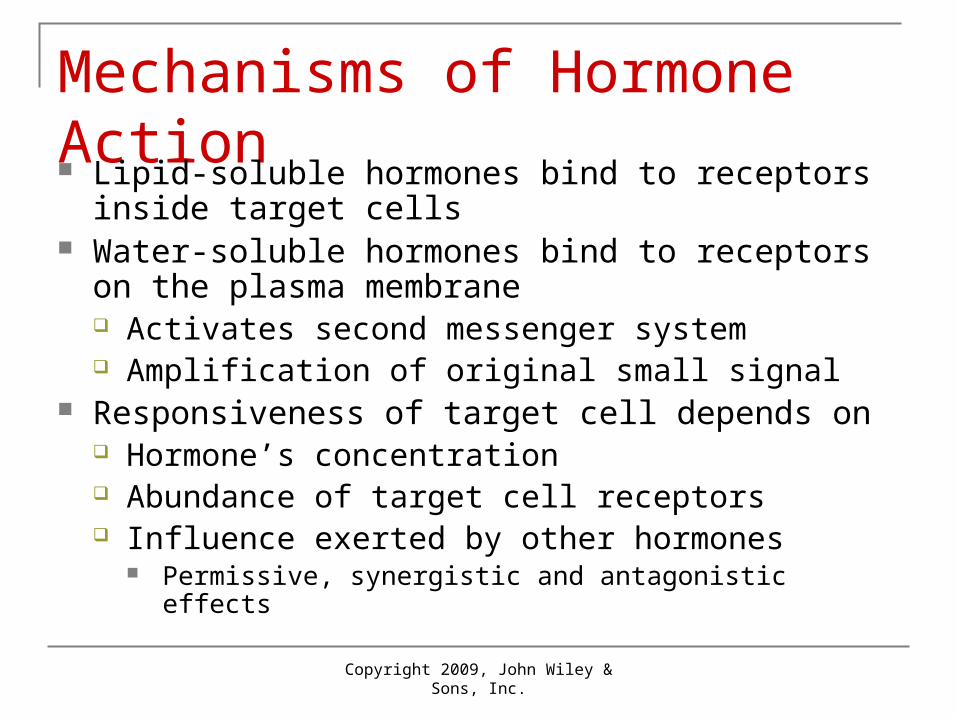
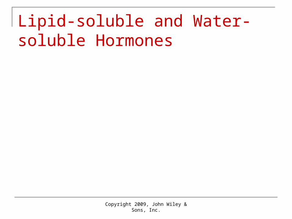
Mechanisms of Hormone Action Lipid-soluble hormones bind to receptors inside target
cells Water-soluble hormones bind to receptors on the
plasma membrane Activates second messenger system Amplification of original small signal
Responsiveness of target cell depends on Hormone’s concentration Abundance of target cell receptors Influence exerted by other hormones
Permissive, synergistic and antagonistic effects
Copyright 2009, John Wiley & Sons, Inc.
Lipid-soluble and Water-soluble Hormones

1 Lipid-solublehormonediffuses into cell
Blood capillary
Target cell
Transportprotein
Free hormone
1 Lipid-solublehormonediffuses into cell
Blood capillary
Activatedreceptor-hormonecomplex altersgene expression
NucleusReceptor
mRNA
DNA
Cytosol
Target cell
Transportprotein
Free hormone
2
1 Lipid-solublehormonediffuses into cell
Blood capillary
Activatedreceptor-hormonecomplex altersgene expression
NucleusReceptor
mRNANewly formedmRNA directssynthesis ofspecific proteinson ribosomes
DNA
Cytosol
Target cell
Transportprotein
Free hormone
Ribosome
2
3
1 Lipid-solublehormonediffuses into cell
Blood capillary
Activatedreceptor-hormonecomplex altersgene expression
NucleusReceptor
mRNANewly formedmRNA directssynthesis ofspecific proteinson ribosomes
DNA
Cytosol
Target cell
New proteins altercell's activity
Transportprotein
Free hormone
Ribosome
Newprotein
2
3
4

Water-solublehormone
Receptor
G protein
Blood capillary
Binding of hormone (first messenger)to its receptor activates G protein,which activates adenylate cyclase
Adenylate cyclase
Target cell
1
Water-solublehormone
Receptor
G protein
cAMP
Second messenger
Activated adenylatecyclase convertsATP to cAMP
Blood capillary
Binding of hormone (first messenger)to its receptor activates G protein,which activates adenylate cyclase
Adenylate cyclase
Target cell
ATP
1
2
Water-solublehormone
Receptor
cAMP serves as asecond messengerto activate proteinkinases
G protein
Protein kinases
cAMP
Second messenger
Activated adenylatecyclase convertsATP to cAMP
Blood capillary
Binding of hormone (first messenger)to its receptor activates G protein,which activates adenylate cyclase
Adenylate cyclase
Target cell
ATP
1
2
3 Activatedproteinkinases
Water-solublehormone
Receptor
cAMP serves as asecond messengerto activate proteinkinases
G protein
Protein kinases
cAMP
Activatedproteinkinases
Second messenger
Activated adenylatecyclase convertsATP to cAMP
Activated proteinkinasesphosphorylatecellular proteins
Blood capillary
Binding of hormone (first messenger)to its receptor activates G protein,which activates adenylate cyclase
Adenylate cyclase
Target cell
ATP
1
2
4
3
Protein— P
ADP
Protein
ATP
Water-solublehormone
Receptor
cAMP serves as asecond messengerto activate proteinkinases
G protein
Protein kinases
cAMP
Activatedproteinkinases
Protein—
Second messenger
Activated adenylatecyclase convertsATP to cAMP
Activated proteinkinasesphosphorylatecellular proteins
Millions of phosphorylatedproteins cause reactions thatproduce physiological responses
Blood capillary
Binding of hormone (first messenger)to its receptor activates G protein,which activates adenylate cyclase
Adenylate cyclase
Target cell
P
ADP
Protein
ATP
ATP
1
2
4
3
5
Water-solublehormone
Receptor
cAMP serves as asecond messengerto activate proteinkinases
G protein
Protein kinases
cAMP
Activatedproteinkinases
Protein—
Second messenger
Phosphodiesteraseinactivates cAMP
Activated adenylatecyclase convertsATP to cAMP
Activated proteinkinasesphosphorylatecellular proteins
Millions of phosphorylatedproteins cause reactions thatproduce physiological responses
Blood capillary
Binding of hormone (first messenger)to its receptor activates G protein,which activates adenylate cyclase
Adenylate cyclase
Target cell
P
ADP
Protein
ATP
ATP
1
2
6
4
3
5
Copyright 2009, John Wiley & Sons, Inc.
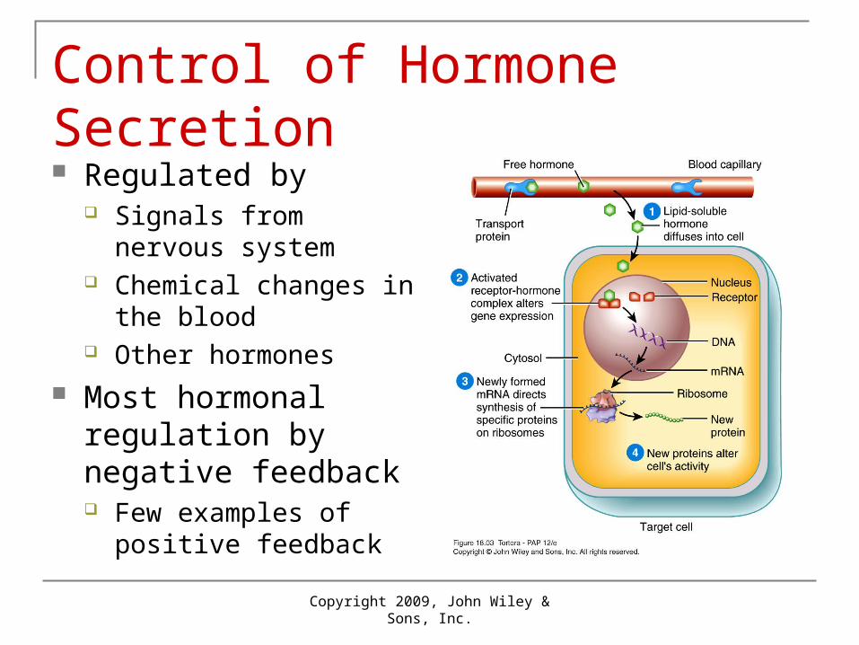
Control of Hormone Secretion Regulated by
Signals from nervous system
Chemical changes in the blood
Other hormones Most hormonal
regulation by negative feedback Few examples of positive
feedback

Copyright 2009, John Wiley & Sons, Inc.
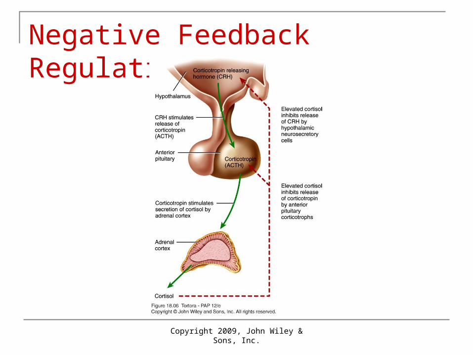
Negative and Positive Feedback systems Negative Feedback systems
Reverses a change in a controlled condition Regulation of blood pressure (force exerted by blood as
it presses again the walls of the blood vessels) excess thyroid hormone turns off hypothalamus excess cortisol turns off hypothalamus and pituitary
Positive Feedback systems Strengthen or reinforce a change in one of the
body’s controlled conditions Normal child birth
Copyright 2009, John Wiley & Sons, Inc.
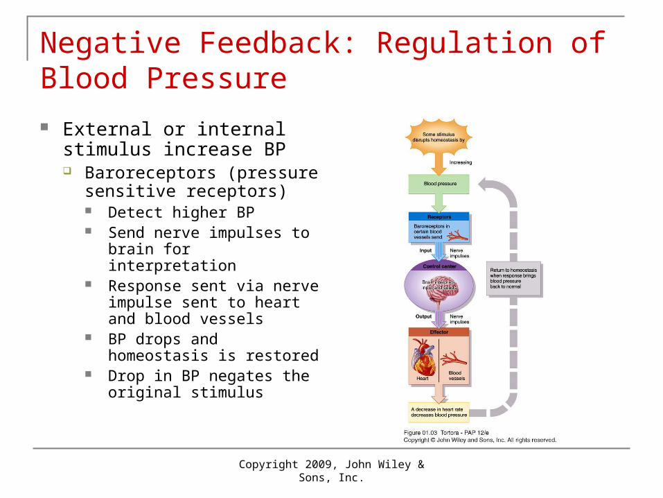
Negative Feedback: Regulation of Blood Pressure
External or internal stimulus increase BP Baroreceptors (pressure
sensitive receptors) Detect higher BP Send nerve impulses to
brain for interpretation Response sent via nerve
impulse sent to heart and blood vessels
BP drops and homeostasis is restored
Drop in BP negates the original stimulus

Copyright 2009, John Wiley & Sons, Inc.
Positive Feedback Systems: Normal Childbirth Uterine contractions cause
vagina to open Stretch-sensitive receptors in
cervix send impulse to brain Oxytocin is released into the
blood Contractions enhanced and
baby pushes farther down the uterus
Cycle continues to the birth of the baby (no stretching)

Copyright 2009, John Wiley & Sons, Inc.
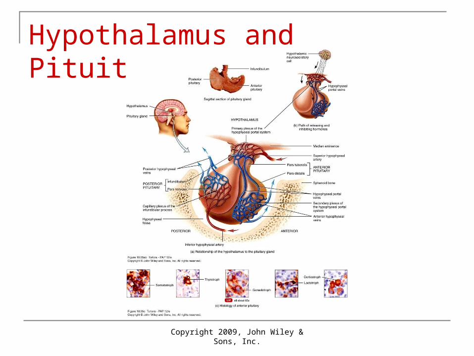
Hypothalamus and Pituitary Gland Hypothalamus is a major link between
nervous and endocrine system Pituitary attached to hypothalamus by
infundibulum Anterior pituitary or adenohypophysis Posterior pituitary or neurohypophysis
Copyright 2009, John Wiley & Sons, Inc.
Hypothalamus and Pituitary Gland

Copyright 2009, John Wiley & Sons, Inc.
Anterior pituitary
Release of hormones stimulated by releasing and inhibiting hormones from the hypothalamus
Also regulated by negative feedback Hypothalamic hormones made by
neurosecretory cells transported by hypophyseal portal system
Anterior pituitary hormones that act on other endocrine systems called tropic hormones

Copyright 2009, John Wiley & Sons, Inc.
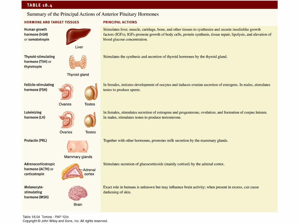
Hormones of the Anterior Pituitary Human growth hormone (hGH) or somatotropin
Stimulates secretion of insulin-like growth factors (IGFs) or somatomedins that promote growth, protein synthesis
Thyroid-stimulating hormone (TSH) or thyrotropin Stimulates synthesis and secretion of thyroid hormones by
thyroid Follicle-stimulating hormone (FSH)
Ovaries initiates development of oocytes, stimulates sperm production by testes
Luteinizing hormone (LH) Ovaries stimulates ovulation, testes stimulates testosterone
production

Copyright 2009, John Wiley & Sons, Inc.
Hormones of the Anterior Pituitary Prolactin (PRL)
Promotes milk secretion by mammary glands Adrenocorticotropic hormone (ACTH) or
corticotropin Stimulates glucocorticoid secretion by adrenal
cortex Melanocyte-stimulating Hormone (MSH)
Unknown role in humans

18_table_03
Copyright 2009, John Wiley & Sons, Inc.
Negative Feedback Regulation
Copyright 2009, John Wiley & Sons, Inc.
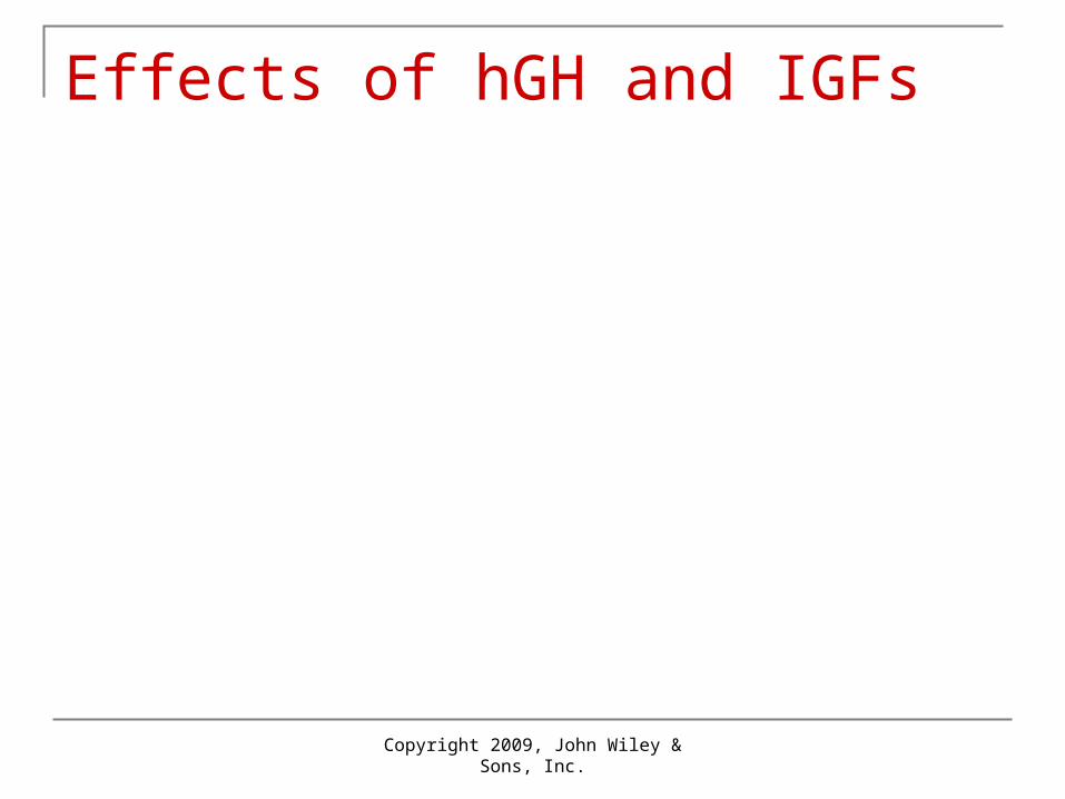
Effects of hGH and IGFs

Low blood glucose(hypoglycemia)stimulates release of
GHRH
1 Low blood glucose(hypoglycemia)stimulates release of
hGH
GHRH stimulatessecretionof hGH bysomatotrophs
GHRH
1
2
Low blood glucose(hypoglycemia)stimulates release of
hGH
GHRH stimulatessecretionof hGH bysomatotrophs
GHRH
hGH and IGFs speedup breakdown of liverglycogen into glucose,which enters the bloodmore rapidly
1
3
2
Low blood glucose(hypoglycemia)stimulates release of
hGH
GHRH stimulatessecretionof hGH bysomatotrophs
GHRH
hGH and IGFs speedup breakdown of liverglycogen into glucose,which enters the bloodmore rapidly
Blood glucose levelrises to normal(about 90 mg/100 mL)
1
3
4
2
Low blood glucose(hypoglycemia)stimulates release of
hGH
GHRH stimulatessecretionof hGH bysomatotrophs
GHRH
hGH and IGFs speedup breakdown of liverglycogen into glucose,which enters the bloodmore rapidly
Blood glucose levelrises to normal(about 90 mg/100 mL)
If blood glucosecontinues to increase,hyperglycemia inhibitsrelease of GHRH
1
3
4
5
2
Anteriorpituitary
Low blood glucose(hypoglycemia)stimulates release of
High blood glucose(hyperglycemia)stimulates release of
hGH
GHRH stimulatessecretionof hGH bysomatotrophs
GHIHGHRH
hGH and IGFs speedup breakdown of liverglycogen into glucose,which enters the bloodmore rapidly
Blood glucose levelrises to normal(about 90 mg/100 mL)
If blood glucosecontinues to increase,hyperglycemia inhibitsrelease of GHRH
1 6
3
4
5
2
Anteriorpituitary
GHIH inhibitssecretion ofhGH bysomatotrophs
Low blood glucose(hypoglycemia)stimulates release of
High blood glucose(hyperglycemia)stimulates release of
hGH
GHRH stimulatessecretionof hGH bysomatotrophs
GHIHGHRH
hGH and IGFs speedup breakdown of liverglycogen into glucose,which enters the bloodmore rapidly
Blood glucose levelrises to normal(about 90 mg/100 mL)
If blood glucosecontinues to increase,hyperglycemia inhibitsrelease of GHRH
1 6
7
3
4
5
2 GHIH inhibitssecretion ofhGH bysomatotrophs
Low blood glucose(hypoglycemia)stimulates release of
High blood glucose(hyperglycemia)stimulates release of
Anteriorpituitary
hGH
GHRH stimulatessecretionof hGH bysomatotrophs
GHIHGHRH
A low level of hGH andIGFs decreases the rateof glycogen breakdownin the liver and glucoseenters the blood moreslowly
hGH and IGFs speedup breakdown of liverglycogen into glucose,which enters the bloodmore rapidly
Blood glucose levelrises to normal(about 90 mg/100 mL)
If blood glucosecontinues to increase,hyperglycemia inhibitsrelease of GHRH
1 6
7
83
4
5
2 GHIH inhibitssecretion ofhGH bysomatotrophs
Low blood glucose(hypoglycemia)stimulates release of
High blood glucose(hyperglycemia)stimulates release of
Anteriorpituitary
hGH
GHRH stimulatessecretionof hGH bysomatotrophs
GHIHGHRH
A low level of hGH andIGFs decreases the rateof glycogen breakdownin the liver and glucoseenters the blood moreslowly
Blood glucose levelfalls to normal(about 90 mg/100 mL)
hGH and IGFs speedup breakdown of liverglycogen into glucose,which enters the bloodmore rapidly
Blood glucose levelrises to normal(about 90 mg/100 mL)
If blood glucosecontinues to increase,hyperglycemia inhibitsrelease of GHRH
1 6
7
8
9
3
4
5
2 GHIH inhibitssecretion ofhGH bysomatotrophs
Low blood glucose(hypoglycemia)stimulates release of
High blood glucose(hyperglycemia)stimulates release of
Anteriorpituitary
hGH
GHRH stimulatessecretionof hGH bysomatotrophs
GHIHGHRH
A low level of hGH andIGFs decreases the rateof glycogen breakdownin the liver and glucoseenters the blood moreslowly
Blood glucose levelfalls to normal(about 90 mg/100 mL)
hGH and IGFs speedup breakdown of liverglycogen into glucose,which enters the bloodmore rapidly
Blood glucose levelrises to normal(about 90 mg/100 mL)
If blood glucosecontinues to increase,hyperglycemia inhibitsrelease of GHRH
If blood glucosecontinues to decrease,hypoglycemia inhibitsrelease of GHIH
1 6
7
8
9
10
3
4
5
2
18_table_04

Copyright 2009, John Wiley & Sons, Inc.
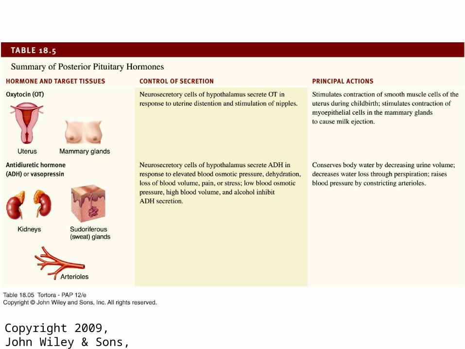
Posterior pituitary
Does not synthesize hormones Stores and releases hormones made by the
hypothalamus Transported along hypothalamohypophyseal tract
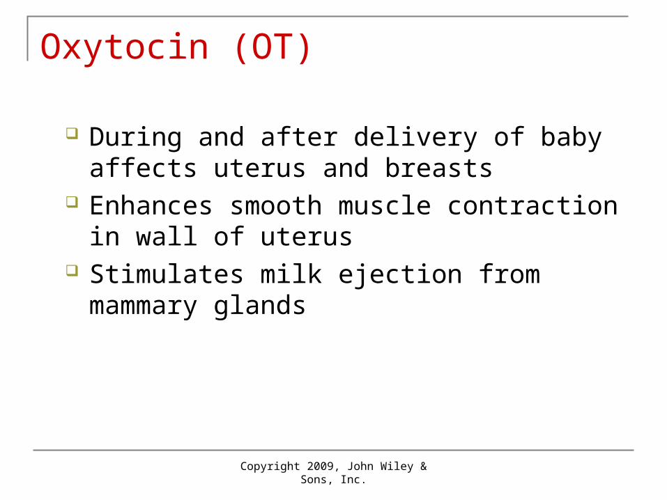
Oxytocin (OT) Antidiuretic hormone (ADH) or vasopressin

Copyright 2009, John Wiley & Sons, Inc.
Hypothalamohypophyseal tract
Copyright 2009, John Wiley & Sons, Inc.
Oxytocin (OT)
During and after delivery of baby affects uterus and breasts
Enhances smooth muscle contraction in wall of uterus
Stimulates milk ejection from mammary glands
Copyright 2009, John Wiley & Sons, Inc.
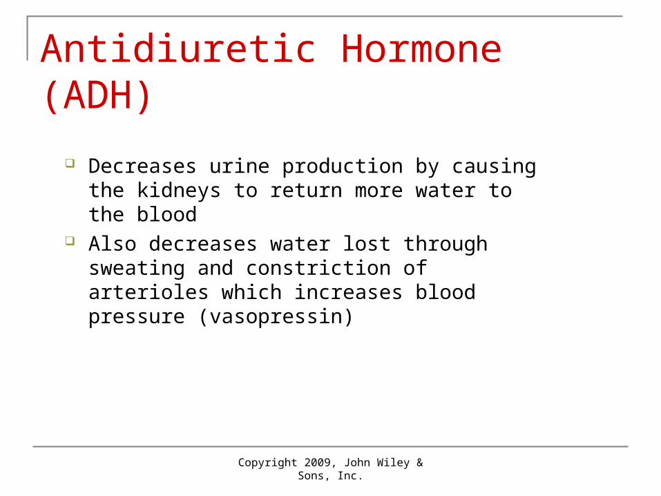
Antidiuretic Hormone (ADH)
Decreases urine production by causing the kidneys to return more water to the blood
Also decreases water lost through sweating and constriction of arterioles which increases blood pressure (vasopressin)
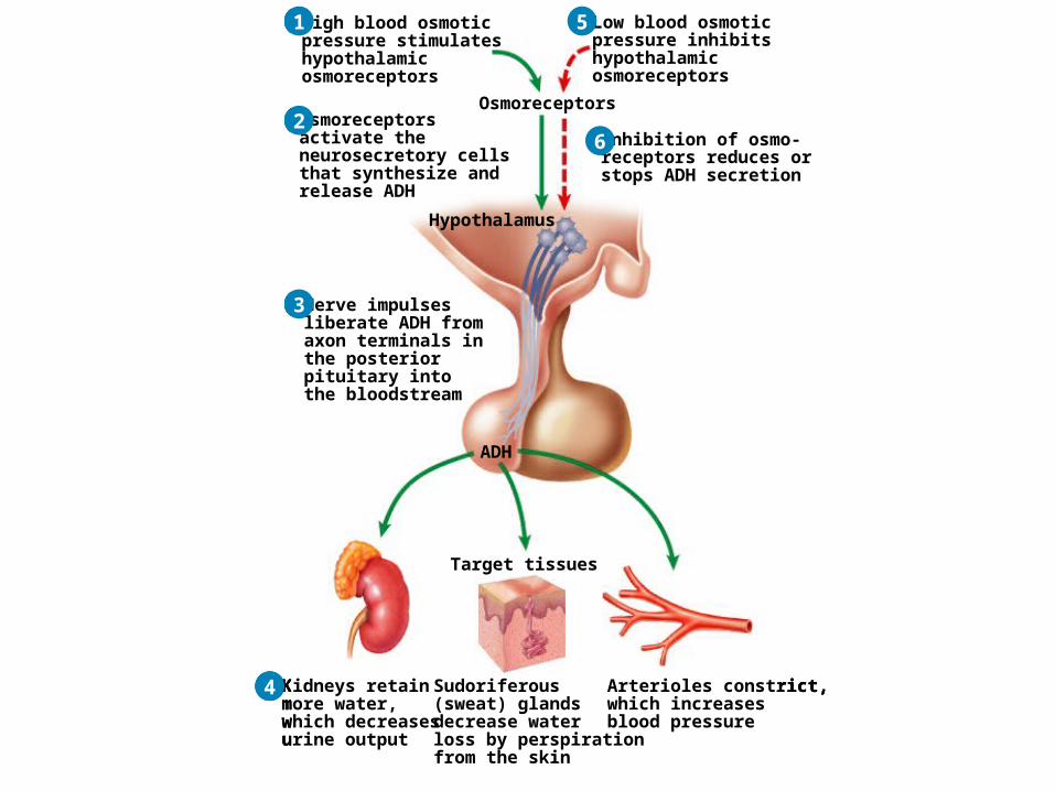
Osmoreceptors
High blood osmoticpressure stimulateshypothalamicosmoreceptors
1
Osmoreceptors
High blood osmoticpressure stimulateshypothalamicosmoreceptors
Osmoreceptorsactivate theneurosecretory cellsthat synthesize andrelease ADH
Hypothalamus
1
2Osmoreceptors
High blood osmoticpressure stimulateshypothalamicosmoreceptors
Nerve impulsesliberate ADH fromaxon terminals inthe posteriorpituitary intothe bloodstream
Osmoreceptorsactivate theneurosecretory cellsthat synthesize andrelease ADH
Hypothalamus
ADH
1
2
3
Osmoreceptors
High blood osmoticpressure stimulateshypothalamicosmoreceptors
Nerve impulsesliberate ADH fromaxon terminals inthe posteriorpituitary intothe bloodstream
Osmoreceptorsactivate theneurosecretory cellsthat synthesize andrelease ADH
Hypothalamus
Sudoriferous(sweat) glandsdecrease waterloss by perspirationfrom the skin
Arterioles constrict,which increasesblood pressure
Kidneys retainmore water,which decreasesurine output
ADH
Target tissues
1
2
3
4
Osmoreceptors
High blood osmoticpressure stimulateshypothalamicosmoreceptors
Low blood osmoticpressure inhibitshypothalamicosmoreceptors
Nerve impulsesliberate ADH fromaxon terminals inthe posteriorpituitary intothe bloodstream
Osmoreceptorsactivate theneurosecretory cellsthat synthesize andrelease ADH
Hypothalamus
Sudoriferous(sweat) glandsdecrease waterloss by perspirationfrom the skin
Arterioles constrict,which increasesblood pressure
Kidneys retainmore water,which decreasesurine output
ADH
Target tissues
1
2
3
4
5
Osmoreceptors
High blood osmoticpressure stimulateshypothalamicosmoreceptors
Low blood osmoticpressure inhibitshypothalamicosmoreceptors
Nerve impulsesliberate ADH fromaxon terminals inthe posteriorpituitary intothe bloodstream
Osmoreceptorsactivate theneurosecretory cellsthat synthesize andrelease ADH
Hypothalamus
Inhibition of osmo-receptors reduces orstops ADH secretion
Sudoriferous(sweat) glandsdecrease waterloss by perspirationfrom the skin
Arterioles constrict,which increasesblood pressure
Kidneys retainmore water,which decreasesurine output
ADH
Target tissues
1
2
3
4
5
6
18_table_05
Copyright 2009, John Wiley & Sons, Inc.
Copyright 2009, John Wiley & Sons, Inc.
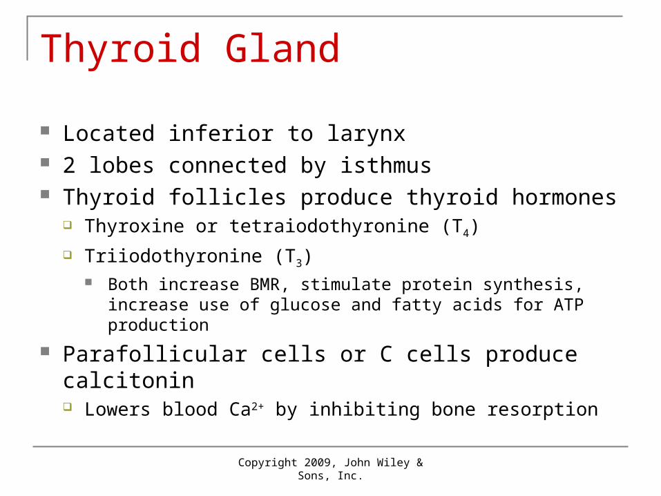
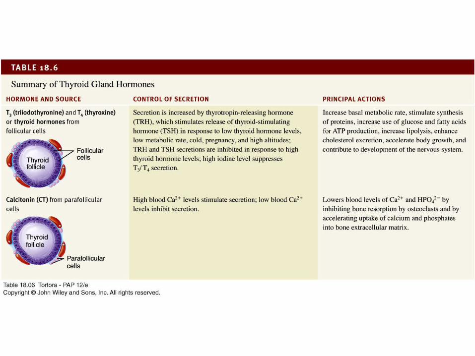
Thyroid Gland
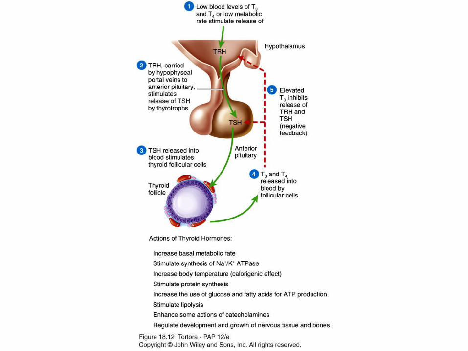
Located inferior to larynx 2 lobes connected by isthmus Thyroid follicles produce thyroid hormones
Thyroxine or tetraiodothyronine (T4)
Triiodothyronine (T3) Both increase BMR, stimulate protein synthesis, increase use
of glucose and fatty acids for ATP production
Parafollicular cells or C cells produce calcitonin Lowers blood Ca2+ by inhibiting bone resorption

Copyright 2009, John Wiley & Sons, Inc.
Thyroid Gland
Copyright 2009, John Wiley & Sons, Inc.
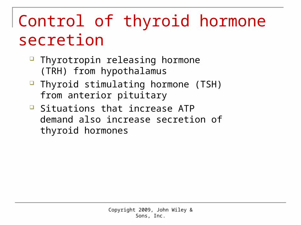
Control of thyroid hormone secretion
Thyrotropin releasing hormone (TRH) from hypothalamus
Thyroid stimulating hormone (TSH) from anterior pituitary
Situations that increase ATP demand also increase secretion of thyroid hormones
18_12
18_table_06
Copyright 2009, John Wiley & Sons, Inc.
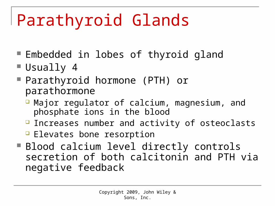
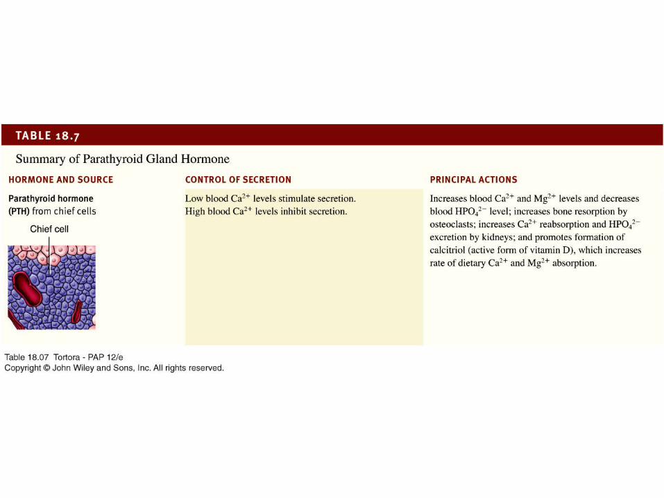
Parathyroid Glands
Embedded in lobes of thyroid gland Usually 4 Parathyroid hormone (PTH) or parathormone
Major regulator of calcium, magnesium, and phosphate ions in the blood
Increases number and activity of osteoclasts Elevates bone resorption
Blood calcium level directly controls secretion of both calcitonin and PTH via negative feedback

Copyright 2009, John Wiley & Sons, Inc.
Parathyroid Glands
Copyright 2009, John Wiley & Sons, Inc.
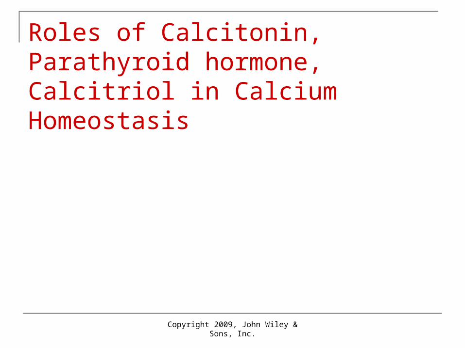
Roles of Calcitonin, Parathyroid hormone, Calcitriol in Calcium Homeostasis

1 High level of Ca2+ in bloodstimulates thyroid glandparafollicular cells to release more CT.
1 High level of Ca2+ in bloodstimulates thyroid glandparafollicular cells to release more CT.
CALCITONIN inhibitsosteoclasts, thus decreasingblood Ca2+ level.
2
1 High level of Ca2+ in bloodstimulates thyroid glandparafollicular cells to release more CT.
Low level of Ca2+ in bloodstimulates parathyroid gland chief cells to release more PTH.
CALCITONIN inhibitsosteoclasts, thus decreasingblood Ca2+ level.
3
2
1 High level of Ca2+ in bloodstimulates thyroid glandparafollicular cells to release more CT.
Low level of Ca2+ in bloodstimulates parathyroid gland chief cells to release more PTH.
CALCITONIN inhibitsosteoclasts, thus decreasingblood Ca2+ level.
PARATHYROID HORMONE (PTH)promotes release of Ca2+ frombone extracellular matrix intoblood and slows loss of Ca2+ in urine, thus increasing bloodCa2+ level.
3
4 2
1
PTH also stimulatesthe kidneys to releaseCALCITRIOL.
High level of Ca2+ in bloodstimulates thyroid glandparafollicular cells to release more CT.
Low level of Ca2+ in bloodstimulates parathyroid gland chief cells to release more PTH.
CALCITONIN inhibitsosteoclasts, thus decreasingblood Ca2+ level.
PARATHYROID HORMONE (PTH)promotes release of Ca2+ frombone extracellular matrix intoblood and slows loss of Ca2+ in urine, thus increasing bloodCa2+ level.
3
4 25
1
CALCITRIOL stimulatesincreased absorption ofCa2+ from foods, whichincreases blood Ca2+ level.
PTH also stimulatesthe kidneys to releaseCALCITRIOL.
High level of Ca2+ in bloodstimulates thyroid glandparafollicular cells to release more CT.
Low level of Ca2+ in bloodstimulates parathyroid gland chief cells to release more PTH.
CALCITONIN inhibitsosteoclasts, thus decreasingblood Ca2+ level.
PARATHYROID HORMONE (PTH)promotes release of Ca2+ frombone extracellular matrix intoblood and slows loss of Ca2+ in urine, thus increasing bloodCa2+ level.
3
4 25
6
18_table_07

Copyright 2009, John Wiley & Sons, Inc.
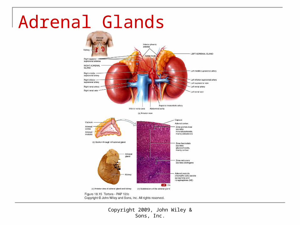
Adrenal Glands
2 structurally and functionally distinct regions Adrenal cortex
Mineralocorticoids affect mineral homeostasis Glucocorticoids affect glucose homeostasis
cortisol Androgens have masculinzing effects
Dehydroepiandrosterone (DHEA) only important in females Adrenal medulla
Modified sympathetic ganglion of autonomic nervous system
Intensifies sympathetic responses Epinephrine and norepinephrine
Copyright 2009, John Wiley & Sons, Inc.
Adrenal Glands

Mineralocorticoids
Aldosterone Regulates homeostasis of:
two mineral ions sodium conservation potassium secretion
blood pressure blood volume excretion of H+
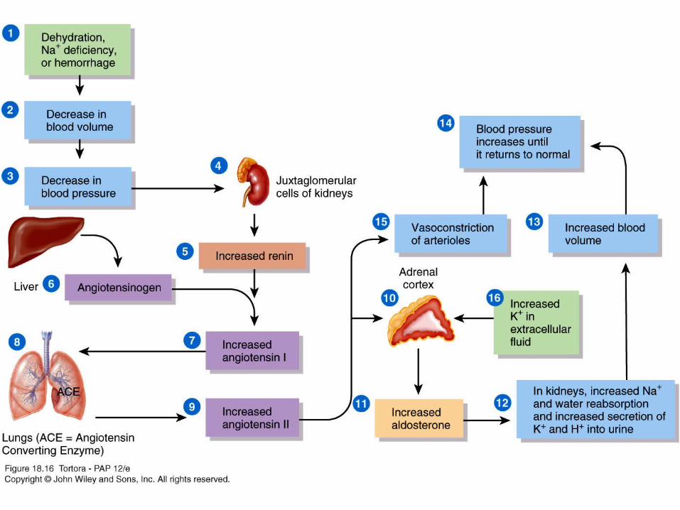
18_16

Glucocorticoids
Cortisol (hydrocortisone), corticosterone, and cortisone
Following effects:1. protein breakdown
2. glucose formation / gluconeogenesis
3. lipolysis
4. stress resistance
5. anti-inflammatory effects
6. depression of immune responses
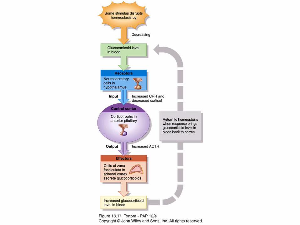
18_17
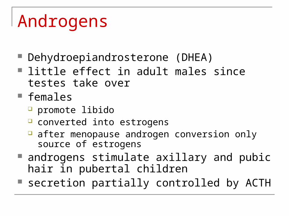
Androgens
Dehydroepiandrosterone (DHEA) little effect in adult males since testes take over females
promote libido converted into estrogens after menopause androgen conversion only source of
estrogens androgens stimulate axillary and pubic hair in
pubertal children secretion partially controlled by ACTH

18_table_08

Copyright 2009, John Wiley & Sons, Inc.
Pancreas
Copyright 2009, John Wiley & Sons, Inc.
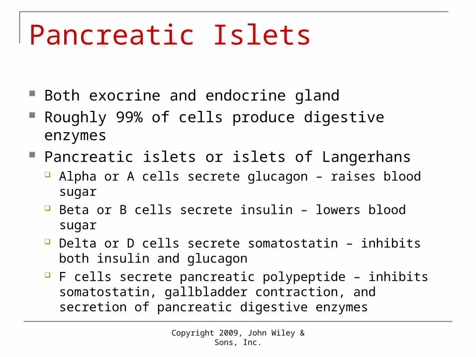
Pancreatic Islets
Both exocrine and endocrine gland Roughly 99% of cells produce digestive enzymes Pancreatic islets or islets of Langerhans
Alpha or A cells secrete glucagon – raises blood sugar Beta or B cells secrete insulin – lowers blood sugar Delta or D cells secrete somatostatin – inhibits both insulin
and glucagon F cells secrete pancreatic polypeptide – inhibits
somatostatin, gallbladder contraction, and secretion of pancreatic digestive enzymes

Copyright 2009, John Wiley & Sons, Inc.
Negative Feedback Regulation of Glucagon and Insulin

Low blood glucose(hypoglycemia)stimulates alphacells to secrete
1
GLUCAGON
Glucagon acts onhepatocytes(liver cells) to:
• convert glycogen into glucose (glycogenolysis)• form glucose from lactic acid and certain amino acids (gluconeogenesis)
Low blood glucose(hypoglycemia)stimulates alphacells to secrete
GLUCAGON
1
2 Glucagon acts onhepatocytes(liver cells) to:
• convert glycogen into glucose (glycogenolysis)• form glucose from lactic acid and certain amino acids (gluconeogenesis)
Glucose releasedby hepatocytesraises blood glucoselevel to normal
Low blood glucose(hypoglycemia)stimulates alphacells to secrete
GLUCAGON
1
2
3
Glucagon acts onhepatocytes(liver cells) to:
• convert glycogen into glucose (glycogenolysis)• form glucose from lactic acid and certain amino acids (gluconeogenesis)
Glucose releasedby hepatocytesraises blood glucoselevel to normal
If blood glucosecontinues to rise,hyperglycemia inhibitsrelease of glucagon
Low blood glucose(hypoglycemia)stimulates alphacells to secrete
GLUCAGON
1
2
3
4
Glucagon acts onhepatocytes(liver cells) to:
• convert glycogen into glucose (glycogenolysis)• form glucose from lactic acid and certain amino acids (gluconeogenesis)
Glucose releasedby hepatocytesraises blood glucoselevel to normal
If blood glucosecontinues to rise,hyperglycemia inhibitsrelease of glucagon
Low blood glucose(hypoglycemia)stimulates alphacells to secrete
High blood glucose(hyperglycemia)stimulates beta cellsto secrete
GLUCAGON
1 5
2
3
4
INSULIN
Insulin acts on variousbody cells to:
• accelerate facilitated diffusion of glucose into cells• speed conversion of glucose into glycogen (glycogenesis)• increase uptake of amino acids and increase protein synthesis• speed synthesis of fatty acids (lipogenesis)• slow glycogenolysis• slow gluconeogenesis
Glucagon acts onhepatocytes(liver cells) to:
• convert glycogen into glucose (glycogenolysis)• form glucose from lactic acid and certain amino acids (gluconeogenesis)
Glucose releasedby hepatocytesraises blood glucoselevel to normal
If blood glucosecontinues to rise,hyperglycemia inhibitsrelease of glucagon
Low blood glucose(hypoglycemia)stimulates alphacells to secrete
High blood glucose(hyperglycemia)stimulates beta cellsto secrete
INSULINGLUCAGON
1 5
2
3
4
6 Insulin acts on variousbody cells to:
• accelerate facilitated diffusion of glucose into cells• speed conversion of glucose into glycogen (glycogenesis)• increase uptake of amino acids and increase protein synthesis• speed synthesis of fatty acids (lipogenesis)• slow glycogenolysis• slow gluconeogenesis
Blood glucose level falls
Glucagon acts onhepatocytes(liver cells) to:
• convert glycogen into glucose (glycogenolysis)• form glucose from lactic acid and certain amino acids (gluconeogenesis)
Glucose releasedby hepatocytesraises blood glucoselevel to normal
If blood glucosecontinues to rise,hyperglycemia inhibitsrelease of glucagon
Low blood glucose(hypoglycemia)stimulates alphacells to secrete
High blood glucose(hyperglycemia)stimulates beta cellsto secrete
INSULINGLUCAGON
1 5
2
3
4
6
7
Insulin acts on variousbody cells to:
• accelerate facilitated diffusion of glucose into cells• speed conversion of glucose into glycogen (glycogenesis)• increase uptake of amino acids and increase protein synthesis• speed synthesis of fatty acids (lipogenesis)• slow glycogenolysis• slow gluconeogenesis
If blood glucose continuesto fall, hypoglycemiainhibits release ofinsulin
Blood glucose level falls
Glucagon acts onhepatocytes(liver cells) to:
• convert glycogen into glucose (glycogenolysis)• form glucose from lactic acid and certain amino acids (gluconeogenesis)
Glucose releasedby hepatocytesraises blood glucoselevel to normal
If blood glucosecontinues to rise,hyperglycemia inhibitsrelease of glucagon
Low blood glucose(hypoglycemia)stimulates alphacells to secrete
High blood glucose(hyperglycemia)stimulates beta cellsto secrete
INSULINGLUCAGON
1 5
2
3
4
6
7
8
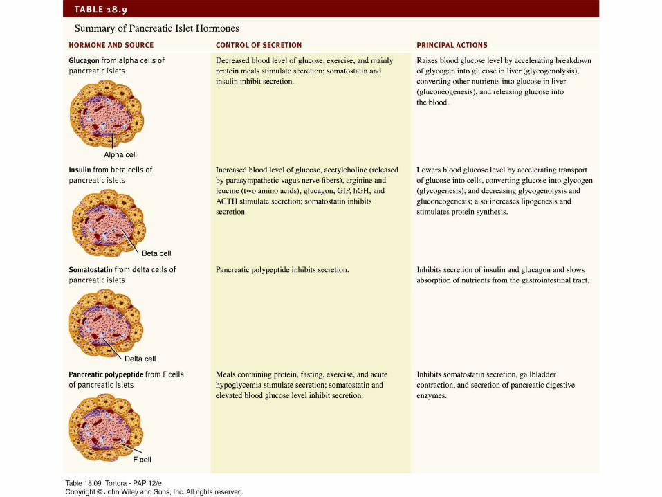
18_table_09
Copyright 2009, John Wiley & Sons, Inc.
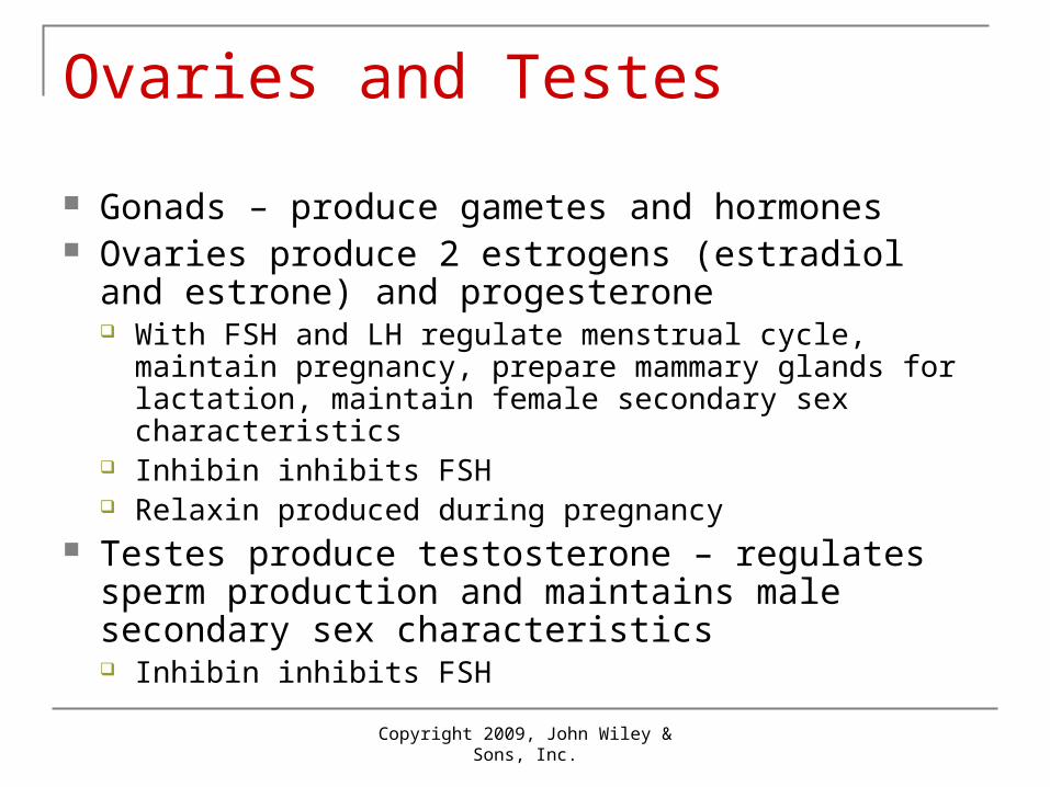
Ovaries and Testes
Gonads – produce gametes and hormones Ovaries produce 2 estrogens (estradiol and estrone)
and progesterone With FSH and LH regulate menstrual cycle, maintain
pregnancy, prepare mammary glands for lactation, maintain female secondary sex characteristics
Inhibin inhibits FSH Relaxin produced during pregnancy
Testes produce testosterone – regulates sperm production and maintains male secondary sex characteristics Inhibin inhibits FSH

Copyright 2009, John Wiley & Sons, Inc.
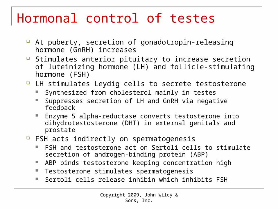
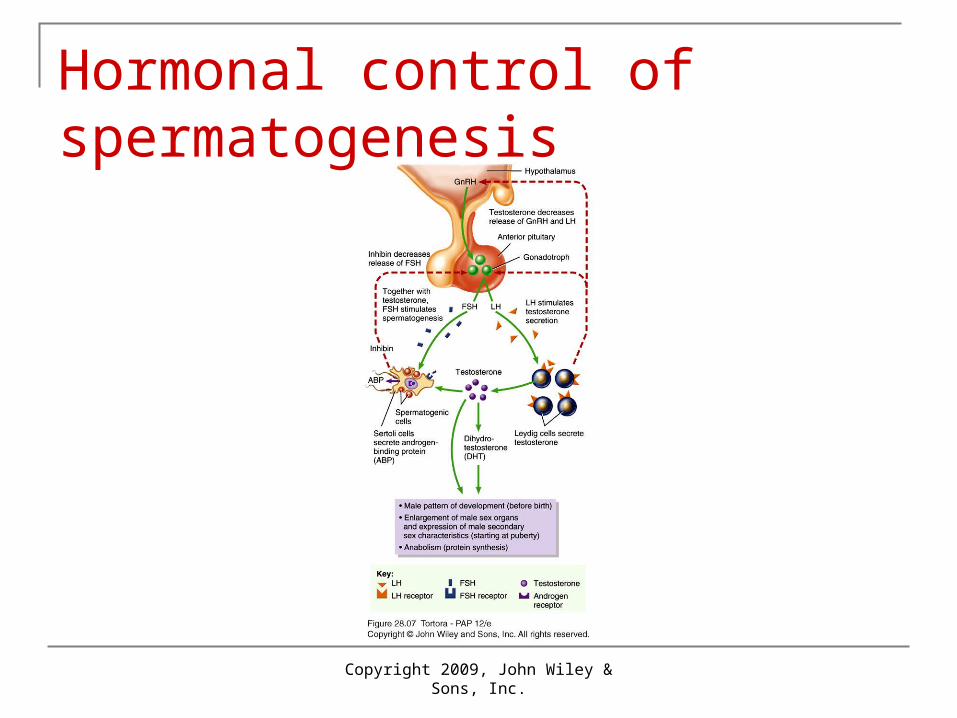
Hormonal control of testes
At puberty, secretion of gonadotropin-releasing hormone (GnRH) increases
Stimulates anterior pituitary to increase secretion of luteinizing hormone (LH) and follicle-stimulating hormone (FSH)
LH stimulates Leydig cells to secrete testosterone Synthesized from cholesterol mainly in testes Suppresses secretion of LH and GnRH via negative feedback Enzyme 5 alpha-reductase converts testosterone into
dihydrotestosterone (DHT) in external genitals and prostate FSH acts indirectly on spermatogenesis
FSH and testosterone act on Sertoli cells to stimulate secretion of androgen-binding protein (ABP)
ABP binds testosterone keeping concentration high Testosterone stimulates spermatogenesis Sertoli cells release inhibin which inhibits FSH
Copyright 2009, John Wiley & Sons, Inc.
Hormonal control of spermatogenesis
Copyright 2009, John Wiley & Sons, Inc.
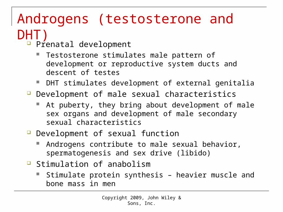
Androgens (testosterone and DHT)
Prenatal development Testosterone stimulates male pattern of development or
reproductive system ducts and descent of testes DHT stimulates development of external genitalia
Development of male sexual characteristics At puberty, they bring about development of male sex organs
and development of male secondary sexual characteristics Development of sexual function
Androgens contribute to male sexual behavior, spermatogenesis and sex drive (libido)
Stimulation of anabolism Stimulate protein synthesis – heavier muscle and bone mass
in men

Copyright 2009, John Wiley & Sons, Inc.
Negative feedback regulates testosterone production
Copyright 2009, John Wiley & Sons, Inc.
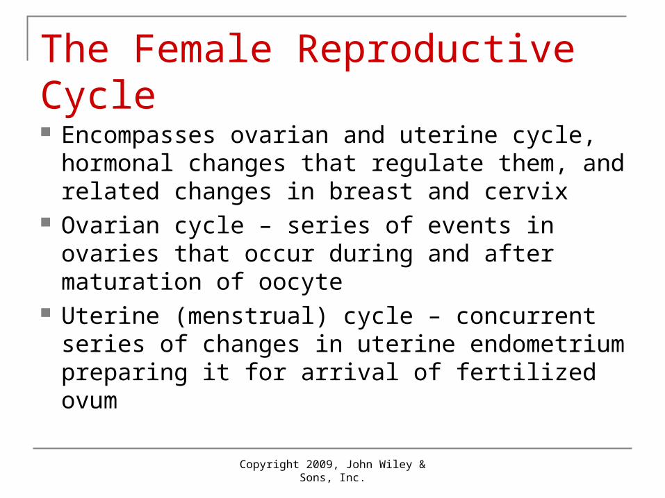
The Female Reproductive Cycle Encompasses ovarian and uterine cycle,
hormonal changes that regulate them, and related changes in breast and cervix
Ovarian cycle – series of events in ovaries that occur during and after maturation of oocyte
Uterine (menstrual) cycle – concurrent series of changes in uterine endometrium preparing it for arrival of fertilized ovum
Copyright 2009, John Wiley & Sons, Inc.
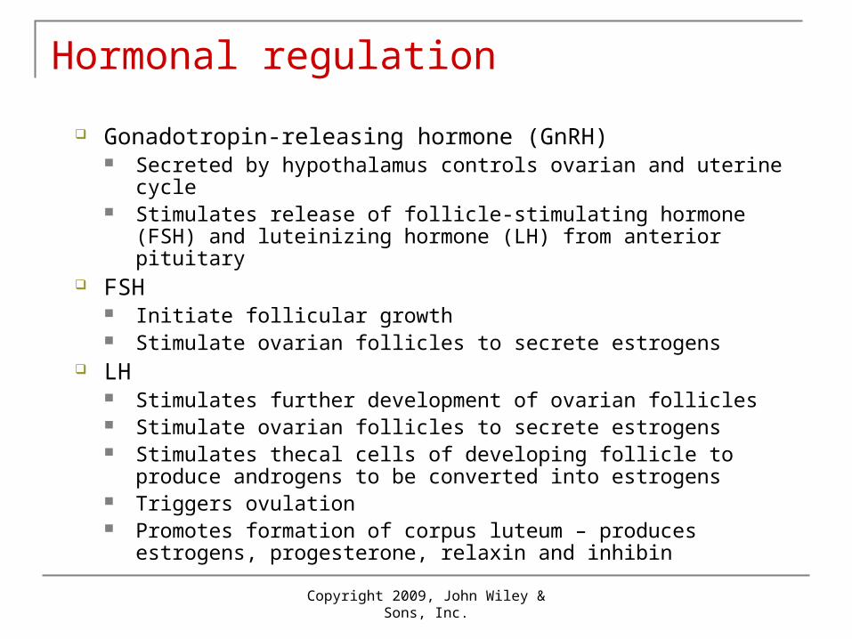
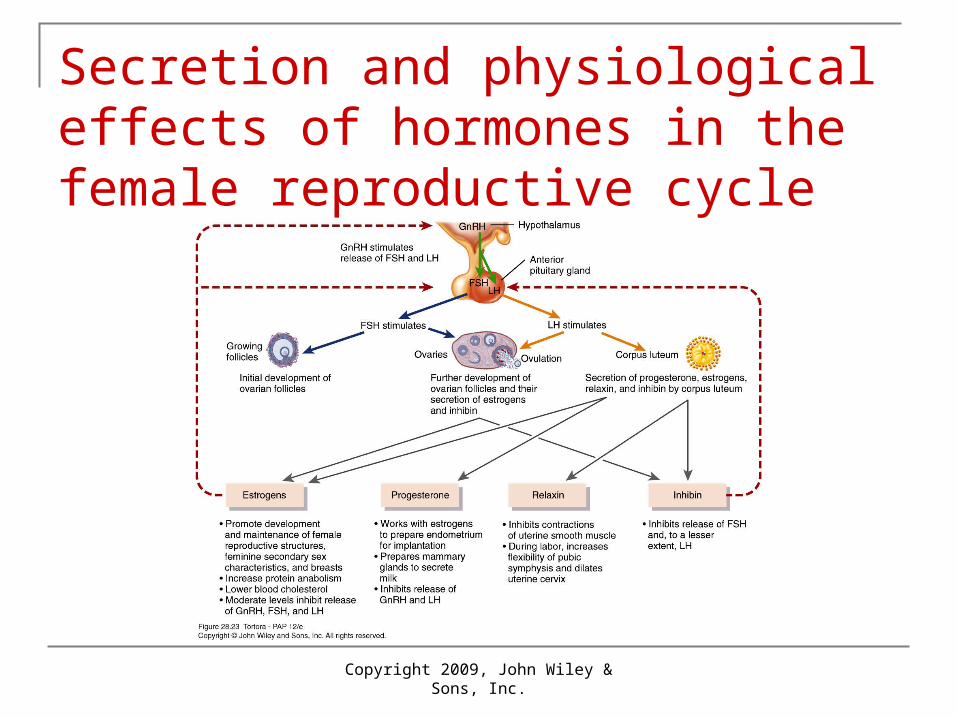
Hormonal regulation
Gonadotropin-releasing hormone (GnRH) Secreted by hypothalamus controls ovarian and uterine cycle Stimulates release of follicle-stimulating hormone (FSH) and
luteinizing hormone (LH) from anterior pituitary FSH
Initiate follicular growth Stimulate ovarian follicles to secrete estrogens
LH Stimulates further development of ovarian follicles Stimulate ovarian follicles to secrete estrogens Stimulates thecal cells of developing follicle to produce androgens to
be converted into estrogens Triggers ovulation Promotes formation of corpus luteum – produces estrogens,
progesterone, relaxin and inhibin
Copyright 2009, John Wiley & Sons, Inc.
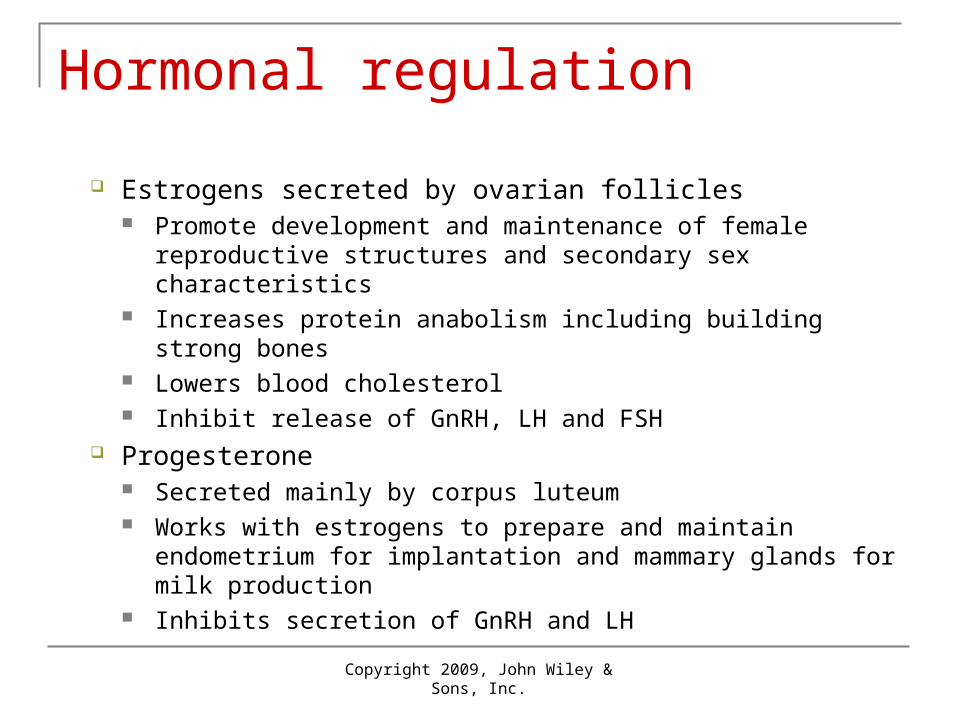
Hormonal regulation
Estrogens secreted by ovarian follicles Promote development and maintenance of female
reproductive structures and secondary sex characteristics Increases protein anabolism including building strong bones Lowers blood cholesterol Inhibit release of GnRH, LH and FSH
Progesterone Secreted mainly by corpus luteum Works with estrogens to prepare and maintain endometrium
for implantation and mammary glands for milk production Inhibits secretion of GnRH and LH

Copyright 2009, John Wiley & Sons, Inc.
Hormonal regulation
Relaxin Produced by corpus luteum Relaxes uterus by inhibiting contraction of myometrium At end of pregnancy, increases flexibility of pubic
symphysis and dilates uterine cervix Inhibin
Secreted by granulosa cells of growing follicles and by corpus luteum
Inhibits secretion of FSH and LH
Copyright 2009, John Wiley & Sons, Inc.
Secretion and physiological effects of hormones in the female reproductive cycle

Copyright 2009, John Wiley & Sons, Inc.
4 Phases
Typical duration 24-35 days Assume a duration of 28 days
1. Menstrual phase
2. Preovulatory phase
3. Ovulation
4. Postovulatory phase

Copyright 2009, John Wiley & Sons, Inc.
The female reproductive cycle
Copyright 2009, John Wiley & Sons, Inc.
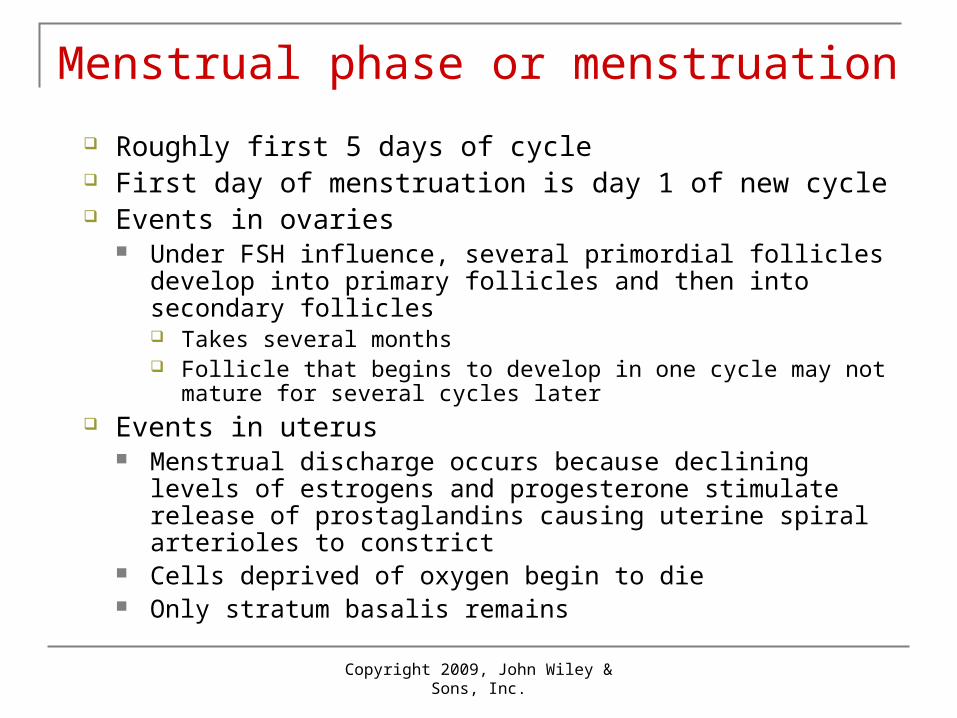
Menstrual phase or menstruation
Roughly first 5 days of cycle First day of menstruation is day 1 of new cycle Events in ovaries
Under FSH influence, several primordial follicles develop into primary follicles and then into secondary follicles Takes several months Follicle that begins to develop in one cycle may not mature for
several cycles later Events in uterus
Menstrual discharge occurs because declining levels of estrogens and progesterone stimulate release of prostaglandins causing uterine spiral arterioles to constrict
Cells deprived of oxygen begin to die Only stratum basalis remains
Copyright 2009, John Wiley & Sons, Inc.
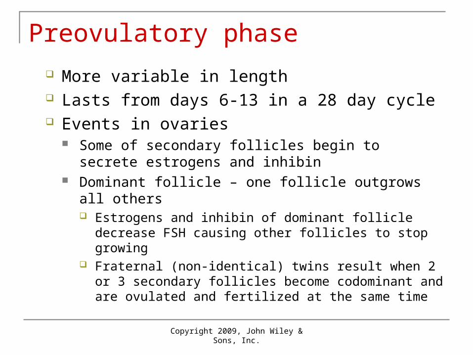
Preovulatory phase
More variable in length Lasts from days 6-13 in a 28 day cycle Events in ovaries
Some of secondary follicles begin to secrete estrogens and inhibin
Dominant follicle – one follicle outgrows all others Estrogens and inhibin of dominant follicle decrease FSH
causing other follicles to stop growing Fraternal (non-identical) twins result when 2 or 3 secondary
follicles become codominant and are ovulated and fertilized at the same time

Copyright 2009, John Wiley & Sons, Inc.
Preovulatory phase
Normally, one dominant follicle becomes the mature (graafian) follicle
In ovarian cycle, menstrual and preovulatory phases are termed follicular phase because follicles are growing
Events in uterus Estrogens stimulate repair of endometrium Cells of stratum basalis undergo mitosis to form new
stratum functionalis Thickness of endometrium doubles In uterine cycle, preovulatory phase is the proliferative
phase because endometrium is proliferating
Copyright 2009, John Wiley & Sons, Inc.
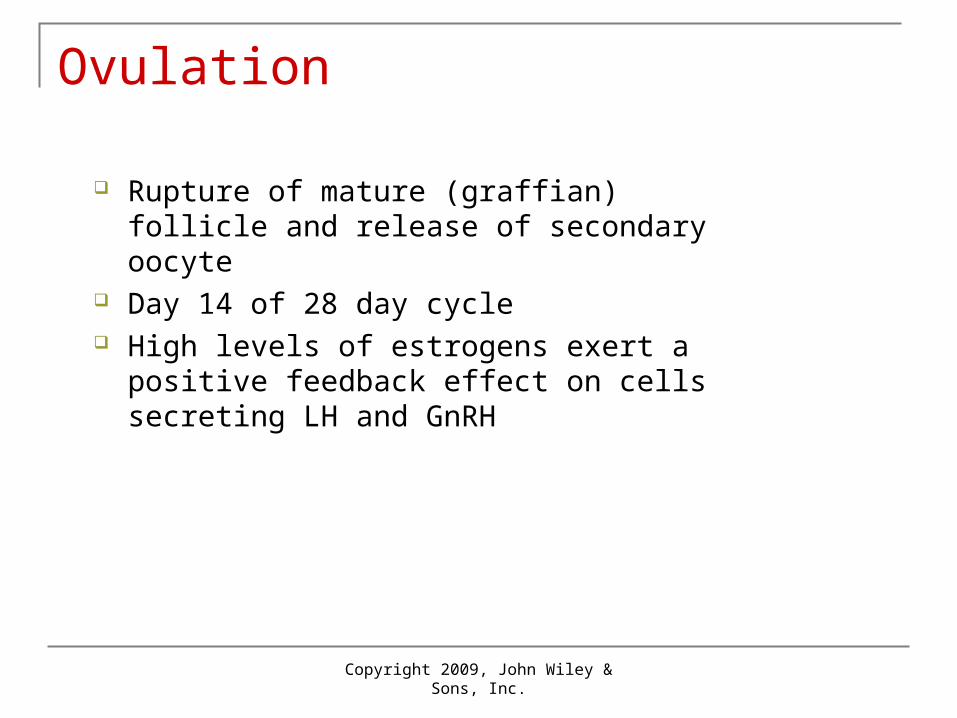
Ovulation
Rupture of mature (graffian) follicle and release of secondary oocyte
Day 14 of 28 day cycle High levels of estrogens exert a positive
feedback effect on cells secreting LH and GnRH

High levels ofestrogens fromalmost maturefollicle stimulaterelease of moreGnRH and LH
Hypothalamus
Anterior pituitary
Ovary
Corpus hemorrhagicum(ruptured follicle)
Almost mature(graafian) follicle
LH
GnRH
1 High levels ofestrogens fromalmost maturefollicle stimulaterelease of moreGnRH and LH
Hypothalamus
Anterior pituitary
GnRH promotesrelease of FSHand more LH
Ovary
Corpus hemorrhagicum(ruptured follicle)
Almost mature(graafian) follicle
LH
GnRH
1
2
High levels ofestrogens fromalmost maturefollicle stimulaterelease of moreGnRH and LH
LH surgebrings aboutovulation
Ovulatedsecondaryoocyte
Hypothalamus
Anterior pituitary
GnRH promotesrelease of FSHand more LH
Ovary
Corpus hemorrhagicum(ruptured follicle)
Almost mature(graafian) follicle
LH
GnRH
1
2
3

Copyright 2009, John Wiley & Sons, Inc.
Postovulatory phase
Duration most constant of phases Lasts for 14 days in 28 day cycle (day 15-28) Events in one ovary
After ovulation, mature follicle collapses to form corpus luteum under the influence of LH
Secretes progesterone, estrogen, relaxin and inhibin In the ovarian cycle, this is the luteal phase

Copyright 2009, John Wiley & Sons, Inc.
Corpus luteum
If oocyte not fertilized, corpus luteum lasts 2 weeks Degenerates in corpus albicans As levels of progesterone, estrogens and inhibin decrease,
release of GnRH, FSH, and LH rise due to loss of negative feedback
Follicular growth resume as new ovarian cycle begins If oocyte is fertilized, corpus luteum lasts more than 2
weeks Human chorionic gonadotropin (hCG) produced by chorion
of embryo about 8 days after fertilization stimulates corpus luteum

Copyright 2009, John Wiley & Sons, Inc.
Events in uterus
Progesterone and estrogens produced by corpus luteum promote growth of endometrium
Because of secretory activity of endometrial glands, this is the secretory phase of uterine cycle
Changes peak about 1 week after ovulation when a fertilized ovum might arrive in uterus
If fertilization does not occur, levels of progesterone and estrogens decline due to degeneration of corpus luteum
Withdrawal of estrogens and progesterone causes menstruation

Copyright 2009, John Wiley & Sons, Inc.
Hormonal interactions in the ovarian and uterine cycles
Copyright 2009, John Wiley & Sons, Inc.
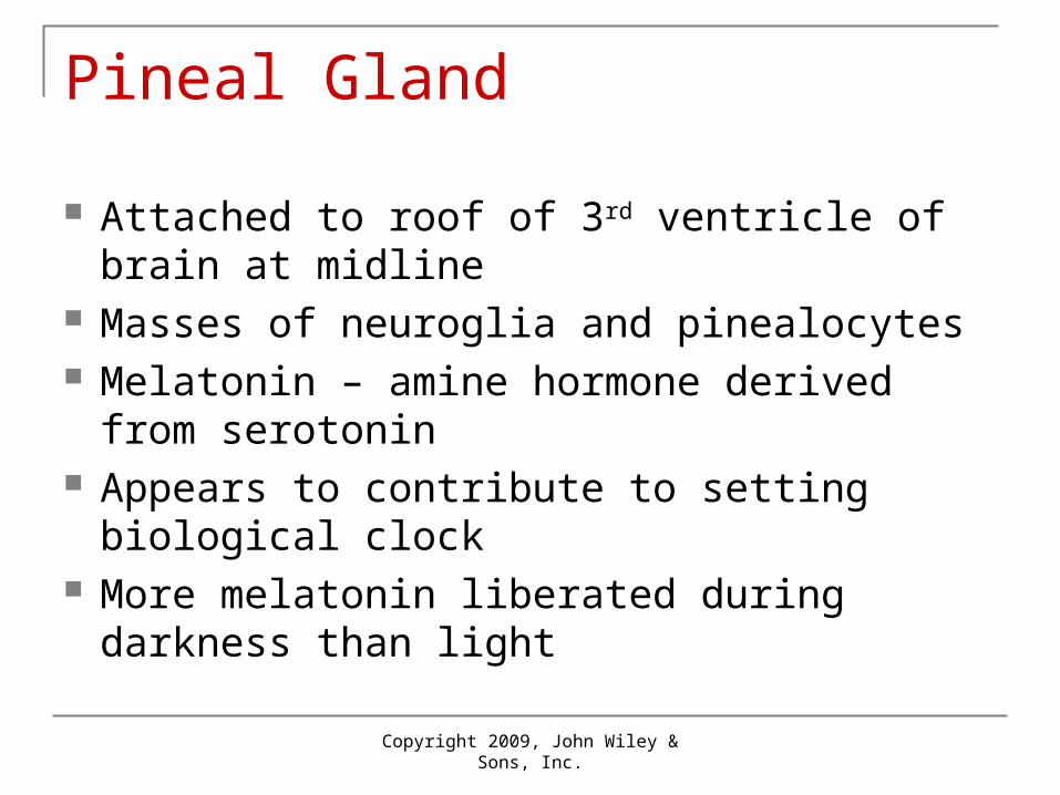
Pineal Gland
Attached to roof of 3rd ventricle of brain at midline
Masses of neuroglia and pinealocytes Melatonin – amine hormone derived from
serotonin Appears to contribute to setting biological
clock More melatonin liberated during darkness
than light


Eicosanoids
Two families prostaglandins leukotrienes
found in all body cells except red blood cells act as local hormones (paracrine and/or
autocrine) in response to mechanical and chemical stimuli
synthesized by clipping 20 carbon arachidonic acid from membrane phospholipids
Eicosanoids
thromboxane is modified PG that constricts blood vessels and activates platelets
rapid, short acting bind receptors on target cells stimulate or inhibit second messenger LTs stimulate chemotaxis and mediate
inflammation

Eicosanoids
PGs alter: smooth muscle contraction glandular secretion blood flow reproductive processes platelet function respiration nerve impulses lipid metabolism immune response
Eicosanoids
PGs promote: inflammation fever pain intensity

Copyright 2009, John Wiley & Sons, Inc.
The Stress Response
Eustress in helpful stress / Distress is harmful Body’s homeostatic mechanisms attempt to
counteract stress Stressful conditions can result in stress response or
general adaptation syndrome (GAS) 3 stages – initial flight-or-fight, slower resistance reaction,
eventually exhaustion Prolonged exposure to cortisol can result in wasting of
muscles, suppression of immune system, ulceration of GI tract, and failure of pancreatic beta cells

Copyright 2009, John Wiley & Sons, Inc.
Stress Response

Copyright 2009, John Wiley & Sons, Inc.
End of Chapter 18
Copyright 2009 John Wiley & Sons, Inc.All rights reserved. Reproduction or translation of this work beyond that permitted in section 117 of the 1976 United States Copyright Act without express permission of the copyright owner is unlawful. Request for further information should be addressed to the Permission Department, John Wiley & Sons, Inc. The purchaser may make back-up copies for his/her own use only and not for distribution or resale. The Publishers assumes no responsibility for errors, omissions, or damages caused by the use of theses programs or from the use of the information herein.