Copyright 2007 by Saunders/Elsevier. All rights reserved. Chapter 16: Digestive System: Oral Cavity...
9
Copyright 2007 by Saunders/Elsevier. All rights reserved. Chapter 16: Digestive System: Oral Cavity Color Textbook of Histology, 3rd ed. Gartner & Hiatt Copyright 2007 by Saunders/Elsevier. All rights reserved.
-
Upload
basil-hicks -
Category
Documents
-
view
215 -
download
0
Transcript of Copyright 2007 by Saunders/Elsevier. All rights reserved. Chapter 16: Digestive System: Oral Cavity...

Copyright 2007 by Saunders/Elsevier. All rights reserved.
Chapter 16:
Digestive System:Oral Cavity
Color Textbook of Histology, 3rd ed.
Gartner & Hiatt Copyright 2007 by Saunders/Elsevier. All rights reserved.
Copyright 2007 by Saunders/Elsevier. All rights reserved.
Oral Cavity
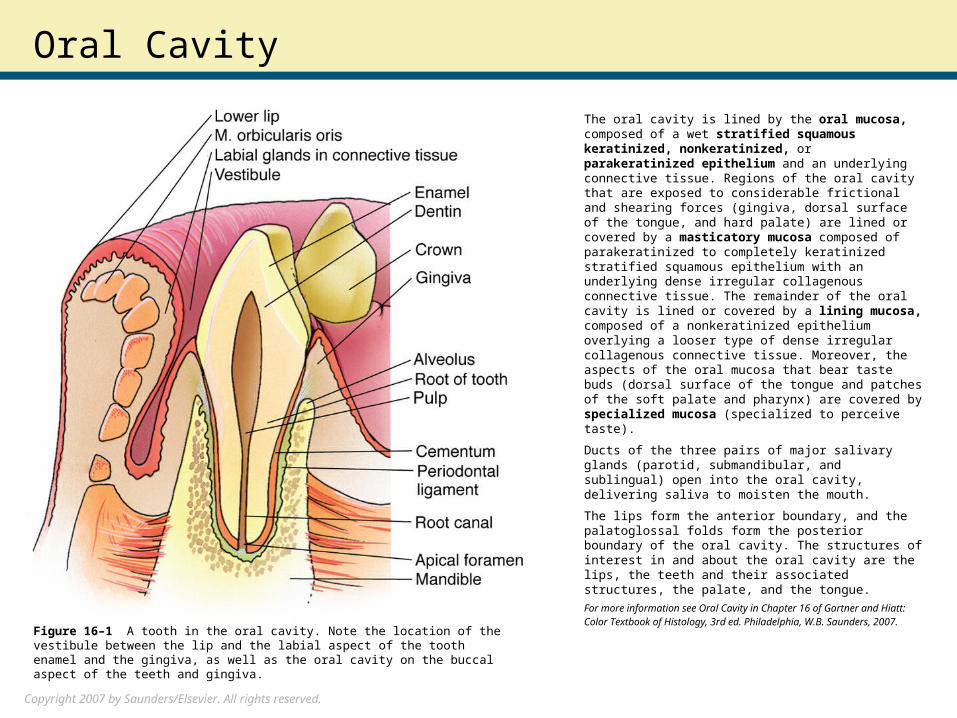
The oral cavity is lined by the oral mucosa, composed of a wet stratified squamous keratinized, nonkeratinized, or parakeratinized epithelium and an underlying connective tissue. Regions of the oral cavity that are exposed to considerable frictional and shearing forces (gingiva, dorsal surface of the tongue, and hard palate) are lined or covered by a masticatory mucosa composed of parakeratinized to completely keratinized stratified squamous epithelium with an underlying dense irregular collagenous connective tissue. The remainder of the oral cavity is lined or covered by a lining mucosa, composed of a nonkeratinized epithelium overlying a looser type of dense irregular collagenous connective tissue. Moreover, the aspects of the oral mucosa that bear taste buds (dorsal surface of the tongue and patches of the soft palate and pharynx) are covered by specialized mucosa (specialized to perceive taste).
Ducts of the three pairs of major salivary glands (parotid, submandibular, and sublingual) open into the oral cavity, delivering saliva to moisten the mouth.
The lips form the anterior boundary, and the palatoglossal folds form the posterior boundary of the oral cavity. The structures of interest in and about the oral cavity are the lips, the teeth and their associated structures, the palate, and the tongue.
For more information see Oral Cavity in Chapter 16 of Gartner and Hiatt: Color Textbook of Histology, 3rd ed. Philadelphia, W.B. Saunders, 2007.
Figure 16–1 A tooth in the oral cavity. Note the location of the vestibule between the lip and the labial aspect of the tooth enamel and the gingiva, as well as the oral cavity on the buccal aspect of the teeth and gingiva.
Copyright 2007 by Saunders/Elsevier. All rights reserved.
Lip
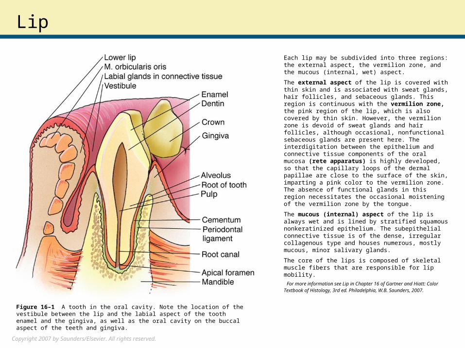
Each lip may be subdivided into three regions: the external aspect, the vermilion zone, and the mucous (internal, wet) aspect.
The external aspect of the lip is covered with thin skin and is associated with sweat glands, hair follicles, and sebaceous glands. This region is continuous with the vermilion zone, the pink region of the lip, which is also covered by thin skin. However, the vermilion zone is devoid of sweat glands and hair follicles, although occasional, nonfunctional sebaceous glands are present here. The interdigitation between the epithelium and connective tissue components of the oral mucosa (rete apparatus) is highly developed, so that the capillary loops of the dermal papillae are close to the surface of the skin, imparting a pink color to the vermilion zone. The absence of functional glands in this region necessitates the occasional moistening of the vermilion zone by the tongue.
The mucous (internal) aspect of the lip is always wet and is lined by stratified squamous nonkeratinized epithelium. The subepithelial connective tissue is of the dense, irregular collagenous type and houses numerous, mostly mucous, minor salivary glands.
The core of the lips is composed of skeletal muscle fibers that are responsible for lip mobility.
For more information see Lip in Chapter 16 of Gartner and Hiatt: Color Textbook of Histology, 3rd ed. Philadelphia, W.B. Saunders, 2007.
Figure 16–1 A tooth in the oral cavity. Note the location of the vestibule between the lip and the labial aspect of the tooth enamel and the gingiva, as well as the oral cavity on the buccal aspect of the teeth and gingiva.
Copyright 2007 by Saunders/Elsevier. All rights reserved.
Teeth
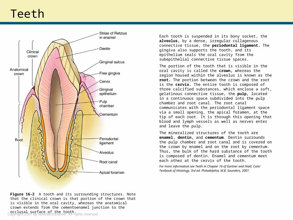
Each tooth is suspended in its bony socket, the alveolus, by a dense, irregular collagenous connective tissue, the periodontal ligament. The gingiva also supports the tooth, and its epithelium seals the oral cavity from the subepithelial connective tissue spaces.
The portion of the tooth that is visible in the oral cavity is called the crown, whereas the region housed within the alveolus is known as the root. The portion between the crown and the root is the cervix. The entire tooth is composed of three calcified substances, which enclose a soft, gelatinous connective tissue, the pulp, located in a continuous space subdivided into the pulp chamber and root canal. The root canal communicates with the periodontal ligament space via a small opening, the apical foramen, at the tip of each root. It is through this opening that blood and lymph vessels as well as nerves enter and leave the pulp.
The mineralized structures of the tooth are enamel, dentin, and cementum. Dentin surrounds the pulp chamber and root canal and is covered on the crown by enamel and on the root by cementum. Thus, the bulk of the hard substance of the tooth is composed of dentin. Enamel and cementum meet each other at the cervix of the tooth.
For more information see Teeth in Chapter 16 of Gartner and Hiatt: Color Textbook of Histology, 3rd ed. Philadelphia, W.B. Saunders, 2007.
Figure 16–2 A tooth and its surrounding structures. Note that the clinical crown is that portion of the crown that is visible in the oral cavity, whereas the anatomical crown extends from the cementoenamel junction to the occlusal surface of the tooth.
Copyright 2007 by Saunders/Elsevier. All rights reserved.
Odontogenesis
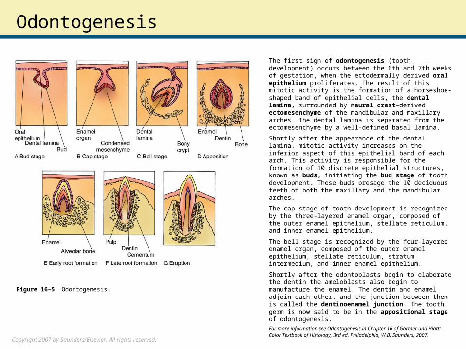
The first sign of odontogenesis (tooth development) occurs between the 6th and 7th weeks of gestation, when the ectodermally derived oral epithelium proliferates. The result of this mitotic activity is the formation of a horseshoe-shaped band of epithelial cells, the dental lamina, surrounded by neural crest–derived ectomesenchyme of the mandibular and maxillary arches. The dental lamina is separated from the ectomesenchyme by a well-defined basal lamina.
Shortly after the appearance of the dental lamina, mitotic activity increases on the inferior aspect of this epithelial band of each arch. This activity is responsible for the formation of 10 discrete epithelial structures, known as buds, initiating the bud stage of tooth development. These buds presage the 10 deciduous teeth of both the maxillary and the mandibular arches.
The cap stage of tooth development is recognized by the three-layered enamel organ, composed of the outer enamel epithelium, stellate reticulum, and inner enamel epithelium.
The bell stage is recognized by the four-layered enamel organ, composed of the outer enamel epithelium, stellate reticulum, stratum intermedium, and inner enamel epithelium.
Shortly after the odontoblasts begin to elaborate the dentin the ameloblasts also begin to manufacture the enamel. The dentin and enamel adjoin each other, and the junction between them is called the dentinoenamel junction. The tooth germ is now said to be in the appositional stage of odontogenesis.
For more information see Odontogenesis in Chapter 16 of Gartner and Hiatt: Color Textbook of Histology, 3rd ed. Philadelphia, W.B. Saunders, 2007.
Figure 16–5 Odontogenesis.
Copyright 2007 by Saunders/Elsevier. All rights reserved.
Periodontal Ligament and Alveolus
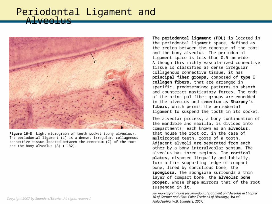
The periodontal ligament (PDL) is located in the periodontal ligament space, defined as the region between the cementum of the root and the bony alveolus. The periodontal ligament space is less than 0.5 mm wide. Although this richly vascularized connective tissue is classified as dense irregular collagenous connective tissue, it has principal fiber groups, composed of type I collagen fibers, that are arranged in specific, predetermined patterns to absorb and counteract masticatory forces. The ends of the principal fiber groups are embedded in the alveolus and cementum as Sharpey’s fibers, which permit the periodontal ligament to suspend the tooth in its socket.
The alveolar process, a bony continuation of the mandible and maxilla, is divided into compartments, each known as an alveolus, that house the root or, in the case of multirooted teeth, roots of a tooth. Adjacent alveoli are separated from each other by a bony interalveolar septum. The alveolus has three regions. The cortical plates, disposed lingually and labially, form a firm supporting ledge of compact bone, lined by cancellous bone, the spongiosa. The spongiosa surrounds a thin layer of compact bone, the alveolar bone proper, whose shape mirrors that of the root suspended in it.
For more information see Periodontal Ligament and Alveolus in Chapter 16 of Gartner and Hiatt: Color Textbook of Histology, 3rd ed. Philadelphia, W.B. Saunders, 2007.
Figure 16–8 Light micrograph of tooth socket (bony alveolus). The periodontal ligament (L) is a dense, irregular, collagenous connective tissue located between the cementum (C) of the root and the bony alveolus (A) (´132).

Copyright 2007 by Saunders/Elsevier. All rights reserved.
Tongue
The tongue is the largest structure in the oral cavity. Its extreme mobility is due to the large intertwined mass of skeletal muscle fibers that compose its bulk. The muscle fibers may be classified into two groups: those that originate outside the tongue, the extrinsic muscles, and those that originate in and insert into the tongue, the intrinsic muscles. The extrinsic muscles are responsible for moving the tongue in and out of the mouth as well as from side to side, whereas the intrinsic muscles alter the shape of the tongue.
The tongue has a dorsal surface, a ventral surface, and two lateral surfaces. The dorsal surface is observed to have two unequal regions, the larger anterior two thirds and the smaller posterior one third. The two regions are separated from one another by a shallow, V-shaped groove, the sulcus terminalis, whose apex points posteriorly and contains a deep concavity, the foramen cecum.
The dorsal surface of the posterior one third of the tongue is uneven because of the presence of the lingual tonsils. The most posterior portion of the tongue is known as the root of the tongue. Lingual papillae, most of which project above the surface, cover the anterior two thirds of the tongue’s dorsal surface.
For more information see Tongue in Chapter 16 of Gartner and Hiatt: Color Textbook of Histology, 3rd ed. Philadelphia, W.B. Saunders, 2007.
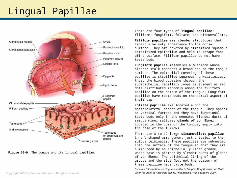
Figure 16–9 The tongue and its lingual papillae.
Copyright 2007 by Saunders/Elsevier. All rights reserved.
Lingual Papillae
There are four types of lingual papillae: filiform, fungiform, foliate, and circumvallate.
Filiform papillae are slender structures that impart a velvety appearance to the dorsal surface. They are covered by stratified squamous keratinized epithelium and help to scrape food off a surface. Filiform papillae do not have taste buds.
Fungiform papilla resembles a mushroom whose slender stalk connects a broad cap to the tongue surface. The epithelial covering of these papillae is stratified squamous nonkeratinized; thus, the blood coursing through the subepithelial capillary loops is evident as red dots distributed randomly among the filiform papillae on the dorsum of the tongue. Fungiform papillae have taste buds on the dorsal aspect of their cap.
Foliate papillae are located along the posterolateral aspect of the tongue. They appear as vertical furrows and they have functional taste buds only in the neonate. Slender ducts of serous minor salivary glands of von Ebner, located in the core of the tongue, empty into the base of the furrows.
There are 8 to 12 large circumvallate papillae in a V-shaped arrangement just anterior to the sulcus terminalis. These papillae are submerged into the surface of the tongue so that they are surrounded by an epithelially lined groove, whose base is pierced by slender ducts of glands of von Ebner. The epithelial lining of the groove and the side (but not the dorsum) of these papillae have taste buds.
For more information see Lingual papillae in Chapter 16 of Gartner and Hiatt: Color Textbook of Histology, 3rd ed. Philadelphia, W.B. Saunders, 2007.
Figure 16–9 The tongue and its lingual papillae.
Copyright 2007 by Saunders/Elsevier. All rights reserved.
Taste Buds
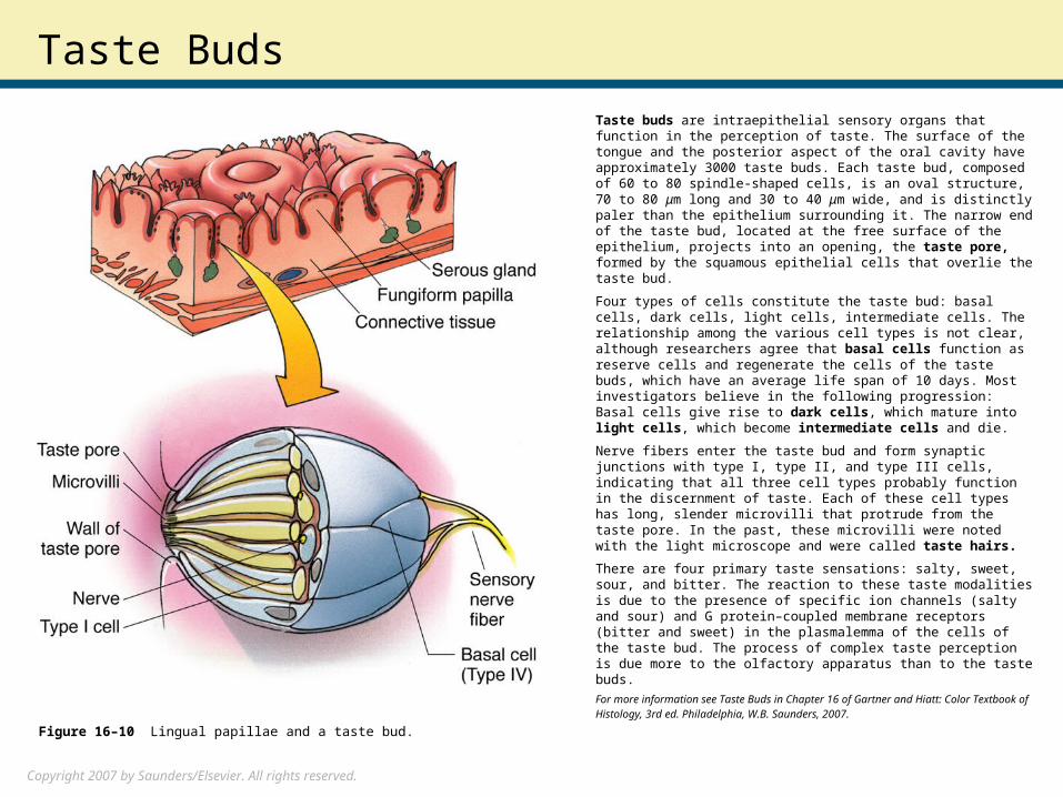
Taste buds are intraepithelial sensory organs that function in the perception of taste. The surface of the tongue and the posterior aspect of the oral cavity have approximately 3000 taste buds. Each taste bud, composed of 60 to 80 spindle-shaped cells, is an oval structure, 70 to 80 μm long and 30 to 40 μm wide, and is distinctly paler than the epithelium surrounding it. The narrow end of the taste bud, located at the free surface of the epithelium, projects into an opening, the taste pore, formed by the squamous epithelial cells that overlie the taste bud.
Four types of cells constitute the taste bud: basal cells, dark cells, light cells, intermediate cells. The relationship among the various cell types is not clear, although researchers agree that basal cells function as reserve cells and regenerate the cells of the taste buds, which have an average life span of 10 days. Most investigators believe in the following progression: Basal cells give rise to dark cells, which mature into light cells, which become intermediate cells and die.
Nerve fibers enter the taste bud and form synaptic junctions with type I, type II, and type III cells, indicating that all three cell types probably function in the discernment of taste. Each of these cell types has long, slender microvilli that protrude from the taste pore. In the past, these microvilli were noted with the light microscope and were called taste hairs.
There are four primary taste sensations: salty, sweet, sour, and bitter. The reaction to these taste modalities is due to the presence of specific ion channels (salty and sour) and G protein–coupled membrane receptors (bitter and sweet) in the plasmalemma of the cells of the taste bud. The process of complex taste perception is due more to the olfactory apparatus than to the taste buds.
For more information see Taste Buds in Chapter 16 of Gartner and Hiatt: Color Textbook of Histology, 3rd ed. Philadelphia, W.B. Saunders, 2007. Figure 16–10 Lingual papillae and a taste bud.