
Copyright © 2006 by Mosby, Inc. Slide 1 Chapter 18 Fungal Diseases of the Lung Figure 18-1. Fungal...
35
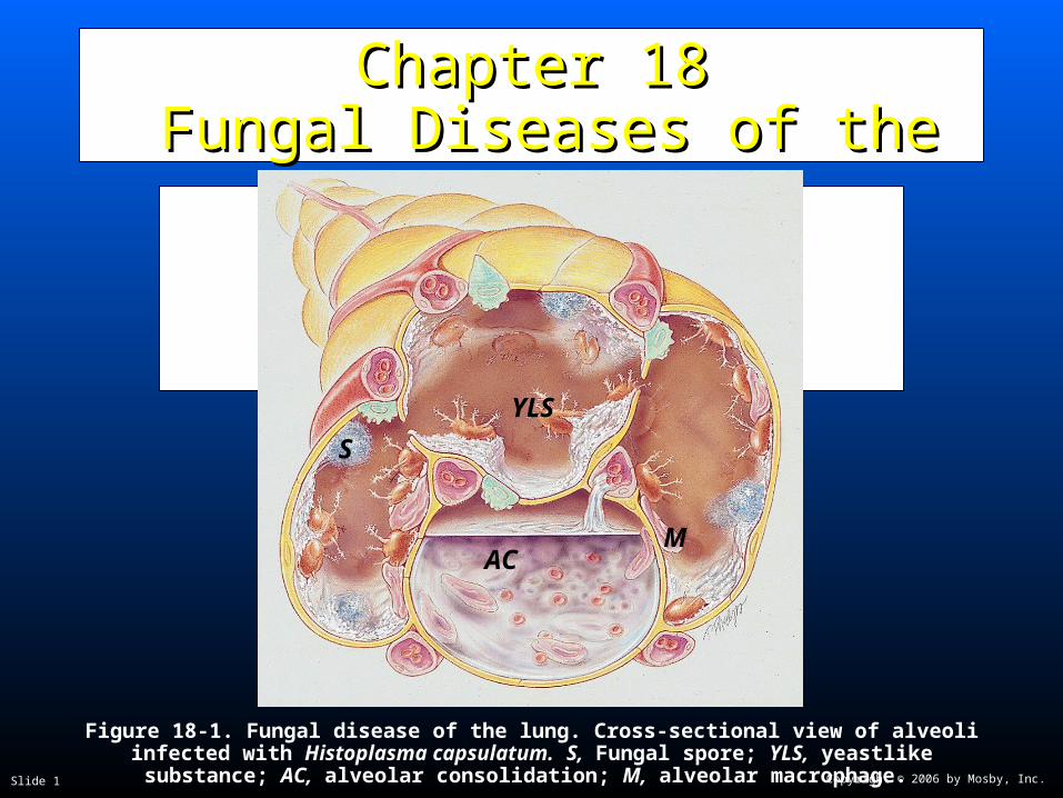
Copyright © 2006 by Mosby, Inc. Slide 1 Chapter 18 Chapter 18 Fungal Diseases of the Fungal Diseases of the Lung Lung Figure 18-1. Fungal disease of the lung. Cross-sectional view of alveoli infected with Histoplasma capsulatum. S, Fungal spore; YLS, yeastlike substance; AC, alveolar consolidation; M, alveolar macrophage. AC S YLS M
-
Upload
raina-souter -
Category
Documents
-
view
216 -
download
2
Transcript of Copyright © 2006 by Mosby, Inc. Slide 1 Chapter 18 Fungal Diseases of the Lung Figure 18-1. Fungal...
Copyright © 2006 by Mosby, Inc.Slide 1
Chapter 18Chapter 18 Fungal Diseases of the Lung Fungal Diseases of the Lung
Figure 18-1. Fungal disease of the lung. Cross-sectional view of alveoli infected with Histoplasma capsulatum. S, Fungal spore; YLS, yeastlike substance; AC, alveolar
consolidation; M, alveolar macrophage.
AC
S
YLS
M

Copyright © 2006 by Mosby, Inc.Slide 2
Anatomic Alterations of the LungsAnatomic Alterations of the Lungs
Alveolar consolidationAlveolar consolidation
Alveolar-capillary destructionAlveolar-capillary destruction
Granuloma formationGranuloma formation
Cavity formationCavity formation
Fibrosis of the lung parenchymaFibrosis of the lung parenchyma
Airway secretionsAirway secretions
Copyright © 2006 by Mosby, Inc.Slide 3
EtiologyEtiology
Histoplasmosis Histoplasmosis (most common fungal disease(most common fungal diseasein the United States)in the United States)
Screening and diagnosisScreening and diagnosis Fungal cultureFungal culture
Fungal stainFungal stain
SerologySerology
Copyright © 2006 by Mosby, Inc.Slide 4
EtiologyEtiology
CoccidioidomycosisCoccidioidomycosis
Screening and diagnosisScreening and diagnosis Direct visualization of distinctive spherulesDirect visualization of distinctive spherules
Blood test that detects antibodies of the fungusBlood test that detects antibodies of the fungus
Culture of the organismCulture of the organism
Copyright © 2006 by Mosby, Inc.Slide 5
EtiologyEtiology
BlastomycosisBlastomycosis
Screening and diagnosis and diagnosis Direct visualization of yeast in sputum smearsDirect visualization of yeast in sputum smears
Culture of the fungusCulture of the fungus

Copyright © 2006 by Mosby, Inc.Slide 6
EtiologyEtiology
Opportunistic pathogensOpportunistic pathogens
Candida albicansCandida albicans
Cryptococcus neoformansCryptococcus neoformans
AspergillusAspergillus
Copyright © 2006 by Mosby, Inc.Slide 7
Overview of the Cardiopulmonary Overview of the Cardiopulmonary Clinical Manifestations Clinical Manifestations
Associated with Associated with FUNGAL DISEASES OF THE LUNGFUNGAL DISEASES OF THE LUNG
The following clinical manifestations result from The following clinical manifestations result from the pathophysiologic mechanisms caused (or the pathophysiologic mechanisms caused (or activated) by activated) by Alveolar ConsolidationAlveolar Consolidation (see (see Figure 9-8)Figure 9-8), , and and Increased Alveolar-Capillary Increased Alveolar-Capillary Membrane ThicknessMembrane Thickness (see Figure 9-9)—the (see Figure 9-9)—the major anatomic alterations of the lungs major anatomic alterations of the lungs associated with fungal diseases of the lung (see associated with fungal diseases of the lung (see Figure 18-1). Figure 18-1).
Copyright © 2006 by Mosby, Inc.Slide 8
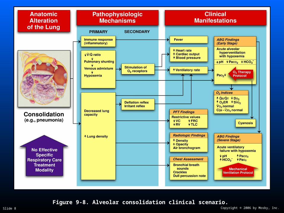
Figure 9-8. Alveolar consolidation clinical scenario.

Copyright © 2006 by Mosby, Inc.Slide 9
Figure 9-9. Increased alveolar-capillary membrane thickness clinical scenario.

Copyright © 2006 by Mosby, Inc.Slide 10
Clinical Data Obtained at the Clinical Data Obtained at the Patient’s BedsidePatient’s Bedside
Vital signsVital signs
Increased respiratory rateIncreased respiratory rate
Increased heart rate, cardiac output, Increased heart rate, cardiac output, blood pressureblood pressure

Copyright © 2006 by Mosby, Inc.Slide 11
Clinical Data Obtained at the Clinical Data Obtained at the Patient’s BedsidePatient’s Bedside
Chest pain/decreased chest expansionChest pain/decreased chest expansion
CyanosisCyanosis
Digital clubbingDigital clubbing
Peripheral edema and distentionPeripheral edema and distention Distended neck veinsDistended neck veins
Pitting edemaPitting edema
Enlarged and tender liverEnlarged and tender liver

Copyright © 2006 by Mosby, Inc.Slide 12
Digital Clubbing
Figure 2-46. Digital clubbing.Figure 2-46. Digital clubbing.

Copyright © 2006 by Mosby, Inc.Slide 13
DistendedDistendedNeck VeinsNeck Veins
Figure 2-48. Distended neck veins (Figure 2-48. Distended neck veins (arrowsarrows).).

Copyright © 2006 by Mosby, Inc.Slide 14
Figure 2-47. Pitting edema. From Bloom A, Ireland J: Figure 2-47. Pitting edema. From Bloom A, Ireland J: Color atlas of diabetesColor atlas of diabetes, ed 2,, ed 2,London, 1992, Mosby-Wolfe.London, 1992, Mosby-Wolfe.

Copyright © 2006 by Mosby, Inc.Slide 15
Clinical Data Obtained at the Clinical Data Obtained at the Patient’s BedsidePatient’s Bedside
Cough, sputum production, and hemoptysisCough, sputum production, and hemoptysis
Chest assessment findingsChest assessment findings Increased tactile and vocal fremitusIncreased tactile and vocal fremitus
Dull percussion noteDull percussion note
Bronchial breath soundsBronchial breath sounds
Crackles, rhonchi, and wheezingCrackles, rhonchi, and wheezing
Pleural friction rubPleural friction rub
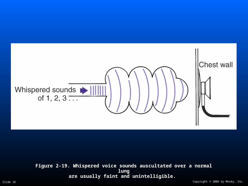
Whispered pectoriloquyWhispered pectoriloquy

Copyright © 2006 by Mosby, Inc.Slide 16
Figure 2-11. Figure 2-11. A short, dull, or flat percussion note is typically produced over areas A short, dull, or flat percussion note is typically produced over areas of alveolar consolidation.of alveolar consolidation.

Copyright © 2006 by Mosby, Inc.Slide 17
Figure 2-16. Figure 2-16. Auscultation of bronchial breath sounds over a consolidated lung Auscultation of bronchial breath sounds over a consolidated lung unit.unit.
Copyright © 2006 by Mosby, Inc.Slide 18
Figure 2-19. Figure 2-19. Whispered voice sounds auscultated over a normal lungWhispered voice sounds auscultated over a normal lungare usually faint and unintelligible.are usually faint and unintelligible.

Copyright © 2006 by Mosby, Inc.Slide 19
Clinical Data Obtained from Clinical Data Obtained from Laboratory Tests and Special Laboratory Tests and Special
ProceduresProcedures

Copyright © 2006 by Mosby, Inc.Slide 20
Pulmonary Function Study: Pulmonary Function Study: Expiratory Maneuver FindingsExpiratory Maneuver Findings
FVC FEVT FEF25%-75% FEF200-1200
N or N or N
PEFR MVV FEF50% FEV1%
N N or N N or
FVC FEVT FEF25%-75% FEF200-1200
N or N or N
PEFR MVV FEF50% FEV1%
N N or N N or

Copyright © 2006 by Mosby, Inc.Slide 21
Pulmonary Function Study: Pulmonary Function Study: Lung Volume and Capacity Findings Lung Volume and Capacity Findings
VT RV FRC TLC
N or
VC IC ERV RV/TLC%
N
VT RV FRC TLC
N or
VC IC ERV RV/TLC%
N
Copyright © 2006 by Mosby, Inc.Slide 22
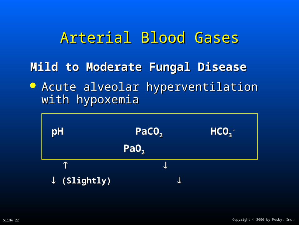
Arterial Blood GasesArterial Blood Gases
Mild to Moderate Fungal DiseaseMild to Moderate Fungal Disease
Acute alveolar hyperventilation with Acute alveolar hyperventilation with hypoxemiahypoxemia
pH PaCO2 HCO3- PaO2
(Slightly)
pH PaCO2 HCO3- PaO2
(Slightly)

Copyright © 2006 by Mosby, Inc.Slide 23
Time and Progression of Disease Time and Progression of Disease
100100
5050
3030
8080
00
PaCO2
1010
2020
4040
Alveolar HyperventilationAlveolar Hyperventilation
6060
7070
9090 Point at which PaO2 declines enough to stimulate peripheral oxygen receptors
Point at which PaO2 declines enough to stimulate peripheral oxygen receptors
PaO2
Disease OnsetDisease OnsetP
aO2
or
PaC
O2
PaO
2 o
r P
aCO
2
Figure 4-2. PaO2 and PaCO2 trends during acute alveolar hyperventilation.

Copyright © 2006 by Mosby, Inc.Slide 24
Arterial Blood GasesArterial Blood Gases
Severe Fungal Disease with PulmonarySevere Fungal Disease with PulmonaryFibrosisFibrosis
Chronic ventilatory failure with hypoxemiaChronic ventilatory failure with hypoxemia
pH PaCO2 HCO3- PaO2
Normal (Significantly)
pH PaCO2 HCO3- PaO2
Normal (Significantly)

Copyright © 2006 by Mosby, Inc.Slide 25
Time and Progression of DiseaseTime and Progression of Disease
100100
5050
3030
80
0
PaO2
1010
2020
4040
Alveolar HyperventilationAlveolar Hyperventilation
6060
7070
9090Point at which PaO2 declines enough to stimulate peripheral oxygen receptors
Point at which PaO2 declines enough to stimulate peripheral oxygen receptors
PaCO 2
Chronic Ventilatory Failure Chronic Ventilatory FailureDisease OnsetDisease Onset
Point at which disease becomes severe and patient begins to become fatigued
Point at which disease becomes severe and patient begins to become fatigued
Pa0
2 o
r P
aC0 2
Pa0
2 o
r P
aC0 2
Figure 4-7. PaO2 and PaCO2 trends during acute or chronic ventilatory failure.

Copyright © 2006 by Mosby, Inc.Slide 26
Acute Ventilatory Changes on Acute Ventilatory Changes on Chronic Ventilatory FailureChronic Ventilatory Failure
Acute alveolar hyperventilation on chronic Acute alveolar hyperventilation on chronic ventilatory failureventilatory failure
Acute ventilatory failure on chronic ventilatory Acute ventilatory failure on chronic ventilatory failurefailure
Copyright © 2006 by Mosby, Inc.Slide 27
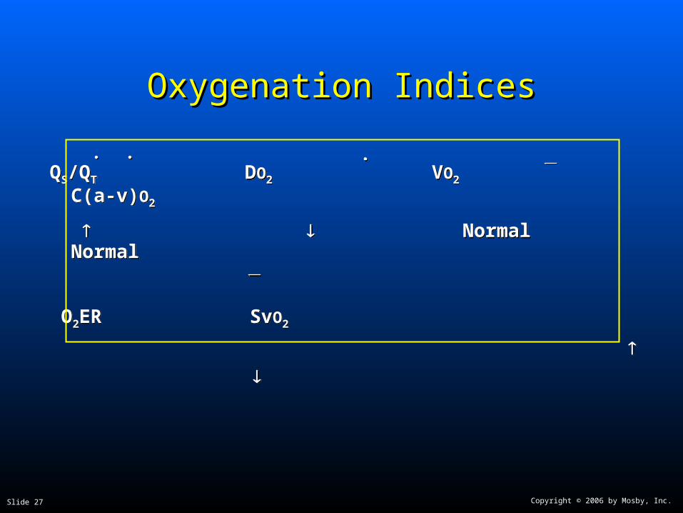
Oxygenation IndicesOxygenation Indices
QS/QT DO2 VO2 C(a-v)O2
Normal Normal
O2ER SvO2
QS/QT DO2 VO2 C(a-v)O2
Normal Normal
O2ER SvO2
Copyright © 2006 by Mosby, Inc.Slide 28
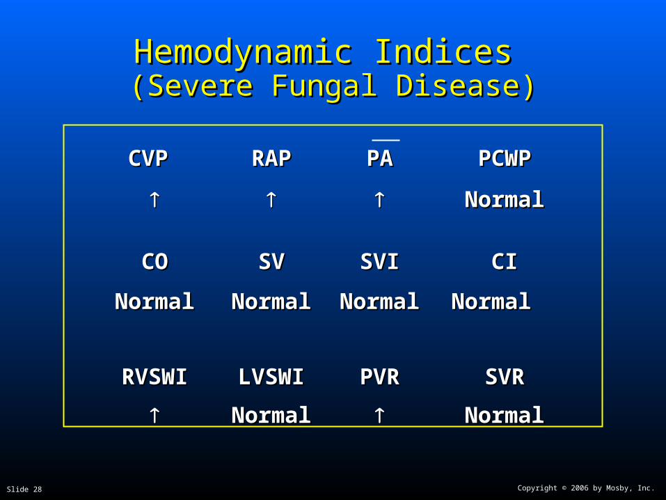
Hemodynamic Indices Hemodynamic Indices (Severe Fungal Disease)(Severe Fungal Disease)
CVP CVP RAPRAP PAPA PCWPPCWP
NormalNormal
COCO SVSV SVISVI CICI
NormalNormal NormalNormal NormalNormal Normal Normal
RVSWIRVSWI LVSWILVSWI PVRPVR SVRSVR
NormalNormal NormalNormal
Copyright © 2006 by Mosby, Inc.Slide 29
Abnormal Laboratory Tests Abnormal Laboratory Tests and Proceduresand Procedures
See Etiology and Primary Pathogen See Etiology and Primary Pathogen sections in this chaptersections in this chapter

Copyright © 2006 by Mosby, Inc.Slide 30
Radiologic FindingsRadiologic Findings
Chest radiographChest radiograph
Increased opacityIncreased opacity
Cavity formationCavity formation
Pleural effusionPleural effusion
Calcification and fibrosisCalcification and fibrosis
Right ventricular enlargementRight ventricular enlargement

Copyright © 2006 by Mosby, Inc.Slide 31
Figure 18-2. Acute inhalational histoplasmosis in an otherwise healthy patient. This young man Figure 18-2. Acute inhalational histoplasmosis in an otherwise healthy patient. This young man developed fever and cough after tearing down an old barn. The study shows bilateral hilar developed fever and cough after tearing down an old barn. The study shows bilateral hilar
adenopathy. (From Armstrong P et al: adenopathy. (From Armstrong P et al: Imaging of diseases of the chest,Imaging of diseases of the chest, ed 2, St. Louis, 1995, Mosby.) ed 2, St. Louis, 1995, Mosby.)

Copyright © 2006 by Mosby, Inc.Slide 32
Figure 18-3. Histoplasmoma, showing a well-defined spherical nodule. The central portion of Figure 18-3. Histoplasmoma, showing a well-defined spherical nodule. The central portion of the nodule shows calcification. (From Armstrong P et al: the nodule shows calcification. (From Armstrong P et al: Imaging of diseases of the chest,Imaging of diseases of the chest,
ed 2, St. Louis, 1995, Mosby.)ed 2, St. Louis, 1995, Mosby.)

Copyright © 2006 by Mosby, Inc.Slide 33
General Management of General Management of Fungal DiseaseFungal Disease
Pharmacologic agents Pharmacologic agents
Amphotericin B (Fungizone)Amphotericin B (Fungizone)
Itraconazole (Sporanox)Itraconazole (Sporanox)
Fluconazole (Diflucan)Fluconazole (Diflucan)

Copyright © 2006 by Mosby, Inc.Slide 34
General Management of General Management of Fungal DiseaseFungal Disease
Respiratory care treatment protocolsRespiratory care treatment protocols
Oxygen therapy protocolOxygen therapy protocol
Bronchopulmonary hygiene therapy protocolBronchopulmonary hygiene therapy protocol
Hyperinflation therapy protocolHyperinflation therapy protocol
Mechanical ventilation protocolMechanical ventilation protocol

Copyright © 2006 by Mosby, Inc.Slide 35
Classroom DiscussionClassroom DiscussionCase Study: Case Study:
Fungal Diseases of the LungFungal Diseases of the Lung


![Dr. Saleem Shaikh ORAL FUNGAL INFECTION. COMMON FUNGAL INFECTIONS Candidiasis Histoplasmosis [Darlings disease] – Common lung Blastomycosis [Gilchrists.](https://static.fdocuments.net/doc/165x107/5697bff31a28abf838cbc4a5/dr-saleem-shaikh-oral-fungal-infection-common-fungal-infections-candidiasis.jpg)

![CVIA · cardiac fungal infection [1], evidence of invasive lung aspergil-losis supported disease dissemination, as in previous case stud - ies [4,7]. On imaging, fungal heart disease](https://static.fdocuments.net/doc/165x107/601a052fbebcea3c2916bb94/cvia-cardiac-fungal-infection-1-evidence-of-invasive-lung-aspergil-losis-supported.jpg)

![4D Model Generator of the Human Lung, ³/XQJ &HU ......The lung consists of about three hundred millions of alveoli, tiny air bags with the size of about 0.3 mm [5]. The pulmonary](https://static.fdocuments.net/doc/165x107/5eb475bcd190f971f74a6032/4d-model-generator-of-the-human-lung-xqj-hu-the-lung-consists-of.jpg)


