Copyright © 2003 Pearson Education, Inc. publishing as Benjamin Cummings Nuclei (yellow) and actin...
49
Copyright © 2003 Pearson Education, Inc. publishing as Benjamin Cummings • Nuclei (yellow) and actin (red) Figure 4.6x
-
Upload
walter-carroll -
Category
Documents
-
view
213 -
download
0
Transcript of Copyright © 2003 Pearson Education, Inc. publishing as Benjamin Cummings Nuclei (yellow) and actin...

Copyright © 2003 Pearson Education, Inc. publishing as Benjamin Cummings
• Nuclei (yellow) and actin (red)
Figure 4.6x

Copyright © 2003 Pearson Education, Inc. publishing as Benjamin Cummings
• network of protein fibers
The cell’s internal skeleton helps organize its structure and activities
THE CYTOSKELETON
Figure 4.17A

Copyright © 2003 Pearson Education, Inc. publishing as Benjamin Cummings
• Microfilaments of actin enable cells to change shape and move
• Intermediate filaments reinforce the cell and anchor certain organelles
• Microtubules
– give the cell rigidity
– provide anchors for organelles
– act as tracks for organelle movement

Copyright © 2003 Pearson Education, Inc. publishing as Benjamin Cummings
MICROFILAMENT
Figure 4.17B
INTERMEDIATEFILAMENT
MICROTUBULE
Actin subunit Fibrous subunitsTubulinsubunit
7 nm 10 nm25 nm

Copyright © 2003 Pearson Education, Inc. publishing as Benjamin Cummings
QuickTime™ and aCinepak decompressorare needed to see this picture.

Copyright © 2003 Pearson Education, Inc. publishing as Benjamin Cummings
• A cilia or flagellum is composed of a core of microtubules wrapped in plasma membrane
• Eukaryotes have “9+2” structure
How do cilia and flagella move?
Cilia and flagella move when microtubules bend

Copyright © 2003 Pearson Education, Inc. publishing as Benjamin Cummings
Figure 4.18A
FLAGELLUM
Outer microtubule doublet
Plasmamembrane
Centralmicrotubules
Outer microtubule doublet
Plasmamembrane
Electron micrograph of sections:
Flagellum
Basal body
Basal body(structurally identical to centriole)

Copyright © 2003 Pearson Education, Inc. publishing as Benjamin Cummings
Slide 43QuickTime™ and aCinepak decompressorare needed to see this picture.

polarhead
nonpolartails
P –
Phospholipid bilayer
hydrophobic molecules hydrophilic molecules cytosol

Copyright © 2003 Pearson Education, Inc. publishing as Benjamin Cummings
Surfaces allow exchange of signals and molecules.
• Plant cells connect by plasmodesmata
Cell surfaces protect, support, and join cells

Copyright © 2003 Pearson Education, Inc. publishing as Benjamin Cummings
Figure 4.19A
Vacuole
Layers of one plant cell wall
Walls of two adjacent plant cells
PLASMODESMATA
Cytoplasm
Plasma membrane

Copyright © 2003 Pearson Education, Inc. publishing as Benjamin Cummings
• Animal cells - extracellular matrix
– sticky layer of glycoproteins
– binds cells together in tissues
– can also protect and support cells

Copyright © 2003 Pearson Education, Inc. publishing as Benjamin Cummings
• Tight junctions can bind cells together into leakproof sheets
• Anchoring junctions link animal cells
• Gap junctions allow substances to flow from cell to cell
TIGHTJUNCTION
ANCHORING JUNCTION
GAPJUNCTION
Plasma membranes ofadjacent cells
ExtracellularmatrixFigure 4.19B

Copyright © 2003 Pearson Education, Inc. publishing as Benjamin Cummings
Eukaryotic organelles fall into 4 functional groups
• 1. Manufacture and transport – dependent on network of membranes
- Nucleus
- Ribosomes
- Rough ER
- Smooth ER
- Golgi apparatus

Copyright © 2003 Pearson Education, Inc. publishing as Benjamin Cummings
2. Breakdown – all single-membrane sacs
• Lysosomes (in animals, some protists)
• Peroxisomes
• Vacuoles (plants)

Copyright © 2003 Pearson Education, Inc. publishing as Benjamin Cummings
3. Energy Processing – involves extensive membranes embedded with enzymes
• Chloroplasts
• Mitochondria

Copyright © 2003 Pearson Education, Inc. publishing as Benjamin Cummings
4. Support, Movement, Communication
• Cytoskeleton – includes cilia, flagella, filaments, microtubules
• Cell walls
• Extracellular matrix
• Cell junctions


What do these have in common?
• HIV infection
• Transplanted organs
• Communication between neurons
• Drug addiction
• Cystic fibrosis
• hypercholesteremia

Copyright © 2003 Pearson Education, Inc. publishing as Benjamin Cummings
• selectively permeable
• hold teams of enzymes
Membranes organize the chemical activities of cells
Cytoplasm
Figure 5.10
Plasma membrane
• Contact between cell and environment
• Keeps useful materials inside and harmful stuff outside
• Allows transport, communication in both directions

Plasma membrane components
Phospholipid bilayer
Cholesterol
Proteins
Glycocalyx

polarhead
nonpolartails
P –
Phospholipid bilayer
hydrophobic molecules hydrophilic molecules cytosol

THE PLASMA MEMBRANE
phospholipids cholesterol
cytoskeletonperipheralprotein
integralprotein
Cholesterol blocks some small molecules, adds fluidity

• Membrane Proteins– span entire membrane or lie on
either side– Purposes
• Structural Support• Recognition• Communication• Transport

• Glycocalyx
– Composed of sugars protruding from lipids and proteins
– Functions
• Binding sites for proteins
• Lubricate cells.
• Stick cells down.

Copyright © 2003 Pearson Education, Inc. publishing as Benjamin Cummings
• Many membrane proteins are enzymes
Figure 5.13
• Some proteins function as receptors for chemical messages from other cells
– The binding of a messenger to a receptor may trigger signal transduction
Enzyme activity Signal transduction
Messenger molecule
Receptor
Activated molecule

Copyright © 2003 Pearson Education, Inc. publishing as Benjamin Cummings
• The plasma membrane of an animal cell
Fibers of the extracellular matrix
Figure 5.12
Glycoprotein Carbohydrate (of glycoprotein)
Microfilaments of the cytoskeleton
Phospholipid
Cholesterol
Proteins
CYTOPLASM
Glycolipid

• Diffusion and Gradients
– Diffusion = movement of molecules from region of higher to lower concentration.
– Osmosis = diffusion of water across a membrane
Copyright © 2003 Pearson Education, Inc. publishing as Benjamin Cummings
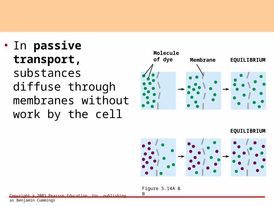
• In passive transport, substances diffuse through membranes without work by the cell
EQUILIBRIUMMolecule of dye
Figure 5.14A & B
Membrane
EQUILIBRIUM

free water molecule: can fit through pore
bound water moleculesclustered around sugar:cannot fit through pore
pore
sugar
H2O
bagbursts
selectively permeable membrane
water molecule
pure water
sugar molecule
(a)
selectively permeable membrane
(b)

Copyright © 2003 Pearson Education, Inc. publishing as Benjamin Cummings
• water travels from an area of higher concentration to an area of lower water concentration
Osmosis = diffusion of water across a membraneHypotonicsolution
Figure 5.15
Solutemolecule
HYPOTONIC SOLUTION
Hypertonic solution
Selectivelypermeablemembrane
HYPERTONIC SOLUTION
Selectivelypermeablemembrane
NET FLOW OF WATER
Solute molecule with cluster of water molecules
Water molecule

Copyright © 2003 Pearson Education, Inc. publishing as Benjamin Cummings
• Osmosis causes cells to shrink in a hypertonic solution and swell in a hypotonic solution
Water balance between cells and their surroundings is crucial to organisms
osmoregulation = control of water balance

isotonic solution hypertonic solution hypotonic solution
10 microns
equal movement of waterinto and out of cells
net water movement out of cells
net water movement into cells

QuickTime™ and aCinepak decompressorare needed to see this picture.
Copyright © 2003 Pearson Education, Inc. publishing as Benjamin Cummings
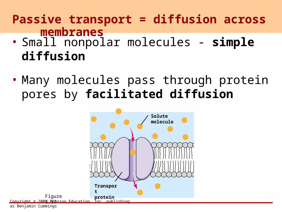
• Small nonpolar molecules - simple diffusion
• Many molecules pass through protein pores by facilitated diffusion
Passive transport = diffusion across membranes
Figure 5.17
Solutemolecule
Transportprotein

Copyright © 2003 Pearson Education, Inc. publishing as Benjamin Cummings
• transport proteins needed
• against a concentration gradient
• requires energy (ATP)
Active transport

Copyright © 2003 Pearson Education, Inc. publishing as Benjamin Cummings
• Active transport in two solutes across a membrane
• Na+/K+ pump
• Protein shape changeFigure 5.18
Transportprotein
1
FLUIDOUTSIDECELL
Firstsolute
First solute, inside cell, binds to protein
Phosphorylated transport protein
2 ATP transfers phosphate to protein
3 Protein releases solute outside cell
4 Second solute binds to protein
Second solute
5 Phosphate detaches from protein
6 Protein releases second solute into cell

Copyright © 2003 Pearson Education, Inc. publishing as Benjamin Cummings
exocytosis = vesicle fuses with the membrane and expels its contents
Exocytosis and endocytosis transport large molecules
Figure 5.19A
FLUID OUTSIDE CELL
CYTOPLASM
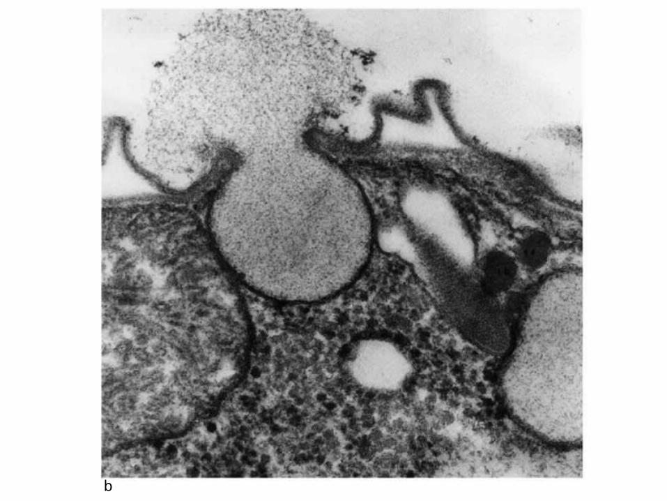
b

Copyright © 2003 Pearson Education, Inc. publishing as Benjamin Cummings
– or the membrane may fold inward, trapping material from the outside (endocytosis)
Figure 5.19B

food particle
particle enclosed in vesicle
phagocytosis
1 32
Phagocytosis, “cell eating”
—How the human immune system ingests whole bacteria or one-celled creatures eat.

Copyright © 2003 Pearson Education, Inc. publishing as Benjamin Cummings
QuickTime™ and aTIFF (Uncompressed) decompressorare needed to see this picture.

(cytoplasm)
vesicle containing extracellular fluid
pinocytosis
2

extracellular fluid
plasma membrane
vesiclecytosol
receptors captured molecules
coatedpit
vesicle
bacterium pseudopodium
vesicle

Receptor-mediated endocytosis

Copyright © 2003 Pearson Education, Inc. publishing as Benjamin Cummings
• Cholesterol can accumulate in the blood if membranes lack cholesterol receptors
Figure 5.20
LDL PARTICLEPhospholipid outer layer
Protein
Cholesterol
Plasma membraneCYTOPLASM
Receptor protein
Vesicle


What do these have in common?
• HIV infection
• Transplanted organs
• Communication between neurons
• Drug addiction
• Cystic fibrosis
• hypercholesteremia