
Controlling Hospital-Acquired Infection: Focus on the Role ... · Controlling Hospital-Acquired...
26
Controlling Hospital-Acquired Infection: Focus on the Role of the Environment and New Technologies for Decontamination Stephanie J. Dancer Department of Microbiology, Hairmyres Hospital, East Kilbride, Lanarkshire, Scotland, United Kingdom SUMMARY ..................................................................................................................................................665 INTRODUCTION ............................................................................................................................................666 CLEANING AND HAI ........................................................................................................................................667 MRSA .....................................................................................................................................................667 VRE .......................................................................................................................................................668 C. difficile ..................................................................................................................................................669 Acinetobacter .............................................................................................................................................669 Multidrug-Resistant Gram-Negative Bacilli ...............................................................................................................670 Pseudomonas and Stenotrophomonas spp. ..............................................................................................................671 Norovirus .................................................................................................................................................672 MANUAL CLEANING: PROCESS AND EQUIPMENT ........................................................................................................672 Routine Cleaning Practices ...............................................................................................................................672 Noncritical Surfaces ......................................................................................................................................673 Critical Surfaces ...........................................................................................................................................673 Clinical Equipment .......................................................................................................................................673 Terminal (Deep) Cleaning ................................................................................................................................674 Microfiber versus Cotton .................................................................................................................................674 Contamination of Cleaning Equipment and Liquids .....................................................................................................675 Benefits of Physically Removing Soil ......................................................................................................................675 AUTOMATED DECONTAMINATION DEVICES ..............................................................................................................675 Steam Cleaning...........................................................................................................................................676 Ozone ....................................................................................................................................................676 UV Light ..................................................................................................................................................676 HINS ......................................................................................................................................................677 Hydrogen Peroxide .......................................................................................................................................677 Comparison between UV Light and Hydrogen Peroxide Systems........................................................................................677 ANTIMICROBIAL SURFACES ................................................................................................................................678 Antiadhesive Surfaces ....................................................................................................................................678 Antimicrobial Coatings ..................................................................................................................................678 Triclosan ...............................................................................................................................................678 Silver ...................................................................................................................................................679 Copper .................................................................................................................................................679 Bacteriophage-modified surfaces......................................................................................................................679 Polycationic antimicrobial surfaces ....................................................................................................................679 Light-activated antimicrobial surfaces .................................................................................................................679 Current Concerns over Antimicrobial Surfaces ...........................................................................................................679 HOW TO MEASURE CLEANLINESS .........................................................................................................................680 Microbiological Methods .................................................................................................................................680 ATP Bioluminescence Systems ...........................................................................................................................680 HOW TO MEASURE CLEANING ............................................................................................................................680 Fluorescent Markers ......................................................................................................................................680 ATP Bioluminescence Systems ...........................................................................................................................681 Observation, Supervision, and Education of Housekeeping Staff.........................................................................................681 DISCUSSION ................................................................................................................................................681 Current Unanswered Questions ..........................................................................................................................681 CONCLUSION ...............................................................................................................................................682 REFERENCES ................................................................................................................................................682 AUTHOR BIO ................................................................................................................................................690 SUMMARY There is increasing interest in the role of cleaning for managing hospital-acquired infections (HAI). Pathogens such as vancomy- cin-resistant enterococci (VRE), methicillin-resistant Staphylo- coccus aureus (MRSA), multiresistant Gram-negative bacilli, no- rovirus, and Clostridium difficile persist in the health care environment for days. Both detergent- and disinfectant-based Address correspondence to [email protected]. Copyright © 2014, American Society for Microbiology. All Rights Reserved. doi:10.1128/CMR.00020-14 October 2014 Volume 27 Number 4 Clinical Microbiology Reviews p. 665– 690 cmr.asm.org 665 on April 19, 2020 by guest http://cmr.asm.org/ Downloaded from
Transcript of Controlling Hospital-Acquired Infection: Focus on the Role ... · Controlling Hospital-Acquired...
Controlling Hospital-Acquired Infection: Focus on the Role of theEnvironment and New Technologies for Decontamination
Stephanie J. Dancer
Department of Microbiology, Hairmyres Hospital, East Kilbride, Lanarkshire, Scotland, United Kingdom
SUMMARY . . . . . . . . . . . . . . . . . . . . . . . . . . . . . . . . . . . . . . . . . . . . . . . . . . . . . . . . . . . . . . . . . . . . . . . . . . . . . . . . . . . . . . . . . . . . . . . . . . . . . . . . . . . . . . . . . . . . . . . . . . . . . . . . . . . . . . . . . . . . . . . . . .665INTRODUCTION . . . . . . . . . . . . . . . . . . . . . . . . . . . . . . . . . . . . . . . . . . . . . . . . . . . . . . . . . . . . . . . . . . . . . . . . . . . . . . . . . . . . . . . . . . . . . . . . . . . . . . . . . . . . . . . . . . . . . . . . . . . . . . . . . . . . . . . . . . . .666CLEANING AND HAI . . . . . . . . . . . . . . . . . . . . . . . . . . . . . . . . . . . . . . . . . . . . . . . . . . . . . . . . . . . . . . . . . . . . . . . . . . . . . . . . . . . . . . . . . . . . . . . . . . . . . . . . . . . . . . . . . . . . . . . . . . . . . . . . . . . . . . . .667
MRSA . . . . . . . . . . . . . . . . . . . . . . . . . . . . . . . . . . . . . . . . . . . . . . . . . . . . . . . . . . . . . . . . . . . . . . . . . . . . . . . . . . . . . . . . . . . . . . . . . . . . . . . . . . . . . . . . . . . . . . . . . . . . . . . . . . . . . . . . . . . . . . . . . . . . .667VRE . . . . . . . . . . . . . . . . . . . . . . . . . . . . . . . . . . . . . . . . . . . . . . . . . . . . . . . . . . . . . . . . . . . . . . . . . . . . . . . . . . . . . . . . . . . . . . . . . . . . . . . . . . . . . . . . . . . . . . . . . . . . . . . . . . . . . . . . . . . . . . . . . . . . . . .668C. difficile . . . . . . . . . . . . . . . . . . . . . . . . . . . . . . . . . . . . . . . . . . . . . . . . . . . . . . . . . . . . . . . . . . . . . . . . . . . . . . . . . . . . . . . . . . . . . . . . . . . . . . . . . . . . . . . . . . . . . . . . . . . . . . . . . . . . . . . . . . . . . . . . . .669Acinetobacter . . . . . . . . . . . . . . . . . . . . . . . . . . . . . . . . . . . . . . . . . . . . . . . . . . . . . . . . . . . . . . . . . . . . . . . . . . . . . . . . . . . . . . . . . . . . . . . . . . . . . . . . . . . . . . . . . . . . . . . . . . . . . . . . . . . . . . . . . . . . .669Multidrug-Resistant Gram-Negative Bacilli . . . . . . . . . . . . . . . . . . . . . . . . . . . . . . . . . . . . . . . . . . . . . . . . . . . . . . . . . . . . . . . . . . . . . . . . . . . . . . . . . . . . . . . . . . . . . . . . . . . . . . . . . . . . . . .670Pseudomonas and Stenotrophomonas spp. . . . . . . . . . . . . . . . . . . . . . . . . . . . . . . . . . . . . . . . . . . . . . . . . . . . . . . . . . . . . . . . . . . . . . . . . . . . . . . . . . . . . . . . . . . . . . . . . . . . . . . . . . . . . . .671Norovirus . . . . . . . . . . . . . . . . . . . . . . . . . . . . . . . . . . . . . . . . . . . . . . . . . . . . . . . . . . . . . . . . . . . . . . . . . . . . . . . . . . . . . . . . . . . . . . . . . . . . . . . . . . . . . . . . . . . . . . . . . . . . . . . . . . . . . . . . . . . . . . . . .672
MANUAL CLEANING: PROCESS AND EQUIPMENT . . . . . . . . . . . . . . . . . . . . . . . . . . . . . . . . . . . . . . . . . . . . . . . . . . . . . . . . . . . . . . . . . . . . . . . . . . . . . . . . . . . . . . . . . . . . . . . . . . . . . . . .672Routine Cleaning Practices . . . . . . . . . . . . . . . . . . . . . . . . . . . . . . . . . . . . . . . . . . . . . . . . . . . . . . . . . . . . . . . . . . . . . . . . . . . . . . . . . . . . . . . . . . . . . . . . . . . . . . . . . . . . . . . . . . . . . . . . . . . . . . .672Noncritical Surfaces . . . . . . . . . . . . . . . . . . . . . . . . . . . . . . . . . . . . . . . . . . . . . . . . . . . . . . . . . . . . . . . . . . . . . . . . . . . . . . . . . . . . . . . . . . . . . . . . . . . . . . . . . . . . . . . . . . . . . . . . . . . . . . . . . . . . . .673Critical Surfaces. . . . . . . . . . . . . . . . . . . . . . . . . . . . . . . . . . . . . . . . . . . . . . . . . . . . . . . . . . . . . . . . . . . . . . . . . . . . . . . . . . . . . . . . . . . . . . . . . . . . . . . . . . . . . . . . . . . . . . . . . . . . . . . . . . . . . . . . . . .673Clinical Equipment . . . . . . . . . . . . . . . . . . . . . . . . . . . . . . . . . . . . . . . . . . . . . . . . . . . . . . . . . . . . . . . . . . . . . . . . . . . . . . . . . . . . . . . . . . . . . . . . . . . . . . . . . . . . . . . . . . . . . . . . . . . . . . . . . . . . . . .673Terminal (Deep) Cleaning . . . . . . . . . . . . . . . . . . . . . . . . . . . . . . . . . . . . . . . . . . . . . . . . . . . . . . . . . . . . . . . . . . . . . . . . . . . . . . . . . . . . . . . . . . . . . . . . . . . . . . . . . . . . . . . . . . . . . . . . . . . . . . . .674Microfiber versus Cotton . . . . . . . . . . . . . . . . . . . . . . . . . . . . . . . . . . . . . . . . . . . . . . . . . . . . . . . . . . . . . . . . . . . . . . . . . . . . . . . . . . . . . . . . . . . . . . . . . . . . . . . . . . . . . . . . . . . . . . . . . . . . . . . . .674Contamination of Cleaning Equipment and Liquids . . . . . . . . . . . . . . . . . . . . . . . . . . . . . . . . . . . . . . . . . . . . . . . . . . . . . . . . . . . . . . . . . . . . . . . . . . . . . . . . . . . . . . . . . . . . . . . . . . . . .675Benefits of Physically Removing Soil. . . . . . . . . . . . . . . . . . . . . . . . . . . . . . . . . . . . . . . . . . . . . . . . . . . . . . . . . . . . . . . . . . . . . . . . . . . . . . . . . . . . . . . . . . . . . . . . . . . . . . . . . . . . . . . . . . . . . .675
AUTOMATED DECONTAMINATION DEVICES . . . . . . . . . . . . . . . . . . . . . . . . . . . . . . . . . . . . . . . . . . . . . . . . . . . . . . . . . . . . . . . . . . . . . . . . . . . . . . . . . . . . . . . . . . . . . . . . . . . . . . . . . . . . . .675Steam Cleaning. . . . . . . . . . . . . . . . . . . . . . . . . . . . . . . . . . . . . . . . . . . . . . . . . . . . . . . . . . . . . . . . . . . . . . . . . . . . . . . . . . . . . . . . . . . . . . . . . . . . . . . . . . . . . . . . . . . . . . . . . . . . . . . . . . . . . . . . . . .676Ozone . . . . . . . . . . . . . . . . . . . . . . . . . . . . . . . . . . . . . . . . . . . . . . . . . . . . . . . . . . . . . . . . . . . . . . . . . . . . . . . . . . . . . . . . . . . . . . . . . . . . . . . . . . . . . . . . . . . . . . . . . . . . . . . . . . . . . . . . . . . . . . . . . . . .676UV Light . . . . . . . . . . . . . . . . . . . . . . . . . . . . . . . . . . . . . . . . . . . . . . . . . . . . . . . . . . . . . . . . . . . . . . . . . . . . . . . . . . . . . . . . . . . . . . . . . . . . . . . . . . . . . . . . . . . . . . . . . . . . . . . . . . . . . . . . . . . . . . . . . .676HINS . . . . . . . . . . . . . . . . . . . . . . . . . . . . . . . . . . . . . . . . . . . . . . . . . . . . . . . . . . . . . . . . . . . . . . . . . . . . . . . . . . . . . . . . . . . . . . . . . . . . . . . . . . . . . . . . . . . . . . . . . . . . . . . . . . . . . . . . . . . . . . . . . . . . . .677Hydrogen Peroxide . . . . . . . . . . . . . . . . . . . . . . . . . . . . . . . . . . . . . . . . . . . . . . . . . . . . . . . . . . . . . . . . . . . . . . . . . . . . . . . . . . . . . . . . . . . . . . . . . . . . . . . . . . . . . . . . . . . . . . . . . . . . . . . . . . . . . . .677Comparison between UV Light and Hydrogen Peroxide Systems. . . . . . . . . . . . . . . . . . . . . . . . . . . . . . . . . . . . . . . . . . . . . . . . . . . . . . . . . . . . . . . . . . . . . . . . . . . . . . . . . . . . . . . .677
ANTIMICROBIAL SURFACES . . . . . . . . . . . . . . . . . . . . . . . . . . . . . . . . . . . . . . . . . . . . . . . . . . . . . . . . . . . . . . . . . . . . . . . . . . . . . . . . . . . . . . . . . . . . . . . . . . . . . . . . . . . . . . . . . . . . . . . . . . . . . . . .678Antiadhesive Surfaces . . . . . . . . . . . . . . . . . . . . . . . . . . . . . . . . . . . . . . . . . . . . . . . . . . . . . . . . . . . . . . . . . . . . . . . . . . . . . . . . . . . . . . . . . . . . . . . . . . . . . . . . . . . . . . . . . . . . . . . . . . . . . . . . . . . .678Antimicrobial Coatings . . . . . . . . . . . . . . . . . . . . . . . . . . . . . . . . . . . . . . . . . . . . . . . . . . . . . . . . . . . . . . . . . . . . . . . . . . . . . . . . . . . . . . . . . . . . . . . . . . . . . . . . . . . . . . . . . . . . . . . . . . . . . . . . . .678
Triclosan . . . . . . . . . . . . . . . . . . . . . . . . . . . . . . . . . . . . . . . . . . . . . . . . . . . . . . . . . . . . . . . . . . . . . . . . . . . . . . . . . . . . . . . . . . . . . . . . . . . . . . . . . . . . . . . . . . . . . . . . . . . . . . . . . . . . . . . . . . . . . . .678Silver . . . . . . . . . . . . . . . . . . . . . . . . . . . . . . . . . . . . . . . . . . . . . . . . . . . . . . . . . . . . . . . . . . . . . . . . . . . . . . . . . . . . . . . . . . . . . . . . . . . . . . . . . . . . . . . . . . . . . . . . . . . . . . . . . . . . . . . . . . . . . . . . . . .679Copper. . . . . . . . . . . . . . . . . . . . . . . . . . . . . . . . . . . . . . . . . . . . . . . . . . . . . . . . . . . . . . . . . . . . . . . . . . . . . . . . . . . . . . . . . . . . . . . . . . . . . . . . . . . . . . . . . . . . . . . . . . . . . . . . . . . . . . . . . . . . . . . . .679Bacteriophage-modified surfaces. . . . . . . . . . . . . . . . . . . . . . . . . . . . . . . . . . . . . . . . . . . . . . . . . . . . . . . . . . . . . . . . . . . . . . . . . . . . . . . . . . . . . . . . . . . . . . . . . . . . . . . . . . . . . . . . . . . . . .679Polycationic antimicrobial surfaces . . . . . . . . . . . . . . . . . . . . . . . . . . . . . . . . . . . . . . . . . . . . . . . . . . . . . . . . . . . . . . . . . . . . . . . . . . . . . . . . . . . . . . . . . . . . . . . . . . . . . . . . . . . . . . . . . . . .679Light-activated antimicrobial surfaces . . . . . . . . . . . . . . . . . . . . . . . . . . . . . . . . . . . . . . . . . . . . . . . . . . . . . . . . . . . . . . . . . . . . . . . . . . . . . . . . . . . . . . . . . . . . . . . . . . . . . . . . . . . . . . . . .679
Current Concerns over Antimicrobial Surfaces . . . . . . . . . . . . . . . . . . . . . . . . . . . . . . . . . . . . . . . . . . . . . . . . . . . . . . . . . . . . . . . . . . . . . . . . . . . . . . . . . . . . . . . . . . . . . . . . . . . . . . . . . . .679HOW TO MEASURE CLEANLINESS . . . . . . . . . . . . . . . . . . . . . . . . . . . . . . . . . . . . . . . . . . . . . . . . . . . . . . . . . . . . . . . . . . . . . . . . . . . . . . . . . . . . . . . . . . . . . . . . . . . . . . . . . . . . . . . . . . . . . . . . .680
Microbiological Methods . . . . . . . . . . . . . . . . . . . . . . . . . . . . . . . . . . . . . . . . . . . . . . . . . . . . . . . . . . . . . . . . . . . . . . . . . . . . . . . . . . . . . . . . . . . . . . . . . . . . . . . . . . . . . . . . . . . . . . . . . . . . . . . . .680ATP Bioluminescence Systems . . . . . . . . . . . . . . . . . . . . . . . . . . . . . . . . . . . . . . . . . . . . . . . . . . . . . . . . . . . . . . . . . . . . . . . . . . . . . . . . . . . . . . . . . . . . . . . . . . . . . . . . . . . . . . . . . . . . . . . . . . .680
HOW TO MEASURE CLEANING . . . . . . . . . . . . . . . . . . . . . . . . . . . . . . . . . . . . . . . . . . . . . . . . . . . . . . . . . . . . . . . . . . . . . . . . . . . . . . . . . . . . . . . . . . . . . . . . . . . . . . . . . . . . . . . . . . . . . . . . . . . .680Fluorescent Markers . . . . . . . . . . . . . . . . . . . . . . . . . . . . . . . . . . . . . . . . . . . . . . . . . . . . . . . . . . . . . . . . . . . . . . . . . . . . . . . . . . . . . . . . . . . . . . . . . . . . . . . . . . . . . . . . . . . . . . . . . . . . . . . . . . . . . .680ATP Bioluminescence Systems . . . . . . . . . . . . . . . . . . . . . . . . . . . . . . . . . . . . . . . . . . . . . . . . . . . . . . . . . . . . . . . . . . . . . . . . . . . . . . . . . . . . . . . . . . . . . . . . . . . . . . . . . . . . . . . . . . . . . . . . . . .681Observation, Supervision, and Education of Housekeeping Staff. . . . . . . . . . . . . . . . . . . . . . . . . . . . . . . . . . . . . . . . . . . . . . . . . . . . . . . . . . . . . . . . . . . . . . . . . . . . . . . . . . . . . . . . .681
DISCUSSION . . . . . . . . . . . . . . . . . . . . . . . . . . . . . . . . . . . . . . . . . . . . . . . . . . . . . . . . . . . . . . . . . . . . . . . . . . . . . . . . . . . . . . . . . . . . . . . . . . . . . . . . . . . . . . . . . . . . . . . . . . . . . . . . . . . . . . . . . . . . . . . .681Current Unanswered Questions . . . . . . . . . . . . . . . . . . . . . . . . . . . . . . . . . . . . . . . . . . . . . . . . . . . . . . . . . . . . . . . . . . . . . . . . . . . . . . . . . . . . . . . . . . . . . . . . . . . . . . . . . . . . . . . . . . . . . . . . . .681
CONCLUSION . . . . . . . . . . . . . . . . . . . . . . . . . . . . . . . . . . . . . . . . . . . . . . . . . . . . . . . . . . . . . . . . . . . . . . . . . . . . . . . . . . . . . . . . . . . . . . . . . . . . . . . . . . . . . . . . . . . . . . . . . . . . . . . . . . . . . . . . . . . . . . .682REFERENCES . . . . . . . . . . . . . . . . . . . . . . . . . . . . . . . . . . . . . . . . . . . . . . . . . . . . . . . . . . . . . . . . . . . . . . . . . . . . . . . . . . . . . . . . . . . . . . . . . . . . . . . . . . . . . . . . . . . . . . . . . . . . . . . . . . . . . . . . . . . . . . . .682AUTHOR BIO . . . . . . . . . . . . . . . . . . . . . . . . . . . . . . . . . . . . . . . . . . . . . . . . . . . . . . . . . . . . . . . . . . . . . . . . . . . . . . . . . . . . . . . . . . . . . . . . . . . . . . . . . . . . . . . . . . . . . . . . . . . . . . . . . . . . . . . . . . . . . . . .690
SUMMARY
There is increasing interest in the role of cleaning for managinghospital-acquired infections (HAI). Pathogens such as vancomy-cin-resistant enterococci (VRE), methicillin-resistant Staphylo-coccus aureus (MRSA), multiresistant Gram-negative bacilli, no-rovirus, and Clostridium difficile persist in the health careenvironment for days. Both detergent- and disinfectant-based
Address correspondence to [email protected].
Copyright © 2014, American Society for Microbiology. All Rights Reserved.
doi:10.1128/CMR.00020-14
October 2014 Volume 27 Number 4 Clinical Microbiology Reviews p. 665– 690 cmr.asm.org 665
on April 19, 2020 by guest
http://cmr.asm
.org/D
ownloaded from

cleaning can help control these pathogens, although difficultieswith measuring cleanliness have compromised the quality of pub-lished evidence. Traditional cleaning methods are notoriously in-efficient for decontamination, and new approaches have beenproposed, including disinfectants, steam, automated dispersalsystems, and antimicrobial surfaces. These methods are difficult toevaluate for cost-effectiveness because environmental data are notusually modeled against patient outcome. Recent studies have re-ported the value of physically removing soil using detergent, com-pared with more expensive (and toxic) disinfectants. Simplecleaning methods should be evaluated against nonmanual disin-fection using standardized sampling and surveillance. Givenworldwide concern over escalating antimicrobial resistance, it isclear that more studies on health care decontamination are re-quired. Cleaning schedules should be adapted to reflect clinicalrisk, location, type of site, and hand touch frequency and shouldbe evaluated for cost versus benefit for both routine and outbreaksituations. Forthcoming evidence on the role of antimicrobial sur-faces could supplement infection prevention strategies for healthcare environments, including those targeting multidrug-resistantpathogens.
INTRODUCTION
There has been much debate over the infection risk to patientsfrom contaminated health care surfaces (1). It is now recog-
nized that the environment may facilitate transmission of severalimportant health care-associated pathogens, including vancomy-cin-resistant enterococci (VRE), Clostridium difficile, Acinetobac-ter spp., methicillin-resistant Staphylococcus aureus (MRSA) andnorovirus (2–6). These pathogens are frequently shed by patientsand staff, whereupon they contaminate surfaces for days and in-crease the risk of acquisition for other patients (7–14) (Table 1).Environmental screening confirms repeated contamination ofitems, equipment, and general sites in bed spaces and rooms ofcolonized or infected patients and often throughout multiple clin-ical areas in a health care institution (15). Health care workers’hands are liable to touch these contaminated surfaces during pa-tient care, which increases the risk of onward transmission to oth-ers (15, 16). Unrecognized environmental reservoirs may also actas a focus for outbreaks or ongoing sporadic transmission (17).Recent studies suggest that the risk of acquiring VRE, MRSA,Acinetobacter spp., Pseudomonas spp., or C. difficile is increased if anew admission is placed in a room previously occupied by a pa-tient known to be colonized or infected with one of these patho-gens (18–23). This provides some support for a key environmentalrole in pathogen transmission. Survival characteristics of individ-ual species or strains on floors and other surfaces could determinethe degree of infection risk for patients from inadequately cleanedrooms or bed spaces (7, 24).
Keeping hospitals clean has long been regarded as an estheticnecessity. This has no doubt helped justify the effort and resourcesinvolved, since the evidence confirming links between infectionrisk and contaminated hospitals has only just begun to accumulate(24, 25). In the United Kingdom, cleaning services in the 1990swere an easy target for cost savings in the absence of robust scien-tific evidence (24, 26–28). The number of housekeeping staff de-creased sharply, along with substantial reductions in cleaninghours. Basic cleaning was not thought to be critical for infectioncontrol and thus provided an opportunity for cost-cutting (24,28). From the late 1990s and early 2000s, however, there was a
rapid increase in hospital-acquired MRSA infections in the UnitedKingdom. This generated much interest in all aspects of pathogentransmission during health care, including pathogen survival andthe possibility of environmental reservoirs. Hospital cleaning sud-denly became a focus for patients and politicians alike, supportedby burgeoning studies confirming the benefits from enhancedcleaning and decontamination during routine and costly outbreaksituations (23, 29, 30). Now, both national agencies and localhealth boards have revisited housekeeping policies to reflect newawareness of the importance of basic hospital hygiene, along withformal monitoring, feedback to cleaners, and surveillance of keyenvironmental pathogens (31, 32). While this recognition is wel-come, there are still many controversial issues regarding the placeof cleaning for controlling hospital-acquired infection (HAI),compared with, for example, patient screening, isolation, handhygiene, and antimicrobial stewardship. Current evidence levelscan be, and are, challenged over quantity and quality (33–35).
Across the world, the cleaning process itself is subject to debateover frequencies, methods, equipment, benchmarks, monitoring,and standards for surface cleanliness (1, 17). Cleaning policiesvary considerably, even within the same health district, and relyheavily upon available resources and political support. While af-fluent countries debate routine use of nontouch cleaning ma-chines, underdeveloped countries struggle to provide clean water,basic equipment, and cleaning staff. Scientists and clinical micro-biologists continue to argue over the value of detergent cleaning(e.g., in the United Kingdom and northern Europe) as opposed todisinfectants (in the United States and Australia) (26, 27, 36, 37).There are governmental targets for HAI rates in some countries,which have helped prioritize infection control, including environ-mental cleaning practices. In the United States, penalties may beimposed on hospitals that report preventable HAI and poor envi-ronmental hygiene. The latter is more usually based upon patientexperience and perceptions of cleanliness rather than scientificmeasurement (38, 39) The package of incentives, financial sanc-tions, and public reporting requirements no doubt affects opera-tional behaviors and outcomes in hospitals subjected to manda-tory inspection.
There are additional issues concerning cleaners themselves, inthat many of them receive little or no training for what they aresupposed to be doing, and they lack the career progression en-joyed by most other professions (17). There are fewer opportuni-ties for advancement in housekeeping positions, often com-
TABLE 1 Survival times and infectious doses retrieved or extrapolatedfrom published studiesa
Organism Survival timeInfectiousdose
Methicillin-resistantStaphylococcus aureus
7 days–�7 mo 4 CFU
Acinetobacter 3 days–�5 mo 250 CFUClostridium difficile �5 mo 5 sporesVancomycin-resistant Enterococcus 5 days–�4 mo �103 CFUEscherichia coli 2 h–16 mo 102-105 CFUKlebsiella 2 h–�30 mo 102 CFUNorovirus 8 h–7 days �20 virionsa Survival times and infectious doses of a range of pathogens according to, orextrapolated from, original studies, some of which involved animal-based research (2,7–14).
Dancer
666 cmr.asm.org Clinical Microbiology Reviews
on April 19, 2020 by guest
http://cmr.asm
.org/D
ownloaded from
pounded by language and literacy problems. The status ofcleaning personnel, depicted by lower pay scales and basic condi-tions, does not necessarily reflect the physical cleaning effort andpersonal risks required to protect patients from hospital patho-gens. Janitorial, housekeeping, and domestic staff are regularlyconfronted by risk of injury, poisoning, or scalding from cleaningequipment and fluids, as well as infection risks from cleaning fa-cilities accommodating patients with transmissible pathogens(17).
This review examines the key evidence for basic cleaning as amajor intervention in protecting patients from HAI. Methods forboth manual and automated cleaning are presented and dis-cussed, along with the disinfectant debate, range of antimicrobialsurfaces, and the need for surface-level standards and routinemonitoring. Much of the evidence originates from affluent coun-tries, with United Kingdom cleaning policies chosen to illustratespecific points. Cleaning may be regarded as the most basic infec-tion control activity performed in 21st century hospitals, but itremains of crucial importance, and a great deal more work is re-quired to establish how best to deliver it in a timely and cost-effective manner.
CLEANING AND HAI
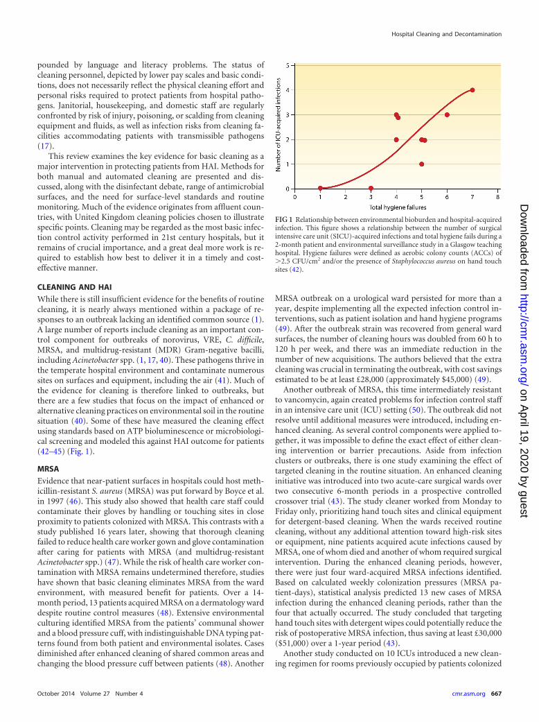
While there is still insufficient evidence for the benefits of routinecleaning, it is nearly always mentioned within a package of re-sponses to an outbreak lacking an identified common source (1).A large number of reports include cleaning as an important con-trol component for outbreaks of norovirus, VRE, C. difficile,MRSA, and multidrug-resistant (MDR) Gram-negative bacilli,including Acinetobacter spp. (1, 17, 40). These pathogens thrive inthe temperate hospital environment and contaminate numeroussites on surfaces and equipment, including the air (41). Much ofthe evidence for cleaning is therefore linked to outbreaks, butthere are a few studies that focus on the impact of enhanced oralternative cleaning practices on environmental soil in the routinesituation (40). Some of these have measured the cleaning effectusing standards based on ATP bioluminescence or microbiologi-cal screening and modeled this against HAI outcome for patients(42–45) (Fig. 1).
MRSA
Evidence that near-patient surfaces in hospitals could host meth-icillin-resistant S. aureus (MRSA) was put forward by Boyce et al.in 1997 (46). This study also showed that health care staff couldcontaminate their gloves by handling or touching sites in closeproximity to patients colonized with MRSA. This contrasts with astudy published 16 years later, showing that thorough cleaningfailed to reduce health care worker gown and glove contaminationafter caring for patients with MRSA (and multidrug-resistantAcinetobacter spp.) (47). While the risk of health care worker con-tamination with MRSA remains undetermined therefore, studieshave shown that basic cleaning eliminates MRSA from the wardenvironment, with measured benefit for patients. Over a 14-month period, 13 patients acquired MRSA on a dermatology warddespite routine control measures (48). Extensive environmentalculturing identified MRSA from the patients’ communal showerand a blood pressure cuff, with indistinguishable DNA typing pat-terns found from both patient and environmental isolates. Casesdiminished after enhanced cleaning of shared common areas andchanging the blood pressure cuff between patients (48). Another
MRSA outbreak on a urological ward persisted for more than ayear, despite implementing all the expected infection control in-terventions, such as patient isolation and hand hygiene programs(49). After the outbreak strain was recovered from general wardsurfaces, the number of cleaning hours was doubled from 60 h to120 h per week, and there was an immediate reduction in thenumber of new acquisitions. The authors believed that the extracleaning was crucial in terminating the outbreak, with cost savingsestimated to be at least £28,000 (approximately $45,000) (49).
Another outbreak of MRSA, this time intermediately resistantto vancomycin, again created problems for infection control staffin an intensive care unit (ICU) setting (50). The outbreak did notresolve until additional measures were introduced, including en-hanced cleaning. As several control components were applied to-gether, it was impossible to define the exact effect of either clean-ing intervention or barrier precautions. Aside from infectionclusters or outbreaks, there is one study examining the effect oftargeted cleaning in the routine situation. An enhanced cleaninginitiative was introduced into two acute-care surgical wards overtwo consecutive 6-month periods in a prospective controlledcrossover trial (43). The study cleaner worked from Monday toFriday only, prioritizing hand touch sites and clinical equipmentfor detergent-based cleaning. When the wards received routinecleaning, without any additional attention toward high-risk sitesor equipment, nine patients acquired acute infections caused byMRSA, one of whom died and another of whom required surgicalintervention. During the enhanced cleaning periods, however,there were just four ward-acquired MRSA infections identified.Based on calculated weekly colonization pressures (MRSA pa-tient-days), statistical analysis predicted 13 new cases of MRSAinfection during the enhanced cleaning periods, rather than thefour that actually occurred. The study concluded that targetinghand touch sites with detergent wipes could potentially reduce therisk of postoperative MRSA infection, thus saving at least £30,000($51,000) over a 1-year period (43).
Another study conducted on 10 ICUs introduced a new clean-ing regimen for rooms previously occupied by patients colonized
FIG 1 Relationship between environmental bioburden and hospital-acquiredinfection. This figure shows a relationship between the number of surgicalintensive care unit (SICU)-acquired infections and total hygiene fails during a2-month patient and environmental surveillance study in a Glasgow teachinghospital. Hygiene failures were defined as aerobic colony counts (ACCs) of�2.5 CFU/cm2 and/or the presence of Staphylococcus aureus on hand touchsites (42).
Hospital Cleaning and Decontamination
October 2014 Volume 27 Number 4 cmr.asm.org 667
on April 19, 2020 by guest
http://cmr.asm
.org/D
ownloaded from

with MRSA or VRE (51). The new regimen included a bucketmethod for soaking cleaning cloths and feedback to cleaners usingfluorescent markers. Although the study was quasi-experimental,environmental monitoring showed decreased contamination ofroom surfaces with MRSA and VRE after initiating the enhancedcleaning (27% versus 45% of cleaned rooms from baseline). Overthe same period, patient acquisition of MRSA was reduced by 49%(and that of VRE by 29%) following the augmented cleaning pack-age (P � 0.001 for both) (51).
Two recent studies report decreased rates of MRSA followingimplementation of a control bundle including targeted screeningof patients, environmental sampling, hand hygiene, laboratorymethods, and enhanced decontamination of patient rooms. Thefirst used a pulsed xenon UV device (PX-UV) in three Americanhospitals, with an overall total of 777 beds in the study hospitals(52). Following identification of colonized patients, a 5-day topi-cal clearance protocol was performed, which, along with PX-UV,ultimately reduced the rate of hospital-acquired MRSA acquisi-tion by 56% across the whole health care system after 6 months(P � 0.001) (52).
The second study evaluated the effect of hydrogen peroxide(HP) decontamination alongside patient screening for MRSA in a300-bed Australian hospital (53). This study ran for 6 years, ratherthan 6 months, and used a retrospective before-and-after designto assess detergent cleaning versus hydrogen peroxide decontam-ination of rooms recently occupied by MRSA patients. Targetedenvironmental screening was performed after room cleaningalongside ongoing surveillance of patient acquisition of MRSAthroughout the hospital. Newly identified patients were isolatedand placed on contact precautions but were not offered a topicalclearance regimen. MRSA was recovered from 25% of rooms fol-lowing detergent cleaning and from 19% of rooms after exposureto hydrogen peroxide (P � 0.001). There was a 3.5% reduction inthe overall proportion of rooms demonstrating persistent MRSAcontamination after using hydrogen peroxide (P � 0.08). Overthe 6 years, the incidence of MRSA acquisition was reduced from9.0 to 5.3 per 10,000 patient-days between detergent and disinfec-tant periods, respectively (P � 0.001).
Both of these studies concluded that enhanced decontamina-tion methods contributed toward decreased MRSA rates, but fur-ther work on the individual effects of PX-UV and hydrogen per-oxide is warranted (52, 53). As before, the proportional effectfrom additional screening and other package components in con-junction with introduction of disinfectant or UV light could notbe accurately determined.
VRE
It is well known that vancomycin-resistant enterococci (VRE) cansurvive long term in the hospital environment (7). Multiple clean-ing practices fail to remove VRE from a range of sites, despite useof powerful disinfectants (54–57). There are reports showing thatsurfaces remain contaminated with VRE when cleaning cloths arereused on sequential surfaces, when there is inadequate contacttime between a surface and applied disinfectant, and when itemsor surface are sprayed and wiped over, rather than being activelyscrubbed (3, 18, 55, 58). Such persistence is not exclusive to VRE,since other pathogens also survive the cleaning process, but VREseems to be particularly adept at withstanding repeated attemptsat disinfection, including double bleach-based cleaning (54–56).
Current protocols using disinfectants can be effective if near-
patient surfaces, such as bed rails, and frequently touched sur-faces, such as door handles, are physically scrubbed at least oncedaily. There is evidence that more conscientious cleaning can con-trol VRE (3, 51, 57). A sentinel study in 2006 demonstrated theimpact of improved cleaning on VRE transmission in a medicalICU, first as a single intervention and then alongside a hand hy-giene initiative (58). Targeting cleaning efficiency decreased bothsurface contamination from VRE and the number of patients ac-quiring the organism; following this with a hand hygiene programfurther reduced surface cultures of VRE and patient acquisition tothe lowest levels gained. There was also less VRE on health careworker hands (58).
Escalating VRE cases in a Brazilian hospital prompted a range ofactivities, including emphasis on environmental cleaning, contactprecautions, and the introduction of an educational program(59). Improvements in cleaning included use of bleach for bath-room surfaces and 70% alcohol for furniture and patient equip-ment. The overall package helped prevent dissemination of VREthroughout the hospital, including intensive care, with a decreasein acquisition rate from 1.49 to 0.33 (P � 0.001) (59). Bleach-based terminal cleaning was used for an earlier study to controlVRE in a hemato-oncology unit, again as part of an interventionpackage (57).
Another “bundle” of interventions, including thorough clean-ing and surface screening cultures, was implemented in threeICUs by a team in South Korea (60). Clinical and surveillancecultures identified 50 patients with VRE during the outbreak,most of whom (n � 46) had vancomycin-resistant Enterococcusfaecium (VREF). During the first 2 months of the outbreak, PFGEanalysis of VREF isolates revealed six main strain types, with re-lated clusters between two of these. Housekeeping staff used 5%sodium hypochlorite to clean all surfaces three times a day. Theoutbreak finally came to a halt 5 months after implementing thepackage of interventions, with a reduction in the weekly preva-lence rate from 9.1/100 to 0.6/100 patient-days (60).
A comparable study described implementation of a multicom-ponent package, also based on bleach disinfection, as a response toincreasing numbers of patients with VRE (61). Additional clean-ing supervisors were appointed to manage the introduction anddelivery of a standardized cleaning regimen using a novel productcontaining detergent and sodium hypochlorite (1,000 ppm). Al-cohol-based hand hygiene was encouraged, along with sleevelessaprons instead of long-sleeved gowns and gloves. VRE coloniza-tion and/or infection and surface contamination were comparedbefore and after implementation of the infection control package.There was a 24.8% reduction (P � 0.001) in the number of newpatients colonized with VRE and a 66.4% reduction (P � 0.012) inenvironmental contamination, despite a similar proportion of pa-tients already colonized on admission. While VRE bacteremia de-creased by over 80% (P � 0.001), the rate of vancomycin-suscep-tible enterococcal bacteremia did not change during the study(P � 0.54). Susceptible enterococcal infection may well derivefrom the patient’s own endogenous flora, whereas resistant en-terococci are more likely to be acquired from persistent surfacereservoirs. The “bleach-clean” package encouraged the decline innew VRE acquisition among particularly vulnerable patientsalongside an overall reduction in VRE bacteremia rate throughoutthe hospital (61).
Extreme environmental survival demonstrated by VRE offersan explanation for the increased risk of VRE acquisition for pa-
Dancer
668 cmr.asm.org Clinical Microbiology Reviews
on April 19, 2020 by guest
http://cmr.asm
.org/D
ownloaded from
tients placed in a room previously occupied by an individual col-onized or infected with VRE (19, 51). The clinical and environ-mental effects of hydrogen peroxide vapor (HPV) for roomdisinfection were assessed following discharge of patients withMRSA, C. difficile, multiresistant Gram-negative bacilli, and VRE.The risk of acquiring MRSA, C. difficile, and multiresistant Gram-negative rods was not significantly reduced after HPV decontam-ination, but patients admitted into HPV-treated rooms were 80%less likely to acquire VRE (62). This suggests that eradication ofpersistent reservoirs of VRE may be particularly important forcontrolling acquisition risk. Cleaning and disinfection should bemade a priority for managing VRE and possibly more so than forother hospital pathogens.
C. difficile
The benefits of cleaning for controlling C. difficile are well estab-lished (6, 63). The use of chlorine-releasing disinfectants forrooms contaminated with C. difficile reduces the amount of sporesin the environment, with additional evidence suggesting that thisaffects recurrence and transmission of C. difficile-associated infec-tion (CDI) (64). There is particularly good evidence for moreconcentrated products, especially those releasing higher levels offree chlorine (e.g., 5,000 mg/liter). The benefits of chlorinatedproducts are more obvious in units with high rates of CDI (e.g.,those for care of the elderly, stroke rehabilitation, etc.) or if used inconjunction with an outbreak. It should be noted that the overallefficiency of disinfectants for eliminating environmental sporesand lowering CDI rates is dependent upon a number of factors,including knowledge and training of cleaning staff, contact time ofdisinfectants, and overall time allocated to staff for cleaning. Spe-cific strains of C. difficile may also exhibit inherent or acquiredproperties that make them more resilient to disinfection attempts(64, 65).
A study published in 2007 evaluated additional bleach cleaningin two ICUs following an increase in patients with C. difficile (66).The extra cleaning was delivered to all parts of one ICU, includingrooms used only by staff. Clinical equipment was cleaned withhypochlorite-containing cloths twice a day. The second unit in-troduced enhanced bleach cleaning in isolation rooms accommo-dating patients already infected with C. difficile. Both units wit-nessed a decrease in infection rates over the next few months,which remained at a lower level for at least 2 years after the bleachcleaning program (66).
Increased rates of CDI in three American hospitals prompted achange of disinfectant for terminal room cleaning (67). After dis-charge of infected patients, all room surfaces from ceiling to floorwere wiped over with towels soaked in dilute bleach instead of theusual quaternary ammonium product. The prevalence density ofC. difficile fell by 48%, with a prolonged and significant reductionin the overall rate of hospital-acquired CDI. Another group im-plemented 0.55% bleach wipes for daily cleaning of two medicalunits with a high incidence of C. difficile (44). There were 31 pa-tients who acquired C. difficile on the wards before the interven-tion and 4 cases afterwards on these wards over the following year,representing a 7-fold decrease in C. difficile cases. There were noother interventions introduced other than targeted cleaning withbleach wipes (44).
A systematic cleaning and disinfection program was assessed byscreening frequently touched surfaces for the presence of C. diffi-cile in CDI rooms after cleaning (68). Three sequential interven-
tions were introduced over a 21-month period: (i) fluorescentmarkers placed at key sites for the purposes of monitoring andfeedback to cleaners, (ii) use of automated UV equipment forenhanced disinfection, and (iii) support from a designated teamresponsible for daily assessment of terminally cleaned CDI rooms.The fluorescent marker strategy improved the cleaning quality offrequently touched sites from 47% to 81% (P � 0 0.0001). Thenumber of screened sites positive for C. difficile decreased by 14%(P � 0.024), 48% (P � 0.001), and 89% (P � 0.006) for interven-tions 1, 2, and 3, respectively, compared with prestudy levels. Pos-itive cultures after disinfection were recovered from two-thirds ofCDI rooms before the study began, whereas during periods 1, 2,and 3, the percentages of CDI rooms with positive cultures afterdisinfection fell by 57%, 35%, and 7%, respectively (68).
More support for the role of cleaning and disinfection in con-trolling CDI comes from a recent English study (69). The teamfitted a statistical breakpoint model against incidence rates oflikely hospital-acquired C. difficile in a university hospital from2002 to 2009 and in a district general hospital from 2005 to 2009.The most important infection control interventions during theseperiods were placed within appropriate categories (antibiotics,cleaning, isolation, and other) for both hospitals and mappedagainst breakpoints identified by the models. The breakpointswere found to correspond with novel cleaning practices ratherthan any of the other control interventions. Statistical modelingpermitted a means of assessing the impact of different interven-tions and showed that additional or enhanced cleaning activitieswere most likely to be responsible for incremental reductions inrates of C. difficile at both hospitals (69).
While cleaning and decontamination strategies clearly have aneffect on patient acquisition rates, it should be remembered thatantimicrobial policies can also be very effective for controlling C.difficile. Severe restrictions on first-line use of cephalosporins andquinolones in a district general hospital reduced acquisition ofnosocomial C. difficile by 77% (2.398 to 0.549 cases/1,000 patientbeds) (70). The antibiotic policy resulted in an immediate de-crease in CDI without any additional infection control interven-tions. In this study, antibiotic stewardship, not cleaning, was fun-damental in controlling C. difficile (70). Beneficial effects ofstewardship can be assessed by spatiotemporal modeling, whichsuggests that protecting the patient from C. difficile acquisitionthrough careful antibiotic choice is more likely to benefit infectioncontrol than attempts at curtailing transmission once a patient issymptomatic (71). Faced with a septic patient, however, it is notalways possible to restrict antibiotics or choose agents less likely toencourage CDI. Under these circumstances, stringent environ-mental decontamination should be maintained in order to pre-vent ongoing transmission.
Acinetobacter
Many studies have emphasized the importance of cleaning in con-trolling outbreaks of Acinetobacter spp., particularly those causedby multiresistant strains in critical care units (4, 72, 73). One studydescribes an outbreak due to multiresistant strains of A. bauman-nii involving more than 30 patients in two ICUs (4). Epidemicstrains were identified from environmental reservoirs throughoutboth of the affected ICUs, which ultimately required completeclosure for terminal disinfection in order to bring the outbreak toan end (4). Another study reported a prolonged outbreak in aneurosurgical ICU, which prompted ongoing environmental
Hospital Cleaning and Decontamination
October 2014 Volume 27 Number 4 cmr.asm.org 669
on April 19, 2020 by guest
http://cmr.asm
.org/D
ownloaded from

sampling in order to identify any persistent reservoirs (74). Theepidemic strain was frequently isolated from hand touch sites be-side patients, with a clear association demonstrated between thelevels of surface contamination and new patient acquisition. Theauthors stated that comprehensive cleaning is fundamental forcontrolling Acinetobacter outbreaks in ICU settings, although themost appropriate cleaning practices in the routine situation re-main ill-defined (74).
One further study involving spread of a multiresistant A. bau-mannii strain in a critical care unit also provides environmentalsampling data during an outbreak affecting over 60 patients (75).Once again, there appeared to be a relationship between the num-ber of positive environmental cultures and new patient cases. Theauthors stated that systematic screening allowed them to targetcleaning resources in order to gain control of the outbreak (75).
An investigation following a sudden increase in the number ofchildren acquiring Acinetobacter on a pediatric burn ward identi-fied the role of frequently handled clinical equipment as an out-break reservoir (76). The outbreak occurred after it was decided toinstall computers beside every child’s bed. Environmental screen-ing identified the organism on several surfaces in the children’srooms, including the plastic covers on top of the computer key-boards. Until the outbreak occurred, there had been no recom-mendation for including bedside computers and their compo-nents in the routine cleaning specification. Targeted infectioncontrol measures were introduced, which included decontamina-tion of the plastic covers and mandatory glove use for staff beforehandling the computers. These simple measures were effective instopping the outbreak (76).
A 3-year prospective study took place in intensive and coronarycare units in order to evaluate a bundle of interventions aimed atreducing long-term drug-resistant Acinetobacter (77). The inter-ventions included a hand hygiene program, patient surveillance,barrier precautions, contact isolation, cohorting affected patients,and intensive cleaning with sodium hypochlorite (1:100) (77).The rate of A. baumannii colonization and/or infection was 3.6cases per 1,000 patient-days before the interventions were intro-duced, with the rate then decreasing by 66% to 1.2 cases per 1,000patient-days (P � 0.001) by the end of the first year. The rate wasfurther reduced by 76% to 0.85 cases per 1,000 patient-days (P �0.001) 2 years later (77).
Another outbreak of Acinetobacter in an ICU affected 18 pa-tients and was traced to a sink in one of the patient rooms (78).Identification of the sink trap as the reservoir suggested that thewhole of the horizontal drainage system could be potentially con-taminated. Application of a bleaching protocol eradicated the res-ervoir and curtailed further acquisition of MDR A. baumannii.However, there were additional infection control measures intro-duced at the same time, which included contact isolation for everypatient identified with MDR A. baumannii, hand hygiene training,additional nurse teaching, use of an alcohol hand gel, and directobservation of cleaning in the ICU (78). Once again, it is impos-sible to extricate the contribution of reservoir decontaminationwhen several interventions were initiated simultaneously as partof an outbreak control package.
One further study provides evidence to support the importanceof cleaning in controlling outbreaks of Acinetobacter (79). As withmost of the studies described, this outbreak also occurred in anICU, and an extremely resistant outbreak strain resisted carbap-enem antibiotics. Carbapenem-resistant A. baumannii was grown
from multiple environmental samples during the outbreak, in-cluding a mattress, a vital signs monitor, near-patient horizontalsurfaces, computer components, and a glucometer. After failureof thorough cleaning attempts with detergent and alcohol wipes, acommercial oxidizing disinfectant (Virkon S [50% potassiumperoxomonosulfate, 15% sodium alkyl benzene sulfonate, and 5%sulfamic acid]) was selected for enhanced cleaning. The introduc-tion of Virkon-based cleaning rapidly brought the outbreak to aclose. The authors were uneasy about the temporal association,because epidemics can resolve of their own accord. Furthermore,they did not audit cleaning effectiveness, hand hygiene compli-ance, antimicrobial consumption, or other potentially confound-ing factors. However, the sudden and sustained decrease in thenumber of cases of infection with a carbapenem-resistant A. bau-mannii strain after implementing use of a powerful new disinfec-tant is compelling (79).
It appears that even stringent manual cleaning with disinfectiondoes not necessarily eliminate Acinetobacter completely from theenvironment. The reasons for this are unknown but probably in-clude poor cleaning practices, missing high-risk sites, overwhelm-ing bioburden, and tolerance to, or misuse of, disinfectants (80,81). In another study, surfaces in rooms occupied by patients col-onized with A. baumannii remained contaminated with the or-ganism despite disinfectant-based cleaning (81). This study alsoreported contamination of rooms accommodating patients notpreviously shown to have any recent cultures of A. baumannii,suggesting long-term persistence in the near-patient environ-ment. There was a significant reduction in Acinetobacter contam-ination following disinfection, but over half the rooms that werepositive prior to cleaning still harbored the organism on a range ofsurfaces after cleaning (81).
Multidrug-Resistant Gram-Negative Bacilli
While the role of cleaning in controlling Acinetobacter outbreaks isnow accepted, the same cannot be said in relation to outbreaks ofmultidrug-resistant (MDR) Gram-negative bacilli. As for any out-break, enhanced cleaning usually comes as part of an overall bun-dle of activities in reaction to cross-infection incidents (1). Thereare, however, plenty of reports detailing coliforms associated withdiscrete items of equipment, specific environmental reservoirs, ora particular product or practice during outbreak investigations(24). Finding a single reservoir and eradicating it usually stops anoutbreak, and a positive outcome would naturally encourage pub-lication (82–85). Terminating an outbreak caused by single-source contamination is much easier to achieve than implement-ing a widespread cleaning regimen that has to cover a multitude ofdiverse items and surfaces.
Away from the outbreak situation, it has long been assumedthat Gram-negative bacteria survive poorly on surfaces. Thismeans that any environmental contribution toward HAI by thisgroup of organisms has not been widely investigated. Recent workhas challenged this, and there is a growing consensus that envi-ronmental cleanliness could be just as important for controllingtransmission of MDR coliforms as it is for MRSA and other or-ganisms (86, 87). This is supported by studies showing that Esch-erichia coli and Klebsiella spp. may survive desiccation for morethan a year and Serratia marcescens for several months (7). Thereare additional reports demonstrating persistence of MDR coli-forms throughout a variety of health care environments, withsome evidence that MDR Klebsiella is recovered from surfaces
Dancer
670 cmr.asm.org Clinical Microbiology Reviews
on April 19, 2020 by guest
http://cmr.asm
.org/D
ownloaded from

more often than MDR E. coli (15, 88–91). One recent studyscreened the near-patient environment beside patients previouslyidentified with carbapenem-resistant Enterobacteriaceae (CRE)and found that about 25% of the sites tested were contaminated,presumably by the patients’ own organisms (90). This study alsodemonstrated that both timing of sampling and local cleaningstrategies could affect data on the frequency of environmentalcontamination by CRE. This is no doubt true for other environ-mental pathogens.
Other than sampling and cleaning practices, it is possible that alack of evidence for viable MDR coliforms and correspondinginfection risk posed by hospital surfaces is due to insensitivescreening methods (90, 92). A targeted recovery strategy was usedto sample frequently touched surfaces situated beside patients col-onized by MDR coliforms (light switch, bed rail, bedside locker,and mattress cover) and two sites in nearby bathrooms shared bypatients (shower handrails and sink faucets) (92). Environmentalscreening next to one of these patients recovered MDR Klebsiellapneumoniae from four of six sites sampled, all of which were in-distinguishable from the strain obtained from the same patient’surine. The sites contaminated with the MDR strain were eitherbeside this patient or from the adjacent communal bathroom.Given the low recovery rates, limited detection, and relativelyshort survival times (1.5 to 2 h), isolating even small numbers ofMDR coliforms suggested a relatively high initial burden on sur-faces. Contamination probably occurred within a short time be-fore sampling (92).
Hospital sinks represent one of the most frequently implicatedreservoirs for MDR Gram-negative bacilli, including MDR coli-forms (93, 94). K. pneumoniae strains demonstrating prolongedsurvival within plumbing components are also more likely to har-bor extended-spectrum �-lactamases (95). Persistent reservoirs ofresistant K. pneumoniae were detected from multiple sites associ-ated with a contaminated sink in a large Scottish hospital (83).More recently, four patients in a neurosurgical ICU acquiredMDR K. pneumoniae thought to have originated from anothercontaminated sink during a 7-month period (85). Removal andreplacement of the sink and related pipes and upgrading the prac-tices for sink usage and decontamination brought the outbreak toan end. A protracted clonal outbreak of multiresistant IMP-8-producing Klebsiella oxytoca in a Spanish ICU was finally termi-nated by removing sinks, drain and trap components, and eventhe horizontal system connecting all suspected sinks (96). If theusual control measures fail to terminate an outbreak, then alter-native and/or unusual reservoirs should always be considered,particularly when preliminary environmental screening is nega-tive.
Another outbreak of MDR Klebsiella was linked with tippingpatient fluids down the nearest available sink rather than takingclinical waste to the designated sluice further away (97). A recentaudit of sinks in ICU rooms suggested that lower rates of sinkcontamination are significantly associated with daily bleach disin-fection, as well as restricting sinks for hand washing only and notroutine disposal of fluid waste from patients (94).
Yet another outbreak of resistant K. pneumoniae highlights therisks of reusing disposable equipment (84).This outbreak in-volved neonates, most of whom were infected just after birth orwithin a few days of hospitalization. Cases occurred among thosebabies receiving mucous aspiration due to respiratory distress.Although a new aspiration tube was used for each separate baby, it
was cleaned only by rinsing in a bowl of tap water between aspi-ration episodes for the same baby. The bowl was not routinelycleaned, and the water was left unchanged between babies. Notsurprisingly, the water was found to be contaminated with thesame resistant K. pneumoniae strain (84).
The lack of evidence for benefit from general surface cleaningalone for MDR Gram-negative organisms, even as a response to anoutbreak, is well recognized (98). There is a recent report emphasiz-ing additional cleaning following recovery of a carbapenemase-pro-ducing K. pneumoniae from patients in a United Kingdom hospital(99). Chlorine-based cleaning was implemented throughout theward, including patient-related items. Additional cleaning wasonly one component of the overall infection control strategy,however, along with a urinary catheter care bundle, tagging ofpatient notes, improved hand hygiene, and contact precautionsfor all cases (99). Another report describes an educational inter-vention to improve environmental cleaning and hand hygiene inan 11-bed gastrointestinal surgical ICU (100). There may wellhave been an underlying outbreak at the start of this initiative,since a high proportion of patients appeared to be already colo-nized. Following the introduction of terminal cleaning with glu-taraldehyde, single-use equipment, barrier precautions, and handhygiene improvements, the number of patients colonized withMDR Enterobacteriaceae decreased from 70% to 40%, attributedto the overall interventional package (100).
Pseudomonas and Stenotrophomonas spp.
Despite lack of evidence for defined transmission pathways, thereare studies suggesting that water sources provide a reservoir forPseudomonas and Stenotrophomonas spp. in the health care envi-ronment (101). These opportunistic organisms pose a risk of col-onization and infection for particularly vulnerable patients. Oneprevious study showed that Pseudomonas aeruginosa may betransmitted from contaminated sinks to hands during hand wash-ing (102). While survival on dry surfaces may only be transient,persistent reservoirs of these organisms can be traced to biofilmadherent to surfaces on sinks, sink traps, pipes, water lines, andhospital drains (103, 104). Biofilm is made up of a multifacetedmatrix of living organisms, which contaminates internal plumb-ing and provides a long-term reservoir for water-associated or-ganisms, including pathogens. The biofilm structure itself is resil-ient and situated on multiple surfaces inside traps, pipes, andinternal water filters. Bacteria present within biofilm are morelikely to be able to withstand chlorine-containing and other typesof disinfectants. They are also likely to demonstrate an increasedcapacity for antimicrobial resistance (95, 105).
Various outbreak investigations have shown that recovery ofPseudomonas and Stenotrophomonas maltophilia from watersources and adjacent surfaces can be linked with indistinguishablestrains cultured from patient specimens (106–108). An outbreakof Burkholderia cepacia on a pediatric unit was traced to sinks andwas thought to be associated with the presence of aerator filtersfitted to the taps (109). Faucet aerators have also been implicatedin an outbreak of S. maltophilia in a surgical ICU, with pulsed-field gel electrophoresis (PFGE) illustrating indistinguishablestrains from patients and aerators (106). For this reason, aeratorsshould be replaced with flow straighteners in health care premises.
Exposing biofilm to chlorine-containing products is the usualreaction to disinfection attempts, but even prolonged irrigationfails to remove all adherent biofilm. Reliable control requires
Hospital Cleaning and Decontamination
October 2014 Volume 27 Number 4 cmr.asm.org 671
on April 19, 2020 by guest
http://cmr.asm
.org/D
ownloaded from
stringent and repeated cleaning strategies, aimed at physical dis-ruption of the biofilm lining the internal surfaces of affected watersystems (108, 110). These are often right beside patients in theclinical environment and difficult, or even impossible, to access.Infection control initiatives require close collaboration betweenstructural facilities, clinical, and housekeeping staffs in order tosafely replace components or remove persistent biofilm. Totaleradication is rarely achieved, but regular inspection and repeatedcleaning followed by chlorine-based or similar disinfection willhinder further cases. Long-term control of Pseudomonas andStenotrophomonas is dependent upon integration of an effectivecleaning strategy into a targeted maintenance program (17, 101).
Norovirus
While the environmental role in the transmission of norovirus isdifficult to prove, the most convincing evidence comes from out-breaks where groups in a common setting with no known directcontact have been sequentially affected. The best examples of thesecome from outbreaks occurring outside hospitals. One report in-volves a single aircraft on which a single passenger vomited duringa long-haul flight (111). Over the next 6 days, flight attendantsworking on the aircraft in multiple flight sectors developed gas-troenteritis. Analysis of specimens from these aircrew attendantsdemonstrated an unusual norovirus genotype. The only possibleexposure was working in the cabin environment, since there wereno other opportunities for person-to-person transmission (111).
Another study describes an outbreak linked to a public concerthall (112). More than 300 people developed gastroenteritis duringa five-day period after a concert attendee vomited in the hall. Thehighest risk occurred among people seated closest to the seat be-longing to the original attendee. Similar events were recorded on acruise ship, where six consecutive cruises were affected (113).While crew members may have carried the virus between cruises,it is highly likely that the linked series of outbreaks was due toenvironmental persistence of infectious norovirus. These inci-dents suggest that without scrupulous cleaning following a singleincident, outbreaks will commence, escalate, or even resume.
Outbreaks of norovirus can be particularly ferocious in closedor semiclosed communities, such as transport vehicles and a vari-ety of public venues (114, 115). Sudden and widespread outbreakscan escalate without warning in nursing and residential homes,schools, hotels, and prisons (114, 116–118). CDC reported anoutbreak of norovirus in a primary school that affected over 100staff members and pupils (117). The investigation following thisoutbreak identified person-to-person contact as a major factor inviral transmission, but there was evidence that the environmentwas also implicated. Despite intensive cleaning with bleach soonafter notification, norovirus was recovered from computer com-ponents in a frequently used classroom the next day. The environ-mental strain was indistinguishable from that retrieved fromsymptomatic patients. Public health staff excluded symptomaticcases from the school, advised hand hygiene improvements, andorganized additional 1:50 bleach cleaning of environmental sitesthat might have been overlooked during the original disinfectionstrategy (117).
The role of cleaning in the control of norovirus outbreaks inhospitals and other health care facilities is unquestioned (5, 116).Indistinguishable genotypes of norovirus from ward surfaces andpatients have been reported, with viable virus apparently surviv-ing enhanced cleaning (119). One recent study identified norovi-
rus reservoirs from expected sites near bathroom showers andtoilets, but ward-based screening also demonstrated viral contam-ination of near-patient sites and a wide range of clinical equip-ment, including blood pressure and pulse oximeter machines,thermometers, notes trolleys, and even soap and alcohol gel con-tainers. Persistent viral reservoirs place new admissions at contin-ued risk of norovirus acquisition. Indeed, overloaded health carefacilities may experience prolonged outbreaks, especially if con-fronted with a higher throughput of patients lacking prior expo-sure (119).
All cleaning specifications, particularly regarding toilets andbathrooms, should use chlorine-based disinfectants at an appro-priate concentration for norovirus outbreaks. Detergent-basedcleaning is not sufficient to eliminate norovirus from the environ-ment (120). A recent in vitro study measured residual contamina-tion of surfaces with norovirus after detergent cleaning with orwithout a disinfectant (121). The authors concluded that cleaningwith liquid soap followed by a 1,000-ppm chlorine wipe generallyproduced the lowest level of persistent contamination. The infec-tivity index of norovirus, however, meant that even the low levelsachieved after a two-tier approach would still represent a risk forhand contact transmission. The authors suggested lengtheningthe contact time between chlorinated disinfectant and contami-nated surfaces to a minimum of 5 min, since this reduced residuallevels of virus to less than those capable of causing infection (121).Translating the results from this study to the clinical environmentposes a challenge, since leaving disinfectants on surfaces for even 5min in a busy ward may not be practical.
MANUAL CLEANING: PROCESS AND EQUIPMENT
Routine Cleaning Practices
In hospitals, environmental surfaces are routinely cleaned, orcleaned and disinfected, according to predetermined cleaning pol-icies (e.g., hourly, daily, twice weekly, etc.) or when surfaces ap-pear visibly dirty, if there are spillages, and always after patientdischarge (31, 122). The type and frequency of routine cleaningdepend upon clinical risk, patient turnover, intensity of peopletraffic, and surface characteristics. Frequent and stringent clean-ing specifications are applied to areas within operating theaters,intensive care units, transplant wards, and so-called “clean”rooms, where sterile medications are decanted and/or processed.Hospital kitchens, restaurants, and cafes also require targeted fre-quent cleaning, as do the laboratories and staff on-call rooms. Lesscomprehensive cleaning regimens are carried out for corridorsand stairwells, offices and waiting rooms, and selected outpatient,storage, general purpose, and entrance areas.
All hospitals should provide a written specification of cleaningservices and their delivery for all areas of the hospital, whetherprovided by in-house or externally contracted staff (31, 122, 123).These should be reviewed on a regular basis by cleaning supervi-sors, hospital managers, and structural facilities and infectioncontrol personnel. Recent recommendations on innovation andresearch in infection control support the opportunity for hospitalsto test new cleaning and decontamination technologies and pub-lish their findings (124).
In the United Kingdom, routine cleaning is performed manu-ally, with basic equipment, including buckets, mops, brushes,brooms, wipes, and cloths (31, 122). Electrical equipment in-cludes vacuum cleaners, floor polishers, and scrubbing machines.
Dancer
672 cmr.asm.org Clinical Microbiology Reviews
on April 19, 2020 by guest
http://cmr.asm
.org/D
ownloaded from

Surfaces fall into two general categories: critical and noncriticalsurfaces. The latter encompass sites such as floors, furniture, softfurnishings (including curtains), doors, wall fixtures, ledges andshelves, radiators, ceilings and walls, grilles and other ventilationcomponents, cupboards, etc. Critical surfaces include those thatare frequently touched or handled, such as handles, buttons,switches, computer keyboards, and bed controls, and noninvasiveclinical equipment, such as electrocardiogram (ECG) machines,blood pressure cuffs, patient hoists, stethoscopes, and intravenousdrip stands.
Noncritical Surfaces
Neutral detergent is used to lift soil, using disposable or reusablematerials. Over 80% of the bacterial load on hospital floors can beremoved by detergent-based cleaning only (125). Water used formop rinsing usually becomes increasingly contaminated duringthis process, especially if used repeatedly without changing or ifsurfaces are heavily soiled and/or have not been cleaned within theprevious 24 h. The water then serves as a medium for spreadingmicrobes around the environment. It should be routinely dis-carded in favor of fresh detergent solutions between bed spaces orevery 15 min, whichever is sooner (122). Disinfectants can be usedfor floors in high-risk clinical areas, although there is no evidencethat any microbial reduction persists for substantially longer pe-riods than that achieved by detergent alone (26, 27).
Mop heads may be disposable, with the length of time and/orareas of use specified; if not, they are employed for a particularduty, e.g., operating theater, before being bagged and sent fordecontamination, usually on a daily basis (122). Failure to ade-quately decontaminate reusable materials permits survival of mi-crobes, including spores, which may then contaminate the nextsurface to be cleaned. This may occur despite use of disinfectants,since certain organisms can resist the effect of specific chemicalagents either naturally, through acquired resistance, or protectedby biofilm (126–128).
Both detergent and disinfectant wipes and cloths can be used towipe over noncritical surfaces on a routine basis, with disposableproducts obviating the need for decontamination (122). Cleaningstaff require education on which product can be used for whichsurface and how long a wipe or cloth should be used before dis-posal. As a general guide, one wipe or cloth can be used for non-critical surfaces in one room or bed space, not including bathroomareas. Cleaning materials for the latter should always be kept sep-arate from those used for other ward surfaces (122). Disposablewipes are quick and easy to use but may leave excess moisture orresidues on surfaces, which can attract additional soil and ulti-mately spoil the finished appearance. They may also be expensiveand cause allergic reactions among housekeepers, with or withoutprotective clothing, including gloves.
Automated assistance includes vacuum and steam cleaners aswell as floor scrubbers and polishers. Use of a vacuum cleanerbefore wet mopping reduces overall soil, which may otherwise bespread around during the mopping process (24). Scrubbing ma-chines achieve a high standard of cleanliness for floors and areoften used for cleaning operating theaters on a routine basis (125).There is a longer-term beneficial microbiological effect seen afterusing these machines, but they tend to be cumbersome as well aslabor-intensive (125).
Critical Surfaces
Frequently touched items such as telephones, handles, taps, lightswitches, levers, knobs, buttons, keyboards, push plates, toys, etc.,are found in most health care institutions. Repeated handling in-creases the risk of contamination by pathogens, which then leadsto hand-based transmission. These items are likely to benefit fromenhanced cleaning, including disinfection (123, 129). High-touchsites or surfaces can be identified through direct observation orenvironmental screening using fluorescent or other markers(130).
A study performed in 1999 described the inoculation of a tele-phone handle in the middle of a neonatal ICU using fragments ofcauliflower mosaic virus. Over the ensuing week, the study teamtracked dispersal of the viral pieces around the unit between handtouch sites (131). Before inoculating the telephone, over 30 sitesfor sampling were chosen in each of six patient rooms according tothe risk of direct or indirect transmission of pathogens. These sitesincluded equipment buttons, handles, computers, patient charts,and hand lotion dispensers. Over half (58%) of the sites screenedin the room containing the inoculated telephone were persistentlycontaminated with the DNA marker. The number of sites positivefor viral markers peaked at 8 h (78%) before declining to 23% 1week later. Around 18% of sites were positive in the remaining fiverooms throughout the week, with a similar decline. The mostcommonly contaminated sites in all six rooms were personnelhands, computers, blood gas analyzers, door and telephone han-dles, control buttons and knobs, patient monitors, and medicalcharts (131). Such data specifically highlight the areas that wouldbenefit from more frequent cleaning or disinfection. The recogni-tion of high-risk sites for potential pathogen transmission utilizesprinciples employed by the food industry, whereby a monitoringframework is constructed specifically to prevent contaminationduring food production (132). This framework is based on a haz-ard analysis critical control point (HACCP) system and aims toeliminate risk through a variety of integrated control strategies(132).
Near-patient hand touch sites constitute the bulk of critical sur-faces in a ward. Routine decontamination is usually includedwithin institutional cleaning policies, including designated tasksfor a range of staff. This can vary between occupied and nonoccu-pied beds, electrical and nonelectrical items, and clinical and non-clinical equipment, all of which illustrates cleaning complexitiesand the potential for fragmented responsibility between house-keeping, nursing, and other clinical staff. Daily attention with de-tergent wipes may be sufficient to control bioburden on an acute-care ward, but high-risk sites in intensive care units may requiremore frequent attention (133). Two studies have clearly shownhow MRSA rapidly recontaminates high-touch sites in the ICUsetting after cleaning (134, 135).
Clinical Equipment
Given the range and types of clinical equipment available in to-day’s hospitals, it is beyond the scope of this review to describe andcompare decontamination strategies for all items that might befound on a ward. There is, however, an important decontamina-tion principle related to clinical equipment that applies to anyitem used for patients excluded from routine domestic specifica-tion (136, 137). All clinical equipment should be cleaned and/ordecontaminated before and after use for all patients regardless of
Hospital Cleaning and Decontamination
October 2014 Volume 27 Number 4 cmr.asm.org 673
on April 19, 2020 by guest
http://cmr.asm
.org/D
ownloaded from

how often it is used, where it is used, or what it is. An item in-tended for patient use should be inspected carefully before it isemployed, and if prior cleaning is not evident by either notifica-tion or obvious soiling, then it should be immediately cleanedaccording to local policies. Many hospitals now ask staff to flagspecific pieces of equipment to show that they were appropriatelycleaned and/or decontaminated after use. This is especially impor-tant for items such as commodes and other nondisposable appa-ratus used for toileting and therefore at high risk of contamina-tion. There are other utensils that might come into contact withblood and/or body fluids, e.g., pulse oximeters, thermometers,blood sugar test kits, saturation probes, etc., and these should alsobe subjected to stringent cleaning and disinfection before and af-ter use. Doctors’ stethoscopes have long been the subject of clean-ing audits and remain a likely source of contamination for a rangeof microbial flora (138). Wiping with alcohol is effective for de-contaminating stethoscopes, but it appears that even this simpleprocedure is abandoned, ignored, or forgotten when staff areoverworked. Indeed, all hygienic practices are consistently chal-lenged on a busy ward (139).
A recent unannounced audit conducted on an acute-care warddiscovered various amounts of organic soil on many items of clin-ical equipment (136). The authors used ATP bioluminescence tomeasure soil and found that 84% of sampled items exceeded thebenchmark value provided by the device manufacturer. The auditidentified several items of equipment on the ward that lacked anydesignated cleaning responsibility, and these tended to showhigher levels of contamination. The results are comparable withdata from a previous audit, which reported a pooled mean of86.8% contamination of equipment, although this study used mi-crobiological sampling methods rather than ATP biolumines-cence (137).
In many hospitals nowadays, nurses have adopted or taken on arange of duties originally performed by doctors, e.g., intravenousline insertion, prescribing, and catheter manipulation. Giventhese specialist tasks, it is understandable that basic cleaning hasbeen overlooked following the current shift in professional re-sponsibilities (136, 140). Cleaning duties do not necessarily rep-resent an appropriate use of time for highly trained nurses (141).Housekeeping staff are expected to comply with policies that oftenlack detailed guidance for each and every item found on a ward.Furthermore, they are not usually trained to decontaminate elec-trical items or clinical equipment (31, 122). Taking these changestogether, there is a risk that frequently used equipment and so-called forgotten sites will accumulate soil, including opportunisticpathogens. Since only a few spores of C. difficile or CFU of S.aureus can initiate infection at a vulnerable site, persistent con-tamination of soiled items provides a continued risk to patients(Table 1) (2, 10). It is likely that numerous items of clinical equip-ment in health care settings receive only sporadic cleaning atten-tion or, perhaps, none at all. Cleaning and decontamination re-sponsibilities for all staff, including medical staff, should beregularly reviewed, along with appropriate and repeated trainingprograms (136, 140).
Terminal (Deep) Cleaning
Terminal or deep cleaning is performed following patient dis-charge (122). If the patient was known to be colonized or infectedwith a specific pathogen, then the cleaning regimen is usually aug-mented with disinfectant at a specified strength depending upon
the pathogen. Methods vary, but a terminal clean usually includesinitial removal of all detachable objects from the room, includingbedding, screens, and/or curtains. Lighting and ventilation com-ponents on the ceiling are dusted or wiped over, followed by cur-tain rails and the upper surfaces of highly placed fixtures and fit-tings. All other sites and surfaces are then cleaned downward tofloor level. Items and equipment removed from the room arewiped over with detergent cloths, alcohol wipes, or disinfectantbefore being replaced.
A terminal clean also implies removal of curtains, drapes, andscreens for laundering or cleaning; fixed blinds may be wiped overin situ. While housekeeping staff are assigned to deliver terminalcleaning of bed spaces and patient rooms, nurses, nursing auxil-iaries, and clinical support workers usually have responsibility forclinical equipment and electrical appliances, including beds. Thisdivision of labor creates confusion over who cleans what, unlessclear contractual obligations are provided (136, 140). A flexibleapproach in terms of responsibility between nurses and house-keeping services must be adopted to ensure that patient care is notcompromised and that the environment and equipment are cor-rectly cleaned without undue delay. At present, cleaned rooms andbed spaces are routinely inspected by eye before admission inUnited Kingdom and most state-run health care systems. Lesssubjective methods of cleanliness assessment have yet to becomewidely incorporated into routine monitoring of the health careenvironment other than for research purposes (129).
Microfiber versus Cotton
Most hospitals prefer cotton-based cloths and mop heads for con-tinued use, since these can be repeatedly washed at high temper-atures (�90°C). There are many types of cleaning cloths, however,with microfiber products now proving popular among cleaningstaff (142). Ultramicrofiber (UMF) cloths are made of a combina-tion of polyamide and polyester, which absorb particles of soilthrough static attraction. Dust and organisms become firmly at-tached to the synthetic fibers and tend to persist within the cloththroughout the cleaning process. A range of different types ofdamp microfiber cloths were recently evaluated for their ability toremove pathogens, including C. difficile spores, MRSA, and E. coli(143). Single-use damp microfiber cloths demonstrated a meanlog10 reduction of 2.21 after cleaning, with smaller reductions ob-tained after repeated use on a series of contaminated surfaces.
An in vitro study was performed to evaluate and compare reus-able (rayon fiber) J-cloths against UMF cloths for eliminatingAcinetobacter spp., MRSA, K. oxytoca, and C. difficile spores fromhospital surfaces (144). UMF cloths were significantly better thanJ-cloths for removing pathogens from tiles, new and used lami-nated worktops, and stainless steel surfaces. These cloths generallyeradicated most, if not all, cultivable bacteria or C. difficile sporesfrom the surfaces tested, while standard J-cloths did not. Thisincluded used laminate surfaces, which can provide hidden reser-voirs for bacteria within surface microfissures (144). The resultsdiffered from those of an earlier study using ordinary microfiber,which reported less striking cleaning abilities depending upontype of product tested (142). The authors attributed the divergentresults to dissimilar structures and lengths of fibers in the clothsused in the two studies, but there were differences in study design(144). The previous study used microbiological methods and ATPbioluminescence to assess cleaning efficacy, whereas the later
Dancer
674 cmr.asm.org Clinical Microbiology Reviews
on April 19, 2020 by guest
http://cmr.asm
.org/D
ownloaded from

UMF study tested surfaces using ATP bioluminescence only (142,144).
In contrast, another study has showed that microfiber cloths areonly marginally more efficient for removal of soil and associatedmicrobes than cotton cloths in the presence of organic matter(145). For surfaces without soil, no significant difference has beenfound between cotton and microfiber cloths (142, 143, 145). Thus,the final choice between traditional and microfiber cloths forcleaning purposes rests with those with purchasing responsibili-ties. It is hoped that these individuals take advice from staff whoactually perform the cleaning themselves. Microfiber products aretoo expensive for single use, so continued use should be subjectedto a cost-benefit analysis. Furthermore, decontamination of thecloths is required after cleaning, since pathogens, includingspores, may adhere to the synthetic fibers. Exposure to bleach andother disinfectants potentially damages some microfiber productsand shortens their life span, so it is important to check manufac-turers’ recommendations before purchase (146).
Contamination of Cleaning Equipment and Liquids
Comprehensive cleaning schedules are seriously compromised ifcleaning equipment or liquids are contaminated. Poor choice ofcleaning methods or products or inadequate maintenance ofequipment will result in environmental contamination of the verysurfaces that need attention. There are numerous examples ofcleaning cloths, including those made of microfiber, that merelydistribute organisms across surfaces instead of removing them(26, 120, 142, 147–149). Enterococci, including VRE, seem to beparticularly difficult to eliminate from contaminated cloths (51,147, 148).
Cleaning equipment is also vulnerable to contamination fromhospital pathogens and this encourages further dispersal through-out the hospital environment (24, 82, 150, 151). Disinfectants aresupposedly better at killing environmental organisms than deter-gent-based agents, but some pathogens are able to survive expo-sure to specific biocides (152). Both multidrug-resistant S. marc-escens and extremely drug-resistant strains of K. pneumoniae havedemonstrated increasing tolerance to chlorhexidine (153, 154).Other cleaning fluids can become contaminated with Gram-neg-ative bacilli during use, with some formulations apparently en-couraging acquisition of resistance elements by Gram-negativeorganisms (126, 155).
Microorganisms will exploit an inadequately cleaned niche toexchange genetic material coding for antimicrobial resistance andother survival mechanisms (156–158). This could include resis-tance or tolerance to disinfectants. Once established, these hardystrains may ultimately infect debilitated patients (157). Hospitalwastewater has been shown to harbor KPC-2-producing K. pneu-moniae, suggesting widespread contamination throughout thehealth care environment (159).
Benefits of Physically Removing Soil
While most hospitals would use disinfectants for cleaning roomsor areas around colonized or infected patients, there have been anumber of recent papers that suggest that physical removal ofbioburden, rather than biocidal activity, is integral to the cleaningprocess (160–164). This was first suggested over 50 years ago,when it was shown that reduced surface contamination followingdisinfectant exposure appeared to be due to the cleaning activity,rather than the killing activity, of the products tested (165, 166).
Two studies published in 2012 found that norovirus (human andMNV1 strains) could be substantially reduced on hard surfacesafter wiping from one to six times using a range of inocula andmaterial wipes (121, 149). This supports the increasingly popularpremise that physical removal could challenge routine use of dis-infectants for controlling surface microbes (163, 164) (Fig. 2).This may be related to the fact that the presence of organic soil ona surface will impede the microbicidal activity of a disinfectant,but this is not the only explanation. Frequent physical removal ofbioburden using detergent-based cleaning methods needs to becompared and contrasted with application of biocides for costbenefits as well as longer-term efficacy and environmental issues(167).
There is no doubt that detergents are less toxic than powerfuldisinfectants as well as less likely to encourage accumulation anddispersal of tolerant or resistant genes among hospital strains(167, 168). A study performed recently demonstrates the effect ofdetergent-based cleaning over a 48-h period for high-risk hospitalsurfaces (133). The study measured the total bioburden and pres-ence of S. aureus, including MRSA, using standardized methods.The results suggest that wiping over near-patient surfaces once aday with single-use detergent wipes might be sufficient to protectpatients from environmental pathogen reservoirs in a nonout-break situation. Disinfectant wipes add cost without necessarilygreater efficacy at pathogen removal (169).
AUTOMATED DECONTAMINATION DEVICES
Given increasing awareness of the role of cleaning, recent innova-tions have tried to improve the scope and quality of cleaning prac-tices in the health care environment (170). There is a continuingrisk of transmission from pathogens within residual bioburden ifsurfaces remain uncleaned or receive inadequate cleaning. Carlingand coworkers applied a transparent gel to selected surfaces inmore than 1,000 patient rooms in 23 acute-care hospitals beforecleaning in order to assess the quality of housekeeping services.The gel is easily cleaned, difficult to detect, stable, and nontoxic,and it fluoresces when exposed to hand-held UV light (170). If UV
FIG 2 How should we clean clinical equipment? This figure shows data froma study examining three different methods for cleaning a dental chair. Clean-ing (wipe-rinse method) using a sodium lauryl sulfate-based detergent dem-onstrated equivalence to use of a disposable barrier and bleach disinfection forreducing MRSA contamination on a dental chair (164). (Photo courtesy of S.Petti.)
Hospital Cleaning and Decontamination
October 2014 Volume 27 Number 4 cmr.asm.org 675
on April 19, 2020 by guest
http://cmr.asm
.org/D
ownloaded from
inspection detects persistent gel on a surface after cleaning, it isassumed that the site did not receive sufficient cleaning attention.The overall cleaning compliance following gel application wasonly 49% (range, 35% to 81%), expressed as a percentage of eval-uated surfaces. With this sort of information, it is not surprisingthat several manufacturers are developing automated room disin-fection units that demonstrate superior decontamination of envi-ronmental objects and surfaces. These systems deliver variousmicrobicidal products, including germicidal light, hydrogen per-oxide, steam, and ozone (171–175).
Automated technologies may offer enhanced decontamina-tion, but they cannot replace routine daily cleaning. Organic soil,liquids, waste, and litter must still be removed from floors andsurfaces before disinfectant agents are released. Furthermore,these machines can usually be used only for terminal or dischargecleaning because the products are either too toxic for patients(e.g., hydrogen peroxide), constitute a safety risk (e.g., steam), orare better suited to work in empty rooms (e.g., UV light).
Steam Cleaning
Steam vapor machines are rapidly effective against a wide range ofpathogens, notably VRE, MRSA, and Gram-negative bacilli, in-cluding P. aeruginosa. Initial inocula of 7 log10 selected organismsare reduced to undetectable levels in less than 5 s following expo-sure to steam (176). The total surface bioburden from hospitalsurfaces is decreased by more than 90%, along with almost com-plete elimination of pathogens (177). While solid rubbish shouldalways be removed before this type of disinfection, steam can bedirectly applied onto a wide variety of soft and hard surfaces with-out prior cleaning (175).
Reports of the efficacy of steam cleaning are few in the literature,but there are examples of benefit from using steam in both routineand outbreak situations. An outbreak of norovirus occurred ontwo wards at the same time in an Australian hospital (178). Twodifferent cleaning protocols were instituted for each ward: oneward received detergent and bleach (1,000 ppm sodium hypo-chlorite plus contact time of 10 min) as a sequential 2-stepmethod, and the other was provided with steam technology. Thesteam component was applied using microfiber cloths and mopsfor terminal cleaning. The advantages from using steam werefewer cleaning hours, no toxic chemicals or dry-cleaning costs,and 90% less water consumption. The end result was visually su-perior, with clear support from cleaning staff. Microfiber-steamtechnology also proved to be a highly effective method of decon-tamination in an outbreak situation, with the same advantages asreported for routine cleaning (161, 178).
Concern has been expressed over some aspects of the steamtechnology for routine hospital cleaning (179). The use of steam todecontaminate hand touch sites such as knobs, buttons, switches,and computers, including those on electrical appliances, presentsobvious practical problems. If a hospital implements steam-basedcleaning in preference to other methods, there is a risk that thesehigh-risk sites might miss out on appropriate cleaning. It is alsothe case that the temperature of steam at delivery may rapidlydissipate depending upon the type and conductivity of exposedsurfaces. This has implications for the length of time that surfaceorganisms are exposed to applied steam. A previous study showedthat steam cleaning of curtains on a disused ward proved difficultto implement because there was no indication which areas had
received sufficient steam exposure, and pathogens were recoveredbefore and after the process (125).
Ultimately, steam delivered to surfaces turns into water. Resid-ual moisture constitutes a risk of slips and falls for patients, staff,and visitors, although superheated steam is less likely to leave wa-ter on exposed floors and other surfaces (179). Steam cleaningpresents further problems when cleaning a crowded ward, becausethere may be difficulties gaining access to sites beside a bed-riddenpatient. There are also time pressures for busy wards, which com-promise effective cleaning of a bed space if cleaners have onlyminutes to deliver the service. In addition, carelessly handledequipment represents a continued risk of burns and scalds forboth handlers and persons nearby, including patients. Inhalationof the vapor could potentially aggravate breathing problems instaff or patients with respiratory conditions (179).
Some hospitals have adopted a rolling program of steam clean-ing commodes, beds (nonelectrical), and other furniture in non-clinical areas. Steam also offers a useful cleaning strategy for publictoilets in hospitals and elsewhere. Steam systems should generallybe used only in well-ventilated areas, since repeated buildup ofcondensate could influence the environmental bioburden as wellas damage the internal fabric. Depending upon the type of equip-ment and surfaces selected, there remains a need for comprehen-sive risk assessment of aerosolized pathogens from the vaporizingprocess (179).
Ozone
Ozone is a potent oxidizing agent which has limited impact onbacterial spores and fungi but is highly effective against vegetativebacterial cells (180, 181). While it is relatively cheap to produce, itis both toxic and potentially corrosive for metals and rubber de-spite rapid dissociation into oxygen. There are consequently onlya few studies reporting its use in health care settings (171).
One recent study demonstrated benefit when ozone was incor-porated into laundry decontamination. A hospital laundry systemusing ozone resulted in a 5 log10 reduction of E. coli and totalcoliform count present in rinse water (182). Two other studieshave reported that ozone has potential as a gaseous decontami-nant for controlling environmental C. difficile, with various re-sults. The first showed that C. difficile could be reduced by �4log10 on various surfaces using a standard delivery of 25 ppmozone for 20 min at 90% relative humidity (183). The secondfound that a 3 log10 reduction in C. difficile spores was obtainedfollowing 25 ppm ozone for 75 min (184). In a domestic setting, anestimated concentration of 12 ppm was needed to eradicate MRSAfrom home surfaces (180). An earlier study used a gaseous ozonegenerator for decontaminating hospital side rooms previously oc-cupied by MRSA patients (174). Concentrations of 0.14 ppm wereachieved for different lengths of time, which failed to eradicateenvironmental MRSA and also initiated respiratory symptomsamong exposed staff.
UV Light
UV irradiation has been investigated as a potential decontaminantagainst environmental pathogens, including disinfection of sur-faces, instruments, and air (185). UV light severs the molecularbonds in DNA at specific wavelengths in order to exert its micro-bicidal effect. UV-C light has a specific wavelength found between200 and 270 nm (usually 254 nm), which itself falls within thegermicidal segment of the electromagnetic spectrum (200 to 320
Dancer
676 cmr.asm.org Clinical Microbiology Reviews
on April 19, 2020 by guest
http://cmr.asm
.org/D
ownloaded from

nm). Investigations of the effects of UV irradiation should con-sider the interaction between several different parameters, nota-bly, time of exposure, lamp position in relation to the irradiatedsurface, barriers between the light source and target surface, in-tensity of emitted light, and extent and flow of air movement.These could all influence the overall effect of UV-C irradiation onsurfaces.
There have been several studies in the last few years examiningthe effect of UV light as a potential decontamination strategy forhealth care environments. Nerandzic et al. described the effects ofa fully automated UV-C system against hospital pathogens (186).The device was tested in the laboratory and patient rooms and wasshown to significantly reduce C. difficile, VRE, and MRSA con-tamination on frequently handled hospital surfaces. The samegroup investigated a hand-held version delivering UV-C irradia-tion (185 to 230 nm) against pathogens in the laboratory, in pa-tient rooms, and on surfaces of items such as keyboards and por-table medical equipment located outside patient rooms (187).While the device significantly reduced C. difficile and MRSA, or-ganic matter on hospital surfaces that were not manually cleanedbefore irradiation clearly impeded the overall effect. This meansthat routine cleaning practices should still be carried out, even if ahospital chooses to implement routine decontamination usingUV technology.
Another study describes the decontamination effect of a porta-ble pulsed UV light device and its impact on work load whenintroduced into a hospital ward (188). Using pulsed UV for rou-tine once-daily disinfection of ward surfaces halved the number ofhousekeeping hours compared with the time taken for manualdisinfection using alcohol wipes. Other studies have shown thatUV-C systems can reduce vegetative bacteria by �3 to 4 log10
within 20 min after inoculation onto a carrier, although 35 to 100min of irradiation is required to reduce C. difficile by �1.7 to 4log10 (189, 190). When surfaces were not directly in line with theUV light source, the systems were not quite as effective.
Some authors have stated that although UV light is microbici-dal, it should not be used as a first-line intervention for decontam-ination but should be considered for use as a supplementary strat-egy depending upon specific needs, e.g., high or escalating HAIrates (185). There are several factors to consider before imple-menting routine UV-C technology; these include overall costs,installation, hospital layout and design, integration into house-keeping services, management of UV operation (including bulbchoice and longevity), and traditional cleaning and disinfectionpractices. UV light is significantly less effective for sites aroundcorners or shielded by solid items that challenge penetration bylight rays. It may also damage plastics and polymers used in thehealth care environment if repeatedly exposed. At this time, morework is required to evaluate the costs versus benefits, safety, andincremental advantages of UV devices for controlling health care-associated infections.
HINS
High-intensity narrow-spectrum (HINS) light is another light-based disinfection method that has shown wide-ranging microbi-cidal activity (191). HINS light utilizes a narrow bandwidth ofhigh-intensity visible violet light with peak output at 405 nm. Themicrobicidal mechanism is different from that of UV-C, in thatmicrobial inactivation is thought to be due to photoexcitation ofporphyrin molecules within bacterial cells. This encourages the
production of singlet oxygen as well as other highly reactive bac-tericidal compounds (192). One study has evaluated the overalleffect of HINS light for decontaminating the clinical environment,but further work is needed to investigate any benefits on HAI ratesfrom this technology (172).
Hydrogen Peroxide
Several systems which produce hydrogen peroxide (HP) in differ-ent formulations (e.g., HP vapors and dry aerosols) have beenstudied for their potential to decontaminate environmental ob-jects and surfaces in hospital rooms. HP systems are effectiveagainst M. tuberculosis, MRSA, viruses, sporeformers, VRE, andmultiresistant Gram-negative bacilli, including Acinetobacter spp.(53, 73, 193–195). Using a before-and-after design, Boyce andcoworkers showed that introducing HP systems onto high-inci-dence wards was associated with a significant decrease in rates ofCDI (196).
HP systems appear to offer reliable microbicidal activity againstmost, if not all, hospital pathogens, but a number of problemshave been raised in association with these systems. Risks of acci-dental exposure of people, animals, and plants continue, with re-peated use of HP liable to encourage erosion of some plastic andpolymer surfaces or items used in health care environments (197).Disinfection is impeded by residual debris, such as organic soil,liquids, and waste, as well as surface properties, such as linen andother soft materials. The equipment needs to be carefully posi-tioned in order to facilitate optimal exposure, but this may com-promise overstretched clinical staff. Without additional support,there may not be sufficient time to coordinate such preparation orperform it adequately.
Like UV-C systems, HP devices are universally expensive, can-not be used in occupied rooms, and require trained operators.They also need planned integration into decontamination andhousekeeping schedules. Effective HP disinfection may take sev-eral hours to complete a full cycle, which contrasts with the timetaken for traditional discharge cleaning (197). Delivering HP de-contamination may prove difficult in a hospital running at 100%bed occupancy, since any restrictions in bed turnover time couldeasily have an impact on admission capacity (197, 198). Roomscannot be easily closed in today’s crowded hospitals, let alonemultibed bays, complete wards, or specialist units offering 24-hemergency care.
Pottage et al. compared MRSA resistance to HP against com-mercially available spore indicators inoculated onto stainless steelcoupons (199). The recovery of MRSA from test coupons wasbetween 1.5 and 3.5 log10 higher than the quantity of Geobacillusstearothermophilus spores recovered after exposure (P � 0.05).The greater resilience displayed by MRSA may have been due toproduction of catalase, which is presumed to break down HP,leading to reduced efficacy. This highlights the fact that steriliza-tion competencies achieved using standard biological indicatorscannot always be extrapolated to other organisms. Preliminarycleaning of surfaces should always be performed to remove theoriginal bioburden, just as specified for UV light (171, 187).
Comparison between UV Light and Hydrogen PeroxideSystems
HP and UV systems have inevitably drawn comparisons. UV-Cdevices cannot eliminate bioburden on surfaces that are not di-rectly in line with emitted light rays, but they do offer a faster
Hospital Cleaning and Decontamination
October 2014 Volume 27 Number 4 cmr.asm.org 677
on April 19, 2020 by guest
http://cmr.asm
.org/D
ownloaded from

decontamination cycle. This reduces the time period that theroom is unavailable for patient admission (200). HP and UV de-vices decrease microbial contamination in patient rooms, with HPvapor delivery apparently significantly better at removing bacte-rial spores. These differences may be influenced by exposure timeand/or intensity of emissions for both systems and require furtherclarification. Whether superior sporicidal activity is clinically im-portant is unclear, since environmental screening has shown thatthe quantity of spores is relatively low on surfaces near patientswith C. difficile infection. This is also true for VRE and MRSA.There are two recent studies, however, that both report a reduc-tion in C. difficile incidence among patients, the first after intro-ducing a pulsed UV system into a community hospital and thesecond after using pulsed UV in a large academic medical center(201, 202). The latter paper also reported an overall decrease in thenumber of patients acquiring multidrug-resistant organisms de-spite missing a quarter of opportunities to apply the device afterpatient discharge (202).
Innovative technologies offer an alternative strategy for envi-ronmental hygiene purposes, but their logistical complexities,aside from costs of equipment, training, management, and per-sonnel, make it imperative that objective, controlled, and inde-pendent studies be performed in order to establish overall costsversus benefits (203). Furthermore, studies have hitherto concen-trated on efficiency of surface disinfection without specifically ex-amining the effects on airborne pathogens. Rapid disinfection cy-cles may well sterilize hard surfaces without eliminating viableorganisms surviving in the air (41). Concern has been expressedby several authors over the premature incorporation of these sys-tems into routine decontamination schedules (171, 173, 197, 203,204). Cost-effectiveness studies would help health care managerschoose the most appropriate system for their facilities based onevidence rather than advertising (197).
ANTIMICROBIAL SURFACES
While regular and conscientious cleaning is a necessity for elimi-nating pathogens, it is not the only mechanism for keeping sur-faces free from microbes. There are some high-tech solutions cur-rently receiving attention, including the so-called “self-sanitizing”surfaces. The technology was first suggested in 1964, but given thelong-held view that hospital surfaces were not relevant for HAIcontrol, the potential use of antimicrobial surfaces has only justbegun to generate discussion (205, 206). It is possible that treatingor coating hospital surfaces liable to contamination by pathogenscould kill or inhibit microbes in order to disrupt transmission topatients. Hard metals such as copper and silver have long beeninvestigated for their antimicrobial properties, and now noveltechnologies such as light-activated titanium dioxide-containingsurfaces are attracting attention (207–209).
The development of effective antimicrobial surface coatingscould impinge on the risk of cyclical transmission of pathogensbetween surfaces, hands, and air (206). These coatings might deterthe accumulation of microbial bioburden on a surface withoutadditional or increased frequency of cleaning and would thereforecontribute toward hygiene practices in the clinical environment.Stopping a surface from functioning as a microbial reservoir ef-fectively reduces the risk of onward transmission in health careenvironments. The risk from person-to-person transmission re-mains, but this may be tackled by barrier nursing and hand hy-giene programs for staff, visitors, and patients. Self-sanitizing sur-
faces have the ability to supplement manual cleaning, which isitself dependent upon operator time, choice, and ability and thussubject to considerable variation (170, 210).
There are several types of antimicrobial surfaces. A compre-hensive review of these surfaces has been written by KristopherPage and colleagues, from which the following classifications havebeen extracted (206). Antimicrobial surfaces can be placed in twomain categories: first, antiadhesive coatings, and second, antimi-crobial coatings and surface technologies. The latter category con-tains examples such as bacteriophage-modified surfaces, polyca-tionic surfaces, and light-activated coatings (206).
Antiadhesive Surfaces
One approach toward inhibiting microbial contamination is toengineer a surface that prevents microbial adhesion to the deviceor surface. This can be achieved by applying a layer of polyethyleneglycol (PEG) directly onto the surface (211). PEG-coated surfacescreate a hydrophilic interaction against hydrophobic bacterialcells, which impedes microbial attachment. The dynamic proper-ties of surface-bound PEG chains also make it more difficult formicrobes to become attached. Diamond-like carbon (DLC) filmssimilarly repel microbial adhesion and have been used as nontoxicsurface coatings for devices such as joint prostheses or stents(212). Easy-clean surfaces are either exceptionally hydrophilic orhydrophobic, with strongly hydrophobic coatings repelling bac-teria to a much greater extent than glass controls or other com-mercial coated glass products (213). Hydrophilic surfaces encour-age water sheeting and ease of cleaning. Polymers can bemanufactured with zwitterionic head groups, which are also use-ful for inhibiting bacterial adhesion and biofilm formation (214).The zwitterionic head attracts a large amount of water and makesthe material hydrophilic. All these surfaces and easy-clean tech-nologies are compromised in one respect, however, which is theirlack of inherent biocidal properties.
Antimicrobial Coatings
Triclosan. There is a wide range of antimicrobial coatings, some ofwhich are commercially available while others exist only at theresearch stage. Currently available products either are based onorganic antimicrobials impregnated into a specific product, e.g.,Microban (triclosan), or rely on inorganic antimicrobials such asionized silver (Ag�) or copper in different formulations (215).Surfaces that utilize diffusible antimicrobials could potentially in-duce microbial tolerance or even resistance, because the productscontinually leach out active compounds into the environment(206). With Microban products, the antimicrobial diffuses overthe surface to exert antimicrobial activity, making it nonperma-nent. About 75% of antibacterial liquid soaps and 30% of bars alsouse triclosan, which was used only in hospital settings until the1990s. Widespread use may be linked with its presence in nasalsecretions of healthy people, where it appears to be associated withS. aureus nasal colonization (216).
Much concern has been expressed over the development ofresistance to triclosan (217). It has been inferred that triclosanencourages the production of poisonous dioxins following expo-sure to UV light (218). Recently, the FDA (U.S. Food and DrugAdministration) announced a new position on antibacterial soap,including those products containing triclosan (219). Manufactur-ers must show both that their products are safe and that their useis superior to simple washing with conventional soap and water,
Dancer
678 cmr.asm.org Clinical Microbiology Reviews
on April 19, 2020 by guest
http://cmr.asm
.org/D
ownloaded from

or they will have to remove them from sale before 2016. Thisshould also apply to impregnated surfaces. It is possible that thecosts of antibacterial products outweigh any potential benefits,unless proved otherwise.
Silver. Both the Greeks and the Romans favored drinking ves-sels made of silver to make water potable. It is thought that silverions (Ag�) bind to thiol (�SH) groups present in microbial en-zymes and proteins, inactivation of which produces the desiredantimicrobial effect (208). However, Ag� coatings do not main-tain permanent activity despite initial effectiveness. Bacteria canbecome tolerant or even resistant to silver coatings or products(220). They therefore rely on additional diffusible antimicrobials,such as rifampin, to which microbes may also become resistant.Silver has been incorporated into various products, includingcoatings and textiles (221–223). Some coatings demonstrate goodantibacterial activity against planktonic Staphylococcus epidermi-dis and A. baumannii and have been used to inhibit environmentalcontamination as well as colonization of implanted medical cath-eters and other devices (223).
Copper. Copper is also toxic to microbes, and there have beenseveral studies examining the antimicrobial effect of coating hos-pital surfaces with copper (207, 224). There is no doubt that bothcopper and copper alloy surfaces demonstrate a profound antimi-crobial effect. There is even one study that attributes a reducedrate of hospital-acquired infection to the installation of coppercoatings onto near-patient surfaces (45). The study itself reportedonly some of the overall data, which detracted from the overallconclusions, but it is clear that further research in this area iswarranted (225).
Bacteriophage-modified surfaces. There have been recent at-tempts to apply bacteriophages to surfaces in order to controlbioburden (226). Antibiotic resistance capabilities do not neces-sarily protect bacteria from attack by phages. Since only one phageis required to infect a host cell in order to initiate multiple phageproduction, this approach could represent an efficient way of dis-infecting a surface. There are a number of complications, how-ever, mainly due to the inherent specificity of a phage for a partic-ular species of bacteria (206). While this forms the basis oftargeted in vivo therapy, it is less useful for open surfaces due to thewide variety of dynamic bioburden, not necessarily bacterial. Amixture of phages should be applied in order to increase the spec-trum of activity, and this would exclude nonbacterial organisms aswell as rare or unusual pathogens. Furthermore, large or unevensurface areas may impede the distribution of phage solutions anddeter them from reaching their appropriate bacterial target (226).
Phage stability in the environment and storage conditions alsorepresent important issues for study, as well as the potential forphage resistance. Phage formulations and treated surfaces need tobe continually monitored and revised in order to remain effective,which will complicate regulatory approval (206). Finally, thephage concentration of a solution required for effective decon-tamination should be carefully considered, along with the mostappropriate incubation time for a specific phage and target (226).
Polycationic antimicrobial surfaces. Surfaces treated with hy-drophobic negatively charged polycations kill bacteria by causingphysical damage to the cellular envelope. Hydrophobic polymerchain coatings attract bacteria toward the treated surface, result-ing in puncture of the cell wall and subsequent cell death. Recentexamples of this type of surface coating include the polyethyl-eneimines (PEIs) (227). While PEI coatings are thought to be per-
manently microbicidal, their longevity, biotolerance, and me-chanical stability have not been widely investigated. There is noinformation as to whether these coatings will be able to withstandroutine wear and tear in health care settings, including cleaningpractices and disinfectant exposure (206).
Light-activated antimicrobial surfaces. Another surface de-contamination strategy is to use a coating that produces reactiveradicals. Biotoxic radicals, unlike antimicrobial agents, do not tar-get a specific microorganism but exert nonselective effects towarda range of microbes (228). This means that they avoid the poten-tial problem of an organism developing resistance to a specifictreatment. There are two main types of coating that produce re-active species and consequently display antimicrobial properties.The first is based on a photosensitizer immobilized within a coat-ing, and the second is a coating containing a titanium dioxide(TiO2)-based catalyst (209, 229, 230). Both of these are classifiedas light-activated antimicrobial agents.
Various modes of action have been investigated, specificallythe mechanism of photocatalysis and how this results in microbialkilling (206). The effectiveness of TiO2 as a photocatalyst is basedon the rate of production of hydroxyl radicals at the surface of thesemiconductor, although the energy of the light illuminating thesurface is also important. There have been some attempts to ex-amine the performance of these coatings in health care environ-ments. In one study, TiO2 efficacy in preventing MRSA contami-nation in a clinical environment containing MRSA patients wasonly 17.8% (231). The study did show, however, that environ-mental contamination was higher for untreated surfaces (12.1%,versus 4.4% for treated surfaces) and also higher for ad hoc sam-ples taken from an environment exposed to MRSA as opposed toa non-MRSA environment (14.1% versus 3.5%, respectively).Disinfection of a surface by photocatalyzed reactions may be analternative and less toxic approach to using chemical disinfec-tants, but it is important to be certain that these coatings demon-strate long-term efficacy in working health care environments.
Current Concerns over Antimicrobial Surfaces
The utility of antimicrobial surfaces needs careful considerationbefore widespread adoption (17, 232). Current evidence hasshown that these surfaces produce a moderate microbicidal effectonly (�2 log10 pathogen reduction), with no studies yet investi-gating efficacy against pathogens such as C. difficile spores andnorovirus. The resources required for installation in health caresettings and overall cost-effectiveness are unknown. There is in-sufficient information on durability and whether antimicrobialactivity is affected by humidity, temperature, cleaning frequency,and/or the presence of an organic load (233). There are ongoingconcerns over possible toxicity, resistance, and allergenic proper-ties (17, 218, 234). Finally, the relative contribution of self-disin-fecting surfaces toward hand contamination and consequentialrisk of cross-transmission has not been established (232). We donot know which sites, surfaces, and clinical equipment in patientareas should be, or could be, coated with an antimicrobial prod-uct. It is true to say, however, that continued research on thesesurfaces is needed and will no doubt attract much interest frombusiness and industry in the future. There has already been a callfor scientific standards for antimicrobial surfaces in view of therapidly expanding technologies and potential importance of theseproducts (235).
Hospital Cleaning and Decontamination
October 2014 Volume 27 Number 4 cmr.asm.org 679
on April 19, 2020 by guest
http://cmr.asm
.org/D
ownloaded from

HOW TO MEASURE CLEANLINESS
There are a number of scientific methods in use for measuringenvironmental soil, since visual inspection cannot accurately de-termine the infection risk for patients (129, 236, 237). The defini-tion of “clean” requires a validated and risk-assessed strategy toestablish a state of “cleanliness,” rather than the subjective assess-ment currently provided by visual inspection and clipboards(129). Microbiological and chemical (ATP bioluminescence)techniques have long been incorporated into a comprehensiveassessment framework utilized by the food industry, and thesetechniques are now being tested in hospitals (17, 42, 236–239).Measurements from these methods have furnished a range of tan-gible values that can be modeled against the infection risk forpatients over time. Collecting data using microbiological andchemical tools provides an opportunity to choose an appropriatebenchmark for routine surface monitoring. This benchmarkshould signify whether hospital cleanliness levels indicate a clini-cal infection risk or not (42, 240). Health care staff, includinghousekeepers, would welcome an evidence-based cleanlinessstandard, thus allowing them to review, change, or target cleaningpractices before an outbreak becomes inevitable (1). Managerswould benefit from established benchmarks, since they would beable to audit, monitor, and defend practices in both routine andoutbreak situations.
Microbiological Methods
Current microbiological standards include an overall aerobic col-ony count and specific pathogen count for defined surface areashealth care environments (129). Aerobic colony counts of �2.5 to5 CFU per cm2 on hand touch sites and �1 CFU/cm2 hospitalpathogen (e.g., MRSA, VRE, C. difficile, etc.) have been proposedand tested as microbiological benchmarks (42, 43, 236–238, 241).The two benchmarks appear to be related, in that higher levels ofaerobic colonies on hand touch sites are more likely to be associ-ated with the presence of S. aureus and MRSA (237). The stan-dards have been used to systematically measure soil in severalhospital studies but have not yet been validated for routine mon-itoring (42, 133, 242, 243).
Similar counts for food preparation surfaces form the basis ofthe monitoring framework set up by the food industry (129, 132).Retail and food manufacturers, plus a variety of other agencies,use microbiological standards based on the presence or absence ofindicator organisms, identification of which alerts the agency to apotential health risk from the medium monitored (132, 244, 245).These standards also incorporate overall counts of nonpathogenicflora, because the organisms of interest are widely spread through-out time and space (129). The most reliable indicator of environ-mental hygiene in health care premises is the presence of coagu-lase-positive staphylococci, because ubiquitous human carriageand frequent human traffic encourage risk of contamination.Studies investigating the application of microbiological standardsin health care environments have selected both S. aureus andMRSA to help monitor cleanliness (1, 2, 42, 43, 241, 246).
ATP Bioluminescence Systems
ATP bioluminescence systems are provided with various bench-marks depending upon make and model of luminometer and theenvironment to be monitored. The benchmark levels range from25 to 500 relative light units (RLU) for 10- to 100-cm2 health caresurfaces (238, 241, 247). Studies have suggested that some systems
are not sufficiently sensitive to detect very low microbial counts(�10 CFU/cm2), which is of concern given the low numbers ofpathogens required to initiate infection (Table 1) (248, 249).Other studies have investigated possible associations betweenATP and microbiological data by systematically measuring bothdata sets from the same surfaces. One study found that benchmarkcategories of 100 RLU and microbial growth of �2.5 CFU/cm2
were only loosely related, since approximately 60% of combineddata sets agreed as to whether a surface should pass or fail (241).Another examined colony counts and ATP values independentlyagainst cleaning performance using fluorescent markers (250).The data presented suggest that ATP monitoring is more usefulfor detecting the need for cleaning attention, whereas microbio-logical screening provides an indication of the quality of cleaning(250). It is clear that more studies are required in order to establishthe best method for monitoring hospital surfaces in the routinesituation (249, 251).
ATP measurements can be hugely inflated by disinfectants, mi-crofiber products, food and drink spillages, and synthetic plasticsused in cleaning and laundry services (236, 251, 252). Chosenbenchmarks should reflect the risk of infection for different typesof patients accommodated in different clinical areas. Sites andsurfaces in outpatient clinics, hospital corridors, and storage areasdo not necessarily provide the same level of infection risk as sur-faces in a bone marrow transplant unit or hand touch sites besidean ICU patient. After these benchmarks have been established,routine monitoring should be able to highlight problem areas ortrends illustrating the dynamic balance between hospital cleanli-ness, staff deficit, and workload. Most importantly, awareness of asudden accumulation of soil might initiate extra cleaning beforepatients are exposed to a risk of infection or even an outbreak (42,238, 240). As previously stated, several studies have already shownthe association between bioburden on health care surfaces andHAI rates, whether due to overall HAI or specific pathogens (42,43, 45, 63, 74, 253).
HOW TO MEASURE CLEANING
Fluorescent Markers
There are alternative ways of assessing the health care environ-ment, notably monitoring the efforts of cleaning staff rather thanmeasuring residual bioburden on surfaces. Most environmentalfailures are likely due to personnel themselves, not products orpractices (254). Assessment of the cleaning process can be intro-duced by using educational strategies, direct and indirect cleaninginspections, observation, scientific monitoring, and feedback tostaff (17, 58, 170, 247, 255). Any form of environmental monitor-ing is quickly noticed by housekeeping staff, although the effectcan wear off without continued feedback or education (17). Inoc-ulation of key sites using invisible fluorescent markers for laterinspection virtually always improves overall cleaning compliance,with a reduced prevalence of hospital pathogens (255–257). How-ever, cleaners become aware of this type of monitoring, search outfluorescent marks, and then target these for cleaning to the detri-ment of other sites and surfaces (258). Although more research isneeded, a few studies have indicated that the use of fluorescentmarkers is linked with decreased transmission of hospital patho-gens (51, 259).
Dancer
680 cmr.asm.org Clinical Microbiology Reviews
on April 19, 2020 by guest
http://cmr.asm
.org/D
ownloaded from

ATP Bioluminescence Systems
Tangible values and trends over time from bioluminescence-based ATP data have the advantage of immediate and potentiallylonger-term feedback for housekeeping staff (260). The use ofATP monitoring appears to have a pronounced effect on cleaners,especially when they receive educational programs at the sametime (247). Similar to the case for fluorescent marking, house-keeping staff react quickly to an environmental monitoring pro-gram because they are concerned that their jobs may be at risk(17, 58).
Observation, Supervision, and Education of HousekeepingStaff
Several other studies have demonstrated different results after in-stituting direct observation, supervision, and education of staff asthey clean, again often showing reductions of important hospitalpathogens (257, 261–264). There is a concern that these interven-tions might lose impact over time, since cleaning is physicallydemanding, poorly paid, and subject to inadequate staffing (17,265). Furthermore, there tends to be rapid turnover among jani-torial and housekeeping staff, and this may be related to highersickness levels as well as dissatisfaction with pay, status, and con-ditions (17).
Ongoing training, education, and continual evidence-based re-assessment are required as an important part of staff management.It is hoped that the overall status of housekeeping staff improves inparallel with the recognition of the importance of basic cleaning inhealth care environments. Specialized cleaning activities can beagreed to and implemented for staff who wish to assume greaterresponsibility and are prepared to undergo relevant training andassessment. Selected housekeeping staff could potentially managethe decontamination of clinical equipment, traditionally the remitof clinical staff, thus releasing more time for the latter to care forpatients. Perhaps the creation of a new training framework fordifferent levels and competencies of cleaning staff would help raisethe status of cleaners, as well as focus attention on the cleaningresources required to keep health care environments safe for pa-tients.
DISCUSSION
There is no easy way to clean a hospital or to keep it clean, howeverwe define “clean.” Removing visual and invisible dirt from thehospitals of today and for the future requires sufficient trainedstaff, ongoing monitoring, measurement of bioburden, educa-tion, constant upgrading of practice, and two-way communica-tion between those responsible for cleaning and those responsiblefor infection control. The risks of cross-transmission are exagger-ated by heavy workload, understaffing, high bed occupancy rates,and rapid bed turnover (266). Poor ventilation, clutter, and inap-propriate storage further compound the ability to clean surfacesproperly and keep them clean (24). Furthermore, in an era of costcutting, those with cleaning responsibilities cannot hope to de-contaminate all high-risk surfaces as often as required when ahospital is full to capacity and patients with attendant microor-ganisms are transferred between wards (and hospitals) day andnight (17, 267).
Current Unanswered Questions
While most would agree that keeping hospitals “clean” and prior-itizing surface cleaning around the patient are of paramount im-
portance, there are several key questions to which we do not yetknow the correct answers. These can be listed as follows. (i) Howimportant is the choice of cleaning fluid, whether detergentand/or disinfectant? (ii) How much better is microfiber than tra-ditional cloths? (iii) When should we use bleach, and when shouldwe not? (iv) When should we consider the use of automated sys-tems? (v) How should we monitor cleaning? (vi) How should wemonitor cleanliness? (vii) Are current specifications targeting themost contaminated sites? (viii) How often should we clean anoccupied room or bed space?
The debate over detergent- or disinfectant-based cleaning in theroutine situation continues to rage unabated. Ignorance about theeffects, short and long term, of cleaning agents persuades manag-ers to choose powerful kill-all fluids or gases for their hospital asprotection against pathogens and lawsuits. Microbiologists andenvironmentalists argue that the removal of dirt should beachieved without resorting to expensive toxic agents, which maythemselves encourage the appearance and persistence of resistantpathogens in habitually exposed environments. Regarding theproliferation of automated dispersal systems for decontaminationof surfaces, there may be unintended consequences of such newtechnologies, quite apart from the expense involved in introduc-ing them. Advertising and marketing are much less costly thanresearch.
The fact that physical removal may be just as good at removingsoil as disinfectants is supported by several recent studies and em-phasizes the need for more work in order to avoid environmentaland human toxicity from potent disinfectants (160–164) (Fig. 2).A recent study suggests that the effect of detergent cleaning onsurface S. aureus and MRSA lasts longer than the effect seen afterdisinfectant exposure (268) (Fig. 3). Aside from this, first-line useof detergents for routine cleaning saves money as well as negatingany risks from tolerance or resistance among pathogens due todisinfectants (197). Hospitals in the United Kingdom routinelyuse detergent-based cleaning for general surfaces and do not seemto experience the same levels of MDR Acinetobacter and VRE asreported by disinfectant-using hospitals in other countries (3, 19,
FIG 3 Effect of detergent and disinfectant cleaning on total Staphylococcusaureus (methicillin-susceptible S. aureus [MSSA] and methicillin-resistant S.aureus [MRSA]) recovered from hand touch sites on a 30-bed ward over 48 h.This figure shows the effects of detergent (blue line) and disinfectant (red line)on surface S. aureus and MRSA from baseline levels over 48 h on a 30-bed acuteward. Both types of cleaning rapidly reduced the overall staphylococcal bur-den, but recontamination occurred more rapidly after disinfectant exposure.The sites monitored were bedside locker, bed frame, and overbed table, andeach 48-h period for each type of cleaning was repeated three times (276).(Adapted from reference 268.)
Hospital Cleaning and Decontamination
October 2014 Volume 27 Number 4 cmr.asm.org 681
on April 19, 2020 by guest
http://cmr.asm
.org/D
ownloaded from

73, 80, 269). It is true to say that cleaning is not the same as disin-fection, although the two terms are habitually interchanged (270).
There appears to be a link between HAI rates and environmen-tal bioburden, although as yet only a few studies have reported thisand even fewer have investigated it (42, 43, 45, 63, 74, 253). Morework on this relationship is urgently required, since a measurableassociation offers tangible proof for the role of the environment inHAI risk. It also justifies the setting of scientific standards formeasuring microbial soil in order to gauge the cleaning effect andinfection risk for patients.
Current cleaning specifications may not be targeting the correctsites, or, if they are, they may not be applied frequently enough.Cleaning and disinfection should be focused on routine decon-tamination of high-risk surfaces, i.e., the sites more likely to har-bor pathogens and thus facilitating transmission (43, 237, 239).Removing pathogens from handles, switches, buttons, knobs, andother frequently touched (and often forgotten) sites is more likelyto have an impact on patient transmission than cleaning inacces-sible surfaces such as high shelves, ledges, or ceilings or low-touchsurfaces such as walls and window panes (130). Thoughtful con-struction of a specification to prioritize the highest-risk sitesshould also obviate the confusion over who cleans what and howoften an item or surface should be cleaned (133, 136, 140, 271). Inparticular, there is currently no evidence to support the frequencyof cleaning a room or bed space while it is occupied by a patient(133, 135). Cleaning specifications should encompass the fact thatoverall cleaning quality is determined not only by the appliedmethod but also by the appropriateness of the method for the typeof surface treated.
CONCLUSION
The importance of clean hospitals has not been widely accepted asa key component in infection control despite the increasing inter-est in HAI during the latter part of the 20th century (24, 27, 272,273). Now it is finally receiving the attention it deserves (259, 274).No doubt there will be much more evidence forthcoming over thenext few years to support and justify hospital cleaning practices.This is to be welcomed, since it is quite possible that accumulatingdata on environmental reservoirs and pathogen transmission inhealth care environments will also benefit healthy people in theirhomes and the community at large (275). Furthermore, with theadvance of antimicrobial resistance increasing for virtually allpathogens, the science underpinning infection control, includingcleaning, will attain a status hitherto unrecognized. Preventing thetransmission of pathogens will be the main focus of the 21st cen-tury unless we rapidly find alternative methods for treating infec-tion other than antimicrobial chemotherapy (274).
REFERENCES1. Dancer SJ. 2009. The role of hospital cleaning in the control of hospital-
acquired infection. J. Hosp. Infect. 73:378 –385. http://dx.doi.org/10.1016/j.jhin.2009.03.030.
2. Dancer SJ. 2008. Importance of the environment in meticillin-resistant Staphylococcus aureus acquisition: the case for hospital clean-ing. Lancet Infect. Dis. 8:101–113. http://dx.doi.org/10.1016/S1473-3099(07)70241-4.
3. Martinez JA, Ruthazer R, Hansjosten K, Barefoot L, Snydman DR.2003. Role of environmental contamination as a risk factor for acquisi-tion of vancomycin-resistant enterococci in patients treated in a medicalintensive care unit. Archives Int. Med. 163:1905–1912. http://dx.doi.org/10.1001/archinte.163.16.1905.
4. Tankovic J, Legrand P, de Gatines G, Chemineau V, Brun-Buisson C,
Duval J. 1994. Characterisation of a hospital outbreak of imipenem-resistant Acinetobacter baumannii by phenotypic and genotypic typingmethods. J. Clin. Microbiol. 32:2677–2681.
5. Green J, Wright PA, Gallimore CI, Mitchell O, Morgan-Capner P,Brown DWG. 1998. The role of environmental contamination withsmall round structured viruses in a hospital outbreak investigated byreverse-transcriptase polymerase chain reaction assay. J. Hosp. Infect.39:39 – 45. http://dx.doi.org/10.1016/S0195-6701(98)90241-9.
6. Kaatz GW, Gitlin SD, Schaberg DR, Wilson KH, Kauffman CA, SeoSM, Fekety R. 1988. Acquisition of Clostridium difficile from the hospitalenvironment. Am. J. Epidemiol. 127:1289 –1294.
7. Kramer A, Schwebke I, Kampf G. 2006. How long do nosocomialpathogens persist on inanimate surfaces? A systematic review. BMC In-fect. Dis. 6:130. http://dx.doi.org/10.1186/1471-2334-6-130.
8. Wagenvoort JHT, Sluijsmans W, Penders RJR. 2000. Better environ-mental survival of outbreak vs. sporadic MRSA isolates. J. Hosp. Infect.45:231–234. http://dx.doi.org/10.1053/jhin.2000.0757.
9. Chiang SR, Chuang YC, Tang HJ, Chen CC, Chen CH, Lee NY, ChouCH, Ko WC. 2009. Intratracheal colistin sulfate for BALB/c mice withearly pneumonia caused by carbapenem-resistant Acinetobacter bau-mannii. Crit. Care Med. 37:2590 –2595. http://dx.doi.org/10.1097/CCM.0b013e3181a0f8e1.
10. Lawley TD, Clare S, Deakin LJ, Goulding D, Yen JL, Raisen C, BrandtC, Lovell J, Cooke F, Clark TG, Dougan G. 2010. Use of purifiedClostridium difficile spores to facilitate evaluation of health care disinfec-tion regimens. Appl. Environ. Microbiol. 76:6895– 6900. http://dx.doi.org/10.1128/AEM.00718-10.
11. Kjerulf A, Espersen F, Gutschik E, Majcherczyk PA, Hougen HP,Rygaard J, Høiby N. 1998. Serological diagnosis of experimental Entero-coccus faecalis endocarditis. APMIS 106:997–1008. http://dx.doi.org/10.1111/j.1699-0463.1998.tb00252.x.
12. Teunis PF, Moe CL, Liu P, Miller SE, Lindesmith L, Baric RS, LePendu J, Calderon RL. 2008. Norwalk virus: how infectious is it? J. Med.Virol. 80:1468 –1476. http://dx.doi.org/10.1002/jmv.21237.
13. Eaton KA, Friedman DI, Francis GJ, Tyler JS, Young VB, Haeger J,Abu-Ali G, Whittam TS. 2008. Pathogenesis of renal disease due toenterohemorrhagic Escherichia coli in germ-free mice. Infect. Immun.76:3054 –3063. http://dx.doi.org/10.1128/IAI.01626-07.
14. Wandall DA, Arpi M, Wandall JH. 1997. A rat model of non-lethalbacterial infection. APMIS 105:187–191. http://dx.doi.org/10.1111/j.1699-0463.1997.tb00557.x.
15. Lemmen SW, Hafner H, Zolldan D, Stanzel S, Lutticken R. 2004.Distribution of multi-resistant Gram-negative versus Gram-positivebacteria in the hospital inanimate environment. J. Hosp. Infect. 56:191–197. http://dx.doi.org/10.1016/j.jhin.2003.12.004.
16. Bhalla A, Pultz NJ, Gries DM, Ray AJ, Eckstein EC, Aron DC, DonskeyCJ. 2004. Acquisition of nosocomial pathogens on hands after contact withenvironmental surfaces near hospitalised patients. Infect. Control Hosp.Epidemiol. 25:164–167. http://dx.doi.org/10.1086/502369.
17. Dancer SJ. 2011. Hospital Cleaning in the 21st Century. Euro. J. Clin.Microbiol. Infect. Dis. 30:1473–1481. http://dx.doi.org/10.1007/s10096-011-1250-x.
18. Huang SS, Datta R, Platt R. 2006. Risk of acquiring antibiotic-resistantbacteria from prior room occupants. Arch. Intern. Med. 166:1945–1951.http://dx.doi.org/10.1001/archinte.166.18.1945.
19. Drees M, Snydman DR, Schmid CH, Barefoot L, Hansjosten K, VuePM, Cronin M, Nasraway SA, Golan Y. 2008. Prior environmentalcontamination increases the risk of acquisition of vancomycin-resistantenterococci. Clin. Infect. Dis. 46:678 – 685. http://dx.doi.org/10.1086/527394.
20. Wilks M, Wilson A, Warwick S, Price E, Kennedy D, Ely A, Millar MR. 2006.Control of an outbreak of multidrug-resistant Acinetobacter baumanii cal-coaceticus colonization and infection in an intensive care unit (ICU)without closing the ICU or placing patients in isolation. Infect. ControlHosp. Epidemiol. 27:654 – 658. http://dx.doi.org/10.1086/507011.
21. Nseir S, Blazejewski C, Lubret R, Wallet F, Courcol R, Durocher A.2011. Risk of acquiring multi-drug-resistant Gram-negative bacilli fromprior room occupants in the intensive care unit. Clin. Microbiol. Infect.17:1201–1208. http://dx.doi.org/10.1111/j.1469-0691.2010.03420.x.
22. Shaughnessy MK, Micielli RL, DePestel DD, Arndt J, Strachan CL,Welch KB, Chenoweth CE. 2011. Evaluation of hospital room assign-ment and acquisition of Clostridium difficile infection. Infect. ControlHosp. Epidemiol. 32:201–206. http://dx.doi.org/10.1086/658669.
Dancer
682 cmr.asm.org Clinical Microbiology Reviews
on April 19, 2020 by guest
http://cmr.asm
.org/D
ownloaded from

23. Dancer SJ, Carling PC. 2010. All that glistens may be neither gold norclean. J. Hosp. Infect. 76:177–178. http://dx.doi.org/10.1016/j.jhin.2010.06.008.
24. Dancer SJ. 1999. Mopping up hospital infection. J. Hosp. Infect. 43:85–100. http://dx.doi.org/10.1053/jhin.1999.0616.
25. Nightingale F. 1863. Notes on hospitals. Published by Longman, Green,Longman, Roberts, and Green, London, United Kingdom. http://archive.org/details/notesonhospital01nighgoog. Accessed September 2014.
26. Dharan S, Mourouga P, Copin P, Bessmer G, Tschanz B, Pittet D.1999. Routine disinfection of patients’ environmental surfaces: myth orreality? J. Hosp. Infect. 42:113–117. http://dx.doi.org/10.1053/jhin.1999.0567.
27. Dettenkofer M, Wenzler S, Amthor S, Antes G, Motschall E, DaschnerFD. 2004. Does disinfection of environmental surfaces influence noso-comial infection rates? A systematic review. Am. J. Infect. Control 32:84 –89. http://dx.doi.org/10.1016/j.ajic.2003.07.006.
28. Davies S. 2005. Hospital contract cleaning and infection control.UNISON, London, United Kingdom.
29. Chan P, Dipper A, Kelsey P, Harrison J. 2010. Newspaper reporting ofmeticillin-resistant Staphylococcus aureus and ‘the dirty hospital.’ J.Hosp. Infect. 75:318 –322. http://dx.doi.org/10.1016/j.jhin.2010.01.027.
30. Davies S. 2009. Making the connections: contract cleaning and infectioncontrol. UNISON, London, United Kingdom.
31. National Patient Safety Agency. 2007. The national specifications forcleanliness in the NHS: a framework for setting and measuring perfor-mance outcomes. NPSA, London, United Kingdom.
32. Healthcare Commission. 2008. Inspections of cleanliness and infectioncontrol: how well are acute trusts following the hygiene code. http://www.official-documents.gov.uk/document/hc0809/hc07/0718/0718.pdf. Ac-cessed June 2014.
33. Popp W. 2013. Cleaning— on the way to evidence-based knowledge.Healthcare Infect. 18:1–2. http://dx.doi.org/10.1071/HI13004.
34. Harbarth S. 2013. Hospital-based environmental hygiene: prioritiesfor research. Healthcare Infect. 18:49 –50. http://dx.doi.org/10.1071/HI13006.
35. Mitchell B, Dancer SJ, Shaban RZ, Graves N. 2013. Moving forwardwith hospital cleaning. Am. J. Infect. Control 41:1138 –1139. http://dx.doi.org/10.1016/j.ajic.2013.03.307.
36. Cozad A, Jones RD. 2003. Disinfection and the prevention of infectiousdisease. Am. J. Infect. Control 31:243–254. http://dx.doi.org/10.1067/mic.2003.49.
37. Daschner FD, Schuster A, Dettenkofer M, Kümmerer K. 2004. Noroutine surface disinfection. Am. J. Infect. Control 32:513–515. http://dx.doi.org/10.1016/j.ajic.2004.07.007.
38. Trucano M, Kaldenberg D. 2007. The relationship between patientperceptions of hospital practices and facility infection rates: evidencefrom Pennsylvania hospitals. Patient Safety and Quality Healthcare. http://www.psqh.com/enews/0807feature.html. Accessed June 2014.
39. Greaves F, Pape UJ, King D, Darzi A, Majeed A, Wachter RM, MillettC. 2012. Associations between Web-based patient ratings and objectivemeasures of hospital quality. Arch. Intern. Med. 172:435– 436. http://dx.doi.org/10.1001/archinternmed.2011.1675.
40. Donskey CJ. 2013. Does improving surface cleaning and disinfectionreduce health care-associated infections? Am. J. Infect. Control 41:S12–S19. http://dx.doi.org/10.1016/j.ajic.2012.12.010.
41. Hobday RA, Dancer SJ. 2013. Historical and current perspectives on therole of sunlight and natural ventilation for controlling infection. J. Hosp.Infect. 84:271–282. http://dx.doi.org/10.1016/j.jhin.2013.04.011.
42. White L, Dancer SJ, Robertson C, MacDonald J. 2008. Are hygienestandards useful in assessing infection risk? Am. J. Infect. Control 36:381–384. http://dx.doi.org/10.1016/j.ajic.2007.10.015.
43. Dancer SJ, White LF, Lamb J, Girvan EK, Robertson C. 2009. Mea-suring the effect of enhanced cleaning in a UK hospital: a prospectivecross-over study. BMC Med. 7:28. http://dx.doi.org/10.1186/1741-7015-7-28.
44. Orenstein R, Aronhalt KC, McManus JE, Jr, Fedraw LA. 2011. Atargeted strategy to wipe out Clostridium difficile. Infect. Control Hosp.Epidemiol. 32:1137–1139. http://dx.doi.org/10.1086/662586.
45. Salgado CD, Sepkowitz KA, John JF, Cantey JR, Attaway HH, Free-man KD, Sharpe PA, Michels HT, Schmidt MG. 2013. Copper surfacesreduce the rate of healthcare-acquired infections in the intensive careunit. Infect. Control Hosp. Epidemiol. 34:479 – 486. http://dx.doi.org/10.1086/670207.
46. Boyce JM, Potter-Bynoe G, Chenevert C, King T. 1997. Environmentalcontamination due to methicillin-resistant Staphylococcus aureus: possi-ble infection control implications. Infect. Control Hosp. Epidemiol. 18:622– 627. http://dx.doi.org/10.1086/647686.
47. Hess AS, Shardell M, Johnson JK, Thom KA, Roghmann MC, NetzerG, Amr S, Morgan DJ, Harris AD. 2013. A randomized controlled trialof enhanced cleaning to reduce contamination of healthcare workergowns and gloves with multidrug-resistant bacteria. Infect. ControlHosp. Epidemiol. 34:487– 493. http://dx.doi.org/10.1086/670205.
48. Layton MC, Perez M, Heald P, Patterson JE. 1993. An outbreak ofmupirocin-resistant Staphylococcus aureus on a dermatology ward asso-ciated with an environmental reservoir. Infect. Control Hosp. Epide-miol. 14:369 –375. http://dx.doi.org/10.1086/646764.
49. Rampling A, Wiseman S, Davis L, Hyett AP, Walbridge AN, PayneGC, Cornaby AJ. 2001. Evidence that hospital hygiene is important inthe control of methicillin-resistant Staphylococcus aureus. J. Hosp. Infect.49:109 –116. http://dx.doi.org/10.1053/jhin.2001.1013.
50. de Lassence A, Hidri N, Timsit JF, Joly-Guillou ML, Thiery G, BoyerA, Lable P, Blivet A, Kalinowski H, Martin Y, Lajonchere JP, DreyfussD. 2006. Control and outcome of a large outbreak of colonization andinfection with glycopeptide-intermediate Staphylococcus aureus in an in-tensive care unit. Clin. Infect. Dis. 42:170 –178. http://dx.doi.org/10.1086/498898.
51. Datta R, Platt R, Yokoe DS, Huang SS. 2011. Environmental cleaningintervention and risk of acquiring multidrug-resistant organisms fromprior room occupants. Arch. Int. Med. 171:491– 494. http://dx.doi.org/10.1001/archinternmed.2011.64.
52. Simmons S, Morgan M, Hopkins T, Helsabeck K, Stachowiak J,Stibich M. 2013. Impact of a multi-hospital intervention utilisingscreening, hand hygiene education and pulsed xenon ultraviolet (PX-UV) on the rate of hospital associated meticillin resistant Staphylococcusaureus infection. J. Infect. Prevent. 14:172–174. http://dx.doi.org/10.1177/1757177413490813.
53. Mitchell BG, Digney W, Locket P, Dancer SJ. 2014. Controlling me-thicillin-resistant Staphylococcus aureus (MRSA) in a hospital and therole of hydrogen peroxide decontamination: an interrupted time seriesanalysis. BMJ Open 4:e004522. http://dx.doi.org/10.1136/bmjopen-2013-004522.
54. Noble MA, Isaac-Renton JL, Bryce DL. 1998. The toilet as a transmis-sion vector of vancomycin-resistant enterococci. J. Hosp. Infect. 40:237–241. http://dx.doi.org/10.1016/S0195-6701(98)90141-4.
55. Byers KE, Durbin LJ, Simonton BM, Anglim AM, Adal KA, Farr BM.1998. Disinfection of hospital rooms contaminated with vancomycin-resistant Enterococcus faecium. Infect. Control Hosp. Epidemiol. 19:261–264. http://dx.doi.org/10.1086/647806.
56. Lankford MG, Collins S, Youngberg L, Rooney DM, Warren JR,Noskin GA. 2006. Assessment of materials commonly utilized in healthcare: implications for bacterial survival and transmission. Am. J. Infect.Control 34:258 –263. http://dx.doi.org/10.1016/j.ajic.2005.10.008.
57. Sample ML, Gravel D, Oxley C, Toye B, Garber G, Ramotar K. 2002.An outbreak of vancomycin-resistant enterococci in a hematology-oncology unit: control by patient cohorting and terminal cleaning of theenvironment. Infect. Control Hosp. Epidemiol. 23:468 – 470. http://dx.doi.org/10.1086/502088.
58. Hayden MK, Bonten MJ, Blom DW, Lyle EA, van de Vijver DA,Weinstein RA. 2006. Reduction in acquisition of vancomycin-resistantEnterococcus after enforcement of routine environmental cleaning mea-sures. Clin. Infect. Dis. 42:1552–1560. http://dx.doi.org/10.1086/503845.
59. Rossini FA, Fagnani R, Leichsenring ML, Dantas SR, Cardoso LG,Levy CE, Moretti ML, Trabasso P. 2012. Successful prevention of thetransmission of vancomycin-resistant enterococci in a Brazilian publicteaching hospital. Rev. Soc. Bras. Med. Trop. 45:184 –188. http://dx.doi.org/10.1590/S0037-86822012000200009.
60. Yoon YK, Sim HS, Kim JY, Park DW, Sohn JW, Roh KH, Lee SE, KimMJ. 2009. Epidemiology and control of an outbreak of vancomycin-resistant enterococci in the intensive care units. Yonsei Med. J. 50:637–643. http://dx.doi.org/10.3349/ymj.2009.50.5.637.
61. Grabsch EA, Mahony AA, Cameron DR, Martin RD, Heland M,Davey P, Petty M, Xie S, Grayson ML. 2012. Significant reduction invancomycin-resistant enterococcus colonization and bacteraemia afterintroduction of a bleach-based cleaning-disinfection programme. J.Hosp. Infect. 82:234 –242. http://dx.doi.org/10.1016/j.jhin.2012.08.010.
62. Passaretti CL, Otter JA, Reich NG, Myers J, Shepard J, Ross T, Carroll
Hospital Cleaning and Decontamination
October 2014 Volume 27 Number 4 cmr.asm.org 683
on April 19, 2020 by guest
http://cmr.asm
.org/D
ownloaded from

KC, Lipsett P, Perl TM. 2013. An evaluation of environmental decon-tamination with hydrogen peroxide vapor for reducing the risk of patientacquisition of multidrug-resistant organisms. Clin. Infect. Dis. 56:27–35.http://dx.doi.org/10.1093/cid/cis839.
63. Wilcox MH, Fawley WN, Wrigglesworth N, Parnell P, Verity P,Freeman J. 2003. Comparison of the effect of detergent versus hypochlo-rite cleaning on environmental contamination and incidence of Clostrid-ium difficile infection. J. Hosp. Infect. 54:109 –114. http://dx.doi.org/10.1016/S0195-6701(02)00400-0.
64. MacLeod-Glover N, Sadowski C. 2010. Efficacy of cleaning products forClostridium difficile. Can. Fam. Phys. 56:417– 423.
65. Doan L, Forrest H, Fakis A, Craig J, Claxton L, Khare M. 2012. Clinicaland cost-effectiveness of eight disinfection methods for terminal disin-fection of hospital isolation rooms contaminated with Clostridium diffi-cile 027. J. Hosp. Infect. 82:114 –121. http://dx.doi.org/10.1016/j.jhin.2012.06.014.
66. McMullen KM, Zack J, Coopersmith CM, Kollef M, Dubberke E,Warren DK. 2007. Use of hypochlorite solution to decrease rates ofClostridium difficile-associated diarrhoea. Infect. Control Hosp. Epide-miol. 28:205–207. http://dx.doi.org/10.1086/511791.
67. Hacek DM, Ogle AM, Fisher A, Robicsek A, Peterson LR. 2010.Significant impact of terminal room cleaning with bleach on reducingnosocomial Clostridium difficile. Am. J. Infect. Control 38:350 –353. http://dx.doi.org/10.1016/j.ajic.2009.11.003.
68. Sitzlar B, Deshpande A, Fertelli D, Kundrapu S, Sethi AK, DonskeyCJ. 2013. An environmental disinfection odyssey: evaluation of sequen-tial interventions to improve disinfection of Clostridium difficile isolationrooms. Infect. Control Hosp. Epidemiol. 34:459 – 465. http://dx.doi.org/10.1086/670217.
69. Hughes GJ, Nickerson E, Enoch DA, Ahluwalia J, Wilkinson C, AyersR, Brown NM. 2013. Impact of cleaning and other interventions on thereduction of hospital-acquired Clostridium difficile infections in two hos-pitals in England assessed using a breakpoint model. J. Hosp. Infect.84:227–234. http://dx.doi.org/10.1016/j.jhin.2012.12.018.
70. Dancer SJ, Kirkpatrick P, Corcoran DS, Christison F, Farmer D,Robertson C. 2013. Approaching zero: temporal effects of a restrictiveantibiotic policy on hospital-acquired Clostridium difficile, ESBL-producing coliforms and MRSA. Int. J. Antimicrob. Agents 41:137–142.http://dx.doi.org/10.1016/j.ijantimicag.2012.10.013.
71. Starr JM, Campbell A, Renshaw E, Poxton IR, Gibson GJ. 2009.Spatio-temporal stochastic modelling of Clostridium difficile. J. Hosp.Infect. 71:49 –56. http://dx.doi.org/10.1016/j.jhin.2008.09.013.
72. Scerpella EG, Wanger AR, Armitige L, Anderlini P, Ericsson CD. 1995.Nosocomial outbreak caused by a multiresistant clone of Acinetobacterbaumannii: results of the case– control and molecular epidemiologic in-vestigations. Infect. Control Hosp. Epidemiol. 16:92–97. http://dx.doi.org/10.2307/30140949.
73. Chmielarczyk A, Higgins PG, Wojkowska-Mach J, Synowiec E, ZanderE, Romaniszyn D, Gosiewski T, Seifert H, Heczko P, Bulanda M. 2012.Control of an outbreak of Acinetobacter baumannii infections usingvapourized hydrogen peroxide. J. Hosp. Infect. 81:239 –245. http://dx.doi.org/10.1016/j.jhin.2012.05.010.
74. Denton M, Wilcox MH, Parnell P, Green D, Keer V, Hawkey PM,Evans I, Murphy P. 2004. Role of environmental cleaning in controllingan outbreak of Acinetobacter baumannii on a neurosurgical intensive careunit. J. Hosp. Infect. 56:106 –110. http://dx.doi.org/10.1016/j.jhin.2003.10.017.
75. Naranjo JD, Navarro JIV, Busselo MS, Ruíz AM, Hernández JMH,Garmendia MPT, López MIU. 2013. Control of a clonal outbreak ofmultidrug-resistant Acinetobacter baumannii in a hospital of the Basquecountry after the introduction of environmental cleaning led by the sys-tematic sampling from environmental objects. Interdisciplin. Perspect.Infect. Dis. 2013:1–9. http://www.hindawi.com/journals/ipid/2013/582831/. Accessed June 2014.
76. Neely A, Maley MP, Warden GD. 1999. Computer keyboards as reser-voirs for Acinetobacter baumannii in a burn hospital. Clin. Infect. Dis.29:1358 –1359. http://dx.doi.org/10.1086/313463.
77. Apisarnthanarak A, Pinitchai U, Thongphubeth K, Yuekyen C, War-ren DK, Fraser VJ, Thammasat University Pandrug-Resistant Acin-etobacter baumannii Control Group. 2008. A multifaceted interventionto reduce pandrug-resistant Acinetobacter baumannii colonization andinfection in 3 intensive care units in a Thai tertiary care center: a 3-yearstudy. Clin. Infect. Dis. 47:760 –767. http://dx.doi.org/10.1086/591134.
78. La Forgia C, Franke J, Hacek DM, Thomson RB, Jr, Robicsek A,Peterson LR. 2010. Management of a multidrug-resistant Acinetobacterbaumannii outbreak in an intensive care unit using novel environmentaldisinfection: a 38-month report. Am. J. Infect. Control 38:259 –263. http://dx.doi.org/10.1016/j.ajic.2009.07.012.
79. Doidge M, Allworth AM, Woods M, Marshall P, Terry M, O’Brien K,Goh HM, George N, Nimmo GR, Schembri MA, Lipman J, PatersonDL. 2010. Control of an outbreak of carbapenem-resistant Acinetobacterbaumannii in Australia after introduction of environmental cleaningwith a commercial oxidizing disinfectant. Infect. Control Hosp. Epide-miol. 31:418-420. http://dx.doi.org/10.1086/651312.
80. Manian FA, Griesenauer S, Senkel D, Setzer JM, Doll SA, Perry AM,Wiechens M. 2011. Isolation of Acinetobacter baumannii complex andmethicillin-resistant Staphylococcus aureus from hospital rooms follow-ing terminal cleaning and disinfection: can we do better? Infect. ControlHosp. Epidemiol. 32:667– 672. http://dx.doi.org/10.1086/660357.
81. Strassle P, Thom KA, Johnsonm JK, Leekha S, Lissauer M, Zhu J,Harris AD. 2012. The effect of terminal cleaning on environmental con-tamination rates of multidrug-resistant Acinetobacter baumannii. Am. J.Infect. Control 40:1005–1007. http://dx.doi.org/10.1016/j.ajic.2012.05.027.
82. Joynson DHM. 1978. Bowls and bacteria. J. Hyg. Cambr. 80:423– 425.http://dx.doi.org/10.1017/S0022172400024888.
83. Hobson RP, MacKenzie FM, Gould IM. 1996. An outbreak of multiply-resistant Klebsiella pneumoniae in the Grampian region of Scotland. J.Hosp. Infect. 33:249 –262. http://dx.doi.org/10.1016/S0195-6701(96)90011-0.
84. Randrianirina F, Vedy S, Rakotovao D, Ramarokoto CE, RatsitohainaH, Carod JF, Ratsima E, Morillon M, Talarmin A. 2009. Role ofcontaminated aspiration tubes in nosocomial outbreak of Klebsiellapneumoniae producing SHV-2 and CTX-M-15 extended-spectrum�-lactamases. J. Hosp. Infect. 72:23–29. http://dx.doi.org/10.1016/j.jhin.2009.02.004.
85. Starlander G, Melhus A. 2012. Minor outbreak of extended-spectrum�-lactamase-producing Klebsiella pneumoniae in an intensive care unitdue to a contaminated sink. J. Hosp. Infect. 82:122–124. http://dx.doi.org/10.1016/j.jhin.2012.07.004.
86. Khan A, Dancer SJ, Humphreys H. 2012. Priorities in the control ofextended-spectrum �-lactamases (ESBLs) in hospitals. J. Hosp. Infect.82:85–93. http://dx.doi.org/10.1016/j.jhin.2012.06.013.
87. Tacconelli E, Cataldo MA, Dancer SJ, De Angelis G, Falcone M, FrankU, Kahlmeter G, Pan A, Petrosillo N, Rodríguez-Baño J, Singh N,Venditti M, Yokoe DS, Cookson B. 2014. ESCMID guidelines for themanagement of the infection control measures to reduce transmission ofmultidrug-resistant Gram-negative bacteria in hospitalized patients.Clin. Microbiol. Infect. 20(Suppl 1):S1–S55. http://dx.doi.org/10.1111/1469-0691.12427.
88. D’Agata EM, Venkataraman L, DeGirolami P, Samore M. 1999. Mo-lecular epidemiology of ceftazidime-resistant gram-negative bacilli oninanimate surfaces and their role in cross-transmission during nonout-break periods. J. Clin. Microbiol. 37:3065–3067.
89. Kac G, Podglajen I, Vaupre= S, Colardelle N, Buu-Hof A, Gutmann L.2004. Molecular epidemiology of extended spectrum betalactamase pro-ducing Enterobacteriaceae isolated from environmental and clinicalspecimens in a cardiac surgery intensive care unit. Infect. Control Hosp.Epidemiol. 10:852– 855. http://dx.doi.org/10.1086/502308.
90. Lerner A, Adler A, Abu-Hanna J, Meitus I, Navon-Venezia S, CarmeliY. 2013. Environmental contamination by carbapenemase-resistant en-terobacteriaceae. J. Clin. Microbiol. 51:177–181. http://dx.doi.org/10.1128/JCM.01992-12.
91. Guet-Revillet H, Le Monnier A, Breton N, Descamps P, Lecuyer H,Alaabouche I, Bureau C, Nassif X, Zahar JR. 2012. Environmentalcontamination with extended-spectrum beta-lactamases: is there anydifference between Escherichia coli and Klebsiella spp? Am. J. Infect. Con-trol 40:845– 848. http://dx.doi.org/10.1016/j.ajic.2011.10.007.
92. Judge C, Galvin S, Burke L, Thomas T, Humphreys H, Fitzgerald-Hughes D. 2013. Search and you will find: detecting extended-spectrum�-lactamase-producing Klebsiella pneumoniae from a patient’s immedi-ate environment. Infect. Control Hosp. Epidemiol. 34:534 –536. http://dx.doi.org/10.1086/670206.
93. Kotsanas D, Wijesooriya WR, Korman TM, Gillespie EE, Wright L,Snook K, Williams N, Bell JM, Li HY, Stuart RL. 2013. “Down thedrain”: carbapenem-resistant bacteria in intensive care unit patients and
Dancer
684 cmr.asm.org Clinical Microbiology Reviews
on April 19, 2020 by guest
http://cmr.asm
.org/D
ownloaded from

handwashing sinks. Med. J. Aust. 198:267–269. http://dx.doi.org/10.5694/mja12.11757.
94. Roux D, Aubier B, Cochard H, Quentin R, van der Mee-Marquet N,HAI Prevention Group of the Réseau des Hygiénistes du Centre. 2013.Contaminated sinks in intensive care units: an underestimated source ofextended-spectrum beta-lactamase-producing Enterobacteriaceae in thepatient environment. J. Hosp. Infect. 85:106 –111. http://dx.doi.org/10.1016/j.jhin.2013.07.006.
95. Yang D, Zhang Z. 2008. Biofilm-forming Klebsiella pneumoniae strainshave greater likelihood of producing extended-spectrum �-lactamases. J.Hosp. Infect. 68:369 –371. http://dx.doi.org/10.1016/j.jhin.2008.02.001.
96. Vergara-López S, Domínguez MC, Conejo MC, Pascual Á Rodríguez-Baño J. 2013. Wastewater drainage system as an occult reservoir in aprotracted clonal outbreak due to metallo-�-lactamase-producing Kleb-siella oxytoca. Clin. Microbiol. Infect. 19:E490 –E498. http://dx.doi.org/10.1111/1469-0691.12288.
97. Lowe C, Willey B, O’Shaughnessy A, Lee W, Lum M, Pike K, LarocqueC, Dedier H, Dales L, Moore C, McGeer A, Mount Sinai HospitalInfection Control Team. 2012. Outbreak of extended-spectrum �-lac-tamase-producing Klebsiella oxytoca infections associated with contam-inated handwashing sinks. Emerg. Infect. Dis. 18:1242–1247. http://dx.doi.org/10.3201/eid1808.111268.
98. Goddard S, Muller MP. 2011. The efficacy of infection control inter-ventions in reducing the incidence of extended-spectrum �-lactamase-producing Enterobacteriaceae in the non-outbreak setting: a systematicreview. Am. J. Infect. Control 39:599 – 601. http://dx.doi.org/10.1016/j.ajic.2010.09.018.
99. Virgincar N, Iyer S, Stacey A, Maharjan S, Pike R, Perry C, Wyeth J,Woodford N. 2011. Klebsiella pneumoniae producing KPC carbapen-emase in a district general hospital in the UK. J. Hosp. Infect. 78:293–296.http://dx.doi.org/10.1016/j.jhin.2011.03.016.
100. Soulier A, Barbut F, Ollivier JM, Petit JC, Lienhart A. 1995. Decreasedtransmission of Enterobacteriaceae with extended-spectrum beta-lactamases in an intensive care unit by nursing reorganisation. J. Hosp.Infect. 31:89 –97. http://dx.doi.org/10.1016/0195-6701(95)90163-9.
101. Kerr K, Snelling AM. 2009. Pseudomonas aeruginosa: a formidable andever-present adversary. J. Hosp. Infect. 73:338 –344. http://dx.doi.org/10.1016/j.jhin.2009.04.020.
102. Döring G, Jansen S, Noll H, Grupp H, Frank F, Botzenhart K,Magdorf K, Wahn U. 1996. Distribution and transmission of Pseudomo-nas aeruginosa and Burkholderia cepacia in a hospital ward. Pediatr. Pul-monol. 21:90 –100. http://dx.doi.org/10.1002/(SICI)1099-0496(199602)21:2�90::AID-PPUL5�3.0.CO;2-T.
103. Panagea S, Winstanley C, Walshaw MJ, Ledson MJ, Hart CA. 2005.Environmental contamination with an epidemic strain of Pseudomonasaeruginosa in a Liverpool cystic fibrosis centre, and study of its survivalon dry surfaces. J. Hosp. Infect. 59:102–107. http://dx.doi.org/10.1016/j.jhin.2004.09.018.
104. Tall BD, Williams HN, George KS, Gray RT, Walch M. 1995. Bacterialsuccession within a biofilm in water supply lines of dental air-water sy-ringes. Can. J. Microbiol. 41:647– 654. http://dx.doi.org/10.1139/m95-088.
105. Costerton JW, Cheng K-J, Geesey GG, Ladd TI, Nickel JC, DasguptaM, Marrie TJ. 1987. Bacterial biofilms in nature and disease. Annu.Rev. Microbiol. 41:435– 464. http://dx.doi.org/10.1146/annurev.mi.41.100187.002251.
106. Weber DJ, Rutala WA, Blanchet CN, Jordan M, Gergen MF. 1999.Faucet aerators: a source of patient colonisation with Stenotrophomonasmaltophilia. Am. J. Infect. Control 27:59 – 63. http://dx.doi.org/10.1016/S0196-6553(99)70077-5.
107. Doring G, Ulrich M, Muller W, Bitzer J, Schmidt-Koenig L, Münst L,Grupp H, Wolz C, Stern M, Botzenhart K. 1991. Generation of Pseu-domonas aeruginosa aerosols during hand-washing from contaminatedsink drains, transmission to hands of hospital personnel, and its preven-tion by use of a new heating device. Zentralbl. Hyg. 191:494 –505.
108. Hota S, Hirji Z, Stockton K, Lemieux C, Dedier H, Wolfaardt G,Gardam MA. 2009. Outbreak of multi-drug resistant Pseudomonasaeruginosa colonisation and infection secondary to imperfect intensivecare unit room design. Infect. Control Hosp. Epidemiol. 30:25–33. http://dx.doi.org/10.1086/592700.
109. Lucero CA, Cohen AL, Trevino I, Rupp AH, Harris M, Forkan-KellyS, Noble-Wang J, Jensen B, Shams A, Arduino MJ, LiPuma JJ, GerberSI, Srinivasan A. 2011. Outbreak of Burkholderia cepacia complex
among ventilated paediatric patients linked to hospital sinks. Am. J. In-fect. Control 39:775–778. http://dx.doi.org/10.1016/j.ajic.2010.12.005.
110. Rutala WA, Weber DJ. 1997. Inorganic hypochlorite (bleach) use inhealthcare facilities. Clin. Microbiol. Rev. 10:597– 610.
111. Thornley CN, Emslie NA, Sprott TW, Greening GE, Rapana JP. 2011.Recurring norovirus transmission on an airplane. Clin. Infect. Dis. 53:515–520. http://dx.doi.org/10.1093/cid/cir465.
112. Evans MR, Meldrum R, Lane W, Gardner D, Ribeiro CD, GallimoreCI, Westmoreland D. 2002. An outbreak of viral gastroenteritis follow-ing environmental contamination at a concert hall. Epidemiol. Infect.129:355–360.
113. Isakbaeva ET, Widdowson MA, Beard RS, Bulens SN, Mullins J,Monroe SS, Bresee J, Sassano P, Cramer EH, Glass RI. 2005. Norovirustransmission on cruise ship. Emerg. Infect. Dis. 11:154 –158. http://dx.doi.org/10.3201/eid1101.040434.
114. Love SS, Jiang X, Barrett E, Farkas T, Kelly S. 2002. A large hoteloutbreak of Norwalk-like virus gastroenteritis among three large groupsof guests and hotel employees in Virginia. Epidemiol. Infect. 129:127–132.
115. Carling PC, Bruno-Murtha LA, Griffiths JK. 2009. Cruise ship envi-ronmental hygiene and the risk of norovirus infection outbreaks: an ob-jective assessment of 56 vessels over 3 years. Clin. Infect. Dis. 49:1312–1317. http://dx.doi.org/10.1086/606058.
116. Wu HM, Fornek M, Schwab KJ, Chapin AR, Gibson K, Schwab E,Spencer C, Henning K. 2005. A norovirus outbreak at a long-term-carefacility: the role of environmental surface contamination. Infect. ControlHosp. Epidemiol. 26:802– 810. http://dx.doi.org/10.1086/502497.
117. CDC. 2008. Norovirus outbreak in an elementary school—District ofColumbia, February 2007. MMWR Morb. Mortal. Wkly. Rep. 56:1340 –1343.
118. Luchs A, Morillo SG, Ribeiro CD, Vilanova BC, Calux SJ, CarmonaRC, Timenetsky MST. 2011. Gastroenteritis outbreak due to G2P[4]rotavirus and GII norovirus at two correctional facilities in Brazil, 2010.J. Clin. Virol. 51:213–214. http://dx.doi.org/10.1016/j.jcv.2011.04.006.
119. Morter S, Bennet G, Fish J, Richards J, Allen DJ, Nawaz S, Iturriza-Gomara M, Brolly S, Gray J. 2011. Norovirus in the hospital setting:virus introduction and spread within the hospital environment. J. Hosp.Infect. 77:106 –112. http://dx.doi.org/10.1016/j.jhin.2010.09.035.
120. Barker J, Vipond IB, Bloomfield SF. 2004. Effects of cleaning anddisinfection in reducing the spread of norovirus contamination via en-vironmental surfaces. J. Hosp. Infect. 58:42– 49. http://dx.doi.org/10.1016/j.jhin.2004.04.021.
121. Tuladhar E, Hazeleger WC, Koopmans M, Zwietering MH, BeumerRR, Duizer E. 2012. Residual viral and bacterial contamination of sur-faces after cleaning and disinfection. Appl. Environ. Microbiol. 78:7769 –7775. http://dx.doi.org/10.1128/AEM.02144-12.
122. NHS National Services Scotland. 2009. The NHS Scotland nationalcleaning services specification. Health Facilities Scotland, Glasgow, Scot-land.
123. Sehulster L, Chinn RY, CDC, HICPAC. 2003. Guidelines for environ-mental infection control in health-care facilities. Recommendations ofCDC and the Healthcare Infection Control Practices Advisory Commit-tee (HICPAC). MMWR Recomm. Rep. 52:1– 42.
124. National Institute of Health and Clinical Excellence. 2011. PH36.Prevention and control of healthcare-associated infections: guidance.http://www.nice.org.uk/nicemedia/live/13763/59578/59578.pdf. Ac-cessed April 2014.
125. White L, Dancer SJ, Robertson C. 2007. A microbiological evaluation ofhospital cleaning methods. Int. J. Environ. Health Res. 17:285–295. http://dx.doi.org/10.1080/09603120701372433.
126. Hegstad K, Langsrud S, Lunestad BT, Scheie AA, Sunde M,Yazdankhah SP. 2010. Does the wide use of quaternary ammoniumcompounds enhance the selection and spread of antimicrobial resistanceand thus threaten our health? Microb. Drug Resist. 16:91–104. http://dx.doi.org/10.1089/mdr.2009.0120.
127. Bridier A, Briandet R, Thomas V, Dubois-Brissonnet F. 2011. Com-parative biocidal activity of peracetic acid, benzalkonium chloride andortho-phthalaldehyde on 77 bacterial strains. J. Hosp. Infect. 78:208 –213. http://dx.doi.org/10.1016/j.jhin.2011.03.014.
128. Vickery K, Deva A, Jacombs A, Allan J, Valente P, Gosbell IB. 2012.Presence of biofilm containing viable multiresistant organisms despiteterminal cleaning on clinical surfaces in an intensive care unit. J. Hosp.Infect. 80:52–55. http://dx.doi.org/10.1016/j.jhin.2011.07.007.
Hospital Cleaning and Decontamination
October 2014 Volume 27 Number 4 cmr.asm.org 685
on April 19, 2020 by guest
http://cmr.asm
.org/D
ownloaded from
129. Dancer SJ. 2004. How do we assess hospital cleaning? A proposal formicrobiological standards for surface hygiene in hospitals. J. Hosp. In-fect. 56:10 –15. http://dx.doi.org/10.1016/j.jhin.2003.09.017.
130. Smith SJ, Young V, Robertson C, Dancer SJ. 2012. Cross-transmissionaudit of environmental surfaces, clinical equipment and patient: whotouches what? J. Hosp. Infect. 80:206 –211. http://dx.doi.org/10.1016/j.jhin.2011.12.007.
131. Oelberg DG, Joyner SE, Jiang X, Laborde D, Islam MP, Pickering LK.2000. Detection of pathogen transmission in neonatal nurseries usingDNA markers as surrogate indicators. Pediatrics 105:311–315. http://dx.doi.org/10.1542/peds.105.2.311.
132. Griffith C. 2006. HACCP and the management of healthcare associatedinfections: are there lessons to be learnt from other industries? Int. J.Health Care Qual. Assur. Inc. Leadersh. Health Serv. 19:351–367.
133. Bogusz A, Stewart M, Hunter J, Yip B, Reid D, Robertson C, DancerSJ. 2013. How quickly do hospital surfaces become contaminated afterdetergent cleaning? Healthcare Infect. 18:3–9. http://dx.doi.org/10.1071/HI12063.
134. Hardy KJ, Gossain S, Henderson N, Drugan C, Oppenheim BA, GaoF, Hawkey PM. 2007. Rapid recontamination with MRSA of the envi-ronment of an intensive care unit after decontamination with hydrogenperoxide vapour. J. Hosp. Infect. 66:360 –368. http://dx.doi.org/10.1016/j.jhin.2007.05.009.
135. Aldeyab MA, McElnay JC, Elshibly SM, Hughes CM, McDowell DA,McMahon MA, Scott MG, Kearney MP. 2009. Evaluation of the efficacyof a conventional cleaning regimen in removing methicillin-resistantStaphylococcus aureus from contaminated surfaces in an intensive careunit. Infect. Control Hosp. Epidemiol. 30:304 –306. http://dx.doi.org/10.1086/595964.
136. Anderson RE, Young V, Stewart M, Robertson C, Dancer SJ. 2011.Cleanliness audit of clinical surfaces and equipment: who cleans what? J.Hosp. Infect. 78:178 –181. http://dx.doi.org/10.1016/j.jhin.2011.01.030.
137. Schabrun S, Chipchase L. 2006. Healthcare equipment as a source ofnosocomial infection: a systematic review. J. Hosp. Infect. 63:239 –245.http://dx.doi.org/10.1016/j.jhin.2005.10.013.
138. Zachary KC, Bayne PS, Morrison VJ, Ford DS, Silver LC, Hooper DC.2001. Contamination of gowns, gloves, and stethoscopes with vancomy-cin-resistant enterococci. Infect. Control Hosp. Epidemiol. 22:560 –564.http://dx.doi.org/10.1086/501952.
139. Dancer SJ. 2012. Infection control ‘undercover’: a patient experience. J.Hosp. Infect. 80:189 –191. http://dx.doi.org/10.1016/j.jhin.2011.12.003.
140. Dumigan DJ, Boyce JM, Havill NL, Golebiewski M, Balogun O,Rizvani R. 2010. Who is really caring for your environment of care?Developing standardized cleaning procedures and effective monitoringtechniques. Am. J. Infect. Control 38:387–392. http://dx.doi.org/10.1016/j.ajic.2009.07.005.
141. Olesen EM. 2004. Are you ‘too posh to wash’? Nursing 34:57. http://dx.doi.org/10.1097/00152193-200411000-00046.
142. Moore G, Griffith C. 2006. A laboratory evaluation of the decontami-nation properties of microfibre cloths. J. Hosp. Infect. 64:379 –385. http://dx.doi.org/10.1016/j.jhin.2006.08.006.
143. Smith D, Gillanders S, Holah J, Gush C. 2011. Assessing the efficacy ofdifferent microfibre cloths at removing surface microorganisms associ-ated with healthcare-associated infections. J. Hosp. Infect. 78:182–186.http://dx.doi.org/10.1016/j.jhin.2011.02.015.
144. Wren MWD, Rollins MSM, Jeanes A, Hall TJ, Coen PG, Gant VA.2008. Removing bacteria from hospital surfaces: a laboratory compari-son of ultra microfiber and standard cloths. J. Hosp. Infect. 70:265–271.http://dx.doi.org/10.1016/j.jhin.2008.07.017.
145. Mafu AA, Massicotte R, Pichette G, Lafleur S, Lemay MJ, Ahmad D.2013. Influence of surface and cloth characteristics on mechanical re-moval of meticillin-resistant Staphylococcus aureus (MRSA) attached toinanimate environmental surfaces in hospital and healthcare facilities.Int. J. Infect. Control 9:3.
146. Gant VA, Jeanes A, Hall TJ. 2010. Response to: Griffith CJ, Dancer SJ.Hospital cleaning: problems with steam cleaning and microfibre. J.Hosp. Infect. 74:82– 84. http://dx.doi.org/10.1016/j.jhin.2009.09.006.
147. Scott E, Bloomfield SF. 1990. The survival and transfer of microbialcontamination via cloths, hands and utensils. J. Appl. Bacteriol. 68:271–278. http://dx.doi.org/10.1111/j.1365-2672.1990.tb02574.x.
148. Bergen LK, Meyer M, Hog M, Rubenhagen B, Andersen LP. 2009.Spread of bacteria on surfaces when cleaning with microfibre cloths. J.Hosp. Infect. 71:132–137. http://dx.doi.org/10.1016/j.jhin.2008.10.025.
149. Gibson KE, Crandall PG, Ricke SC. 2012. Removal and transfer ofviruses on food contact surfaces by cleaning cloths. Appl. Environ. Mi-crobiol. 78:3037–3044. http://dx.doi.org/10.1128/AEM.00027-12.
150. Engelhart S, Krizek L, Glasmacher A, Fischnaller E, Marklein G, ExnerM. 2002. Pseudomonas aeruginosa outbreak in a haematology-oncologyunit associated with contaminated surface cleaning equipment. J. Hosp.Infect. 52:93–98. http://dx.doi.org/10.1053/jhin.2002.1279.
151. Forder AA. 1973. Buckets and mops in operating theatres. Lancet i:1325.152. Wishart MM, Riley TV. 1976. Infection with Pseudomonas maltophilia:
hospital outbreak due to contaminated disinfectant. Med. J. Aust. 2:710 –712.
153. McAllister TA, Lucas CE, Mocan H, Liddell RH, Gibson BE, Hann IM,Platt DJ. 1989. Serratia marcescens outbreak in a paediatric oncology unittraced to contaminated chlorhexidine. Scott. Med. J. 34:525–528.
154. Naparstek L, Carmeli Y, Chmelnitsky I, Banin E, Navon-Venezia S.2012. Reduced susceptibility to chlorhexidine among extremely-drug-resistant strains of Klebsiella pneumoniae. J. Hosp. Infect. 81:15–19. http://dx.doi.org/10.1016/j.jhin.2012.02.007.
155. Werry C, Lawrence JM, Sanderson PJ. 1988. Contamination of deter-gent cleaning solutions during hospital cleaning. J. Hosp. Infect. 11:44 –49. http://dx.doi.org/10.1016/0195-6701(88)90038-2.
156. Reed CS, Barrett SP, Threlfall EJ, Cheasty T. 1995. Control of infectionwith multiple antibiotic resistant bacteria in a hospital renal unit: thevalue of plasmid characterisation. Epidemiol. Infect. 115:61–70. http://dx.doi.org/10.1017/S095026880005812X.
157. Shlaes DM, Currie-McCumber C, Eanes M, Rotter G, Floyd R. 1986.Gentamicin-resistance plasmids in an intensive care unit. Infect. Control7:355–361.
158. Warnes SL, Highmore CJ, Keevil CW. 2012. Horizontal transfer ofantibiotic resistance genes on abiotic touch surfaces: implications forpublic health. mBio 3(6):e00489-12. http://dx.doi.org/10.1128/mBio.00489-12.
159. Chagas TPG, Seki LM, da Silva DM, Asensi MD. 2011. Occurrence ofKPC-2-producing Klebsiella pneumoniae strains in hospital wastewater. J.Hosp. Infect. 77:281–282. http://dx.doi.org/10.1016/j.jhin.2010.10.008.
160. Gillespie EE, Scott C, Wilson J, Stuart R. 2012. Pilot study to measurecleaning effectiveness in healthcare. Am. J. Infect. Control 40:477– 478.http://dx.doi.org/10.1016/j.ajic.2011.06.010.
161. Gillespie E, Wilson J, Lovegrove A, Scott C, Abernethy M, Kotsanas D,Stuart R. 2013. Environment cleaning without chemicals in clinical set-tings. Am. J. Infect. Control 41:461– 463. http://dx.doi.org/10.1016/j.ajic.2012.07.003.
162. Berendt AE, Turnbull L, Spady D, Rennie R, Forgie SE. 2011. Threeswipes and you’re out: how many swipes are needed to decontaminateplastic with disposable wipes? Am. J. Infect. Control 39:442– 443. http://dx.doi.org/10.1016/j.ajic.2010.08.014.
163. Rutala WA, Gergen MF, Weber DJ. 2012. Efficacy of different cleaningand disinfection methods against Clostridium difficile spores: importanceof physical removal versus sporicidal inactivation. Infect. Control Hosp.Epidemiol. 33:1255–1258. http://dx.doi.org/10.1086/668434.
164. Petti S, Polimeni A, Dancer SJ. 2013. Effect of disposable barriers,disinfection and cleaning on controlling methicillin-resistant Staphylo-coccus aureus environmental contamination. Am. J. Infect. Control 41:836 – 840. http://dx.doi.org/10.1016/j.ajic.2012.09.021.
165. Finegold SM, Sweeney EE, Gaylor DW, Brady D, Miller LG. 1962.Hospital floor decontamination: controlled blind studies in evaluation ofgermicides. Antimicrob. Agents Chemother. 1962:250 –258.
166. Finegold SM, Sweeney EE, Miller LG, Silten J, Brady D, Gaylor DW.1963. Controlled evaluation of environmental decontamination of gen-eral surgical wards. Antimicrob. Agents Chemother. (Bethesda) 161:774 –784.
167. Sattar S. 2010. Promises and pitfalls of recent advances in chemicalmeans of preventing the spread of nosocomial infections by environ-mental surfaces. Am. J. Infect. Control 38:S34 –S40. http://dx.doi.org/10.1016/j.ajic.2010.04.207.
168. Russell AD. 2004. Bacterial adaptation and resistance to antiseptics,disinfectants and preservatives is not a new phenomenon. J. Hosp. Infect.57:97–104. http://dx.doi.org/10.1016/j.jhin.2004.01.004.
169. Sattar SA, Jean-Yves Maillard J-Y. 2013. The crucial role of wiping indecontamination of high-touch environmental surfaces: review of cur-rent status and directions for the future. Am. J. Infect. Control 41:S97–S104. http://dx.doi.org/10.1016/j.ajic.2012.10.032.
Dancer
686 cmr.asm.org Clinical Microbiology Reviews
on April 19, 2020 by guest
http://cmr.asm
.org/D
ownloaded from

170. Carling PC, Parry MF, Von Beheren SM, Healthcare EnvironmentalHygiene Study Group. 2008. Identifying opportunities to enhance en-vironmental cleaning in 23 acute care hospitals. Infect. Control Hosp.Epidemiol. 29:1–7. http://dx.doi.org/10.1086/524329.
171. Davies A, Pottage T, Bennett A, Walker J. 2011. Gaseous and airdecontamination technologies for Clostridium difficile in the healthcareenvironment. J. Hosp. Infect. 77:199 –203. http://dx.doi.org/10.1016/j.jhin.2010.08.012.
172. Maclean M, MacGregor SJ, Anderson JG, Woolsey GA, Coia JE,Hamilton K, Taggart I, Watson SB, Thakker B, Gettinby G. 2010.Environmental decontamination of a hospital isolation room usinghigh-intensity narrow-spectrum light. J. Hosp. Infect. 76:247–251. http://dx.doi.org/10.1016/j.jhin.2010.07.010.
173. Rutala WA, Weber DJ. 2011. Are room decontamination units neededto prevent transmission of environmental pathogens? Infect. ControlHosp. Epidemiol. 32:743–747. http://dx.doi.org/10.1086/661226.
174. Berrington AW, Pedler SJ. 1998. Investigation of gaseous ozone forMRSA decontamination of hospital side-rooms. J. Hosp. Infect. 40:61–65. http://dx.doi.org/10.1016/S0195-6701(98)90026-3.
175. Department of Health. 2007. An integrated approach to hospital clean-ing: microfibre cloth and steam cleaning technology. Department ofHealth, London, United Kingdom.
176. Tanner BD. 2009. Reduction in infection risk through treatment ofmicrobially contaminated surfaces with a novel, portable, saturatedsteam vapor disinfection system. Am. J. Infect. Control 37:20 –27. http://dx.doi.org/10.1016/j.ajic.2008.03.008.
177. Sexton JD, Tanner BD, Maxwell SL, Gerba CP. 2011. Reduction in themicrobial load on high-touch surfaces in hospital rooms by treatmentwith a portable saturated steam vapor disinfection system. Am. J. Infect.Control 39:655– 662. http://dx.doi.org/10.1016/j.ajic.2010.11.009.
178. Abernethy M, Gillespie E, Snook K, Stuart RL. 2013. Microfiber andsteam for environmental cleaning during an outbreak. Am. J. Infect.Control 41:1134 –1135. http://dx.doi.org/10.1016/j.ajic.2013.02.011.
179. Griffith CJ, Dancer SJ. 2009. Hospital cleaning: problems with steamcleaning and microfibre. J. Hosp. Infect. 72:360 –361. http://dx.doi.org/10.1016/j.jhin.2009.04.009.
180. de Boer HE, van Elzelingen-Dekker CM, van Rheenen-Verberg CM,Spanjaard L. 2006. Use of gaseous ozone for eradication of methicillin-resistant Staphylococcus aureus from the home environment of a colo-nized hospital employee. Infect. Control Hosp. Epidemiol. 27:1120 –1122. http://dx.doi.org/10.1086/507966.
181. Foegeding P. 1985. Ozone inactivation of Bacillus and Clostridium sporepopulations and the importance of spore coat to resistance. Food Micro-biol. 2:123–124. http://dx.doi.org/10.1016/S0740-0020(85)80005-8.
182. Cardoso CC, Fiorini JE, Ferriera LR, Gurjao JW, Amaral LA. 2000.Disinfection of hospital laundry using ozone: microbiological evalua-tion. Infect. Control Hosp. Epidemiol. 21:248. http://dx.doi.org/10.1086/503216.
183. Sharma M, Hudson JB. 2008. Ozone gas is an effective and practicalantibacterial agent. Am. J. Infect. Control 36:559 –563. http://dx.doi.org/10.1016/j.ajic.2007.10.021.
184. Moat J, Cargill J, Shone J, Upton M. 2009. Application of a noveldecontamination process using gaseous ozone. Can. J. Microbiol. 55:928 –933. http://dx.doi.org/10.1139/W09-046.
185. Memarzadeh F, Olmsted RN, Bartley JM. 2010. Applications of ultra-violet germicidal irradiation disinfection in health care facilities: effectiveadjunct, but not stand-alone technology. Am. J. Infect. Control 38:S13–S24. http://dx.doi.org/10.1016/j.ajic.2010.04.208.
186. Nerandzic MM, Cadnum JL, Pultz MJ, Donskey CJ. 2010. Evaluationof an automated ultraviolet radiation device for decontamination ofClostridium difficile and other healthcare-associated pathogens in hospi-tal rooms. BMC Infect. Dis. 10:197. http://dx.doi.org/10.1186/1471-2334-10-197.
187. Nerandzic MM, Cadnum JL, Eckart KE, Donskey CJ. 2012. Evaluationof a hand-held far-ultraviolet radiation device for decontamination ofClostridium difficile and other healthcare-associated pathogens. BMC In-fect. Dis. 12:120. http://dx.doi.org/10.1186/1471-2334-12-120.
188. Umezawa K, Asai S, Inokuchi S, Miyachi H. 2012. A comparative studyof the bactericidal activity and daily disinfection housekeeping surfacesby a new portable pulsed UV radiation device. Curr. Microbiol. 64:581–587. http://dx.doi.org/10.1007/s00284-012-0110-y.
189. Rutala WA, Gergen MF, Weber DJ. 2010. Room decontamination by
ultraviolet radiation. Infect. Control Hosp. Epidemiol. 31:1025–1029.http://dx.doi.org/10.1086/656244.
190. Boyce JM, Havill NL, Moore BA. 2011. Terminal decontamination ofpatient rooms using an automated mobile UV light unit. Infect. ControlHosp. Epidemiol. 32:737–742. http://dx.doi.org/10.1086/661222.
191. Maclean M, MacGregor SJ, Anderson JG, Woolsey GA. 2009. Inacti-vation of bacterial pathogens following exposure to light from a 405-nmLED array. Appl. Environ. Microbiol. 75:1932–1937. http://dx.doi.org/10.1128/AEM.01892-08.
192. Hamblin MR, Viveros J, Yang C, Ahmadi A, Ganz RA, Tolkoff MJ.2005. Helicobacter pylori accumulates photoactive porphyrins and iskilled by visible light. Antimicrob. Agents Chemother. 49:2822–2827.http://dx.doi.org/10.1128/AAC.49.7.2822-2827.2005.
193. Falagas ME, Thomaidis PC, Kotsantis IK, Sgouros K, Samonis G,Karageorgopoulos DE. 2011. Airborne hydrogen peroxide for disinfec-tion of the hospital environment and infection control: a systematic re-view. J. Hosp. Infect. 78:171–177. http://dx.doi.org/10.1016/j.jhin.2010.12.006.
194. Hall L, Otter JA, Chewins J, Wengenack NL. 2007. Use of hydrogenperoxide vapor for deactivation of Mycobacterium tuberculosis in a bio-logical safety cabinet and a room. J. Clin. Microbiol. 45:810 – 815. http://dx.doi.org/10.1128/JCM.01797-06.
195. Bentley K, Dove BK, Parks SR, Walker JT, Bennett AM. 2012. Hydro-gen peroxide vapour decontamination of surfaces artificially contami-nated with norovirus surrogate feline calicivirus. J. Hosp. Infect. 80:116 –121. http://dx.doi.org/10.1016/j.jhin.2011.10.010.
196. Boyce JM, Havill NL, Otter JA, McDonald LC, Adams NM, Cooper T,Thompson A, Wiggs L, Killgore G, Tauman A, Noble-Wang J. 2008.Impact of hydrogen peroxide vapor room decontamination on Clostrid-ium difficile environmental contamination and transmission in a health-care setting. Infect. Control Hosp. Epidemiol. 29:723–729. http://dx.doi.org/10.1086/589906.
197. Dancer SJ. 2013. Floor wars: the battle for ‘clean’ surfaces. J. Hosp.Infect. 84:339 –340. http://dx.doi.org/10.1016/j.jhin.2013.01.015.
198. Otter JA, Puchowicz M, Ryan D, Salkeld JA, Cooper TA, Havill NL,Tuozzo K, Boyce JM. 2009. Feasibility of routinely using hydrogenperoxide vapour to decontaminate rooms in a busy United States hospi-tal. Infect. Control Hosp. Epidemiol. 30:574 –577. http://dx.doi.org/10.1086/597544.
199. Pottage T, Macken S, Walker JT, Bennett AM. 2012. Meticillin-resistant Staphylococcus aureus is more resistant to vapourized hydrogenperoxide than commercial Geobacillus stearothermophilus biological in-dicators. J. Hosp. Infect. 80:41– 45. http://dx.doi.org/10.1016/j.jhin.2011.11.001.
200. Havill NL, Moore BA, Boyce JM. 2012. Comparison of the microbio-logical efficacy of hydrogen peroxide vapour and ultraviolet light pro-cesses for room decontamination. Infect. Control Hosp. Epidemiol. 33:507–512. http://dx.doi.org/10.1086/665326.
201. Levin J, Riley LS, Parrish C, English D, Ahn S. 2013. The effect ofportable pulsed xenon ultraviolet light after terminal cleaning on hospi-tal-associated Clostridium difficile infection in a community hospital.Am. J. Infect. Control 41:746 –748. http://dx.doi.org/10.1016/j.ajic.2013.02.010.
202. Haas JP, Menz J, Dusza S, Montecalvo MA. 2014. Implementation andimpact of ultraviolet environmental disinfection in an acute care setting.Am. J. Infect. Control 42:586 –590. http://dx.doi.org/10.1016/j.ajic.2013.12.013.
203. Po JL, Carling PC. 2010. The need for additional investigation of roomdecontamination processes. Infect. Control Hosp. Epidemiol. 31:776 –777. http://dx.doi.org/10.1086/653819.
204. Boyce JM. 2009. New approaches to decontamination of rooms afterpatients are discharged. Infect. Control Hosp. Epidemiol. 30:515–517.http://dx.doi.org/10.1086/598999.
205. Kingston D, Noble WC. 1964. Tests on self-disinfecting surfaces. J. Hyg.(Lond.) 62:519 –531. http://dx.doi.org/10.1017/S0022172400040237.
206. Page K, Wilson M, Parkin IP. 2009. Antimicrobial surfaces and theirpotential in reducing the role of the inanimate environment in the inci-dence of hospital-acquired infections. J. Mater. Chem. 19:3819 –3831.http://dx.doi.org/10.1039/B818698G.
207. Noyce JO, Michels HH, Keevil CW. 2006. Potential use of coppersurfaces to reduce survival of epidemic meticillin-resistant Staphylococ-cus aureus in the healthcare environment. J. Hosp. Infect. 63:289 –297.http://dx.doi.org/10.1016/j.jhin.2005.12.008.
Hospital Cleaning and Decontamination
October 2014 Volume 27 Number 4 cmr.asm.org 687
on April 19, 2020 by guest
http://cmr.asm
.org/D
ownloaded from

208. Lansdown ABG. 2006. Silver in healthcare: an enigma and pathologicalfascination. Bull. R. Coll. Pathol. 133:36 –38.
209. Su W, Wei SS, Hu SQ, Tang JX. 2009. Preparation of TiO(2)/Agcolloids with ultraviolet resistance and antibacterial property using shortchain polyethylene glycol. J. Hazard. Mater. 172:716 –720. http://dx.doi.org/10.1016/j.jhazmat.2009.07.056.
210. Boyce JM, Havill NL, Lipka A, Havill H, Rizvani R. 2010. Variations inhospital daily cleaning performance. Infect. Control Hosp. Epidemiol.31:99 –101. http://dx.doi.org/10.1086/649225.
211. Park KD, Kim YS, Han DK, Kim YH, Lee EHB, Suh H, Choi KS. 1998.Bacterial adhesion on PEG modified polyurethane surfaces. Biomaterials19:851– 859. http://dx.doi.org/10.1016/S0142-9612(97)00245-7.
212. Hauert R. 2003. A review of modified DLC coatings for biological appli-cations. Diam. Rel. Mater. 12:583–589. http://dx.doi.org/10.1016/S0925-9635(03)00081-5.
213. Parkin IP, Palgrave RG. 2005. Self-cleaning coatings. J. Mater. Chem.15:1689 –1695. http://dx.doi.org/10.1039/B412803F.
214. Cheng G, Zhang Z, Chen SF, Bryers JD, Jiang SY. 2007. Inhibition ofbacterial adhesion and biofilm formation on zwitterionic surfaces. Bio-materials 28:4192– 4199. http://dx.doi.org/10.1016/j.biomaterials.2007.05.041.
215. Medlin J. 1997. Germ warfare. Environ. Health Perspect. 105:290 –292.http://dx.doi.org/10.1289/ehp.97105290.
216. Syed AK, Ghosh S, Love NG, Boles BR. Triclosan promotes Staphylo-coccus aureus nasal colonization. mBio 5(2):01015-13. http://dx.doi.org/10.1128/mBio.01015-13.
217. Skovgaard S, Nielsen LN, Larsen MH, Skov RL, Ingmer H, Westh H.2013. Staphylococcus epidermidis isolated in 1965 are more susceptible totriclosan than current isolates. PLoS One 8:e62197. http://dx.doi.org/10.1371/journal.pone.0062197.
218. Sánchez-Prado L, Llompart M, Lores M, Fernández-Alvarez M,García-Jares C, Cela R. 2006. Further research on the photo-SPME oftriclosan. Anal. Bioanal. Chem. 384:1548 –1557. http://dx.doi.org/10.1007/s00216-006-0311-y.
219. FDA. 2013. Taking closer look at ‘antibacterial’ soap. U.S. Food and DrugAdministration, Washington, DC. http://www.fda.gov/forconsumers/consumerupdates/ucm378393.htm. Accessed June 2014.
220. Sütterlin S, Molin Y, Melhus A. 2014. Relatively high prevalence ofgenetic and phenotypic silver resistance among Enterobacter cloacaeisolates, poster P1016. ECCMID, Barcelona, Spain. http://eccmid.meetingxpert.net/ECCMID_699/poster_109206/program.aspx/anchor109206. Accessed June 2014.
221. Taylor L, Phillips P, Hastings R. 2009. Reduction of bacterial contam-ination in a healthcare environment by silver antimicrobial technology. J.Infect. Prevent. 10:6 –12. http://dx.doi.org/10.1177/1757177408099083.
222. O’Hanlon SJ, Enright MC. 2009. A novel bactericidal fabric coating withpotent in vitro activity against meticillin-resistant Staphylococcus aureus(MRSA). Int. J. Antimcrob. Agents 33:427– 431. http://dx.doi.org/10.1016/j.ijantimicag.2008.10.020.
223. Stobie N, Duffy B, Colreavy J, McHale P, Hinder SJ, McCormack DE.2010. Dual-action hygienic coatings: benefits of hydrophobicity and sil-ver ion release for protection of environmental and clinical surfaces. J.Coll. Interface Sci. 345:286 –292. http://dx.doi.org/10.1016/j.jcis.2010.02.009.
224. Casey AL, Adams D, Karpanen TJ, Lambert PA, Cookson BD, Night-ingale P, Miruszenko L, Shillam R, Christian P, Elliott TSJ. 2010. Roleof copper in reducing hospital environment contamination. J. Hosp.Infect. 74:72–77. http://dx.doi.org/10.1016/j.jhin.2009.08.018.
225. Harbarth S, Matthias Dancer SJ. 2013. The environment and health-care-acquired infections: why accurate reporting of results and evalua-tion of biological plausibility are important. Infect. Control Hosp. Epi-demiol. 34:996 –997. http://dx.doi.org/10.1086/671741.
226. Chen LK, Liu YL, Hu A, Chang KC, Lin NT, Lai MJ, Tseng CC. 2013.Potential of bacteriophage �AB2 as an environmental biocontrol agentfor the control of multidrug-resistant Acinetobacter baumannii. BMCMicrobiol. 13:154. http://dx.doi.org/10.1186/1471-2180-13-154.
227. Klibanov AM. 2007. Permanently microbicidal materials coatings. J.Mater. Chem. 17:2479 –2482. http://dx.doi.org/10.1039/B702079A.
228. Wilson M. 2003. Light-activated antimicrobial coating for the continu-ous disinfection of surfaces. Infect. Control Hosp. Epidemiol. 24:782–784. http://dx.doi.org/10.1086/502136.
229. Decraene V, Pratten J, Wilson. 2006. Cellulose acetate containing tolu-idine blue and rose bengal is an effective antimicrobial coating when
exposed to white light. Appl. Environ. Microbiol. 72:4436 – 4439. http://dx.doi.org/10.1128/AEM.02945-05.
230. Matsunaga T, Tomoda R, Nakajima T, Nakamura N, Komine T. 1988.Continuous-sterilization system that uses photosemiconductor pow-ders. Appl. Environ. Microbiol. 54:1330 –1333.
231. Chow WL, Tin AS, Lim WW, Lim J, Kurup A, Ling ML, Tan AL, OngBC. 2013. Efficacy of titanium dioxide compounds in preventing envi-ronmental contamination by meticillin resistant Staphylococcus aureus(MRSA). Int. J. Infect. Control 9:2.
232. Weber DJ, Rutala WA. 2012. Commentary: self-disinfecting surfaces.Infect. Control Hosp. Epidemiol. 33:10 –13. http://dx.doi.org/10.1086/663648.
233. Airey P, Verran J. 2007. Potential use of copper as a hygienic surface;problems associated with cumulative soiling and cleaning. J. Hosp. In-fect. 67:271–277. http://dx.doi.org/10.1016/j.jhin.2007.09.002.
234. Schweizer HP. 2001. Triclosan: a widely used biocide and its link toantibiotics. FEMS Microbiol. Lett. 202:1–7. http://dx.doi.org/10.1111/j.1574-6968.2001.tb10772.x.
235. Ojeil M, Jermann C, Holah JJ, Denyer SP, Maillard JY. 2013. Evalua-tion of new in vitro efficacy test for antimicrobial surface activity reflect-ing UK hospital conditions. J. Hosp. Infect. 85:274 –281. http://dx.doi.org/10.1016/j.jhin.2013.08.007.
236. Malik RE, Cooper RA, Griffith CJ. 2003. Use of audit tools to evaluatethe efficacy of cleaning systems in hospitals. Am. J. Infect. Control 31:181–187. http://dx.doi.org/10.1067/mic.2003.34.
237. Dancer SJ, White L, Robertson C. 2008. Monitoring environmentalcleanliness on two surgical wards. Int. J. Environ. Hygiene. 18:357–364.http://dx.doi.org/10.1080/09603120802102465.
238. Lewis T, Griffith C, Gallo M, Weinbren M. 2008. A modified ATPbenchmark for evaluating the cleaning of some hospital environmentalsurfaces. J. Hosp. Infect. 69:156 –163. http://dx.doi.org/10.1016/j.jhin.2008.03.013.
239. Dancer SJ, Coyne M, Samavedam S, Kennedy J, Wallace P. 2006.MRSA acquisition in an intensive care unit. Am. J. Infect. Control 34:10 –17. http://dx.doi.org/10.1016/j.ajic.2005.08.009.
240. Mitchell B, Wilson F, McGregor A, Dancer SJ. 2013. Methods toevaluate environmental cleanliness in healthcare facilities. HealthcareInfect. 18:23–30. http://dx.doi.org/10.1071/HI12047.
241. Mulvey D, Redding P, Robertson C, Woodall C, Kingsmore P, BedwellD, Dancer SJ. 2011. Finding a benchmark for monitoring hospital clean-liness. J. Hosp. Infect. 77:25–30. http://dx.doi.org/10.1016/j.jhin.2010.08.006.
242. Schmidt MG, Attaway HH, Sharpe PA, John J, Jr, Sepkowitz KA, MorganA, Fairey SE, Singh S, Steed LL, Cantey JR, Freeman KD, Michels HT,Salgado CD. 2012. Sustained reduction of microbial burden on commonhospital surfaces through introduction of copper. J. Clin. Microbiol. 50:2217–2223. http://dx.doi.org/10.1128/JCM.01032-12.
243. Amodio E, Cannova L, Villafrate MR, Merendino AM, Aprea L,Calamusa G. 2014. Analytical performance issues: comparison of ATPbioluminescence and aerobic bacterial count for evaluating surfacecleanliness in an Italian hospital. J. Occup. Environ. Hyg. 11:D23–7. http://dx.doi.org/10.1080/15459624.2013.852281.
244. Kay D, Bartram J, Pruss A, Ashbolt N, Wyer MD, Fleisher JM,Fewtrell L, Rogers A, Rees G. 2004. Derivation of numerical values forthe World Health Organization guidelines for recreational waters. WaterRes. 38:1296 –1304. http://dx.doi.org/10.1016/j.watres.2003.11.032.
245. Pasquarella C, Pitzurra O, Savino A. 2000. The index of microbial aircontamination. J. Hosp. Infect. 46:241–256. http://dx.doi.org/10.1053/jhin.2000.0820.
246. Sherlock O, O’Connell N, Creamer E, Humphreys H. 2009. Is it reallyclean? An evaluation of the efficacy of four methods for determininghospital cleanliness. J. Hosp. Infect. 72:140 –146. http://dx.doi.org/10.1016/j.jhin.2009.02.013.
247. Boyce JM, Havill NL, Dumigan DG, Golebiewski M, Balogun O, RizvaniR. 2009. Monitoring the effectiveness of hospital cleaning practices by use ofan adenosine triphosphate bioluminescence assay. Infect. Control Hosp.Epidemiol. 30:678–684. http://dx.doi.org/10.1086/598243.
248. Aiken ZA, Wilson M, Pratten J. 2011. Evaluation of ATP biolumines-cence assays for potential use in a hospital setting. Infect. Control Hosp.Epidemiol. 32:507–509. http://dx.doi.org/10.1086/659761.
249. Whiteley GS, Derry C, Glasbey T. 2012. The comparative performanceof three brands of portable ATP-bioluminometer intended for use in
Dancer
688 cmr.asm.org Clinical Microbiology Reviews
on April 19, 2020 by guest
http://cmr.asm
.org/D
ownloaded from
hospital infection control. Healthcare Infect. 17:91–97. http://dx.doi.org/10.1071/HI12021.
250. Boyce JM, Havill NL, Havill HL, Mangione E, Dumigan DG, MooreBA. 2011. Comparison of fluorescent marker systems with 2 quantitativemethods of assessing terminal cleaning practices. Infect. Control Hosp.Epidemiol. 32:1187–1193. http://dx.doi.org/10.1086/662626.
251. Malik DJ, Shama G. 2012. Estimating bacterial surface contaminationby means of ATP determinations: 20 pence short of a pound. J. Hosp.Infect. 80:354 –355. http://dx.doi.org/10.1016/j.jhin.2011.12.021.
252. Brown E, Eder AR, Thompson KM. 2010. Do surface and cleaningchemistries interfere with ATP measurement systems for monitoringpatient room hygiene? J. Hosp. Infect. 74:193–195. http://dx.doi.org/10.1016/j.jhin.2009.10.006.
253. Chadwick C, Oppenheim BA. 1996. Cleaning as a cost-effective methodof infection control. Lancet 347:1776. http://dx.doi.org/10.1016/S0140-6736(96)90858-2.
254. Hota B, Blom DW, Lyle EA, Weinstein RA, Hayden MK. 2009.Interventional evaluation of environmental contamination by vancomy-cin-resistant enterococci: failure of personnel, product, or procedure? J.Hosp. Infect. 71:123–131. http://dx.doi.org/10.1016/j.jhin.2008.10.030.
255. Carling PC, Briggs JL, Perkins J, Highlander D. 2006. Improved clean-ing of patient rooms using a new targeting method. Clin. Infect. Dis.42:385–388. http://dx.doi.org/10.1086/499361.
256. Carling P. 2013. Methods for assessing the adequacy of practice andimproving room disinfection. Am. J. Infect. Control 41(Suppl 5):S20 –S25. http://dx.doi.org/10.1016/j.ajic.2013.01.003.
257. Goodman ER, Platt R, Bass R, Onderdonk AB, Yokoe DS, Huang SS.2008. Impact of an environmental cleaning intervention on the presenceof methicillin-resistant Staphylococcus aureus and vancomycin-resistantenterococci on surfaces in intensive care unit rooms. Infect. ControlHosp. Epidemiol. 29:593–599. http://dx.doi.org/10.1086/588566.
258. Kundrapu S, Sunkesula V, Sitzlar BM, Fertelli D, Deshpande A,Donskey CJ. 2014. More cleaning, less screening: evaluation of the timerequired for monitoring versus performing environmental cleaning. In-fect. Control Hosp. Epidemiol. 35:202–204. http://dx.doi.org/10.1086/674852.
259. Carling PC, Huang SS. 2013. Improving healthcare environmentalcleaning and disinfection. Infect. Control Hosp. Epidemiol. 34:507–513.http://dx.doi.org/10.1086/670222.
260. Willis C, Morley R, Westbury J, Greenwood M, Pallett A. 2007.Evaluation of ATP bioluminescence swabbing as a monitoring and train-ing tool for effective hospital cleaning. Br. J. Infect. Control 8:17–21.http://dx.doi.org/10.1177/1469044607083604.
261. Eckstein BC, Adams DA, Eckstein EC, Rao A, Sethi AK, Yadavalli GK,Donskey CJ. 2007. Reduction of Clostridium difficile and vancomycin-resistant Enterococcus contamination of environmental surfaces after anintervention to improve cleaning methods. BMC Infect. Dis. 7:61. http://dx.doi.org/10.1186/1471-2334-7-61.
262. Guerrero DM, Carling PC, Jury LA, Ponnada S, Nerandzic MM,Donskey CJ. 2013. Beyond the Hawthorne effect: reduction of Clostrid-ium difficile environmental contamination through active interventionto improve cleaning practices. Infect. Control Hosp. Epidemiol. 34:524 –526. http://dx.doi.org/10.1086/670213.
263. Rupp ME, Adler A, Schellen M, Cassling K, Fitzgerald T, Sholtz L,Lyden E, Carling P. 2013. The time spent cleaning a hospital room doesnot correlate with the thoroughness of cleaning. Infect. Control Hosp.Epidemiol. 34:100 –102. http://dx.doi.org/10.1086/668779.
264. Munoz-Price LS, Bimbach DJ, Lubarsky DA, Arheart KL, Fajardo-Aquino Y, Rosalsky M, Cleary T, Depascale D, Coro G, Namias N,Carling P. 2012. Decreasing operating room environmental pathogencontamination through improved cleaning practice. Infect. ControlHosp. Epidemiol. 33:897–904. http://dx.doi.org/10.1086/667381.
265. Fitzgerald T, Sholtz LA, Marion N, Turner P, Carling PC, Rupp ME.2012. Maintenance of environmental services cleaning and disinfectionin the ICU after a performance improvement project, poster 15-216. Am.J. Infect. Control 40:e159. http://dx.doi.org/10.1016/j.ajic.2012.04.281.
266. Clements A, Halton K, Graves N, Pettitt A, Morton A, Looke D,Whitby M. 2008. Overcrowding and understaffing in modern health-care systems: key determinants in meticillin-resistant Staphylococcus au-reus transmission. Lancet Infect. Dis. 8:427– 434. http://dx.doi.org/10.1016/S1473-3099(08)70151-8.
267. Donker T, Wallinga J, Grundmann H. 2014. Dispersal of antibiotic-resistant high-risk clones by hospital networks: changing the patient di-rection can make all the difference. J. Hosp. Infect. 86:34 – 41. http://dx.doi.org/10.1016/j.jhin.2013.06.021.
268. Stewart M, Bogusz A, Hunter J, Devanny I, Yip B, Reid D, RobertsonC, Dancer SJ. 2014. Use of electrolysed water to clean near-patient sites:is disinfectant better than detergent?, poster eP269. ECCMID, Barcelona,Spain.
269. Thom KA, Johnson JK, Lee MS, Harris AD. 2011. Environmentalcontamination because of multidrug-resistant Acinetobacter baumanniisurrounding colonized or infected patients. Am. J. Infect. Control 39:711–715. http://dx.doi.org/10.1016/j.ajic.2010.09.005.
270. Dancer SJ, Humphreys H, Carling P. 2011. Disinfection is not the sameas cleaning. Crit. Care Med. 39:1853–1854. http://dx.doi.org/10.1097/CCM.0b013e31821856de.
271. Ptak J, Tostenson L, Kirkland K, Taylor E. 2009. Who is responsible forcleaning that? Am. J. Infect. Control 37:E133–E134. http://dx.doi.org/10.1016/j.ajic.2009.04.180.
272. Haley RW, Quade D, Freeman HE, Bennett JV. 1980. Study on theefficacy of nosocomial infection control (SENIC Project). Summary ofstudy design. Am. J. Epidemiol. 111:472– 485.
273. Maki DG, Alvarado CJ, Hassemer CA, Zilz MA. 1982. Relation of theinanimate hospital environment to endemic nosocomial infection. N.Engl. J. Med. 307:1562–1566. http://dx.doi.org/10.1056/NEJM198212163072507.
274. Dancer SJ. 2013. Infection control in the post-antibiotic era. HealthcareInfect. 18:51– 60. http://dx.doi.org/10.1071/HI12042.
275. Scott E. 2013. Community-based infections and the potential role ofcommon touch surfaces as vectors for the transmission of infectiousagents in home and community settings. Am. J. Infect. Control 41:1087–1092. http://dx.doi.org/10.1016/j.ajic.2013.05.014.
276. Stewart M, Bogusz A, Hunter J, Devanny I, Yip B, Reid D, RobertsonC, Dancer SJ. Use of electrolysed water to clean near-patient sites: isdisinfectant better than detergent? Infect. Control Hosp. Epidemiol., inpress.
Continued next page
Hospital Cleaning and Decontamination
October 2014 Volume 27 Number 4 cmr.asm.org 689
on April 19, 2020 by guest
http://cmr.asm
.org/D
ownloaded from

Stephanie Jane Dancer, B.Sc., MB.BS, M.Sc.,M.D., FRCPath, DTM&H, FRCP (Ed), is a con-sultant medical microbiologist in NHS Lanark-shire, Scotland, United Kingdom. She has beenediting the Journal of Hospital Infection since1996, including a five-year period as editor-in-chief. She trained at St. Bartholomew’s Hospitalin London, followed by postgraduate studies atGuy’s Hospital, where she wrote a thesis on theepidemiology and biochemistry of toxin-pro-ducing staphylococci. She has worked and trav-elled all over the world, including tropical Southeast Asia and the CanadianHigh Arctic, where she resuscitated 30,000-year-old organisms from glacialice. She spent six years as Infection Control Officer for Argyll, West Scotland,before moving to Health Protection Scotland in Glasgow as the inauguralmicrobiologist (2002 to 2005). There, she set up MRSA surveillance for Scot-land, evaluated real-time PCR for MRSA screening, and helped establish theScottish Microbiology Forum. She has been a member of several nationalworking groups on antibiotic prescribing, MRSA, and hospital cleaning andis a current or recent member of NHS Scotland HAI Commodities, UK NICE(infection control), UK HTA (screening and diagnostics), ESCMID groupson control of MRSA and multiresistant Gram-negative bacilli, and ECCMIDconference committees. She has published books, book chapters, and over120 microbiological papers in peer-reviewed journals on hospital cleaning,antimicrobial management, infection, and MRSA. At present, she balancesclinical and editorial duties with research, specifically on the role of antibi-otics, screening, and cleaning in the control of MRSA and other hospitalpathogens.
Dancer
690 cmr.asm.org Clinical Microbiology Reviews
on April 19, 2020 by guest
http://cmr.asm
.org/D
ownloaded from


![N Hospital infection Control. n Nosocomial infections (hospital –acquired infection): An infection acquired in [a] hospital by a patient who was admitted.](https://static.fdocuments.net/doc/165x107/5697c0071a28abf838cc627a/n-hospital-infection-control-n-nosocomial-infections-hospital-acquired.jpg)


