Control of articular synovitis for bone and cartilage ...
11
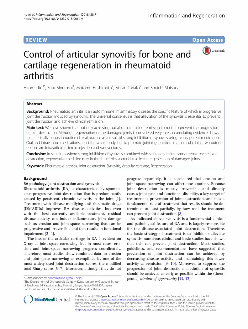
REVIEW Open Access Control of articular synovitis for bone and cartilage regeneration in rheumatoid arthritis Hiromu Ito 1* , Furu Moritoshi 1 , Motomu Hashimoto 2 , Masao Tanaka 2 and Shuichi Matsuda 1 Abstract Background: Rheumatoid arthritis is an autoimmune inflammatory disease, the specific feature of which is progressive joint destruction induced by synovitis. The universal consensus is that alleviation of the synovitis is essential to prevent joint destruction and achieve clinical remission. Main text: We have shown that not only achieving but also maintaining remission is crucial to prevent the progression of joint destruction. Although regeneration of the damaged joints is considered very rare, accumulating evidence shows that it actually occurs in routine clinical practice as a result of strong inhibition of synovitis using highly potent medications. Oral and intravenous medications affect the whole body, but to promote joint regeneration in a particular joint, two potent options are intra-articular steroid injection and synovectomy. Conclusion: In situations where strong inhibition of synovitis combined with self-regeneration cannot repair severe joint destruction, regenerative medicine may in the future play a crucial role in the regeneration of damaged joints. Keywords: Rheumatoid arthritis, Joint destruction, Synovitis, Articular cartilage, Regeneration Background RA pathology: joint destruction and synovitis Rheumatoid arthritis (RA) is characterized by spontan- eous progressive joint destruction that is predominantly caused by persistent, chronic synovitis in the joint [1]. Treatment with disease-modifying anti-rheumatic drugs (DMARDs) improves RA disease activities, but even with the best currently available treatment, residual disease activity can induce inflammatory joint damage such as erosion and joint-space narrowing that can be progressive and irreversible and that results in functional impairment [2–4]. The loss of the articular cartilage in RA is evident on X-ray as joint-space narrowing, but in most cases, ero- sion and joint-space narrowing progress coordinately. Therefore, most studies show combined data for erosion and joint-space narrowing as exemplified by one of the most widely used joint destruction scores, the modified total Sharp score [5–7]. Moreover, although they do not progress separately, it is considered that erosion and joint-space narrowing can affect one another. Because joint destruction is mostly irreversible and directly causes joint pain and functional disability, a key target of treatment is prevention of joint destruction, and it is a fundamental rule of treatment that results should be de- termined, at least partially, by how well the treatment can prevent joint destruction [8]. As indicated above, synovitis is a fundamental clinical and pathological feature of RA and is largely responsible for the disease-associated joint destruction. Therefore, the basic strategy of treatment is to inhibit or alleviate synovitis; numerous clinical and basic studies have shown that this can prevent joint destruction. Most studies, guidelines, and recommendations have suggested that prevention of joint destruction can be achieved by decreasing disease activity and maintaining this lower activity as remission [9, 10]. Moreover, to suppress the progression of joint destruction, alleviation of synovitis should be achieved as early as possible within the (thera- peutic) window of opportunity [11, 12]. * Correspondence: [email protected] 1 The Department of Orthopaedic Surgery, Kyoto University Graduate School of Medicine, 54 Kawahara-cho, Shogoin, Sakyo, Kyoto 606-8507, Japan Full list of author information is available at the end of the article Inflammation and Regeneration © The Author(s). 2018 Open Access This article is distributed under the terms of the Creative Commons Attribution 4.0 International License (http://creativecommons.org/licenses/by/4.0/), which permits unrestricted use, distribution, and reproduction in any medium, provided you give appropriate credit to the original author(s) and the source, provide a link to the Creative Commons license, and indicate if changes were made. The Creative Commons Public Domain Dedication waiver (http://creativecommons.org/publicdomain/zero/1.0/) applies to the data made available in this article, unless otherwise stated. Ito et al. Inflammation and Regeneration (2018) 38:7 https://doi.org/10.1186/s41232-018-0064-y
Transcript of Control of articular synovitis for bone and cartilage ...
REVIEW Open Access
Control of articular synovitis for bone andcartilage regeneration in rheumatoidarthritisHiromu Ito1*, Furu Moritoshi1, Motomu Hashimoto2, Masao Tanaka2 and Shuichi Matsuda1
Abstract
Background: Rheumatoid arthritis is an autoimmune inflammatory disease, the specific feature of which is progressivejoint destruction induced by synovitis. The universal consensus is that alleviation of the synovitis is essential to preventjoint destruction and achieve clinical remission.
Main text: We have shown that not only achieving but also maintaining remission is crucial to prevent the progressionof joint destruction. Although regeneration of the damaged joints is considered very rare, accumulating evidence showsthat it actually occurs in routine clinical practice as a result of strong inhibition of synovitis using highly potent medications.Oral and intravenous medications affect the whole body, but to promote joint regeneration in a particular joint, two potentoptions are intra-articular steroid injection and synovectomy.
Conclusion: In situations where strong inhibition of synovitis combined with self-regeneration cannot repair severe jointdestruction, regenerative medicine may in the future play a crucial role in the regeneration of damaged joints.
Keywords: Rheumatoid arthritis, Joint destruction, Synovitis, Articular cartilage, Regeneration
BackgroundRA pathology: joint destruction and synovitisRheumatoid arthritis (RA) is characterized by spontan-eous progressive joint destruction that is predominantlycaused by persistent, chronic synovitis in the joint [1].Treatment with disease-modifying anti-rheumatic drugs(DMARDs) improves RA disease activities, but evenwith the best currently available treatment, residualdisease activity can induce inflammatory joint damagesuch as erosion and joint-space narrowing that can beprogressive and irreversible and that results in functionalimpairment [2–4].The loss of the articular cartilage in RA is evident on
X-ray as joint-space narrowing, but in most cases, ero-sion and joint-space narrowing progress coordinately.Therefore, most studies show combined data for erosionand joint-space narrowing as exemplified by one of themost widely used joint destruction scores, the modifiedtotal Sharp score [5–7]. Moreover, although they do not
progress separately, it is considered that erosion andjoint-space narrowing can affect one another. Becausejoint destruction is mostly irreversible and directlycauses joint pain and functional disability, a key target oftreatment is prevention of joint destruction, and it is afundamental rule of treatment that results should be de-termined, at least partially, by how well the treatmentcan prevent joint destruction [8].As indicated above, synovitis is a fundamental clinical
and pathological feature of RA and is largely responsiblefor the disease-associated joint destruction. Therefore,the basic strategy of treatment is to inhibit or alleviatesynovitis; numerous clinical and basic studies have shownthat this can prevent joint destruction. Most studies,guidelines, and recommendations have suggested thatprevention of joint destruction can be achieved bydecreasing disease activity and maintaining this loweractivity as remission [9, 10]. Moreover, to suppress theprogression of joint destruction, alleviation of synovitisshould be achieved as early as possible within the (thera-peutic) window of opportunity [11, 12].* Correspondence: [email protected]
1The Department of Orthopaedic Surgery, Kyoto University Graduate Schoolof Medicine, 54 Kawahara-cho, Shogoin, Sakyo, Kyoto 606-8507, JapanFull list of author information is available at the end of the article
Inflammation and Regeneration
© The Author(s). 2018 Open Access This article is distributed under the terms of the Creative Commons Attribution 4.0International License (http://creativecommons.org/licenses/by/4.0/), which permits unrestricted use, distribution, andreproduction in any medium, provided you give appropriate credit to the original author(s) and the source, provide a link tothe Creative Commons license, and indicate if changes were made. The Creative Commons Public Domain Dedication waiver(http://creativecommons.org/publicdomain/zero/1.0/) applies to the data made available in this article, unless otherwise stated.
Ito et al. Inflammation and Regeneration (2018) 38:7 https://doi.org/10.1186/s41232-018-0064-y
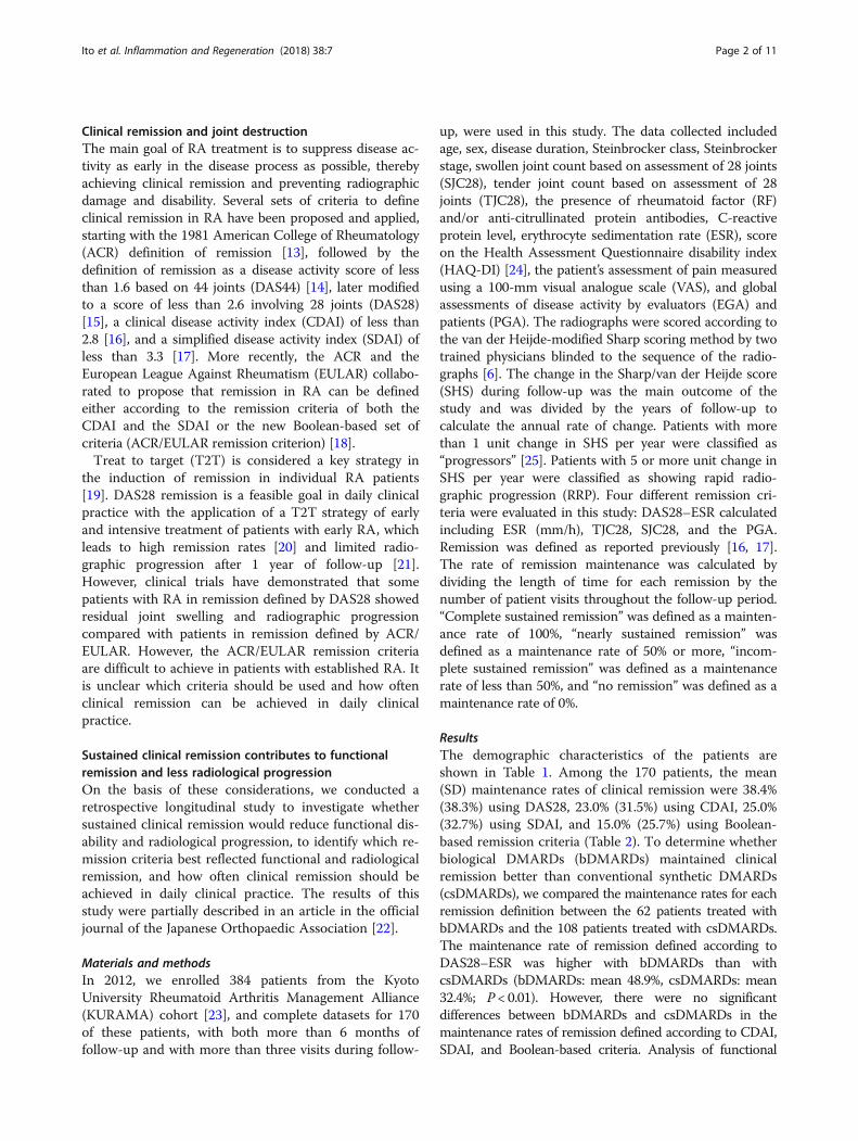
Clinical remission and joint destructionThe main goal of RA treatment is to suppress disease ac-tivity as early in the disease process as possible, therebyachieving clinical remission and preventing radiographicdamage and disability. Several sets of criteria to defineclinical remission in RA have been proposed and applied,starting with the 1981 American College of Rheumatology(ACR) definition of remission [13], followed by thedefinition of remission as a disease activity score of lessthan 1.6 based on 44 joints (DAS44) [14], later modifiedto a score of less than 2.6 involving 28 joints (DAS28)[15], a clinical disease activity index (CDAI) of less than2.8 [16], and a simplified disease activity index (SDAI) ofless than 3.3 [17]. More recently, the ACR and theEuropean League Against Rheumatism (EULAR) collabo-rated to propose that remission in RA can be definedeither according to the remission criteria of both theCDAI and the SDAI or the new Boolean-based set ofcriteria (ACR/EULAR remission criterion) [18].Treat to target (T2T) is considered a key strategy in
the induction of remission in individual RA patients[19]. DAS28 remission is a feasible goal in daily clinicalpractice with the application of a T2T strategy of earlyand intensive treatment of patients with early RA, whichleads to high remission rates [20] and limited radio-graphic progression after 1 year of follow-up [21].However, clinical trials have demonstrated that somepatients with RA in remission defined by DAS28 showedresidual joint swelling and radiographic progressioncompared with patients in remission defined by ACR/EULAR. However, the ACR/EULAR remission criteriaare difficult to achieve in patients with established RA. Itis unclear which criteria should be used and how oftenclinical remission can be achieved in daily clinicalpractice.
Sustained clinical remission contributes to functionalremission and less radiological progressionOn the basis of these considerations, we conducted aretrospective longitudinal study to investigate whethersustained clinical remission would reduce functional dis-ability and radiological progression, to identify which re-mission criteria best reflected functional and radiologicalremission, and how often clinical remission should beachieved in daily clinical practice. The results of thisstudy were partially described in an article in the officialjournal of the Japanese Orthopaedic Association [22].
Materials and methodsIn 2012, we enrolled 384 patients from the KyotoUniversity Rheumatoid Arthritis Management Alliance(KURAMA) cohort [23], and complete datasets for 170of these patients, with both more than 6 months offollow-up and with more than three visits during follow-
up, were used in this study. The data collected includedage, sex, disease duration, Steinbrocker class, Steinbrockerstage, swollen joint count based on assessment of 28 joints(SJC28), tender joint count based on assessment of 28joints (TJC28), the presence of rheumatoid factor (RF)and/or anti-citrullinated protein antibodies, C-reactiveprotein level, erythrocyte sedimentation rate (ESR), scoreon the Health Assessment Questionnaire disability index(HAQ-DI) [24], the patient’s assessment of pain measuredusing a 100-mm visual analogue scale (VAS), and globalassessments of disease activity by evaluators (EGA) andpatients (PGA). The radiographs were scored according tothe van der Heijde-modified Sharp scoring method by twotrained physicians blinded to the sequence of the radio-graphs [6]. The change in the Sharp/van der Heijde score(SHS) during follow-up was the main outcome of thestudy and was divided by the years of follow-up tocalculate the annual rate of change. Patients with morethan 1 unit change in SHS per year were classified as“progressors” [25]. Patients with 5 or more unit change inSHS per year were classified as showing rapid radio-graphic progression (RRP). Four different remission cri-teria were evaluated in this study: DAS28–ESR calculatedincluding ESR (mm/h), TJC28, SJC28, and the PGA.Remission was defined as reported previously [16, 17].The rate of remission maintenance was calculated bydividing the length of time for each remission by thenumber of patient visits throughout the follow-up period.“Complete sustained remission” was defined as a mainten-ance rate of 100%, “nearly sustained remission” wasdefined as a maintenance rate of 50% or more, “incom-plete sustained remission” was defined as a maintenancerate of less than 50%, and “no remission” was defined as amaintenance rate of 0%.
ResultsThe demographic characteristics of the patients areshown in Table 1. Among the 170 patients, the mean(SD) maintenance rates of clinical remission were 38.4%(38.3%) using DAS28, 23.0% (31.5%) using CDAI, 25.0%(32.7%) using SDAI, and 15.0% (25.7%) using Boolean-based remission criteria (Table 2). To determine whetherbiological DMARDs (bDMARDs) maintained clinicalremission better than conventional synthetic DMARDs(csDMARDs), we compared the maintenance rates for eachremission definition between the 62 patients treated withbDMARDs and the 108 patients treated with csDMARDs.The maintenance rate of remission defined according toDAS28–ESR was higher with bDMARDs than withcsDMARDs (bDMARDs: mean 48.9%, csDMARDs: mean32.4%; P < 0.01). However, there were no significantdifferences between bDMARDs and csDMARDs in themaintenance rates of remission defined according to CDAI,SDAI, and Boolean-based criteria. Analysis of functional
Ito et al. Inflammation and Regeneration (2018) 38:7 Page 2 of 11
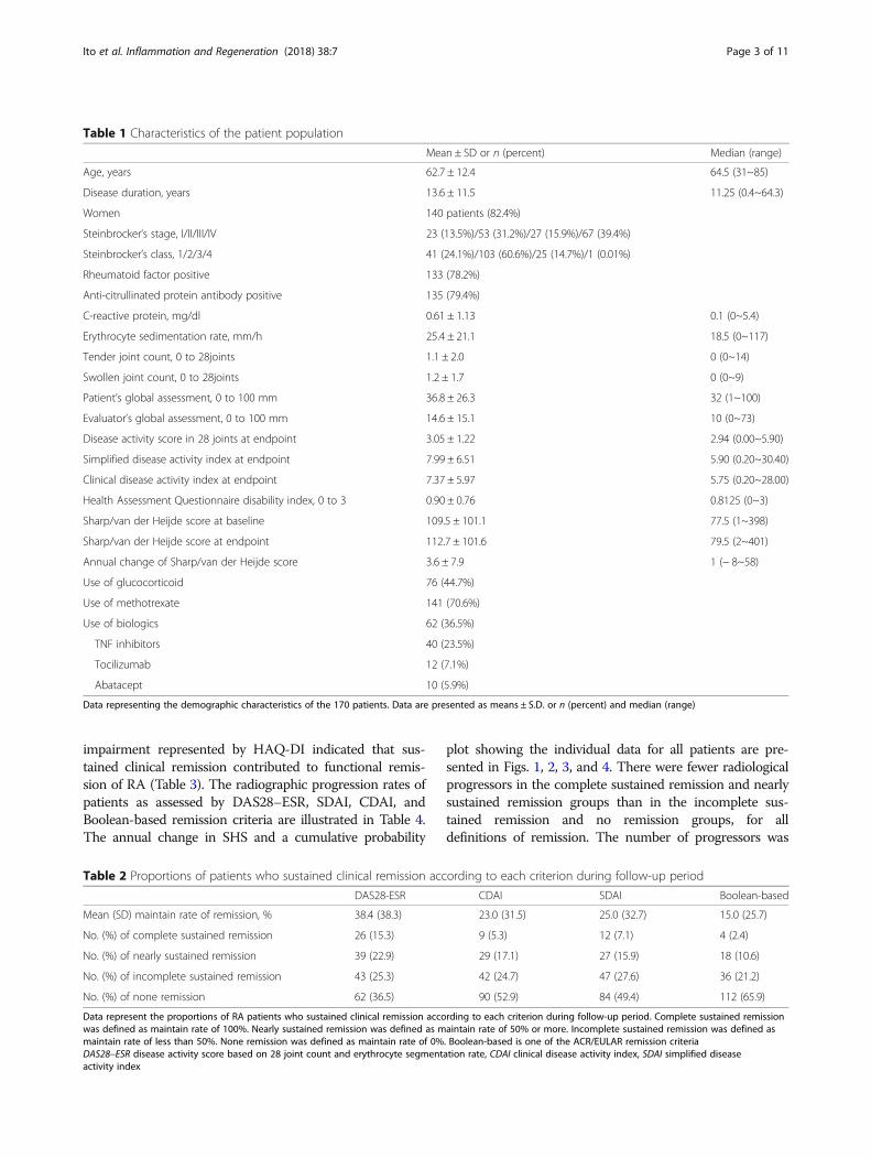
impairment represented by HAQ-DI indicated that sus-tained clinical remission contributed to functional remis-sion of RA (Table 3). The radiographic progression rates ofpatients as assessed by DAS28–ESR, SDAI, CDAI, andBoolean-based remission criteria are illustrated in Table 4.The annual change in SHS and a cumulative probability
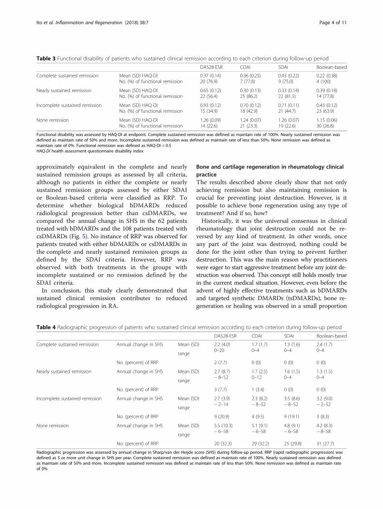
plot showing the individual data for all patients are pre-sented in Figs. 1, 2, 3, and 4. There were fewer radiologicalprogressors in the complete sustained remission and nearlysustained remission groups than in the incomplete sus-tained remission and no remission groups, for alldefinitions of remission. The number of progressors was
Table 1 Characteristics of the patient population
Mean ± SD or n (percent) Median (range)
Age, years 62.7 ± 12.4 64.5 (31~85)
Disease duration, years 13.6 ± 11.5 11.25 (0.4~64.3)
Women 140 patients (82.4%)
Steinbrocker‘s stage, I/II/III/IV 23 (13.5%)/53 (31.2%)/27 (15.9%)/67 (39.4%)
Steinbrocker’s class, 1/2/3/4 41 (24.1%)/103 (60.6%)/25 (14.7%)/1 (0.01%)
Rheumatoid factor positive 133 (78.2%)
Anti-citrullinated protein antibody positive 135 (79.4%)
C-reactive protein, mg/dl 0.61 ± 1.13 0.1 (0~5.4)
Erythrocyte sedimentation rate, mm/h 25.4 ± 21.1 18.5 (0~117)
Tender joint count, 0 to 28joints 1.1 ± 2.0 0 (0~14)
Swollen joint count, 0 to 28joints 1.2 ± 1.7 0 (0~9)
Patient’s global assessment, 0 to 100 mm 36.8 ± 26.3 32 (1~100)
Evaluator’s global assessment, 0 to 100 mm 14.6 ± 15.1 10 (0~73)
Disease activity score in 28 joints at endpoint 3.05 ± 1.22 2.94 (0.00~5.90)
Simplified disease activity index at endpoint 7.99 ± 6.51 5.90 (0.20~30.40)
Clinical disease activity index at endpoint 7.37 ± 5.97 5.75 (0.20~28.00)
Health Assessment Questionnaire disability index, 0 to 3 0.90 ± 0.76 0.8125 (0~3)
Sharp/van der Heijde score at baseline 109.5 ± 101.1 77.5 (1~398)
Sharp/van der Heijde score at endpoint 112.7 ± 101.6 79.5 (2~401)
Annual change of Sharp/van der Heijde score 3.6 ± 7.9 1 (− 8~58)
Use of glucocorticoid 76 (44.7%)
Use of methotrexate 141 (70.6%)
Use of biologics 62 (36.5%)
TNF inhibitors 40 (23.5%)
Tocilizumab 12 (7.1%)
Abatacept 10 (5.9%)
Data representing the demographic characteristics of the 170 patients. Data are presented as means ± S.D. or n (percent) and median (range)
Table 2 Proportions of patients who sustained clinical remission according to each criterion during follow-up period
DAS28-ESR CDAI SDAI Boolean-based
Mean (SD) maintain rate of remission, % 38.4 (38.3) 23.0 (31.5) 25.0 (32.7) 15.0 (25.7)
No. (%) of complete sustained remission 26 (15.3) 9 (5.3) 12 (7.1) 4 (2.4)
No. (%) of nearly sustained remission 39 (22.9) 29 (17.1) 27 (15.9) 18 (10.6)
No. (%) of incomplete sustained remission 43 (25.3) 42 (24.7) 47 (27.6) 36 (21.2)
No. (%) of none remission 62 (36.5) 90 (52.9) 84 (49.4) 112 (65.9)
Data represent the proportions of RA patients who sustained clinical remission according to each criterion during follow-up period. Complete sustained remissionwas defined as maintain rate of 100%. Nearly sustained remission was defined as maintain rate of 50% or more. Incomplete sustained remission was defined asmaintain rate of less than 50%. None remission was defined as maintain rate of 0%. Boolean-based is one of the ACR/EULAR remission criteriaDAS28–ESR disease activity score based on 28 joint count and erythrocyte segmentation rate, CDAI clinical disease activity index, SDAI simplified diseaseactivity index
Ito et al. Inflammation and Regeneration (2018) 38:7 Page 3 of 11
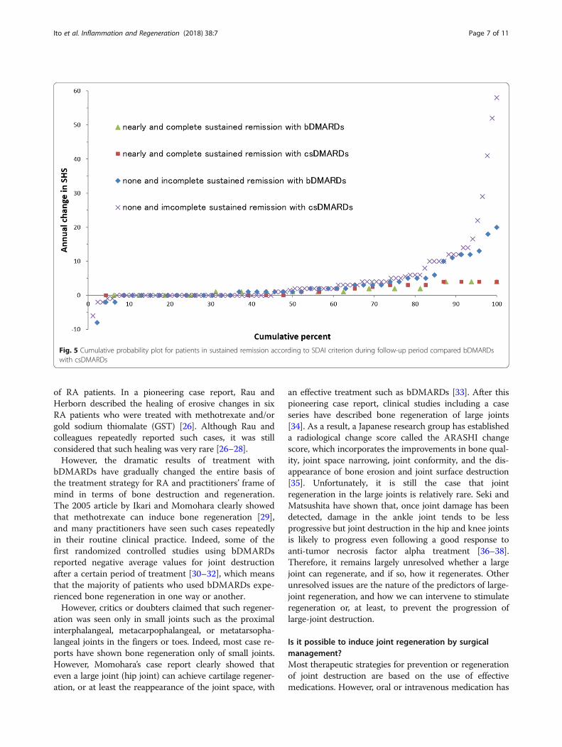
approximately equivalent in the complete and nearlysustained remission groups as assessed by all criteria,although no patients in either the complete or nearlysustained remission groups assessed by either SDAIor Boolean-based criteria were classified as RRP. Todetermine whether biological bDMARDs reducedradiological progression better than csDMARDs, wecompared the annual change in SHS in the 62 patientstreated with bDMARDs and the 108 patients treated withcsDMARDs (Fig. 5). No instance of RRP was observed forpatients treated with either bDMARDs or csDMARDs inthe complete and nearly sustained remission groups asdefined by the SDAI criteria. However, RRP wasobserved with both treatments in the groups withincomplete sustained or no remission defined by theSDAI criteria.In conclusion, this study clearly demonstrated that
sustained clinical remission contributes to reducedradiological progression in RA.
Bone and cartilage regeneration in rheumatology clinicalpracticeThe results described above clearly show that not onlyachieving remission but also maintaining remission iscrucial for preventing joint destruction. However, is itpossible to achieve bone regeneration using any type oftreatment? And if so, how?Historically, it was the universal consensus in clinical
rheumatology that joint destruction could not be re-versed by any kind of treatment. In other words, onceany part of the joint was destroyed, nothing could bedone for the joint other than trying to prevent furtherdestruction. This was the main reason why practitionerswere eager to start aggressive treatment before any joint de-struction was observed. This concept still holds mostly truein the current medical situation. However, even before theadvent of highly effective treatments such as bDMARDsand targeted synthetic DMARDs (tsDMARDs), bone re-generation or healing was observed in a small proportion
Table 4 Radiographic progression of patients who sustained clinical remission according to each criterion during follow-up period
DAS28-ESR CDAI SDAI Boolean-based
Complete sustained remission Annual change in SHS Mean (SD) 2.2 (4.0)0–20
1.7 (1.7)0–4
1.3 (1.6)0–4
2.4 (1.7)0–4
range
No. (percent) of RRP 2 (7.7) 0 (0) 0 (0) 0 (0)
Nearly sustained remission Annual change in SHS Mean (SD) 2.7 (8.7)− 8–52
1.7 (2.5)0–12
1.6 (1.5)0–4
1.3 (1.5)0–4
range
No. (percent) of RRP 3 (7.7) 1 (3.4) 0 (0) 0 (0)
Incomplete sustained remission Annual change in SHS Mean (SD) 2.7 (3.9)− 2–14
2.3 (8.2)− 8–52
3.5 (8.6)− 8–52
3.2 (9.0)− 2–52
range
No. (percent) of RRP 9 (20.9) 4 (9.5) 9 (19.1) 3 (8.3)
None remission Annual change in SHS Mean (SD) 5.5 (10.3)− 6–58
5.1 (9.1)− 6–58
4.8 (9.1)− 6–58
4.2 (8.3)− 8–58
range
No. (percent) of RRP 20 (32.3) 29 (32.2) 25 (29.8) 31 (27.7)
Radiographic progression was assessed by annual change in Sharp/van der Heijde score (SHS) during follow-up period. RRP (rapid radiographic progression) wasdefined as 5 or more unit change in SHS per year. Complete sustained remission was defined as maintain rate of 100%. Nearly sustained remission was definedas maintain rate of 50% and more. Incomplete sustained remission was defined as maintain rate of less than 50%. None remission was defined as maintain rateof 0%
Table 3 Functional disability of patients who sustained clinical remission according to each criterion during follow-up period
DAS28-ESR CDAI SDAI Boolean-based
Complete sustained remission Mean (SD) HAQ-DINo. (%) of functional remission
0.37 (0.14)20 (76.9)
0.36 (0.25)7 (77.8)
0.43 (0.22)9 (75.0)
0.22 (0.38)4 (100)
Nearly sustained remission Mean (SD) HAQ-DINo. (%) of functional remission
0.65 (0.12)22 (56.4)
0.30 (0.13)25 (86.2)
0.33 (0.14)22 (81.5)
0.39 (0.18)14 (77.8)
Incomplete sustained remission Mean (SD) HAQ-DINo. (%) of functional remission
0.93 (0.12)15 (34.9)
0.70 (0.12)18 (42.9)
0.71 (0.11)21 (44.7)
0.43 (0.12)23 (63.9)
None remission Mean (SD) HAQ-DINo. (%) of functional remission
1.26 (0.09)14 (22.6)
1.24 (0.07)21 (23.3)
1.26 (0.07)19 (22.6)
1.15 (0.06)30 (26.8)
Functional disability was assessed by HAQ-DI at endpoint. Complete sustained remission was defined as maintain rate of 100%. Nearly sustained remission wasdefined as maintain rate of 50% and more. Incomplete sustained remission was defined as maintain rate of less than 50%. None remission was defined asmaintain rate of 0%. Functional remission was defined as HAQ-DI < 0.5HAQ-DI health assessment questionnaire disability index
Ito et al. Inflammation and Regeneration (2018) 38:7 Page 4 of 11
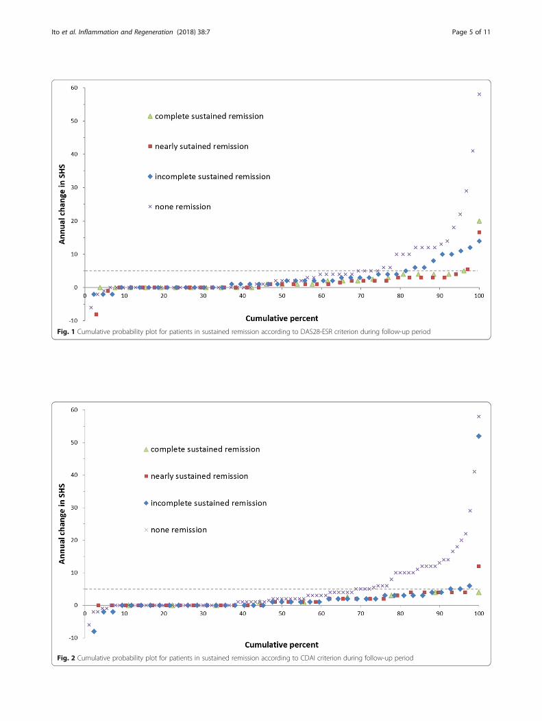
Fig. 1 Cumulative probability plot for patients in sustained remission according to DAS28-ESR criterion during follow-up period
Fig. 2 Cumulative probability plot for patients in sustained remission according to CDAI criterion during follow-up period
Ito et al. Inflammation and Regeneration (2018) 38:7 Page 5 of 11
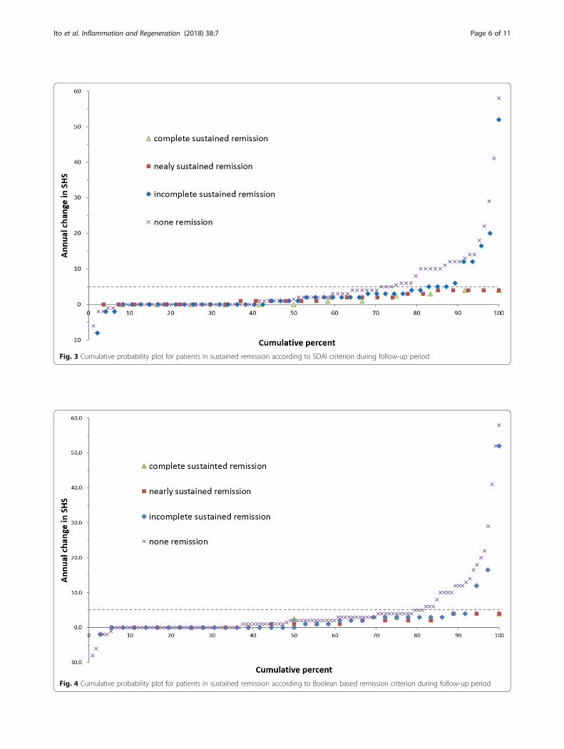
Fig. 3 Cumulative probability plot for patients in sustained remission according to SDAI criterion during follow-up period
Fig. 4 Cumulative probability plot for patients in sustained remission according to Boolean based remission criterion during follow-up period
Ito et al. Inflammation and Regeneration (2018) 38:7 Page 6 of 11
of RA patients. In a pioneering case report, Rau andHerborn described the healing of erosive changes in sixRA patients who were treated with methotrexate and/orgold sodium thiomalate (GST) [26]. Although Rau andcolleagues repeatedly reported such cases, it was stillconsidered that such healing was very rare [26–28].However, the dramatic results of treatment with
bDMARDs have gradually changed the entire basis ofthe treatment strategy for RA and practitioners’ frame ofmind in terms of bone destruction and regeneration.The 2005 article by Ikari and Momohara clearly showedthat methotrexate can induce bone regeneration [29],and many practitioners have seen such cases repeatedlyin their routine clinical practice. Indeed, some of thefirst randomized controlled studies using bDMARDsreported negative average values for joint destructionafter a certain period of treatment [30–32], which meansthat the majority of patients who used bDMARDs expe-rienced bone regeneration in one way or another.However, critics or doubters claimed that such regener-
ation was seen only in small joints such as the proximalinterphalangeal, metacarpophalangeal, or metatarsopha-langeal joints in the fingers or toes. Indeed, most case re-ports have shown bone regeneration only of small joints.However, Momohara’s case report clearly showed thateven a large joint (hip joint) can achieve cartilage regener-ation, or at least the reappearance of the joint space, with
an effective treatment such as bDMARDs [33]. After thispioneering case report, clinical studies including a caseseries have described bone regeneration of large joints[34]. As a result, a Japanese research group has establisheda radiological change score called the ARASHI changescore, which incorporates the improvements in bone qual-ity, joint space narrowing, joint conformity, and the dis-appearance of bone erosion and joint surface destruction[35]. Unfortunately, it is still the case that jointregeneration in the large joints is relatively rare. Seki andMatsushita have shown that, once joint damage has beendetected, damage in the ankle joint tends to be lessprogressive but joint destruction in the hip and knee jointsis likely to progress even following a good response toanti-tumor necrosis factor alpha treatment [36–38].Therefore, it remains largely unresolved whether a largejoint can regenerate, and if so, how it regenerates. Otherunresolved issues are the nature of the predictors of large-joint regeneration, and how we can intervene to stimulateregeneration or, at least, to prevent the progression oflarge-joint destruction.
Is it possible to induce joint regeneration by surgicalmanagement?Most therapeutic strategies for prevention or regenerationof joint destruction are based on the use of effectivemedications. However, oral or intravenous medication has
Fig. 5 Cumulative probability plot for patients in sustained remission according to SDAI criterion during follow-up period compared bDMARDswith csDMARDs
Ito et al. Inflammation and Regeneration (2018) 38:7 Page 7 of 11
an effect on the whole body, i.e., it diffuses throughout thewhole body and may therefore be less effective in aparticular joint. One of the potent options for treating aparticular joint is intra-articular injection. Indeed, severalstudies indicate that intra-articular injection of steroid ishighly effective and comparable to bDMARDs for the alle-viation of disease activity [39]. For example, a preliminaryreport showed that in osteoarthritis, intrajoint injection ofbDMARDs can achieve a better response than injection ofhyaluronan [40]. Moreover, a recent study showed thatintra-articular glucocorticoids in combination with metho-trexate can induce bone regeneration in some cases of RA,although this response is relatively rare [41]. Future investi-gations should determine which patients should receiveintra-articular steroid injection and its optimal timing.Another possible approach to this issue is surgical
intervention. Joint regeneration, especially cartilageregeneration, has been widely investigated over the lastthree decades. We recently published a report of scaffold-less hyaline cartilaginous tissue derived from inducedpluripotent stem cells [42]. However, despite committed,long-term efforts worldwide, clinically useful hyalinecartilage regeneration has not yet been achieved. To over-come this highly problematic threshold to achieving jointregeneration, the most plausible treatment strategy is toinduce or assist the patient’s own ability to regeneratebone and cartilage. In the case of inflammatory arthritis
such as RA, the reduction of synovitis or the surgical re-moval of inflammatory synovia is one plausible option.We have experienced one such case in the past.The patient, a 21-year-old woman, presented to our
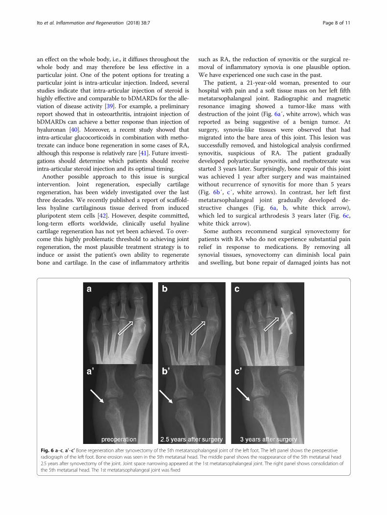
hospital with pain and a soft tissue mass on her left fifthmetatarsophalangeal joint. Radiographic and magneticresonance imaging showed a tumor-like mass withdestruction of the joint (Fig. 6a′, white arrow), which wasreported as being suggestive of a benign tumor. Atsurgery, synovia-like tissues were observed that hadmigrated into the bare area of this joint. This lesion wassuccessfully removed, and histological analysis confirmedsynovitis, suspicious of RA. The patient graduallydeveloped polyarticular synovitis, and methotrexate wasstarted 3 years later. Surprisingly, bone repair of this jointwas achieved 1 year after surgery and was maintainedwithout recurrence of synovitis for more than 5 years(Fig. 6b′, c′, white arrows). In contrast, her left firstmetatarsophalangeal joint gradually developed de-structive changes (Fig. 6a, b, white thick arrow),which led to surgical arthrodesis 3 years later (Fig. 6c,white thick arrow).Some authors recommend surgical synovectomy for
patients with RA who do not experience substantial painrelief in response to medications. By removing allsynovial tissues, synovectomy can diminish local painand swelling, but bone repair of damaged joints has not
Fig. 6 a–c, a′–c′ Bone regeneration after synovectomy of the 5th metatarsophalangeal joint of the left foot. The left panel shows the preoperativeradiograph of the left foot. Bone erosion was seen in the 5th metatarsal head. The middle panel shows the reappearance of the 5th metatarsal head2.5 years after synovectomy of the joint. Joint space narrowing appeared at the 1st metatarsophalangeal joint. The right panel shows consolidation ofthe 5th metatarsal head. The 1st metatarsophalangeal joint was fixed
Ito et al. Inflammation and Regeneration (2018) 38:7 Page 8 of 11
been expected. This case provides evidence thatsynovectomy can induce bone repair of a damaged jointin a patient with early RA. To the best of our knowledge,this is the very first report that synovectomy indeedstimulated joint regeneration. Pinder previously reportedthat synovectomy with drilling of areas of articularcartilage loss showed cartilage regeneration and relief ofsymptoms [43]. However, since then, no other report hasshown similar results by any surgical procedures. Thereason of his success may be that he probably conductedthis procedure in patients with very low disease activity.But the regeneration potential of the joint should bepaid with full attention even in RA patients in thecurrent medication.Also, the molecular mechanisms of how the regener-
ation occurs have been investigated and proposed, whichhas attracted huge attention from basic researchers.Several review articles recently summarized the proposedmechanism of bone remodeling in RA that proinflammatorycytokines such as TNF alpha stimulates the production ofDKK-1 family and soluble frizzled related protein, suggestingthat inhibition of such cytokines downregulates thoseproteins and revives bone formation processes [44, 45].Wehmeyer et al. recently lay stress on the importance ofstromal cells which release Wnt antagonists such as scler-ostin and DKK-1 under inflammatory conditions [44, 46].Taken together, blocking proinflammatory cytokines orremoval of synovial tissues producing such cytokines canregain the balance of bone resorption and formation andcan stimulate bone regeneration. Adding to the suppres-sion of proinflammatory cytokines or cells, suppression ofWnt antagonists or stromal cells may be a potent thera-peutic option in the future.
Future perspectivesIn inflammatory arthritis, synovitis causes bone andcartilage destruction as described above. One of themost crucial requirements for regeneration of thedestroyed joint is alleviation of synovitis. This can beachieved by use of adequate medication as soon as pos-sible after the diagnosis of the disease. When the jointhas the ability to regenerate the destroyed bone and/orarticular cartilage, self-regeneration should occur afteralleviation of the synovitis. However, regenerativemedicine will have a crucial role in treatment when thisability is lost, or when the destruction is too severe to beovercome. Although it is still uncertain what kind oftreatment options will be available in routine clinicalpractice, regenerative medicine should be able to rescuethe damaged joint using potent cell therapies.
ConclusionsTo prevent joint destruction in inflammatory arthritissuch as RA, the universal consensus is to treat, to
alleviate synovitis and to achieve clinical remission. Ourstudy shows that maintaining remission is also crucial toprevent the progression of joint destruction. Althoughregeneration of the damaged joint has been consideredto occur very rarely, accumulating evidence shows thatit can actually occur in routine clinical practice afterstrong inhibition of synovitis with highly potent medica-tions. Two potent options other than oral or intravenousmedication for inducing joint regeneration in a particu-lar joint would be intra-articular steroid injection andsynovectomy. In the future, regenerative medicine couldplay a crucial role in inducing regeneration of damagedjoints after synovitis is effectively inhibited when self-regeneration cannot overcome severe destruction.
AbbreviationsACR: American College of Rheumatology; bDMARD: Biological DMARD;CDAI: Clinical disease activity index; csDMARD: Conventional syntheticDMARD; DAS28: Disease activity score involving a 28 joints; DAS44: Diseaseactivity score based on 44 joints; DMARD: Disease-modifying anti-rheumaticdrug; EGA: Global assessments of disease activity by evaluators; ESR: Erythrocytesedimentation rate; EULAR: The European League Against Rheumatism; HAQ-DI: The Health Assessment Questionnaire disability index; KURAMA: The KyotoUniversity Rheumatoid Arthritis Management Alliance; PGA: Global assessmentsof disease activity by patients; RA: Rheumatoid arthritis; RF: Rheumatoid factor;RRP: Rapid radiographic progression; SDAI: Simplified disease activity index;SHS: Sharp/van der Heijde score; SJC28: Swollen joint count based onassessment of 28 joints; T2T: Treat to target; TJC28: Tender joint count basedon assessment of 28 joints; tsDMARD: Targeted synthetic DMARD; VAS: Visualanalogue scale
AcknowledgementsThe authors thank Drs. Tsuneyo Mimori, Takao Fujii, Chicashi Terao, NoriyukiYamakawa, Kohei Nishitani, Kosaku Murakami, and Hiroyuki Yoshitomi fortheir thoughtful discussion and technical assistance.
Availability of data and materialsThe data and material are accessible by any parties through thecorresponding author (HI).
Authors’ contributionsHI, MF, and SM contributed to the study conception and design. MF contributedto the analysis of the data. HI, MF, MH, and MT contributed to the acquisition ofdata. HI and MF contributed to the interpretation of the data. HI and MF draftedthe article. HI, MF, MH, MT, SM revised the article and gave final approval. Allauthors read and approved the final manuscript.
Ethics approval and consent to participateWritten informed consent for this study was obtained from all participatingpatients. This study was designed in accordance with the Helsinkideclaration and was approved by the ethics committee of Kyoto UniversityGraduate School and Faculty of Medicine (R0357).
Consent for publicationAll of the listed authors declared the consent for publication.
Competing interestsThe Department of Advanced Medicine for Rheumatic Diseases is supportedby Nagahama City, Shiga, Japan, and four pharmaceutical companies (MitsubishiTanabe Pharma Co., Chugai Pharmaceutical Co. Ltd, UCB Japan Co. Ltd, andAYUMI Pharmaceutical Co.). KURAMA cohort study is supported by a grant fromDaiichi Sankyo Co. Ltd. This study is conducted as investigator initiate study. HIhas received a research grant and/or speaker fee from Bristol-Myers, Astellas, andEli Lily. MH has received research grants from Astellas and Daiichi-Sankyo. MT hasreceived research grants from Astellas, Abbvie, Pfizer, and Taisho-Toyama. MF andSM declared no conflicts of interest. The sponsors were not involved in the studydesign; in the collection, analysis, and interpretation of data; in the writing of this
Ito et al. Inflammation and Regeneration (2018) 38:7 Page 9 of 11
manuscript; or in the decision to submit the article for publication. The authors,their immediate families, and any research foundations with which they areaffiliated have not received any financial payments or other benefits from anycommercial entity related to the subject of this article.
Publisher’s NoteSpringer Nature remains neutral with regard to jurisdictional claims inpublished maps and institutional affiliations.
Author details1The Department of Orthopaedic Surgery, Kyoto University Graduate Schoolof Medicine, 54 Kawahara-cho, Shogoin, Sakyo, Kyoto 606-8507, Japan. 2TheDepartment of Advanced Medicine for Rheumatic Diseases, Kyoto UniversityGraduate School of Medicine, Kyoto, Japan.
Received: 8 January 2018 Accepted: 7 March 2018
References1. Terao C, Hashimoto M, Yamamoto K, Murakami K, Ohmura K, Nakashima R,
et al. Three groups in the 28 joints for rheumatoid arthritissynovitis—analysis using more than 17,000 assessments in the KURAMAdatabase. PLoS One. 2013;8(3):e59341.
2. Keystone EC, Haraoui B, Guerette B, Mozaffarian N, Liu S, Kavanaugh A.Clinical, functional, and radiographic implications of time to treatmentresponse in patients with early rheumatoid arthritis: a posthoc analysis ofthe PREMIER study. J Rheumatol. 2014;41(2):235–43.
3. Scott DL, Pugner K, Kaarela K, Doyle DV, Woolf A, Holmes J, et al. The linksbetween joint damage and disability in rheumatoid arthritis. Rheumatology(Oxford). 2000;39:122–32.
4. Welsing PM, Van Gestel AM, Swinkels HL, Kiemeney LA, Van Riel PL. Therelationship between disease activity, joint destruction, and functional capacityover the course of rheumatoid arthritis. Arthritis Rheum. 2001;44:2009–17.
5. Sharp JT, Young DY, Bluhm GB, Brook A, Brower AC, Corbett M, Decker JL,Genant HK, Gofton JP, Goodman N, et al. How many joints in the handsand wrists should be included in a score of radiologic abnormalities used toassess rheumatoid arthritis? Arthritis Rheum. 1985;28(12):1326–35.
6. van der Heijde D. How to read radiographs according to the Sharp/van derHeijde method. J Rheumatol. 1999;26(3):743–5.
7. van der Heijde D, Dankert T, Nieman F, Rau R, Boers M. Reliability and sensitivityto change of a simplification of the Sharp/van der Heijde radiological assessmentin rheumatoid arthritis. Rheumatology (Oxford). 1999;38(10):941–7.
8. Smolen JS, Aletaha D, Redlich K. The pathogenesis of rheumatoid arthritis:new insights from old clinical data? Nat Rev Rheumatol. 2012;8(4):235–43.
9. Smolen JS, Landewé R, Bijlsma J, Burmester G, Chatzidionysiou K, DougadosM, Nam J, Ramiro S, Voshaar M, van Vollenhoven R, Aletaha D, Aringer M,Boers M, Buckley CD, Buttgereit F, Bykerk V, Cardiel M, Combe B, Cutolo M,van Eijk-Hustings Y, Emery P, Finckh A, Gabay C, Gomez-Reino J, Gossec L,Gottenberg JE, Hazes JMW, Huizinga T, Jani M, Karateev D, Kouloumas M,Kvien T, Li Z, Mariette X, McInnes I, Mysler E, Nash P, Pavelka K, Poór G,Richez C, van Riel P, Rubbert-Roth A, Saag K, da Silva J, Stamm T, TakeuchiT, Westhovens R, de Wit M, van der Heijde D. EULAR recommendations forthe management of rheumatoid arthritis with synthetic and biologicaldisease-modifying antirheumatic drugs: 2016 update. Ann Rheum Dis.2017;76(6):960–77.
10. Singh JA, Saag KG, Bridges SL Jr, Akl EA, Bannuru RR, Sullivan MC, VaysbrotE, McNaughton C, Osani M, Shmerling RH, Curtis JR, Furst DE, Parks D,Kavanaugh A, O'Dell J, King C, Leong A, Matteson EL, Schousboe JT,Drevlow B, Ginsberg S, Grober J, St Clair EW, Tindall E, Miller AS, McAlindonT, American College of Rheumatology. 2015 American College ofRheumatology guideline for the treatment of rheumatoid arthritis. ArthritisCare Res (Hoboken). 2016;68(1):1–25.
11. van Steenbergen HW, da Silva JAP, Huizinga TWJ, van der Helm-van MilAHM. Preventing progression from arthralgia to arthritis: targeting the rightpatients. Nat Rev Rheumatol. 2018;14(1):32–41.
12. van Nies JA, Tsonaka R, Gaujoux-Viala C, Fautrel B, van der Helm-van Mil AH.Evaluating relationships between symptom duration and persistence ofrheumatoid arthritis: does a window of opportunity exist? Results on the Leidenearly arthritis clinic and ESPOIR cohorts. Ann Rheum Dis. 2015;74(5):806–12.
13. Pinals RS, Masi AT, Larsen RA. Preliminary criteria for clinical remission inrheumatoid arthritis. Arthritis Rheum. 1981;24:1308–15.
14. Van der Heijde DMFM, van ‘ t Hof MA, van Riel PLCM, Theunisse LM,Lubberts EW, van Leeuwen MA, van Rijswijk MH, van de LBA P. Judgingdisease activity in clinical practice in rheumatoid arthritis: first step in thedevelopment of a disease activity score. Ann Rheum Dis. 1990;49:916–20.
15. Prevoo ML, van ‘t Hof MA, Kuper HH, van Leeuwen MA, van de Putte LB,van Riel PL. Modified disease activity scores that include twenty-eight-jointcounts: development and validation in a prospective longitudinal study ofpatients with rheumatoid arthritis. Arthritis Rheum. 1995;38:44–8.
16. Aletaha D, Nell VP, Stamm T, et al. Acute phase reactants add little tocomposite disease activity indices for rheumatoid arthritis: validation of aclinical activity score. Arthritis Res Ther. 2005;7:R796–806.
17. Aletaha D, Smolen JS. The simplified disease activity index (SDAI) andclinical disease activity index (CDAI) to monitor patients in standard clinicalcare. Best Pract Res Clin Rheumatol. 2007;21:663–75.
18. Felson DT, Smolen JS, Wells G, et al. American College of Rheumatology/European League against Rheumatism provisional definition of remission inrheumatoid arthritis for clinical trials. Ann Rheum Dis. 2011;70:404–13.
19. Smolen JS, Aletaha D, Bijlsma JW, Breedveld FC, Boumpas D, Burmester G,et al. Treating rheumatoid arthritis to target: recommendations of aninternational task force. Ann Rheum Dis. 2010;69:631–7.
20. Schipper LG, Vermeer M, Kuper HH, Hoekstra MO, Haagsma CJ, Broeder AA,et al. A tight control treatment strategy aiming for remission in earlyrheumatoid arthritis is more effective than usual care treatment in dailyclinical practice: a study of two cohorts in the Dutch Rheumatoid ArthritisMonitoring registry. Ann Rheum Dis. 2012;71:845–50.
21. Vermeer M, Kuper HH, Hoekstra M, Haagsma CJ, Posthumus MD, Brus HL, etal. Implementation of a treat-to-target strategy in very early rheumatoidarthritis: results of the Dutch Rheumatoid Arthritis Monitoring remissioninduction cohort study. Arthritis Rheum. 2011;63:2865–72.
22. Ito H, Furu M, Matsuda S. What affects joint destruction in rheumatoidarthritis -effect of sustained remission ratio and biological disease modifyinganti-rheumatic drugs. J Jpn Orthop Assoc. 2018;92(5):(in press). [in theJapanese].
23. Furu M, Hashimoto M, Ito H, Fujii T, Terao C, Yamakawa N, Yoshitomi H,Ogino H, Ishikawa M, Matsuda S, Mimori T. Discordance and accordancebetween patient’s and physician’s assessments in rheumatoid arthritis.Scand J Rheumatol. 2014;43(4):291–5.
24. Fries JF, Spitz P, Kraines RG, Holman HR. Measurement of patient outcomein arthritis. Arthritis Rheum. 1980;23:137–45.
25. van der Heijde D, Landewé R, van Vollenhoven R, et al. Level ofradiographic damage and radiographic progression are determinants ofphysical function: a longitudinal analysis of the TEMPO trial. Ann Rheum Dis.2008;67:1267–70.
26. Rau R, Herborn G. Healing phenomena of erosive changes in rheumatoidarthritis patients undergoing disease-modifying antirheumatic drug therapy.Arthritis Rheum. 1996;39(1):162–8.
27. Rau R, Wassenberg S, Herborn G, Perschel WT, Freitag G. Identification ofradiologic healing phenomena in patients with rheumatoid arthritis. JRheumatol. 2001;28(12):2608–15.
28. Wassenberg S, Rau R. Radiographic healing with sustained clinical remissionin a patient with rheumatoid arthritis receiving methotrexate monotherapy.Arthritis Rheum. 2002;46(10):2804–7.
29. Ikari K, Momohara S. Images in clinical medicine. Bone changes inrheumatoid arthritis. N Engl J Med. 2005;353:e13.
30. Sharp JT, Van Der Heijde D, Boers M, Boonen A, Bruynesteyn K, Emery P,Genant HK, Herborn G, Jurik A, Lassere M, McQueen F, Østergaard M, Peterfy C,Rau R, Strand V, Wassenberg S, Weissman B, Subcommittee on Healing ofErosions of the OMERACT Imaging Committee. Repair of erosions inrheumatoid arthritis does occur. Results from 2 studies by the OMERACTSubcommittee on Healing of Erosions. J Rheumatol. 2003;30(5):1102–7.
31. Lipsky PE, van der Heijde DM, St Clair EW, Furst DE, Breedveld FC, Kalden JR,Smolen JS, Weisman M, Emery P, Feldmann M, Harriman GR, Maini RN; Anti-Tumor Necrosis Factor Trial in Rheumatoid Arthritis with Concomitant TherapyStudy Group. Infliximab and methotrexate in the treatment of rheumatoidarthritis. Anti-Tumor Necrosis Factor Trial in Rheumatoid Arthritis withConcomitant Therapy Study Group. N Engl J Med 2000;343(22):1594-1602.
32. Klareskog L, van der Heijde D, de Jager JP, Gough A, Kalden J, Malaise M,Martín Mola E, Pavelka K, Sany J, Settas L, Wajdula J, Pedersen R, FatenejadS, Sanda M, TEMPO (Trial of Etanercept and Methotrexate with RadiographicPatient Outcomes) study investigators. Therapeutic effect of thecombination of etanercept and methotrexate compared with each
Ito et al. Inflammation and Regeneration (2018) 38:7 Page 10 of 11
treatment alone in patients with rheumatoid arthritis: double-blindrandomised controlled trial. Lancet. 2004;363(9410):675–81.
33. Momohara S, Tanaka E, Iwamoto T, Ikari K, Yamanaka H. Reparativeradiological changes of a large joint after adalimumab for rheumatoidarthritis. Clin Rheumatol. 2011;30(4):591–2.
34. Kanbe K, Oh K, Chiba J, Inoue Y, Taguchi M, Yabuki A. Efficacy ofgolimumab for preventing large joint destruction in patients withrheumatoid arthritis as determined by the ARASHI score. Mod Rheumatol.2017;27(6):938–45.
35. Kaneko A, Matsushita I, Kanbe K, Arai K, Kuga Y, Abe A, Matsumoto T,Nakagawa N, Nishida K. Development and validation of a new radiographicscoring system to evaluate bone and cartilage destruction and healing oflarge joints with rheumatoid arthritis: ARASHI (assessment of rheumatoidarthritis by scoring of large joint destruction and healing in radiographicimaging) study. Mod Rheumatol. 2013;23(6):1053–62.
36. Seki E, Matsushita I, Sugiyama E, Taki H, Shinoda K, Hounoki H, Motomura H,Kimura T. Radiographic progression in weight-bearing joints of patientswith rheumatoid arthritis after TNF-blocking therapies. Clin Rheumatol.2009;28(4):453–60.
37. Matsushita I, Motomura H, Seki E, Kimura T. Radiographic changes andfactors associated with subsequent progression of damage in weight-bearing joints of patients with rheumatoid arthritis under TNF-blockingtherapies-three-year observational study. Mod Rheumatol. 2017;27(4):570–5.
38. Nakajima A, Aoki Y, Sonobe M, Takahashi H, Saito M, Terayama K, NakagawaK. Radiographic progression of large joint damage in patients withrheumatoid arthritis treated with biological disease-modifying anti-rheumatic drugs. Mod Rheumatol. 2016;26(4):517–21.
39. Axelsen MB, Eshed I, Hørslev-Petersen K, Stengaard-Pedersen K, Hetland ML,Møller J, Junker P, Pødenphant J, Schlemmer A, Ellingsen T, Ahlquist P,Lindegaard H, Linauskas A, Dam MY, Hansen I, Horn HC, Ammitzbøll CG,Jørgensen A, Krintel SB, Raun J, Krogh NS, Johansen JS, Østergaard M,OPERA study group. A treat-to-target strategy with methotrexate and intra-articular triamcinolone with or without adalimumab effectively reduces MRIsynovitis, osteitis and tenosynovitis and halts structural damage progressionin early rheumatoid arthritis: results from the OPERA randomised controlledtrial. Ann Rheum Dis. 2015;74(5):867–75.
40. Ohtori S, Orita S, Yamauchi K, Eguchi Y, Ochiai N, Kishida S, Kuniyoshi K,Aoki Y, Nakamura J, Ishikawa T, Miyagi M, Kamoda H, Suzuki M, Kubota G,Sakuma Y, Oikawa Y, Inage K, Sainoh T, Sato J, Shiga Y, Abe K, Fujimoto K,Kanamoto H, Toyone T, Inoue G, Takahashi K. Efficacy of direct injection ofetanercept into knee joints for pain in moderate and severe kneeosteoarthritis. Yonsei Med J. 2015;56(5):1379–83.
41. Hørslev-Petersen K, Hetland ML, Ørnbjerg LM, Junker P, Pødenphant J,Ellingsen T, Ahlquist P, Lindegaard H, Linauskas A, Schlemmer A, Dam MY,Hansen I, Lottenburger T, Ammitzbøll CG, Jørgensen A, Krintel SB, Raun J,Johansen JS, Østergaard M, Stengaard-Pedersen K, OPERA Study-Group.Clinical and radiographic outcome of a treat-to-target strategy usingmethotrexate and intra-articularglucocorticoids with or without adalimumabinduction: a 2-year investigator-initiated, double-blinded, randomised,controlled trial (OPERA). Ann Rheum Dis. 2016;75(9):1645–53.
42. Yamashita A, Morioka M, Yahara Y, Okada M, Kobayashi T, Kuriyama S,Matsuda S, Tsumaki N. Generation of scaffoldless hyaline cartilaginous tissuefrom human iPSCs. Stem Cell Rep. 2015;4(3):404–18.
43. Pinder I. Synovectomy with drilling of the rheumatoid knee. Proc R SocMed. 1974;67(2):107–9.
44. Favero M, Giusti A, Geusens P, Goldring SR, Lems W, Schett G, Bianchi G.OsteoRheumatology: a new discipline? RMD Open. 2015;1(Suppl 1):e000083.
45. Goldring SR. Differential mechanisms of de-regulated bone formation inrheumatoid arthritis and spondyloarthritis. Rheumatology (Oxford). 2016;55(suppl 2):ii56–60.
46. Wehmeyer C, Pap T, Buckley CD, Naylor AJ. The role of stromal cells ininflammatory bone loss. Clin Exp Immunol. 2017;189(1):1–11.
• We accept pre-submission inquiries
• Our selector tool helps you to find the most relevant journal
• We provide round the clock customer support
• Convenient online submission
• Thorough peer review
• Inclusion in PubMed and all major indexing services
• Maximum visibility for your research
Submit your manuscript atwww.biomedcentral.com/submit
Submit your next manuscript to BioMed Central and we will help you at every step:
Ito et al. Inflammation and Regeneration (2018) 38:7 Page 11 of 11