
Contribution of growth differentiation factor 6-dependent cell survival
11
Contribution of growth differentiation factor 6-dependent cell survival to early-onset retinal dystrophies Mika Asai-Coakwell 1 , Lindsey March 2 , Xiao Hua Dai 1 , Michele DuVal 2 , Irma Lopez 6 , Curtis R. French 1 , Jakub Famulski 2 , Elfride De Baere 7 , Peter J. Francis 8 , Periasamy Sundaresan 9 , Yves Sauve ´ 1,3 , Robert K. Koenekoop 6 , Fred B. Berry 4,5 , W. Ted Allison 2,4 , Andrew J. Waskiewicz 2, ∗ and Ordan J. Lehmann 1,4, ∗ 1 Department of Ophthalmology, 2 Department of Biological Sciences, 3 Department of Physiology, 4 Department of Medical Genetics, and 5 Department of Surgery, University of Alberta, Edmonton, Canada T6G 2H7, 6 McGill Ocular Genetics Laboratory and Pediatric Ophthalmology, McGill University Health Centre, Montreal, QC, Canada H3A 1A1, 7 Center for Medical Genetics, Ghent University Hospital, Ghent, Belgium, 8 Casey Eye Institute-OHSU, Portland, OR 97239, USA and 9 Aravind Eye Hospital, Madurai 625 020, India Received December 5, 2012; Revised December 5, 2012; Accepted December 27, 2012 Retinal dystrophies are predominantly caused by mutations affecting the visual phototransduction system and cilia, with few genes identified that function to maintain photoreceptor survival. We reasoned that growth factors involved with early embryonic retinal development would represent excellent candidates for such diseases. Here we show that mutations in the transforming growth factor-b (TGF-b) ligand Growth Differentiation Factor 6, which specifies the dorso-ventral retinal axis, contribute to Leber congenital amaur- osis. Furthermore, deficiency of gdf6 results in photoreceptor degeneration, so demonstrating a connection between Gdf6 signaling and photoreceptor survival. In addition, in both murine and zebrafish mutant models, we observe retinal apoptosis, a characteristic feature of human retinal dystrophies. Treatment of gdf6-defi- cient zebrafish embryos with a novel aminopropyl carbazole, P7C3, rescued the retinal apoptosis without evi- dence of toxicity. These findings implicate for the first time perturbed TGF-b signaling in the genesis of retinal dystrophies, support the study of related morphogenetic genes for comparable roles in retinal disease and may offer additional therapeutic opportunities for genetically heterogeneous disorders presently only treatable with gene therapy. INTRODUCTION Evolutionarily conserved signaling pathways [transforming growth factor-b (TGF-b), Wnt and Hedgehog] pattern the embryo (reviewed in 1); however, our understanding of their contribution to human disease remains incomplete (2,3). One challenge is that early embryonic developmental phenotypes occlude analysis of later gene functions, even though signaling pathway cassettes are utilized repeatedly during development, and mutations cause both early- and late-onset disease. The 20 bone morphogenetic proteins (BMPs) and growth differentiation factors (GDFs) that comprise one TGF-b ligand subfamily (4) perform core roles in axis formation, cell fate determination and patterning (5 – 7). As either homo- or hetero-dimeric ligands (8 – 10) BMP/GDFs activate SMAD phosphorylation (reviewed in 11), as well as multiple non-canonical pathways [mitogen-activated protein kinase, Rho-like GTPase and phos- phatidylinositol-3-kinase/AKT (12)]. Although initially identi- fied through an ability to induce bone and cartilage formation (13) targeted BMP inactivation revealed far wider developmen- tal roles (14), with those in visual development closely parallel- ing embryonic patterning (15). In ocular morphogenesis, BMP/ ∗ To whom correspondence should be addressed. Tel: +1 7804924403 (A.W.), +1 7804928550 (O.L.); Fax: +1 7804929234 (A.W.), +1 7804926934 (O.L.); Email: [email protected] (A.W.), [email protected] (O.L.) # The Author 2013. Published by Oxford University Press. All rights reserved. For Permissions, please email: [email protected] Human Molecular Genetics, 2013, Vol. 22, No. 7 1432–1442 doi:10.1093/hmg/dds560 Advance Access published on January 9, 2013 Downloaded from https://academic.oup.com/hmg/article/22/7/1432/594285 by guest on 05 January 2022
Transcript of Contribution of growth differentiation factor 6-dependent cell survival

Contribution of growth differentiation factor6-dependent cell survival to early-onsetretinal dystrophies
Mika Asai-Coakwell1, Lindsey March2, Xiao Hua Dai1, Michele DuVal2, Irma Lopez6,
Curtis R. French1, Jakub Famulski2, Elfride De Baere7, Peter J. Francis8, Periasamy
Sundaresan9, Yves Sauve1,3, Robert K. Koenekoop6, Fred B. Berry4,5, W. Ted Allison2,4,
Andrew J. Waskiewicz2,∗ and Ordan J. Lehmann1,4,∗
1Department of Ophthalmology, 2Department of Biological Sciences, 3Department of Physiology, 4Department of
Medical Genetics, and 5Department of Surgery, University of Alberta, Edmonton, Canada T6G 2H7, 6McGill Ocular
Genetics Laboratory and Pediatric Ophthalmology, McGill University Health Centre, Montreal, QC, Canada H3A 1A1,7Center for Medical Genetics, Ghent University Hospital, Ghent, Belgium, 8Casey Eye Institute-OHSU, Portland,
OR 97239, USA and 9Aravind Eye Hospital, Madurai 625 020, India
Received December 5, 2012; Revised December 5, 2012; Accepted December 27, 2012
Retinal dystrophies are predominantly caused by mutations affecting the visual phototransduction systemand cilia, with few genes identified that function to maintain photoreceptor survival. We reasoned thatgrowth factors involved with early embryonic retinal development would represent excellent candidates forsuch diseases. Here we show that mutations in the transforming growth factor-b (TGF-b) ligand GrowthDifferentiation Factor 6, which specifies the dorso-ventral retinal axis, contribute to Leber congenital amaur-osis. Furthermore, deficiency of gdf6 results in photoreceptor degeneration, so demonstrating a connectionbetween Gdf6 signaling and photoreceptor survival. In addition, in both murine and zebrafish mutant models,we observe retinal apoptosis, a characteristic feature of human retinal dystrophies. Treatment of gdf6-defi-cient zebrafish embryos with a novel aminopropyl carbazole, P7C3, rescued the retinal apoptosis without evi-dence of toxicity. These findings implicate for the first time perturbed TGF-b signaling in the genesis ofretinal dystrophies, support the study of related morphogenetic genes for comparable roles in retinal diseaseand may offer additional therapeutic opportunities for genetically heterogeneous disorders presently onlytreatable with gene therapy.
INTRODUCTION
Evolutionarily conserved signaling pathways [transforminggrowth factor-b (TGF-b), Wnt and Hedgehog] pattern theembryo (reviewed in 1); however, our understanding of theircontribution to human disease remains incomplete (2,3). Onechallenge is that early embryonic developmental phenotypesocclude analysis of later gene functions, even though signalingpathway cassettes are utilized repeatedly during development,and mutations cause both early- and late-onset disease. The 20bone morphogenetic proteins (BMPs) and growth differentiation
factors (GDFs) that comprise one TGF-b ligand subfamily (4)perform core roles in axis formation, cell fate determinationand patterning (5–7). As either homo- or hetero-dimericligands (8–10) BMP/GDFs activate SMAD phosphorylation(reviewed in 11), as well as multiple non-canonical pathways[mitogen-activated protein kinase, Rho-like GTPase and phos-phatidylinositol-3-kinase/AKT (12)]. Although initially identi-fied through an ability to induce bone and cartilage formation(13) targeted BMP inactivation revealed far wider developmen-tal roles (14), with those in visual development closely parallel-ing embryonic patterning (15). In ocular morphogenesis, BMP/
∗To whom correspondence should be addressed. Tel: +1 7804924403 (A.W.), +1 7804928550 (O.L.); Fax: +1 7804929234 (A.W.), +1 7804926934(O.L.); Email: [email protected] (A.W.), [email protected] (O.L.)
# The Author 2013. Published by Oxford University Press. All rights reserved.For Permissions, please email: [email protected]
Human Molecular Genetics, 2013, Vol. 22, No. 7 1432–1442doi:10.1093/hmg/dds560Advance Access published on January 9, 2013
Dow
nloaded from https://academ
ic.oup.com/hm
g/article/22/7/1432/594285 by guest on 05 January 2022

GDFs specify dorsal fate (16) by activating dorsal targets Tbx5,Aldh1a2 and Efnb2, whilst simultaneously repressing ventralitygenes, Vax2 and Ephb2 (17,18). Opposing the effects ofBMPs, Sonic hedgehog specifies ventral identity (19). In thelast few years, GDFs have been shown to sub-serve comparablefunctions, with Gdf6 positioned at the top of the known hier-archy of retinal genes specifying the dorso-ventral retinal axis(20–22). In zebrafish (20) and Xenopus (8), Gdf6 initiatesdorsal retinal identity, is expressed prior to Bmp4 and lies gen-etically upstream of dorsal retinal patterning genes [bmp4,bmp2b and tbx5 (21,22)].
On the basis of its role patterning the vertical axis of thedeveloping retina, we hypothesized that GDF6 mutationwould underlie a spectrum of retinal disease. We uncoveredevidence that zebrafish Gdf6 plays a role in later eye develop-ment, notably that gdf6 mRNA is expressed in the proliferativeciliary marginal zone with apoptotic cell death observed inmutants. Guided by these new data demonstrating a connec-tion between Gdf6 function and retinal cell proliferation/sur-vival, we selected Leber congenital amaurosis (LCA)[OMIM: 204000], the most severe inherited retinal dystrophyfor analysis. LCA is a clinically and genetically heterogeneousgroup of disorders, characterized by profound congenitalvisual loss, nystagmus and absent full-field electroretinogram(ERG) responses. Representing a common cause of congenitalblindness (prevalence, 1 in 30 000–50 000), it is typicallyautosomal recessively inherited (review in 23). To date, muta-tion of 20 genes account for �65% of LCA [including: photo-transduction (GUCY2D); retinoid cycle (LRAT, RPE65,RDH12); cell maintenance (AIPL1, TULP1, RD3); ciliaryfunction (CEP290, IQCB1, LCA5, RPGRIP1); retinal develop-ment (CRB1, OTX2, CRX) and NAD biosynthesis (NMNAT1)].A subset (RPE65, SPATA7, RPGRIP1, CRB1, CEP290, LRAT,CRX, RDH12) also contribute to more common and later-onsetretinal dystrophies—retinitis pigmentosa [RP (MIM 180100)]or cone-rod dystrophy [CRD (MIM 180020)] (24–28).
Human, murine and zebrafish analyses were employed todemonstrate involvement of one GDF ligand in early-onsetretinal dystrophies. As the first TGF-b signaling pathwaymember associated with retinal dystrophies, this defines an en-tirely new mechanism for onset of such disorders. It alsoimplicates other paralogs as leber congenital amaurosis or ret-initis pigmentosa (LCA or RP) candidates, and the data pre-sented illustrate that mutation of a second GDF ligandinduces comparable phenotypes. Retinal apoptosis wasevident in two animal models, and observation of its inhibitionwith a novel chemical compound may provide additional op-portunities for clinical translation (29,30).
RESULTS
To examine the role of GDF6 in severe retinal dystrophies,DNA samples from 279 LCA and juvenile retinitis pigmentosa(JRP) patients were screened for mutations affecting the twoexons of GDF6. This revealed in one proband, a compoundheterozygous mutation, c.169G.C and c.746A.C, resultingin amino acid changes p.57D.H and p.249A.E (31), re-spectively (Fig. 1A). The proband, whose vision is limitedto detection of hand motions, exhibited the extinguished
ERG typical of the LCA phenotype (Fig. 1B). This individualdoes not have other ocular or systemic phenotypes, but has notundergone the radiological imaging necessary to detect milderGDF6-induced skeletal disease. The proband’s parents eachcarried a single GDF6 variant and exhibited specific ERG ab-normalities [reduced b-wave amplitude (paternal) and delayedrod b-wave implicit time (maternal) (Fig. 1B)], comparablewith those observed in carriers of LCA mutations (32).Three additional heterozygous GDF6 amino acid alterationswere identified in the LCA/JRP cohort: p.E292D(c.876G.A—pedigree II), p.A199T (pedigree III) and againp.A249E (pedigree IV) (Fig. 1C); variants that were eitherabsent from control chromosomes (D57H and E292D: 0 of1500, A199T: 0 of 650) or present at a low prevalence(A249E: 4 of 1500). Two of these are known mutations thatinduce either ocular (A249E: microphthalmia, coloboma;A199T: severe colobomatous microphthalmia with fovealhypoplasia) (31,33) or skeletal disease (A249E: postaxialpolydactyly, Klippel-Feil) (34,35); however, such phenotypeswere not observed in the LCA/JRP patients.
To assess the effects of GDF6 variants on function, we ana-lyzed protein levels and secretion in transfected COS7 cells.These revealed that, compared with wild-type protein levels,the amounts of GDF6-A249E pre-pro-protein and matureligand were reduced in the media (36 and 53%, respectively).A marked reduction in the levels of GDF6-D57H pre-pro-proteinand mature ligand were observed in both cytosolic (80 and 97%)and media (97 and 99%) fractions, with co-transfection of bothGDF6-A249E and -D57H showing reduced mature ligand in themedia (27% decrease), but not cell lysate (Supplementary Ma-terial, Fig. S1). To assess growth factor activity, we utilized re-porter constructs containing two BMP response elements(BREs) from the 1d1 promoter fused to a luciferase reporter(36). Quadruplicate assays, performed on three separate occa-sions, demonstrated that GDF6-A249E and GDF6-D57H acti-vated the reporter at 50 and 24% of the GDF6-WT activity[P , 0.001, Student’s t-test (Fig. 1D)]. These data support amodel whereby compound heterozygosity for A249E andD57H significantly compromises GDF6 activity.
Next we assessed in vitro the functional effect of amino acidalterations discovered in LCA patients with one wild-type andone putatively defective GDF6 allele. These heterozygousGDF6 variants revealed increased levels relative to wild-typeprotein in the whole-cell lysate [full length: GDF6-E292D,37% increase; GDF6-A199T, 47%] and media fractions[mature ligand: GDF6-E292D, 56%; GDF6-A199T, 4%](Fig. 2A). In contrast to our expression data, reporter assaysdemonstrated significantly reduced activation of BRE-luciferase by these variants compared with GDF6-WT(E292D 69% decrease, A199T 56%, P , 0.005, Student’st-test) (Fig. 2B). These functional assays support the heterozy-gous E292D and A199T alterations also contributing to LCA.
To evaluate the effect of mutations in a murine model,Gdf6tm1Lex mice (MGI ID: 3604391) with targeted deletionof Gdf6 exon 2, hereafter referred to as Gdf6+/2, werecrossed to yield homozygous progeny. Since none was gener-ated from 15 litters [Gdf6+/+ n ¼ 39, Gdf6+/2 n ¼ 64], ana-lysis was undertaken at E18 revealing genotypes morereflective of Mendelian ratios [Gdf6+/+ n ¼ 4, Gdf6+/2 n ¼6, Gdf62/2 n ¼ 5; from three pregnancies]. Accordingly,
Human Molecular Genetics, 2013, Vol. 22, No. 7 1433
Dow
nloaded from https://academ
ic.oup.com/hm
g/article/22/7/1432/594285 by guest on 05 January 2022
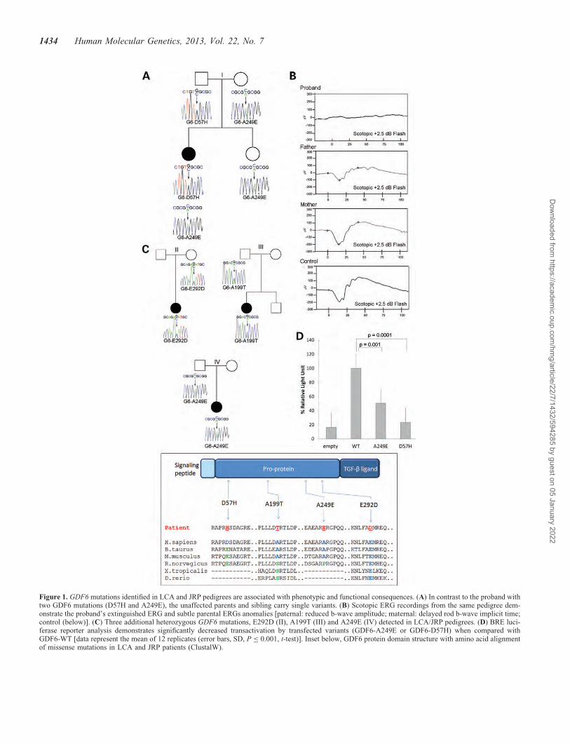
Figure 1. GDF6 mutations identified in LCA and JRP pedigrees are associated with phenotypic and functional consequences. (A) In contrast to the proband withtwo GDF6 mutations (D57H and A249E), the unaffected parents and sibling carry single variants. (B) Scotopic ERG recordings from the same pedigree dem-onstrate the proband’s extinguished ERG and subtle parental ERGs anomalies [paternal: reduced b-wave amplitude; maternal: delayed rod b-wave implicit time;control (below)]. (C) Three additional heterozygous GDF6 mutations, E292D (II), A199T (III) and A249E (IV) detected in LCA/JRP pedigrees. (D) BRE luci-ferase reporter analysis demonstrates significantly decreased transactivation by transfected variants (GDF6-A249E or GDF6-D57H) when compared withGDF6-WT [data represent the mean of 12 replicates (error bars, SD, P ≤ 0.001, t-test)]. Inset below, GDF6 protein domain structure with amino acid alignmentof missense mutations in LCA and JRP patients (ClustalW).
1434 Human Molecular Genetics, 2013, Vol. 22, No. 7
Dow
nloaded from https://academ
ic.oup.com/hm
g/article/22/7/1432/594285 by guest on 05 January 2022

ERGs were recorded in adult heterozygous mutants (Gdf6+/2)and wild-type (Gdf6+/+) littermates (n ¼ 12). A subset ofGdf6+/2 mice (five of nine) exhibited abnormal ERGs withup to 66% decreases in the bipolar cell driven b-wave [purecone (saturated photopic) and mixed rod-cone (saturated sco-topic) (Supplementary Material, Fig. S2)] and 54% decreasesin the photoreceptor mediated a-wave amplitudes. The morepronounced reductions of the b-wave than a-wave amplitudesaccord with preferential inner retina changes observed in het-erozygous patients [parents of LCA probands (Fig. 1B) andothers, described below] (32). Reduced photopic flickerfusion (3–27%), another indicator of inner retinal dysfunction,was also observed in the same five Gdf6+/2 mice. Such find-ings are compatible with a role for Gdf6 in murine and humanretinal function.
In view of the biochemical evidence that human GDF6 var-iants were functionally significant (Figs 1 and 2, SupplementaryMaterial, Fig. S1), and the typically autosomal-recessive natureof LCA and JRP, using exomic next-generation sequencing, wetested the hypothesis that a second TGF-b variant was present inprobands with a single GDF6 mutation. However, due toextreme GC-content, exome sequencing yielded incomplete
coverage across the open-reading frames of BMP ligands(data not shown), preventing identification of the known hetero-zygous mutations and precluding testing of our hypothesis. Inparallel, to determine whether mutation of other TGF-bmembers alters retinal function consistent with a contributionto retinal dystrophies, we examined a pedigree with a well-characterized mutation (GDF3-R266C) in a paralog with signifi-cant roles in retinal development. Notably, GDF3 mutationresults in near identical ocular and skeletal phenotypes toGDF6 (31,34). The hypomorph studied increases the numberof highly conserved cysteine residues in the mature ligand(Supplementary Material, Fig. S3A), and compared with wild-type GDF3 reduces luciferase activation by �50% (34).Each GDF3-R266C heterozygous carrier exhibited abnormalERGs with decreased scotopic b-wave amplitudes, and variablephenotypic severity that is characteristic of human BMP-induced ocular and skeletal phenotypes (29,37,38). Notably,one individual had nearly extinguished scotopic and photopicwaveforms with amplitudes ,10% of normal (SupplementaryMaterial, Fig. S3B) findings consistent with other membersof the TGF-b superfamily contributing to retinal dystrophyphenotypes.
Collectively, the above data demonstrate an increasedprevalence of GDF6 variants in LCA/JRP cases with adisease role supported by the appreciably altered biochemicalfunction, and the ERG anomalies observed with heterozygousmutation of a close paralog. Intrigued by these observations,we sought to analyze the consequence of long-term loss ofGdf6 function on retinal and photoreceptor structure. Weturned to zebrafish, given the ease with which we couldstudy homozygous mutants and the availability of a strain con-taining a p.S55X stop codon mutation (gdf6as327/s327, hereaftercalled gdf6a2/2) (21). Viable homozygous mutants were gen-erated by twice outcrossing to strain AB, with genotypes con-firmed by sequencing (Fig. 3A). Examination of adultgdf6a2/2 mutants revealed microphthalmia, with eyesobscured by overgrown skin (Fig. 3A) and dissection revealingmisshapen irides (Fig. 3B). Histological analysis of gdf6a2/2
mutants at 2 weeks of age demonstrated profound alterationsto the morphology of individual photoreceptor subtypes[Red/green cones (Fig. 3C and D) and UV cones (data notshown)]. Studies at later timepoints revealed loss of normalretinal lamination, as evident from actin and nuclear stains(Fig. 3E and F), together with appreciable disorganization ofMuller glia cells, as assessed by glial fibrillary acidic protein(GFAP) immunohistochemistry (Fig. 3E–H). The increasedGFAP abundance in gdf6a2/2 adult eyes (Fig. 3F) is consist-ent with the gliosis common to neurodegenerative diseases andanimal models thereof (39). Finally, in gdf6a2/2 adults, conephotoreceptors were consistently dysmorphic and reduced inabundance (Fig. 3I–L).
Since photoreceptor death is a key feature of retinal dystro-phies, the level of apoptosis was determined during mouse andzebrafish embryogenesis. At E18, a stage at which the murineretina is divisible into neuroblastic and ganglion cell layers,TUNEL-positive cells were observed in both (Fig. 4A–F)with increased levels of apoptotic signal observed inGdf62/2 and Gdf6+/2 mice compared with wild-type [meanTUNEL-positive cells/section: Gdf62/2 30.4; Gdf6+/2 12.9;Gdf6+/+ 8.3; P ¼ 0.016 and P , 0.0001, Student’s t-test
Figure 2. Evidence of altered biochemical function for heterozygous GDF6variants. (A) Western blot analysis demonstrated increased levels of E292Dand A199T mature ligand compared with wild-type in the cell media. (B)Luciferase reporter analysis revealed decreased activation of the 2xBRE re-porter for both variants compared with wild-type (error bars, SD; P ≤0.0005, t-test). Alpha tubulin and secreted alkaline phosphatase (SEAP) repre-sent controls for cytosolic and secreted proteins, respectively.
Human Molecular Genetics, 2013, Vol. 22, No. 7 1435
Dow
nloaded from https://academ
ic.oup.com/hm
g/article/22/7/1432/594285 by guest on 05 January 2022

(Fig. 4G)]. Substantially increased apoptosis was observed inthe retinas of mutant fish compared with wild-type at 28 hpf[mean a-active caspase-3 signals/eye: gdf62/2 76.4, WT3.34 (P , 0.0001, ANOVA)] (Fig. 4H and K). Comparablefindings were observed using acridine orange to observe apop-totic cells (40). Intrigued by observing increased embryonicretinal apoptosis in murine and zebrafish models of an LCAassociated gene, we next evaluated in gdf62/2 mutants,whether this effect could be ameliorated with a novel anti-apoptotic compound, P7C3. This aminopropyl carbazole isreported to be pro-neurogenic, protecting newborn hippocam-pal neurons from apoptosis (41). P7C3 treatment of gdf62/2
embryos resulted in significantly reduced levels of retinalapoptosis at 28 hpf [70 and 79% reductions at 0.01 and0.1 mM doses, respectively, P , 0.0001, ANOVA (Fig. 4I–M)]. Notably, this effect, evident in replicate experiments,was more pronounced at higher P7C3 concentrations(Fig. 4N). In light of P7C3′’s inhibition of gdf62/2 retinalapoptosis, we next assessed whether there was evidence offunctional rescue, utilizing two distinct assays. The first [visu-ally mediated background adaptation (VBA) (42)] is a neu-roendocrine response in which the detection of ambient lightby retinal ganglion cells results in melanosome contraction(43). Mutants exhibited increased pigmentation on a lightbackground that was unchanged with dimethyl sulfoxide(DMSO), but it partially recovered with P7C3 treatment[gdf6a2/2: DMSO 1 of 22 contracted melanophores; P7C311 of 22; gdf6a+/+ 25 of 25 (Fig. 5)]. The second assay [opto-motor response (OMR)] records the distance travelled in thedirection of motion of a perceived stimulus. This motionevoked assay requires substantially higher levels of retinalsensitivity and is dependent on both visual (photoreceptorand retinal interneuron) and musculo-skeletal function. In con-trast to the VBA result, OMR analysis did not reveal any im-provement in response by gdf6a2/2 mutants with P7C3treatment (Supplementary Material, Fig. S4).
DISCUSSION
Perturbed TGF-b signaling in retinal dystrophies
We report that mutations in GDF6 are associated withearly-onset retinal dystrophies and present comparable find-ings from both zebrafish and murine Gdf6 mutants. By demon-strating involvement in neuronal survival, these data extend
Figure 3. Adult homozygous gdf6a mutant zebrafish exhibit retinal degener-ation. (A–B) Adult gdf6a2/2 zebrafish have small eyes and dysmorphicpupils compared with wild-type [genotypes confirmed by sequencing(right)]. Immunohistochemistry at 2 weeks of age demonstrates that comparedwith wild-type, Red/Green cone photoreceptor morphology is profoundlyaltered with significant shortening of the cone inner segment (C, D). Inadult fish, there is an inflammatory response in the form of abundant GFAPimmunoreactivity, and lack of retinal polarization consistent with neuro-degeneration in adult gdf6a2/2 zebrafish (F, H) compared with wild-type(E, G). Both rod and cone photoreceptors are dysmorphic and decreased inadult gdf6a2/2 zebrafish (J, L) compared with wild-type (I, K). [ONL/INL,outer/inner nuclear layer; GCL, ganglion cell layer; GFAP, GFAP; R/G,red/green cones (scale bars 5 mm)].
1436 Human Molecular Genetics, 2013, Vol. 22, No. 7
Dow
nloaded from https://academ
ic.oup.com/hm
g/article/22/7/1432/594285 by guest on 05 January 2022

Figure 4. Gdf6 murine and zebrafish mutants demonstrate retinal apoptosis. (A–F) Montage of wild-type, Gdf6+/2 and Gdf62/2 retinal sections illustrating thelevel of TUNEL-positive (red) signals in the neuroblastic layer (NBL) and ganglion cell layer (GCL) of E18 murine embryos [Hoechst (blue) co-staining]. Incontrast to the low level of apoptosis in wild-type retina (A), higher levels are evident in heterozygous (B) and homozygous (C) mutant embryos [positive (D)and negative (E) controls]. (F) Low-magnification of entire Gdf62/2 retinal cross section to show localization of TUNEL-positive cells [boxed area shown athigher magnification in C]. (G) The progressive, and statistically significant, increase in the total number of TUNEL-positive signals with increasing Gdf6 nullallele dosage is depicted graphically (P , 0.05, t-test). (H–M) Immunofluorescence with a-active caspase-3 antibody (red) and Hoechst (blue) in 28 hpfembryos. gdf6a mutant retinas display increased apoptosis (H) when compared with non-mutant siblings (K). Treatment with P7C3 reduces the amount of apop-tosis in gdf6a mutant retinas (I and J) when compared with similarly treated non-mutant siblings (L and M). (N) Quantification of the number of anti-activecaspase-3-positive cells in the retina showed non-mutant siblings treated with dimethyl sulfoxide (DMSO), 0.01 mM P7C3 and 0.1 mM P7C3 displayed amean of 3.34 (n ¼ 44), 2.07 (n ¼ 58) and 1.32 (n ¼ 112) apoptosing cells. gdf6a mutants treated as above displayed a mean of 76.41 (n ¼ 34), 23.13 (n ¼16) and 16.09 (n ¼ 33) apoptosing cells, respectively. Error bars, SE; ∗P , 0.00001 (ANOVA). All embryos included in statistics were genotyped via sequencingas being gdf6a mutants or wild-type siblings.
Human Molecular Genetics, 2013, Vol. 22, No. 7 1437
Dow
nloaded from https://academ
ic.oup.com/hm
g/article/22/7/1432/594285 by guest on 05 January 2022
understanding of Gdf6 function from early retinal specifica-tion, involving for the first time TGF-b superfamilymembers in retinal dystrophies. Implication of GDF6, andthrough characteristic ERG anomalies, a second TGF-bmember (Supplementary Material, Fig. S3), will catalyzeevaluation of this gene family in molecularly unexplainedLCA/JRP, particularly for variants in those paralogs withmajor retinal developmental functions. Since these phenotypesaccord closely with Gdf6’s specification of the dorso-ventralretinal axis, evaluating retinal morphogenetic genes forcomparable disease contributions is merited, especially thosespecifying naso-temporal retinal identity [FGFs 3, 8, 18 and19 (37,44,45)].
Despite the numerically small proportion of cases, GDF6’sevolutionarily conserved role provides insight at severallevels. The first relates to variants with significantly reducedbiochemical function, where phenotypic effects are onlydetected in a proportion of heterozygotes on targeted testing(ERGs). This illustrates the eye’s value as a genetic diseasemodel, with electrophysiological testing revealing clinicallysilent retinal phenotypes, and causative alleles contributing
to the singleton LCA cases that have proved recalcitrant togenetic elucidation (Fig. 1). Observation that 50% decreasesin GDF6 signaling are tolerated while either compound hetero-zygous human (A249E and D57H), or homozygous murine orzebrafish null alleles induce severe phenotypes indicate that athreshold level of BMP signaling exists below which pheno-types manifest in all three species. This is supported by theprogressively higher levels of retinal apoptosis with increasingnull Gdf6 allele dosage (Fig. 4G). The therapeutic corollary ofobserving disease phenotypes at high but not low mutationalloads is that therapy only has to increase BMP signalingabove this threshold (and not to 100%) to be effective.
The second feature concerns the elevated level of hetero-zygous functional GDF6 variants in LCA/JRP cases (Figs 1and 2). These are individually insufficient to induce LCA/JRP since the heterozygous parents are overtly unaffected,as are heterozygous murine and zebrafish mutants. However,their increased prevalence, the heterodimerization (46) andretinal developmental functions of other BMP/GDFs(34,38,47–49) plus the ERG anomalies of GDF3 heterozy-gotes, all support the presence of additional disease-causingvariants. Notably, the phenotypes caused by Gdf3 and Gdf6mutation are very similar, with each inducing delayed choroid-al fissure closure or coloboma, retinal developmental anomal-ies and axial skeletal patterning defects in patients and ormodel organisms (22,31,34,47,50,51). The presented datademonstrate that mutation of other BMP/GDFs inducesretinal dysfunction, including extinguished ERGs that pheno-copy the effect of GDF6 mutation (Supplementary Material,Fig. S3), and will facilitate determining whether multi-allelicinheritance of BMP/GDF variants results in sporadicearly-onset retinal dystrophies. An intriguing finding in thisstudy was pleiotropy, with mutations (A249E) that cause post-axial polydactyly, Klippel-Feil or microphthalmia/anophthal-mia/coloboma (MAC), only associated with LCA/JRP. Thisis explicable by buffering of disease phenotypes by the numer-ous ligands and BMP pathway members, leading to context-specific epistasis (52). Coupled with stochastic events, and re-cently identified compensatory autocrine and/or paracrine pro-cesses (53), such mechanisms likely account for theparadoxical ocular phenotypes.
Inhibition of Gdf6-induced apoptosis
TGF-b ligands are multifunctional cytokines that provide pos-itional information to cells and control numerous aspects oftheir development. The profoundly altered cone morphologyinduced by gdf6 mutation (Fig. 3), accords with this role andthe patient LCA/JRP phenotype. In addition, these ligandshave important pro- and anti-apoptotic functions (54–61),with the consistent, pan-retinally increased apoptosis ofmurine and zebrafish homozygous mutants revealing a funda-mental and evolutionarily conserved requirement for Gdf6 inmaintaining retinal cellular populations. This finding accordswith the progressive apoptotic photoreceptor loss and increas-ing visual impairment characteristic of retinal dystrophies(62), and is evident in species with different proportions ofcone photoreceptors (murine 3%, zebrafish 30%). Subsequentefforts concentrated on evaluating whether this neuronal losswas tractable to treatment (41). To date, the applicability of
Figure 5. P7C3 treatment recovers visual background adaptation (VBA) activ-ity in gdf6a mutant embryos. (A–D) Dorsal images of gdf6a mutant (2/2)and non-mutant siblings (+/+) treated with a control dose of DMSO(A, C) or 0.01 mM P7C3 (B, D). Larvae are 7 dpf in all panels. The contri-bution of the presented phenotype in each panel is indicated. DMSO-and P7C3-treated gdf6a+/+ larvae have normal melanophore contraction(A, 100%, n ¼ 25, B, 100%, n ¼ 24) while DMSO-treated gdf6a2/2 larvaehave unresponsive VBA and widely distributed melanophores (C, 95%,n ¼ 21). Treatment of gdf6a2/2 larvae with P7C3 partially recovers VBAand melanophores appear partially contracted (D, 50%, n ¼ 22). (E) Graphof the proportion of embryos that present fully or partially responsive VBAunder the indicated conditions.
1438 Human Molecular Genetics, 2013, Vol. 22, No. 7
Dow
nloaded from https://academ
ic.oup.com/hm
g/article/22/7/1432/594285 by guest on 05 January 2022

anti-apoptotic agents has frequently been restricted by theirbroad mechanisms of action, which include inhibition of fun-damental physiological processes. The significantly reduced acti-vated caspase-3 signaling in P7C3 treated gdf62/2 mutantsprovides immunohistochemical evidence of rescue for a zebrafishLCA model. The paradoxical visual function results are derivedfrom mechanistically distinct assays with log unit differences insensitivity and involvement of different retinal cell types.Coupled with the known lenticular and skeletal phenotypes ofgdf62/2 mutants (22,31), such factors may affect OMR butleave VBA unchanged, potentially explaining the differencesbetween the datasets. In this context, P7C3’s inhibition of neuron-al apoptosis without deleterious effects in different tissues of evo-lutionary disparate species [murine CNS (41) and zebrafishretina], merits further investigation, especially as an effectiveanti-apoptotic agent (63) may synergize with other therapeuticapproaches for enhancing neuronal survival (29,30,64).
Clinical and developmental implications
Of the key conclusions that can be drawn from GDF6’s con-tribution to early-onset retinal dystrophies, the first relates toan invariant feature of BMP signaling from Drosophila tohumans—exquisitely precise spatial and temporal regulationat multiple levels [including heterodimerization with otherBMPs, antagonists and agonists, receptor and co-receptor ex-pression, and Smad phosphorylation (65–69)]. Reinforcementof one ligand’s signaling by another is a common paradigm,as exemplified by Gdf6a’s induction of bmp2b and bmp7a tran-scription (70) leading to enriched BMP mRNA levels in specificregions of the developing embryo. Accordingly, it is improbablethat involvement of TGF-b members in retinal dystrophies isconfined to GDF6, as our GDF3 data demonstrate. A secondconclusion relates to the human ocular phenotypes induced byBMP mutation that have to date been restricted to alterationsin ocular size (microphthalmia or anophthalmia) and tissue mor-phogenesis (coloboma) (31,33,34,50,71–73). This study sig-nificantly extends these roles, suggests that the MACspectrum overlaps with retinal dystrophies and provides asimple means of testing this through ERG recordings of micro-phthalmia patients. In parallel, it implies a more complex inter-play than hitherto appreciated between retinal and globedevelopment, with broader implications for common disordersof ocular size (myopia and hyperopia). A third feature relatesto this study’s implication of a growth factor in LCA, which con-trasts with the photoreceptor-specific nature of most LCA/JRP-causing genes. Finally, one consequence of seemingly dis-parate clinical disorders being caused by alterations to the samedevelopmental pathway is that therapeutic approaches maybenefit a range of disorders. In this context, the finding thatBMP-induced apoptosis can be inhibited with P7C3 may leadto new approaches for treating disorders that still destine thevast majority of patients to visual impairment or blindness.
MATERIALS AND METHODS
Patient analysis
Two hundred and seventy-nine DNA samples from LCA orJRP patients, previously screened for mutations in the
known causative genes, were polymerase chain reaction(PCR) amplified for GDF6 and the products sequenced onan ABI Prism 3100 capillary sequencer (Applied Biosystems)(as previously described) (31). Chromatograms were analyzedusing Sequencher (vs 4.5, GeneCodes) with amino acid se-quence alignments performed using ClustalW (http://www.genome.jp/tools/clustalw/). The prevalence of mutations inwestern Canadian population control samples was determinedby restriction enzyme analysis [BsrBI (A249E), HaeII (D57H),HgaI (A199T), NlaIII (E292D) (New England Biolabs)] (n ¼462 DNA samples) and direct sequencing of both exons (n ¼288 samples). Collection and analysis of DNA samples wasapproved by the University of Alberta Hospital Health Re-search Ethics Board, with informed consent provided by allparticipants.
Western blot and luciferase assaysWild-type GDF6 transcript was generated by PCR of genomicDNA, cloned into pCR4-TOPO (Invitrogen, ON, Canada) andmutations introduced by site-directed mutagenesis. Sub-cloning into pcDNA3.2/V5-DEST (Invitrogen) or CS2+ wasundertaken for western blot and luciferase analyses, respect-ively (31). Western blots were performed on lysates collected48 h after transiently transfecting wild-type GDF6 or the indi-vidual variants into COS7 cells using FuGENE (Roche) aspreviously described (31,34,74). Extracted proteins were sepa-rated on a 15% sodium dodecyl sulfate polyacrylamide gelelectrophoresis gel, transferred to nitrocellulose membranes(BioRad), incubated with primary antibody [anti-V5(1:10 000), secreted alkaline phosphatase (1:5000) ora-tubulin (1:10 000), (AbCam)] and subsequently with anti-mouse or anti-rabbit IgG-HRP [1:5000 (Jackson Laborator-ies)]. After chemoluminescent antibody detection, imageswere analyzed using ImageJ and for quantification, proteinamounts were normalized to alpha-tubulin and secretedalkaline phosphatase (SEAP) levels in the cytosolic andmedia fractions, respectively. For luciferase assays, U2OScells cultured in 24-well plates until 80% confluency weretransfected using FuGENE with either wild-type or variantGDF6, the luciferase reporter gene under the control of a2xBRE (BMP-responsive element) and a pRL-SV40 encodingRenilla luciferase. Cell lysates were collected 48 h post-transfection and luciferase activity quantified [Dual luciferasekit (Promega)] in quadruplicate assays performed on three sep-arate occasions.
Murine analysisGdf6 mutant mice [Gdf6tm1Lex (MGI: 3604391)], previouslygenotyped by sequencing ear-notch derived DNA (31), under-went full-field ERGs at 14–23 months of age with the EspionE2 system (Diagnosys LLC). Briefly, mice were dark-adaptedovernight and responses to a white flash (65008K xenon)were recorded at incremental intensities (19 steps, range:25.22–2.86 log candela s/m2). After photopic adaptation(30 candela/m2), photopic intensity responses were recorded(11 steps, range: 21.22–2.86 log candela s/m2), followedby photopic flicker ERGs, all as previously described (75).For TUNEL analysis, timed pregnancies were used to generateE18 embryos that were collected from euthanized pregnantfemales, with dissected eyes preserved in PFA, paraffin
Human Molecular Genetics, 2013, Vol. 22, No. 7 1439
Dow
nloaded from https://academ
ic.oup.com/hm
g/article/22/7/1432/594285 by guest on 05 January 2022

embedded and sectioned. Apoptosis was detected by terminaldeoxytransferase-mediated dUTP nick end-labeling [TUNELdetection kit (Roche); counterstaining Hoechst 33258 (Invitro-gen)], with standard positive (DNase incubation) and negativecontrols (omission of TdT from reaction buffer). Six retinalsections (≥100 mm apart) were imaged from each eye withconfocal microscopy (Carl Zeiss) to quantify the TUNEL-positive cells per section (statistical analysis, Student’s ttest). All murine procedures and husbandry were approvedby the University of Alberta Animal Policy and Welfare Com-mittee.
Zebrafish analysisAdult gdf6as327/s327 and gdf6a+/+ zebrafish and enucleatedeyes were photographed under a Leica stereomicroscope,with genotyping performed by direct sequencing. Enucleatedeyes were fixed and cryo-preserved using standard methodsprior to immunohistochemistry (full details will be availableon request). Briefly, 10 mm histological sections wereblocked with goat serum, labeled with rhodamine–phalloidinor primary antibodies [to either red/green double cones(zpr1) or GFAP (zfr1)] and fluorescently conjugated secondaryantibodies (Invitrogen), and counterstained (TO-PRO-3 orpropidium iodide). Images were collected using confocal mi-croscopy and pseudocoloured (Zeiss LSM 700 on Axio Obser-ver.Z1). To investigate the effect of P7C3, embryos weretreated from 5 to 28 hpf with either 0.01 mM P7C3, 0.1 mM
P7C3 or DMSO. The level of apoptosis at 28 hpf in dissectedeyes was determined using anti-active caspase-3 antibodystaining (BD Pharmingen) and confocal microscopy (asabove). Since gdf6a mutants do not have an observable pheno-type at 28 hpf, genotypes were determined by PCR and se-quencing. ANOVA analysis was performed with significancevalues at P-value of ,0.00001. For the visual backgroundmediated adaptation assays (VBA), embryos were treatedwith P7C3 from 5 to 48 hpf (the period during which apoptosishas been detected in gdf6a2/2 mutants), transferred to embryomedia until they reached 7 dpf, at which point they were indi-vidually scored for pigmentation (76). For OMR recordings,the distance travelled by individual larvae in response to avisual stimulus was measured (77) with gdf6a2/2 mutantsand non-mutant siblings treated with 0.01 mM P7C3 asdescribed above.
SUPPLEMENTARY MATERIAL
Supplementary Material is available at HMG online.
ACKNOWLEDGEMENTS
We are very grateful to the patients and families whose partici-pation made this research possible. We thank Ming Ye, ErinStrachan, Hao Wang, Aleah McCorry, Sharee Kuny andFrauke Coppieters for technical assistance; Aleah McCorryand Erin Wilson for zebrafish husbandry, Drs Michael Under-hill and Laszlo Patthy for helpful advice and Drs Rod Bremnerand Tsutomu Kume for critically reviewing the manuscript.
Conflict of Interest statement. None declared.
FUNDING
This work was supported by grants from Foundation FightingBlindness Canada (A.J.W. and O.J.L.), Canadian Institutes ofHealth Research (O.J.L.), National Sciences and EngineeringResearch Council of Canada (W.T.A.), Funds for ScientificResearch (E.D.B.), as well as the Canada Research ChairProgram (A.J.W. and O.J.L.).
REFERENCES
1. Arnold, S.J. and Robertson, E.J. (2009) Making a commitment: celllineage allocation and axis patterning in the early mouse embryo. Nat.Rev. Mol. Cell Biol., 10, 91–103.
2. Lie, D.C., Colamarino, S.A., Song, H.J., Desire, L., Mira, H., Consiglio,A., Lein, E.S., Jessberger, S., Lansford, H., Dearie, A.R. et al. (2005)Wnt signalling regulates adult hippocampal neurogenesis. Nature, 437,1370–1375.
3. Mathura, J.R. Jr., Jafari, N., Chang, J.T., Hackett, S.F., Wahlin, K.J.,Della, N.G., Okamoto, N., Zack, D.J. and Campochiaro, P.A. (2000) Bonemorphogenetic proteins-2 and -4: negative growth regulators in adultretinal pigmented epithelium. Invest. Ophthalmol. Vis. Sci., 41, 592–600.
4. Chang, H., Brown, C.W. and Matzuk, M.M. (2002) Genetic analysis ofthe mammalian transforming growth factor-beta superfamily. Endocr.Rev., 23, 787–823.
5. Massague, J., Blain, S.W. and Lo, R.S. (2000) TGFbeta signaling ingrowth control, cancer, and heritable disorders. Cell, 103, 295–309.
6. Massague, J. and Chen, Y.G. (2000) Controlling TGF-beta signaling.Genes Dev., 14, 627–644.
7. Hogan, B.L. (1996) Bone morphogenetic proteins: multifunctionalregulators of vertebrate development. Genes Dev., 10, 1580–1594.
8. Chang, C. and Hemmati-Brivanlou, A. (1999) Xenopus GDF6, anew antagonist of noggin and a partner of BMPs. Development, 126,3347–3357.
9. Israel, D.I., Nove, J., Kerns, K.M., Moutsatsos, I.K. and Kaufman, R.J.(1992) Expression and characterization of bone morphogenetic protein-2in Chinese hamster ovary cells. Growth Factors, 7, 139–150.
10. Aono, A., Hazama, M., Notoya, K., Taketomi, S., Yamasaki, H., Tsukuda,R., Sasaki, S. and Fujisawa, Y. (1995) Potent ectopic bone-inducingactivity of bone morphogenetic protein-4/7 heterodimer. Biochem.Biophys. Res. Commun., 210, 670–677.
11. Miyazono, K., Maeda, S. and Imamura, T. (2005) BMP receptorsignaling: transcriptional targets, regulation of signals, and signalingcross-talk. Cytokine Growth Factor Rev., 16, 251–263.
12. Zhang, Y.E. (2009) Non-Smad pathways in TGF-beta signaling. Cell Res.,19, 128–139.
13. Urist, M.R. (1965) Bone: formation by autoinduction. Science, 150,893–899.
14. Hogan, B.L. (1996) Bone morphogenetic proteins in development.Curr. Opin. Genet. Dev., 6, 432–438.
15. Graff, J.M. (1997) Embryonic patterning: to BMP or not to BMP, that isthe question. Cell, 89, 171–174.
16. Yang, X.J. (2004) Roles of cell-extrinsic growth factors in vertebrate eyepattern formation and retinogenesis. Semin. Cell Dev. Biol., 15, 91–103.
17. Koshiba-Takeuchi, K., Takeuchi, J.K., Matsumoto, K., Momose, T., Uno,K., Hoepker, V., Ogura, K., Takahashi, N., Nakamura, H., Yasuda, K.et al. (2000) Tbx5 and the retinotectum projection. Science, 287,134–137.
18. Sasagawa, S., Takabatake, T., Takabatake, Y., Muramatsu, T. andTakeshima, K. (2002) Axes establishment during eye morphogenesis inXenopus by coordinate and antagonistic actions of BMP4, Shh, and RA.Genesis, 33, 86–96.
19. Lupo, G., Liu, Y., Qiu, R., Chandraratna, R.A., Barsacchi, G., He, R.Q.and Harris, W.A. (2005) Dorsoventral patterning of the Xenopus eye: acollaboration of Retinoid, Hedgehog and FGF receptor signaling.Development, 132, 1737–1748.
20. Rissi, M., Wittbrodt, J., Delot, E., Naegeli, M. and Rosa, F.M. (1995)Zebrafish Radar: a new member of the TGF-beta superfamily definesdorsal regions of the neural plate and the embryonic retina. Mech. Dev.,49, 223–234.
1440 Human Molecular Genetics, 2013, Vol. 22, No. 7
Dow
nloaded from https://academ
ic.oup.com/hm
g/article/22/7/1432/594285 by guest on 05 January 2022

21. Gosse, N.J. and Baier, H. (2009) An essential role for Radar (Gdf6a) ininducing dorsal fate in the zebrafish retina. Proc. Natl Acad. Sci. USA,106, 2236–2241.
22. French, C.R., Erickson, T., French, D.V., Pilgrim, D.B. and Waskiewicz,A.J. (2009) Gdf6a is required for the initiation of dorsal-ventral retinalpatterning and lens development. Dev. Biol., 333, 37–47.
23. den Hollander, A.I., Roepman, R., Koenekoop, R.K. and Cremers, F.P.(2008) Leber congenital amaurosis: genes, proteins and diseasemechanisms. Prog. Retin. Eye Res., 27, 391–419.
24. Sohocki, M.M., Sullivan, L.S., Mintz-Hittner, H.A., Birch, D.,Heckenlively, J.R., Freund, C.L., McInnes, R.R. and Daiger, S.P. (1998)A range of clinical phenotypes associated with mutations in CRX, aphotoreceptor transcription-factor gene. Am. J. Hum. Genet., 63,1307–1315.
25. Morimura, H., Fishman, G.A., Grover, S.A., Fulton, A.B., Berson, E.L.and Dryja, T.P. (1998) Mutations in the RPE65 gene in patients withautosomal recessive retinitis pigmentosa or leber congenital amaurosis.Proc. Natl Acad. Sci. USA, 95, 3088–3093.
26. den Hollander, A.I., Johnson, K., de Kok, Y.J., Klebes, A., Brunner, H.G.,Knust, E. and Cremers, F.P. (2001) CRB1 has a cytoplasmic domain thatis functionally conserved between human and Drosophila. Hum. Mol.
Genet., 10, 2767–2773.27. den Hollander, A.I., Koenekoop, R.K., Yzer, S., Lopez, I., Arends, M.L.,
Voesenek, K.E., Zonneveld, M.N., Strom, T.M., Meitinger, T., Brunner,H.G. et al. (2006) Mutations in the CEP290 (NPHP6) gene are a frequentcause of Leber congenital amaurosis. Am. J. Hum. Genet., 79, 556–561.
28. Perrault, I., Hanein, S., Gerber, S., Barbet, F., Ducroq, D., Dollfus, H.,Hamel, C., Dufier, J.L., Munnich, A., Kaplan, J. et al. (2004) Retinaldehydrogenase 12 (RDH12) mutations in leber congenital amaurosis. Am.
J. Hum. Genet., 75, 639–646.29. Cideciyan, A.V., Hauswirth, W.W., Aleman, T.S., Kaushal, S., Schwartz,
S.B., Boye, S.L., Windsor, E.A., Conlon, T.J., Sumaroka, A., Pang, J.J.et al. (2009) Human RPE65 gene therapy for Leber congenital amaurosis:persistence of early visual improvements and safety at 1 year. Hum. Gene
Ther., 20, 999–1004.
30. Ashtari, M., Cyckowski, L.L., Monroe, J.F., Marshall, K.A., Chung, D.C.,Auricchio, A., Simonelli, F., Leroy, B.P., Maguire, A.M., Shindler, K.S.et al. (2011) The human visual cortex responds to gene therapy-mediatedrecovery of retinal function. J. Clin. Invest., 121, 2160–2168.
31. Asai-Coakwell, M., French, C.R., Ye, M., Garcha, K., Bigot, K., Perera,A.G., Staehling-Hampton, K., Mema, S.C., Chanda, B., Mushegian, A.et al. (2009) Incomplete penetrance and phenotypic variabilitycharacterize Gdf6-attributable oculo-skeletal phenotypes. Hum. Mol.
Genet., 18, 1110–1121.32. Galvin, J.A., Fishman, G.A., Stone, E.M. and Koenekoop, R.K. (2005)
Clinical phenotypes in carriers of Leber congenital amaurosis mutations.Ophthalmology, 112, 349–356.
33. Gonzalez-Rodriguez, J., Pelcastre, E.L., Tovilla-Canales, J.L.,Garcia-Ortiz, J.E., Amato-Almanza, M., Villanueva-Mendoza, C.,Espinosa-Mattar, Z. and Zenteno, J.C. (2010) Mutational screening ofCHX10, GDF6, OTX2, RAX and SOX2 genes in 50 unrelatedmicrophthalmia-anophthalmia-coloboma (MAC) spectrum cases. Br J
Ophthalmol, 94, 1100–1104.34. Ye, M., Berry-Wynne, K.M., Asai-Coakwell, M., Sundaresan, P., Footz,
T., French, C.R., Abitbol, M., Fleisch, V.C., Corbett, N., Allison, W.T.et al. (2010) Mutation of the bone morphogenetic protein GDF3 causesocular and skeletal anomalies. Hum. Mol. Genet., 19, 287–298.
35. Tassabehji, M., Fang, Z.M., Hilton, E.N., McGaughran, J., Zhao, Z., deBock, C.E., Howard, E., Malass, M., Donnai, D., Diwan, A. et al. (2008)Mutations in GDF6 are associated with vertebral segmentation defects inKlippel-Feil syndrome. Hum. Mutat., 29, 1017–1027.
36. Zilberberg, L., ten Dijke, P., Sakai, L.Y. and Rifkin, D.B. (2007) A rapidand sensitive bioassay to measure bone morphogenetic protein activity.BMC Cell Biol., 8, 41.
37. Picker, A., Cavodeassi, F., Machate, A., Bernauer, S., Hans, S., Abe, G.,Kawakami, K., Wilson, S.W. and Brand, M. (2009) Dynamic coupling ofpattern formation and morphogenesis in the developing vertebrate retina.PLoS Biol., 7, e1000214.
38. Furuta, Y. and Hogan, B.L. (1998) BMP4 is essential for lens induction inthe mouse embryo. Genes Dev., 12, 3764–3775.
39. Middeldorp, J. and Hol, E.M. (2011) GFAP in health and disease. Prog.
Neurobiol., 93, 421–443.
40. Uchimoto, T., Nohara, H., Kamehara, R., Iwamura, M., Watanabe, N. andKobayashi, Y. (1999) Mechanism of apoptosis induced by alysosomotropic agent, L-leucyl-L-leucine methyl ester. Apoptosis, 4,357–362.
41. Pieper, A.A., Xie, S., Capota, E., Estill, S.J., Zhong, J., Long, J.M.,Becker, G.L., Huntington, P., Goldman, S.E., Shen, C.H. et al. (2010)Discovery of a proneurogenic, neuroprotective chemical. Cell, 142,39–51.
42. Balm, P.H. and Groneveld, D. (1998) The melanin-concentrating hormonesystem in fish. Ann. N. Y. Acad. Sci., 839, 205–209.
43. Muto, A., Orger, M.B., Wehman, A.M., Smear, M.C., Kay, J.N.,Page-McCaw, P.S., Gahtan, E., Xiao, T., Nevin, L.M., Gosse, N.J. et al.
(2005) Forward genetic analysis of visual behavior in zebrafish. PLoSGenet., 1, e66.
44. Martinez-Morales, J.R., Del Bene, F., Nica, G., Hammerschmidt, M.,Bovolenta, P. and Wittbrodt, J. (2005) Differentiation of the vertebrateretina is coordinated by an FGF signaling center. Dev. Cell, 8, 565–574.
45. Nakayama, Y., Miyake, A., Nakagawa, Y., Mido, T., Yoshikawa, M.,Konishi, M. and Itoh, N. (2008) Fgf19 is required for zebrafish lens andretina development. Dev. Biol., 313, 752–766.
46. Israel, D.I., Nove, J., Kerns, K.M., Kaufman, R.J., Rosen, V., Cox, K.A.and Wozney, J.M. (1996) Heterodimeric bone morphogenetic proteinsshow enhanced activity in vitro and in vivo. Growth Factors, 13,291–300.
47. Kim, J., Wu, H.H., Lander, A.D., Lyons, K.M., Matzuk, M.M. and Calof,A.L. (2005) GDF11 controls the timing of progenitor cell competence indeveloping retina. Science, 308, 1927–1930.
48. Morcillo, J., Martinez-Morales, J.R., Trousse, F., Fermin, Y., Sowden,J.C. and Bovolenta, P. (2006) Proper patterning of the optic fissurerequires the sequential activity of BMP7 and SHH. Development, 133,3179–3190.
49. Sakuta, H., Takahashi, H., Shintani, T., Etani, K., Aoshima, A. and Noda,M. (2006) Role of bone morphogenic protein 2 in retinal patterning andretinotectal projection. J. Neurosci., 26, 10868–10878.
50. Asai-Coakwell, M., French, C.R., Berry, K.M., Ye, M., Koss, R.,Somerville, M., Mueller, R., van Heyningen, V., Waskiewicz, A.J. andLehmann, O.J. (2007) GDF6, a novel locus for a spectrum of oculardevelopmental anomalies. Am. J. Hum. Genet., 80, 306–315.
51. McPherron, A.C., Lawler, A.M. and Lee, S.J. (1999) Regulation ofanterior/posterior patterning of the axial skeleton by growth/differentiation factor 11. Nat. Genet., 22, 260–264.
52. Ramel, M.C. and Hill, C.S. (2012) Spatial regulation of BMP activity.FEBS Lett, 586, 1929–1941.
53. Loeys, B., Lindsay, M., Schepers, D., Bolar, N., Doyle, J., Gallo, E.,Fert-Bober, J., Kempers, M., Fishman, E., Chen, Y. et al. (2012)Loss-of-function mutations in TGFB2 cause Loeys–Dietz syndrome:towards solving the TGFb paradox in aortic aneurysmal disease.Presented at The American Society of Human Genetics, 2012,San Francisco. Abstract #80.
54. Beier, M., Franke, A., Paunel-Gorgulu, A.N., Scheerer, N. and Dunker, N.(2006) Transforming growth factor beta mediates apoptosis in theganglion cell layer during all programmed cell death periods of thedeveloping murine retina. Neurosci. Res., 56, 193–203.
55. Franke, A.G., Gubbe, C., Beier, M. and Duenker, N. (2006) Transforminggrowth factor-beta and bone morphogenetic proteins: cooperative playersin chick and murine programmed retinal cell death. J. Comp. Neurol., 495,263–278.
56. Trousse, F., Esteve, P. and Bovolenta, P. (2001) Bmp4 mediates apoptoticcell death in the developing chick eye. J. Neurosci., 21, 1292–1301.
57. Frank, D.B., Abtahi, A., Yamaguchi, D.J., Manning, S., Shyr, Y., Pozzi,A., Baldwin, H.S., Johnson, J.E. and de Caestecker, M.P. (2005) Bonemorphogenetic protein 4 promotes pulmonary vascular remodeling inhypoxic pulmonary hypertension. Circ. Res., 97, 496–504.
58. Heger, J., Schiegnitz, E., von Waldthausen, D., Anwar, M.M., Piper, H.M.and Euler, G. (2010) Growth differentiation factor 15 acts anti-apoptoticand pro-hypertrophic in adult cardiomyocytes. J. Cell Physiol., 224,120–126.
59. Kiyono, M. and Shibuya, M. (2003) Bone morphogenetic protein 4mediates apoptosis of capillary endothelial cells during rat pupillarymembrane regression. Mol. Cell Biol., 23, 4627–4636.
60. Ehata, S., Hanyu, A., Hayashi, M., Aburatani, H., Kato, Y., Fujime, M.,Saitoh, M., Miyazawa, K., Imamura, T. and Miyazono, K. (2007)Transforming growth factor-beta promotes survival of mammary
Human Molecular Genetics, 2013, Vol. 22, No. 7 1441
Dow
nloaded from https://academ
ic.oup.com/hm
g/article/22/7/1432/594285 by guest on 05 January 2022

carcinoma cells through induction of antiapoptotic transcription factorDEC1. Cancer Res., 67, 9694–9703.
61. Yokouchi, Y., Sakiyama, J., Kameda, T., Iba, H., Suzuki, A., Ueno, N.and Kuroiwa, A. (1996) BMP-2/-4 mediate programmed cell death inchicken limb buds. Development, 122, 3725–3734.
62. Smith, A.J., Bainbridge, J.W. and Ali, R.R. (2009) Prospects for retinal
gene replacement therapy. Trends Genet., 25, 156–165.
63. MacMillan, K.S., Naidoo, J., Liang, J., Melito, L., Williams, N.S.,Morlock, L., Huntington, P.J., Estill, S.J., Longgood, J., Becker, G.L.
et al. (2011) Development of proneurogenic, neuroprotective smallmolecules. J. Am. Chem. Soc., 133, 1428–1437.
64. Mihelec, M., Pearson, R.A., Robbie, S.J., Buch, P.K., Azam, S.A.,Bainbridge, J.W., Smith, A.J. and Ali, R.R. (2011) Long-term
preservation of cones and improvement in visual function following genetherapy in a mouse model of leber congenital amaurosis caused byguanylate cyclase-1 deficiency. Hum. Gene Ther., 22, 1179–1190.
65. Shimmi, O., Umulis, D., Othmer, H. and O’Connor, M.B. (2005)
Facilitated transport of a Dpp/Scw heterodimer by Sog/Tsg leads to robustpatterning of the Drosophila blastoderm embryo. Cell, 120, 873–886.
66. Valera, E., Isaacs, M.J., Kawakami, Y., Izpisua Belmonte, J.C. and Choe,S. (2010) BMP-2/6 heterodimer is more effective than BMP-2 or BMP-6
homodimers as inductor of differentiation of human embryonic stem cells.PLoS One, 5, e11167.
67. Balemans, W. and Van Hul, W. (2002) Extracellular regulation of BMPsignaling in vertebrates: a cocktail of modulators. Dev. Biol., 250,
231–250.
68. Ehrlich, M., Horbelt, D., Marom, B., Knaus, P. and Henis, Y.I. (2011)Homomeric and heteromeric complexes among TGF-beta and BMP
receptors and their roles in signaling. Cell Signal, 23, 1424–1432.
69. Feng, X.H. and Derynck, R. (2005) Specificity and versatility in TGF-betasignaling through Smads. Annu. Rev. Cell Dev. Biol., 21, 659–693.
70. Sidi, S., Goutel, C., Peyrieras, N. and Rosa, F.M. (2003) Maternalinduction of ventral fate by zebrafish radar. Proc. Natl Acad. Sci. USA,100, 3315–3320.
71. Bakrania, P., Efthymiou, M., Klein, J.C., Salt, A., Bunyan, D.J., Wyatt,A., Ponting, C.P., Martin, A., Williams, S., Lindley, V. et al. (2008)Mutations in BMP4 cause eye, brain, and digit developmental anomalies:overlap between the BMP4 and hedgehog signaling pathways. Am. J.Hum. Genet., 82, 304–319.
72. Reis, L.M., Tyler, R.C., Schilter, K.F., Abdul-Rahman, O., Innis, J.W.,Kozel, B.A., Schneider, A.S., Bardakjian, T.M., Lose, E.J., Martin, D.M.et al. (2011) BMP4 loss-of-function mutations in developmental eyedisorders including SHORT syndrome. Hum. Genet., 130, 495–504.
73. Zhang, X., Li, S., Xiao, X., Jia, X., Wang, P., Shen, H., Guo, X. andZhang, Q. (2009) Mutational screening of 10 genes in Chinese patientswith microphthalmia and/or coloboma. Mol. Vis., 15, 2911–2918.
74. Ploger, F., Seemann, P., Schmidt-von Kegler, M., Lehmann, K., Seidel, J.,Kjaer, K.W., Pohl, J. and Mundlos, S. (2008) Brachydactyly type A2associated with a defect in proGDF5 processing. Hum. Mol. Genet., 17,1222–1233.
75. Gilmour, G.S., Gaillard, F., Watson, J., Kuny, S., Mema, S.C., Bonfield,S., Stell, W.K. and Sauve, Y. (2008) The electroretinogram (ERG) of adiurnal cone-rich laboratory rodent, the Nile grass rat (Arvicanthisniloticus). Vis. Res., 48, 2723–2731.
76. Fleisch, V.C. and Neuhauss, S.C. (2006) Visual behavior in zebrafish.Zebrafish, 3, 191–201.
77. Orger, M.B., Gahtan, E., Muto, A., Page-McCaw, P., Smear, M.C. andBaier, H. (2004) Behavioral screening assays in zebrafish. Methods CellBiol., 77, 53–68.
1442 Human Molecular Genetics, 2013, Vol. 22, No. 7
Dow
nloaded from https://academ
ic.oup.com/hm
g/article/22/7/1432/594285 by guest on 05 January 2022


![Statins in the Treatment of Heart Failure: Failed Concept? · per lo Studio della Sopravvivenza ... [survival]) plot and time dependent covariate test • Kaplan Meier Survival curves](https://static.fdocuments.net/doc/165x107/5c6a7c7709d3f20f7f8cb74a/statins-in-the-treatment-of-heart-failure-failed-concept-per-lo-studio-della.jpg)


