Contrast enhanced ultrasound (CEUS) in blunt abdominal trauma ...
7
RESEARCH Open Access Contrast enhanced ultrasound (CEUS) in blunt abdominal trauma Lucio Cagini 1 , Sabrina Gravante 2 , Corrado Maria Malaspina 2 , Elviro Cesarano 3 , Melchiorre Giganti 4 , Alberto Rebonato 2 , Paolo Fonio 5 , Michele Scialpi 2* Abstract In the assessment of polytrauma patient, an accurate diagnostic study protocol with high sensitivity and specificity is necessary. Computed Tomography (CT) is the standard reference in the emergency for evaluating the patients with abdominal trauma. Ultrasonography (US) has a high sensitivity in detecting free fluid in the peritoneum, but it does not show as much sensitivity for traumatic parenchymal lesions. The use of Contrast-Enhanced Ultrasound (CEUS) improves the accuracy of the method in the diagnosis and assessment of the extent of parenchymal lesions. Although the CEUS is not feasible as a method of first level in the diagnosis and management of the polytrauma patient, it can be used in the follow-up of traumatic injuries of abdominal parenchymal organs (liver, spleen and kidneys), especially in young people or children. Introduction Ultrasonography (US) is highly sensitive in the assess- ment of the abdominal trauma, particularly in the detec- tion of intra-abdominal fluid with a percentage varying from 63% to 99% [1.2]; the sensitivity of US is signifi- cantly reduced in the diagnosis of traumatic parenchymal lesions [2,3]. Currently, in the evaluation of patients with abdominal trauma, Computed Tomography (CT) is the reference standard [4]. The introduction in the clinical practice of Contrast-Enhanced Ultrasound (CEUS) has improved the sensitivity of the US in the detection and assessment of the extension of traumatic parenchymal lesions [1,5]. In addition, CEUS exceeded the limits of the B-mode US and the US Color and power-Doppler, and expanded the applications of the method especially in abdominal trauma in children. The first results in the literature indicates the use of CEUS in patients with blunt abdominal trauma after the FAST (Focused Assessment with Sonography in Trauma) or the US, in hemodynamically stable patients with a his- tory of low-energy trauma [1,4,6]. CT is reserved in cases of severe trauma, with clinical suspicion of multiorgan lesions and cases with inconclusive CEUS [6]. In the management of the polytraumic patient it is necessary to plan an effective, efficient and rapid diagnos- tic and therapeutic procedure, in order to reduce the morbidity and mortality of said patient. The Authors assess the current role of CEUS in diagno- sis of blunt abdominal trauma, analyzing limitations and advantages. CEUS: technique The contrast agents eco-amplifiers are able to modify the acoustic impedance of tissues, interacting with ultrasound beams and increasing the echogenicity of the blood. The contrast media (CM) ultrasound (USCA, UltraSound Contrast Agent) consist of microbubbles containing inert gases and surrounded by membrane stabilizers. The power of echogenic microbubbles and acoustic impedance depends on the size of the microbubbles. The microbubbles, unlike the tissues and the free gas, are not simply passive reflectors, but expand and compress in response to the stages of compression and rarefaction of the acoustic wave, with increasingly large hikes in diameter. The non-linear oscillation of microbubbles determines the emission of frequencies of said second har- monic with a frequency which is twice the insonation. Through the use of specific software, low acoustic pres- sures and an algorithm of specific processing, it is possible * Correspondence: [email protected] 2 Radiological and Odontostomatological Sciences, Complex Structure of Radiology, Perugia University, S. Maria della Misericordia Hospital, S. Andrea delle Fratte, 06134 Perugia, Italy Full list of author information is available at the end of the article Cagini et al. Critical Ultrasound Journal 2013, 5(Suppl 1):S9 http://www.criticalultrasoundjournal.com/content/5/S1/S9 © 2013 Cagini et al; licensee BioMed Central Ltd. This is an Open Access article distributed under the terms of the Creative Commons Attribution License (http://creativecommons.org/licenses/by/2.0), which permits unrestricted use, distribution, and reproduction in any medium, provided the original work is properly cited.
Transcript of Contrast enhanced ultrasound (CEUS) in blunt abdominal trauma ...
RESEARCH Open Access
Contrast enhanced ultrasound (CEUS) in bluntabdominal traumaLucio Cagini1, Sabrina Gravante2, Corrado Maria Malaspina2, Elviro Cesarano3, Melchiorre Giganti4,Alberto Rebonato2, Paolo Fonio5, Michele Scialpi2*
Abstract
In the assessment of polytrauma patient, an accurate diagnostic study protocol with high sensitivity and specificityis necessary. Computed Tomography (CT) is the standard reference in the emergency for evaluating the patientswith abdominal trauma. Ultrasonography (US) has a high sensitivity in detecting free fluid in the peritoneum, but itdoes not show as much sensitivity for traumatic parenchymal lesions. The use of Contrast-Enhanced Ultrasound(CEUS) improves the accuracy of the method in the diagnosis and assessment of the extent of parenchymallesions. Although the CEUS is not feasible as a method of first level in the diagnosis and management of thepolytrauma patient, it can be used in the follow-up of traumatic injuries of abdominal parenchymal organs (liver,spleen and kidneys), especially in young people or children.
IntroductionUltrasonography (US) is highly sensitive in the assess-ment of the abdominal trauma, particularly in the detec-tion of intra-abdominal fluid with a percentage varyingfrom 63% to 99% [1.2]; the sensitivity of US is signifi-cantly reduced in the diagnosis of traumatic parenchymallesions [2,3].Currently, in the evaluation of patients with abdominal
trauma, Computed Tomography (CT) is the referencestandard [4]. The introduction in the clinical practice ofContrast-Enhanced Ultrasound (CEUS) has improved thesensitivity of the US in the detection and assessment ofthe extension of traumatic parenchymal lesions [1,5]. Inaddition, CEUS exceeded the limits of the B-mode USand the US Color and power-Doppler, and expanded theapplications of the method especially in abdominaltrauma in children.The first results in the literature indicates the use of
CEUS in patients with blunt abdominal trauma after theFAST (Focused Assessment with Sonography in Trauma)or the US, in hemodynamically stable patients with a his-tory of low-energy trauma [1,4,6]. CT is reserved in cases
of severe trauma, with clinical suspicion of multiorganlesions and cases with inconclusive CEUS [6].In the management of the polytraumic patient it is
necessary to plan an effective, efficient and rapid diagnos-tic and therapeutic procedure, in order to reduce themorbidity and mortality of said patient.The Authors assess the current role of CEUS in diagno-
sis of blunt abdominal trauma, analyzing limitations andadvantages.
CEUS: techniqueThe contrast agents eco-amplifiers are able to modify theacoustic impedance of tissues, interacting with ultrasoundbeams and increasing the echogenicity of the blood. Thecontrast media (CM) ultrasound (USCA, UltraSoundContrast Agent) consist of microbubbles containing inertgases and surrounded by membrane stabilizers.The power of echogenic microbubbles and acoustic
impedance depends on the size of the microbubbles. Themicrobubbles, unlike the tissues and the free gas, are notsimply passive reflectors, but expand and compress inresponse to the stages of compression and rarefactionof the acoustic wave, with increasingly large hikes indiameter. The non-linear oscillation of microbubblesdetermines the emission of frequencies of said second har-monic with a frequency which is twice the insonation.Through the use of specific software, low acoustic pres-sures and an algorithm of specific processing, it is possible
* Correspondence: [email protected] and Odontostomatological Sciences, Complex Structure ofRadiology, Perugia University, S. Maria della Misericordia Hospital, S. Andreadelle Fratte, 06134 Perugia, ItalyFull list of author information is available at the end of the article
Cagini et al. Critical Ultrasound Journal 2013, 5(Suppl 1):S9http://www.criticalultrasoundjournal.com/content/5/S1/S9
© 2013 Cagini et al; licensee BioMed Central Ltd. This is an Open Access article distributed under the terms of the Creative CommonsAttribution License (http://creativecommons.org/licenses/by/2.0), which permits unrestricted use, distribution, and reproduction inany medium, provided the original work is properly cited.

to selectively display the signals from the CM, separatingthe signal of the microbubbles from the one regarding thetissue. This particular signal is identified in real time bymeans of two main algorithms: Pulse Inversion (PI) andContrast Pulse Sequence (CPS) [7,8].The first generation contrast agents, consisted of air-
filled microbubbles: they were particularly fragile andtheir quick and easy break involved a large inter-andintra-individual variation of the signal amplification withshort duration of contrast effect.The second-generation contrast agent (SonoVue -
Bracco), used in our experience, is represented by inertgas-filled microbubbles and denser than air (sulfur hexa-fluoride) and delimited by membranes made of phospholi-pids stabilized, giving high strength and flexibility. Sulphurhexafluoride is eliminated via the respiratory system whilethe membrane phospholipids are metabolized in the liver.This CM is well tolerated (side effects-based incidence ofanaphylactoid reactions have 0.001%), non-nephrotoxic [9]and with short half-life (approximately 12 minutes), butparticular caution should be exercised in patients with car-diac and pulmonary disease [10]. These are characterizedby strong power echogenic microbubbles in size suffi-ciently small (< 7 μm) to be able to pass through the capil-laries but was unable to cross the endothelial fenestrationswith persistence in the blood stream for a relatively longtime [11-14]. In fact, unlike the CM used in CT or MRI,which spread rapidly in the extravascular interstitial space,the ultrasound contrast agent used in pharmacokineticshas the characteristic to remain confined to the vessellumen, without spreading to interstitial level, and thereforeare not filtered in the kidney. The CM is administeredintravenously by bolus injection using needles 18-20Gauge followed by a bolus of approximately 10 ml ofsaline solution [1].
CEUS: pattern of enhancementThe CEUS technique involves the continuous insonation ofthe region of interest after the injection of CM with real-time and continuous evaluation of all the contrastrographicphases (arterial phase, venous phase, and late phase).The CEUS enhancement patterns in each phase are similarto that of CT or MRI; however, due to the different phar-macokinetics of the CM, the late phase of CEUS does notcorrespond to equilibrium phase as described for the extra-cellular CM used in CT [10]. The CEUS findings arerelated to the contrast material distribution and is definedas homogeneous, heterogeneous or absent. On the otherhand, it is difficult to define the degree of the enhancementqualitatively when the parenchyma is considered.The appearance of a normal abdominal parenchymal
organ is homogeneous and hyperechoic in the absence ofdistortions of the echogenicity and vascular structuresclearly distinguishable. Traumas can cause various
parenchymal changes: bruises, lacerations, bruising,bleeding, heart attack or arteriovenous fistulas [1].According to the mechanism of injury, bruises show dif-ferent aspects, ranging from a simple edematous area,ill-defined with ultrasound contrast media, to hypoechoicareas characterized by reduced or absent perfusion. Thelacerations appear as bands of linear or branched markedhypoechogenicity sharply defined, and the clinical courseusually perpendicular to the surface of the organ (anddependent on the force lines of the trauma) and may beassociated with capsular discontinuity of the profile. Theintraparenchymal hematoma is valuable as a heteroge-neously hypoechoic area with poorly defined contours inthe context of which are not recognizable vascular struc-tures; subcapsular hematoma appears as a lenticular areaof absent enhancement surrounding parenchyma inwhich, if actively stocked, is recognizable extravasationCM. The spreading of contrast material within the peri-toneal or retroperitoneal space is indicative of activebleeding [6,14]. The complete avulsion of the vascularpedicle of an organ (e.g. spleen or kidney) is realized withthe complete absence of enhancement of the parenchymain abdominal examination [6]. It is fundamental to thedifferential diagnosis of traumatic parenchymal lesionswith other pathological conditions possible causes ofhypoechoic in ultrasound with contrast medium: e.g. cal-cifications (clearly visible with conventional imaging),pseudoaneurysm, non-traumatic focal parenchymallesions [15].
CEUS: peritoneal traumaThe first diagnostic step in cases of abdominal trauma isthe relief of payment intraperitoneal, indirect sign of par-enchymal injury [4,5]. The hemoperitoneum is oftenassociated with splenic injuries and/or liver and its size isusually related to the severity of the picture.
SpleenThe spleen is the organ most frequently affected intra-abdominal trauma [3]. His exploration may be limitedby the interposition of the coasts and the bloating of thesplenic flexure, particularly at the level of inaccessible sitessuch as the upper pole and the phrenic sub region,especially in uncooperative patients (inability to vary therespiratory phases or decubitus).The splenic arterial phase of enhancement (early start at
12-18 seconds) has a relatively long duration with anaspect of organ uneven and called “zebra” (due to themovement of two circuits with red pulp and white pulp),which makes it difficult if not impossible the identificationof tissue damage. Venous phase (approximately 40-60seconds after i.v. contrast material injection) is accurate inthe detection of traumatic lesions of the spleen (Fig. 1);in the venous phase the normal parenchyma presents a
Cagini et al. Critical Ultrasound Journal 2013, 5(Suppl 1):S9http://www.criticalultrasoundjournal.com/content/5/S1/S9
Page 2 of 7
homogeneous contrast-enhancement of sufficiently longduration (about 5 - 7 minutes) [1,6,15]. Compared to theleft kidney, which exhibits early enhancement but brief,the spleen appears less echogenic and hyperechoic duringthe arterial phase during the late phase.Since the spleen acts as a filter for microbubbles, the
splenic vein and its tributaries exhibit washout about 3minutes after the start of contrast material [4], thus result-ing in only a tenuous enhancement of the vessels due toentrapment intrasplenic. In this case, the venous vascularstructures, soon hypoechoic, can create problems of differ-ential diagnosis with perfusion defects parenchymal post-traumatic; fact, Valentine et al. recommend, when indoubt, a second evaluation with a second bolus of contrastagent [6].Dose: for adults using a range between 0.6 and 1.2 ml.
For children, the dose in ml is calculated using the fol-lowing formula: age/20 [4].
LiverThe liver is the second intraperitoneal parenchymal organinvolved by trauma. CEUS examination, the arterial phaseappears about 10-20 seconds after the injection of contrastmaterial, lasts approximately 15 seconds and is quickly fol-lowed by the venous phase, which lasts about 2 minutes.The late phase lasts until complete clearance of the micro-bubbles from the liver parenchyma (4 - 6 minutes) anddoes not correspond to the equilibrium phase describedfor the extracellular contrast media used in CT or MR [10].Dose: for adults using a range between 1.2 and 2.4 ml.
For children, the dose in ml is calculated using the follow-ing formula: age in years/10. [4]CEUS in the healthy liver parenchyma shows diffuse and
homogeneous hyperechoic in the absence of echotexturedistortions. Traumatic injuries parenchymal are hardlyappreciable examination U.S. [1] and occur at CEUS as
areas of reduced or absent perfusion better highlightedduring the late phase (Fig. 2).The full exploration of the liver can be affected from the
large surface of the liver to explore and limited to the studyof areas not easily accessible (dome and lateral segmentsliver), particularly in uncooperative patients (inability tovary the respiratory phases or decubitus) by interpositionof coasts and bloating stomach and intestines. In addition,the hematoma or laceration severe hepatic entity are noteasily found in the acute phase as yet isoechoic comparedwith adjacent healthy parenchyma. For these reasons, theCT examination remains the imaging modality of refer-ence, in particular in high-energy trauma.
CEUS: retroperitoneal traumaUS is limited in the evaluation of retroperitoneal struc-tures due to their depth and the interposition of thebowel [16].Contrast-enhanced CT is the imaging modality of choice
in diagnosis of retroperitoneal emergencies, providingimportant informations in the detection and the stage ofparenchymal lesions and in definition the type, site andextent of abdominal fluid collections for a proper manage-ment [17,18].
PancreasPancreatic traumatic lesions are less frequent than spleenor liver ones, occurring in less than 2% of the cases. Sincethe high mortality and morbidity rates associated to theselesions, it is essential to come promptly and effectively tothe diagnosis. Usually the pancreas is difficult to exploredue to the interposed intestinal bloating. In literaturevery few studies reported low sensitivity and specificity ofthe US in cases of post-traumatic pancreatic damage [3],asserting the role of CT as a method of choice for thestudy of retroperitoneal organs. US examination may be
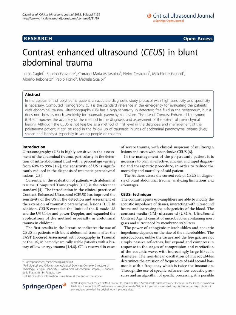
Figure 1 Spleen. Contrast-enhanced CT in venous phase (a), US-B-mode (b) and CEUS (c) in a 40 year- male patient with blunt abdominaltrauma. CT shows a splenic hypodense parenchymal lacerative area (a) not recocognizable by US B-Mode examination (b). CEUS demonstratesplenic hypoechoic lesion corresponding to that of CT.
Cagini et al. Critical Ultrasound Journal 2013, 5(Suppl 1):S9http://www.criticalultrasoundjournal.com/content/5/S1/S9
Page 3 of 7
useful to identify and detect peri-pancreatic collectedfluid, when exploration is feasible. Therefore, currently,CEUS misses indication in the study of post-traumaticpancreatic lesions [19].
KidneyRenal trauma is relatively frequent and represents about5% of the abdominal trauma [19].The kidneys show differ-ent degrees of enhancement in the cortex and the pyra-mids; the cortex almost immediately enhances verybrightly and evenly, while the pyramids enhance diffuselyfrom the periphery to the centre over about 30 seconds.The homogeneous phase of the kidneys generally lasts2-2,5 min: this homogeneous phase (venous phase ornefrographic phase) is still the most effective for detectionof traumatic injuries [1,6]. The recommended dose is thesame as used for the study of the spleen (0,6 ml or ml ofSonoVue: age in years/20). It is necessary to investigatethe kidneys separately with two different boluses of con-trast media [6]. The full exploration of both kidneys isusually hard: the left kidney is sometimes obscured bysuperimposed bowel gas and ribs on images from US eva-luations [3]. Similarly to the liver and spleen, kidney con-tusion lesion can appear as an hypoechoic area withoutclear delimitation; laceration usually appears as a linear orbranched hypoechoic band, perpendicular to the surfaceof the organ. A subcapsular hematoma appears as a non-homogeneous collection surrounding the kidney. In thecase of avulsion of the renal hilum, a total absence of par-enchymal enhancement is found at CEUS. [1,6] (Fig. 3).The rapid-enhancement can generate questions of inter-pretation that can possibly be solved only with a secondinjection of contrast agent [17]. An injection of too high a
dose of contrast media will have a negative effect due tothe intense enhancement, potentially masking the pre-sence of lacerations [6]. Moreover, as much as no micro-bubble excretion into urinary tract is found, CEUSevaluation detects only indirect signs (abdominal fluid)event of accidental bladder, ureter or collecting systemfailure. For the above limits, CT remains the method ofchoice for staging of renal damage [18].
AdrenalTraumatic lesions of the adrenal glands are extremelyrare and often express a high-energy trauma thereforewith high mortality or morbidity. US is particularly usefulin emergency in children: an adrenal traumatic lesion canbe realized in a heterogeneous nodular appearance withincreased echogenicity compared with hepatic or renalparenchyma or as heterogeneous structural alteration ofthe adrenal region. However, the sensitivity of ultrasoundis significantly lower compared to CT, currently consid-ered to be gold standard for diagnosis of adrenal trau-matic lesions [17]. CEUS can play an important role inthe follow-up in selected patients, especially pediatricpatients, to decrease radiation exposure.
Retroperitoneal vesselsTraumatic injury of the abdominal aorta and retoperito-neal great vessels are infrequent but lethal due to theperitoneal or retroperitoneal rapid hemorrhage. Thechances of survival increase with early intervention, so itis essential to get to diagnosis immediately. The retroper-itoneum remains unknown to the FAST examination andpoorly evaluated by US that underestimate free fluid[21-24]. Thus, the CT is the gold standard in case of
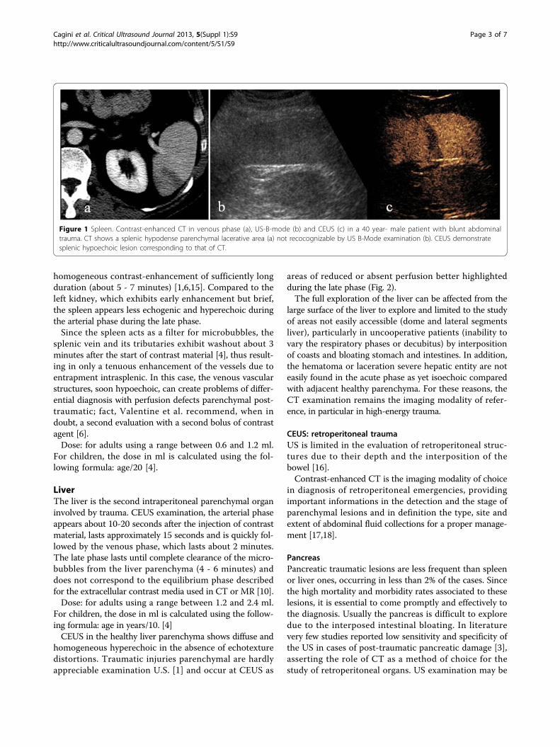
Figure 2 Liver. Contrast-enhanced CT in venous phase (a), US-B-mode (b) and CEUS (c) in a 67 year-old male patient with blunt abdominaltrauma. CT shows a epatic hypodense intra-parenchymal traumatic area (a) not recocognizable by US B-Mode examination (b). CEUSdemonstrate epatic hypoechoic lesion corresponding to that of CT.
Cagini et al. Critical Ultrasound Journal 2013, 5(Suppl 1):S9http://www.criticalultrasoundjournal.com/content/5/S1/S9
Page 4 of 7

abdominal aortic traumatic injuries due to the high sensi-tivity and specificity to detection and staging the injuriesresulting useful for the choice of the appropriate treat-ment [25,26]. However, some studies assume a potentialimprovement of the diagnostic potential of CEUS in thecase of aneurysm rupture for the ability to detect theextravasation of contrast agent [14,27].
DiaphragmTraumatic pulmonary hernia is a complication that mayrarely be associated with blunt abdominal trauma. US candetect the interruption of the echogenic diaphragmaticband or of gaseous artifacts indicating digestive hernia andalso it can identify the presence of hepatic or splenic par-enchyma ectopic. US has a high number of false negativeand is limited in estimating the extent of the lesions. AtCEUS, as at B-mode US examination, the lesions of thediaphragm remain difficult to assess. In the emergency,CT is the gold standard for suspected diaphragmatic inju-ries due to the high spatial resolution and the ability toperform multiplanar reconstructions, which are essentialfor the diagnosis [24,28].
DiscussionUltrasonography (US) is a useful imaging modality in thedetection of traumas, due to its high sensitivity for freeintraperitoneal fluid. The execution is rapid even at thebedside. However, US does not show as much sensitivityfor the detection of solid abdominal organs traumaticinjury; furthermore, the absence of hemoperitoneum doesnot allow the exclusion of the presence of post-traumaticparenchymal injuries [29].
The introduction in clinical practice of contras-enhancedUS (CEUS), increases the sensitivity and accuracy of theUS; hovewer, the CEUS has some limitation in the evalua-tion the parenchymal injuries and retroperitoneum.With respect to parenchymal injuries by CEUS, similarly
to US, some locations (e.g. hepatic dome and the upperpole of the spleen) are poorly inaccessible. CEUS is enableto evaluate simultaneously abdominal parenchyma. Inorder to overcome this limitation, some authors proposeto split contrast material in two or more boluses to studyone or at most two ipsilateral parenchyma. According tothis diagnostic strategy, the kidney must be the first organto be studied because of the early and short enhancementpeak (first two minutes), the same bolus (volume of about2.4 ml) can be used for the study of the liver or the spleenin relation to the late reaching of a homogeneousenhancement (2 to 4 minutes after injection of contrastmaterial) respectively for the abdominal quadrants of theright or left [1]. The study of the renal excretory cavity isnot a limit to the examination of the urinary tract in thepolytrauma patient, due to the lack of renal elimination ofcontrast material used in US.With respect to retroperitoneal injuries similarly to US,
the CEUS is limited by the interposition of the gastricand intestinal bloating, the constitutional habitus of thepatient and in particular, the exploration of the retroperi-toneum, which is sometimes impossible.CEUS should be performed by operators at a high level
competence. In fact administration of an excessive dose ofcontrast agent due to poor experience of the operator canaffect the diagnosis: the dose depends on the target organbut also on the characteristics of the equipment used, the
Figure 3 Kidney. Contrast-enhanced CT in venous phase (a), US-B-mode (b) and CEUS (c) in a 72 year-old female patient with blunt abdominaltrauma. CT shows a renal subcapsular haematoma (a) recocognizable by US B-Mode examination (b).and CEUS corresponding to that of CT.
Cagini et al. Critical Ultrasound Journal 2013, 5(Suppl 1):S9http://www.criticalultrasoundjournal.com/content/5/S1/S9
Page 5 of 7

use of high doses of contrast agent results in excessivehyperechogenicity of the parenchyma which disturbs theidentification of small traumatic injuries [7].Additional limitations of the CEUS are the absence
of three-dimensional scanning, the lack of whole-bodyexploration, extreme difficulty in detecting traumatic boweland mesenteric lesions and the operator dependence thatmakes the technique “subjective”. CT remains the imagingof the choice in trauma due to its high sensitivity and spe-cificity [21,30,31] and the relatively non-invasive and rapidexecution. In polytrauma patients, the use of CEUS wasproposed as a first level examination, after the FAST, inorder to reduce the number of CT examinations [1,4,6].Because the US B-mode is able to demonstrate the pre-sence of intra-abdominal fluid in most cases, but is poorlysensitive in detecting post-traumatic organ damage [3,15],some authors recommend the CEUS in complement ofFAST or the US for the evaluation of liver, spleen and kid-ney trauma [10], others reserve the CEUS at low energyabdominal trauma. In splenic trauma Catalano et al. [15]use CEUS after US, when at US free fluid in the perito-neum is detected, in cases of doubt at US, in cases withpersistent negative at US or laboratory-clinical suspicion ofsplenic injury, reserving CT examination to selectedpatients at a later time. Rhea et al. [32], although they con-sider appropriate the US for the detection of hemoperito-neum, they consider the CT ad an imaging technique witha significant positive impact for the management of traumapatients to reduce the mortality. Concerning pediatricpatients, Benya et al. [29] argue that the US does notexclude negative organ damage, in addition, US examina-tion is not sufficiently helpful in planning a subsequentpossible surgical treatment. In pediatric patients, the exam-ination has greater sensitivity and reference in the manage-ment of trauma is contrast-enhanced CT [17]. Inpolytrauma patients, to avoid diagnostic step that couldresult in the loss of valuable time and the delay of thera-peutic maneuvers, the use of a prompt diagnostic protocolsthat ensures high sensitivity, specificity and diagnosticaccuracy are need [33-35]. In the assessment of post-trau-matic abdominal injuries, literature studies show the sensi-tivity of the FAST and the US of 56% and 68% respectively,with no recognizable organ damage in a percentage vari-able between 16 % and 35% compared to CT [36]. CEUS isan accurate diagnostic tool that should not be consideredas an alternative to CT but rather as a supplement to con-ventional technique useful in cases of inclusive CT [37,38].In the low-energy trauma and in hemodynamically
stable patients, the US can be used as a first-level exam-ination; when US detect intra-abdominal fluid CT exam-ination is need. In the high-energy trauma the use ofUS zas first line diagnostic is superfluous and damagingand the use of CT without and with i.v.c onstrast mate-rial is imperative. In order to reduce the radiation dose,
particularly in young people or children, CEUS has animportant role in the follow-up of conservatively treatedtraumatic injuries of the abdominal parenchymatousorgans (liver, spleen and kidneys) diagnosed by CT[39,40].
Competing interestsThe authors declare that they have no competing interests.
DeclarationsThis article has been published as part of Critical Ultrasound Journal Volume5 Supplement 1, 2013: Topics in emergency abdominal ultrasonography. Thefull contents of the supplement are available online at http://www.criticalultrasoundjournal.com/supplements/5/S1. Publication of thissupplement has been funded by the University of Molise, University ofSiena, University of Cagliari, University of Ferrara and University of Turin.
Author details1Department of Surgical, Radiological and Odontostomatological Sciences,Thoracic Surgery, Perugia University, S. Maria della Misericordia Hospital, S.Andrea delle Fratte, 06134 Perugia, Italy. 2Radiological andOdontostomatological Sciences, Complex Structure of Radiology, PerugiaUniversity, S. Maria della Misericordia Hospital, S. Andrea delle Fratte, 06134Perugia, Italy. 3Radiology Section. Health service. Navy Command of Brindisi,Brindisi, Italy. 4Department of SurgicalSciences, University of Ferrara, Ferrara,Italy. 5Institute of Diagnostic and Interventional Radiology, University of Turin,Turin, Italy.
Published: 15 July 2013
References1. Cokkinos D, Antypa K, Stefanidis K, et al: Contrast-Enhanced Ultrasound for
imaging blunt abdominal trauma – Indication, description of thetechnique and imaging review. Ultraschall in Med 2012, 33:60-67.
2. Valentino M, Serra C, Zironi G, et al: Blunt abdominal trauma: emergencycontrast-enhanced sonography for detection of solid organ injuries. AJR2006, 186:1361-1367.
3. Korner M, Krotz MM, Degenhart C, et al: Current role of emergency US inpatients with major trauma. Radiographics 2008, 28:225-244.
4. Thorelius L: Emergency real-time contrast-enhanced ultrasonography forthe detection of solid organ injuries. Eur Radiol Suppl 2007, 17:107-112.
5. Catalano O, Aiani L, Barozzi L, et al: CEUS in abdominal trauma: multi-center study. Abdom Imaging 2009, 34:225-234.
6. Valentino M, Serra C, Pavlica P, et al: Contrast-Enhanced Ultrasound forblunt abdominal trauma. Semin Ultrasound CT MR 2007, 28(2):130-140.
7. Valentino M, Pavlica P, Barozzi L: Ecografia con mezzo di contrasto neitraumi addominali. Diagnostica per immagini del trauma maggiore.Masson 2010, , 20: 211-216.
8. Bartolotta TV, Midiri M, Scialpi M, et al: Focal nodular hyperplasia innormal and fatty liver: a qualitative and quantitative evaluation withcontrast-enhanced ultrasound. Eur Radiol 2004, 14:583-591.
9. Piscaglia F, Nolsoe C, Dietrich CF, et al: The EFSUMB guidelines andrecommendations on the clinical practice of contrast enhancedultrasound (CEUS): update 2011 on non-hepatic applications. Ultraschallin Med 2012, 33:33-59.
10. Claudon M, Cosgrove D, Albrecht T, et al: Guidelines and good clinicalpractice recommendations for contrast enhanced ultrasound (CEUS) –Update 2008. Ultraschall in Med 2008, 29:28-44.
11. Esposito F, Di Serafino M, Sgambati P, Mercogliano F, Tarantino L,Vallone G, Oresta P: Ultrasound contrast media in paediatric patients: is itan off-label use? Regulatory requirements and radiologist’s liability.Radiol Med 2012, 117(1):148-59.
12. Raimondi F, Migliaro F, Sodano A, Umbaldo A, Romano A, Vallone G,Capasso L: Can neonatal lung ultrasound monitor fluid clearance andpredict the need of respiratory support? Crit Care 2012, 16(6):R220.
13. Reginelli A, Pezzullo MG, Scaglione M, Scialpi M, Brunese L, Grassi R:Gastrointestinal disorders in elderly patients. Radiol Clin North Am 2008,46(4):755-71.
Cagini et al. Critical Ultrasound Journal 2013, 5(Suppl 1):S9http://www.criticalultrasoundjournal.com/content/5/S1/S9
Page 6 of 7
14. Cosgrove D: Ultrasound contrast agent: an overview. Eur J Radiol60(3):324-30.
15. Catalano O, Sandomenico F, Matarazzo I, Siani A: Contrast-EnhancedSonography of the spleen. AJR 2005, 184:1150-1156.
16. Scialpi M, Scaglione M, Volterrani L, et al: Imaging evaluation of postpancreatic surgery. Eur J Radiol 2005, 53(3):417-24.
17. Scialpi M, Mazzei MA, Barberini F, et al: Traumi del surrene. Diagnosticaper immagini del trauma maggiore. Masson 2010, , 18: 193-203.
18. Scialpi M, Scaglione M, Angelelli G, et al: Emergencies in theretroperitoneum: assessment of spread of disease by helical CT.European Journal of Radiology 2004, 50:74-83.
19. Scialpi M, Midiri M, Bartolotta TV, et al: Pancreatic carcinoma versuschronic focal pancreatitis: contrast-enhanced power Dopplerultrasonography findings. Abdom Imaging 2005, 30:222-227.
20. Setola SV, Catalano O, Santodomenico F, Siani A: Contrast-enhancedsonography of the kidney. Abdom Imaging 2007, 32:21-28.
21. Niccoli Asabella A, Di Palo A, Rubini D, Zeppa P, Notaristefano A, Rubini G:Distribution of 18F-FDG in a patient with evolving abdominal aorticaneurysm. Recenti Prog Med 2012, 103(11):552-4.
22. Dialetto G, Reginelli A, Cerrato M, Rossi G, Covino FE, Manduca S, Lassandro F:Endovascular stent-graft treatment of thoracic aortic syndromes: a 7-yearexperience. Eur J Radiol 2007, 64(1):65-72, Epub 2007 Aug 13.
23. Roviello F, Pinto E, Corso G, Pedrazzani C, Caruso S, Filippeschi M, Petrioli R,Marsili S, Mazzei MA, Marrelli D: Safety and potential benefit ofhyperthermic intraperitoneal chemotherapy (HIPEC) in peritonealcarcinomatosis from primary or recurrent ovarian cancer. J Surg Oncol2010, 102(6):663-70, doi: 10.1002/jso.21682.
24. Rosi G, Volterrani L, Macarini L, Cagini L, Cotroneo AR, Scialpi M: Cough-induced intercostal lung herniation successfully diagnosed with imagingtechniques. Recenti Prog Med 2012, 103(11):523-5.
25. Soto JA, e Anderson SW: Multidetector CT of blunt abdominal trauma.Radiology 2012, 265:678-693.
26. Scaglione M, Pinto A, Romano S, et al: Using Multidetector RowComputed Tomography to Diagnose and Stage Pancreatic Carcinoma:the Problems and the Possibilities. JOP. J Pancreas (Online) 2005, 6(1):1-5.
27. Catalano O, Lobianco R, Cusati B, Siani A: Contrast-Enhanced Sonographyfor Diagnosis of Ruptured Abdominal Aortic Aneurysm. American Journalof Roentgenology 2005, 184:423-427.
28. Rotondo A, Catalano O, Grassi R, Scialpi M, Angelelli G: Thoracic CTfindings at hypovolemic shock. Acta Radiol 1998, 39:400-404.
29. Benya EC, Lim-Dunham E, Landrum O, Statter M: Abdominal Sonographyin Examination of Children with Blunt Abdominal Trauma. AJR 2000,174:1613-1616.
30. Salzano A, De Rosa A, Scialpi M, et al: Gunshot wounds of the abdomenstudies by computed tomography. The authors’personal experience in30 cases. Radiol Med 1999, 98(3):168-172.
31. Scaglione M, de Lutio di Castelguidone E, Scialpi M, et al: Blunt trauma tothe gastrointestinal tract and mesentery: is there a role for helical CT inthe decision-making process? European Journal of Radiology 2004,50:67-73.
32. Rhea T, Garza DH, Novelline RA: Controversies in emergency radiology.Emergency Radiology 2004, 10:289-295.
33. Caranci F, Brunese L, Reginelli A, Napoli M, Fonio P, Briganti F: Neckneoplastic conditions in the emergency setting: role of multidetectorcomputed tomography. Semin Ultrasound CT MR 2012, 33(5):443-8.
34. Scardapane A, Lorusso F, Bettocchi S, Moschetta M, Fiume M, Vimercati A,Pepe ML, Angelelli G, Stabile Ianora AA: Deep pelvic endometriosis:accuracy of pelvic MRI completed by MR colonography. Radiol Med 2013,118(2):323-338.
35. Pinto A, Caranci F, Romano L, Carrafiello G, Fonio P, Brunese L: Learningfrom errors in radiology: a comprehensive review. Semin Ultrasound CTMRI 2012, 33:379-382.
36. Catalano O, Lobianco R, Raso MM, Siani A: Blunt hepatic trauma:evaluation with contrast-enhanced sonography. J Ultrasound Med 2005,24:299-310.
37. Catalano O, Lobianco R, Sandomenico F, et al: Real-time contrast-enhanced sonographic imaging in emergency radiology. Radiol Med2004, 108(5-6):454-69.
38. McGahan JP, Horton S, Gerscovich EO, et al: Appearance of solid organinjury with contrast-enhanced sonography in blunt abdominal trauma:preliminary experience. AJR 2006, 187:658-666.
39. Scialpi M, Volterrani L, Mazzei MA, Cappabianca S, Barberini F, Piscioli I,Brunese L, Lupattelli L: Small (< or = 2 cm) atypical hepatichaemangiomas in the non-cirrhotic patient: pattern-based classificationscheme for enhancement at triple-phase helical CT. Radiol Med 2009,114(6):935-47.
40. Rubini G, Altini C, Notaristefano A, Merenda N, Rubini D, Stabile Ianora AA,Giganti M, Niccoli Asabella A: Peritoneal carcinomatosis from ovariancancer: role of 18F-FDG-PET/CT and CA125. Recenti Prog Med 2012,103(11):510-4.
doi:10.1186/2036-7902-5-S1-S9Cite this article as: Cagini et al.: Contrast enhanced ultrasound (CEUS) inblunt abdominal trauma. Critical Ultrasound Journal 2013 5(Suppl 1):S9.
Submit your manuscript to a journal and benefi t from:
7 Convenient online submission
7 Rigorous peer review
7 Immediate publication on acceptance
7 Open access: articles freely available online
7 High visibility within the fi eld
7 Retaining the copyright to your article
Submit your next manuscript at 7 springeropen.com
Cagini et al. Critical Ultrasound Journal 2013, 5(Suppl 1):S9http://www.criticalultrasoundjournal.com/content/5/S1/S9
Page 7 of 7