Contents lists available at ScienceDirect The ... · New perspectives of mitochondrial physiology...
12
The International Journal of Biochemistry & Cell Biology 41 (2009) 1837–1845 Contents lists available at ScienceDirect The International Journal of Biochemistry & Cell Biology journal homepage: www.elsevier.com/locate/biocel Review Capacity of oxidative phosphorylation in human skeletal muscle New perspectives of mitochondrial physiology Erich Gnaiger ∗ Department of General and Transplant Surgery, D. Swarovski Research Laboratory, Medical University of Innsbruck, Innrain 66/6, A-6020 Innsbruck, Austria article info Article history: Available online 2 April 2009 Keywords: Mitochondrial respiratory control High-resolution respirometry Skeletal muscle Q-cycle Q-junction Electron transport Maximal oxygen consumption Pyruvate Glutamate Succinate Tricarboxylic acid cycle abstract Maximal ADP-stimulated mitochondrial respiration depends on convergent electron flow through Com- plexes I + II to the Q-junction of the electron transport system (ETS). In most studies of respiratory control in mitochondrial preparations, however, respiration is limited artificially by supplying substrates for elec- tron input through either Complex I or II. High-resolution respirometry with minimal amounts of tissue biopsy (1–3mg wet weight of permeabilized muscle fibres per assay) provides a routine approach for multiple substrate-uncoupler-inhibitor titrations. Under physiological conditions, maximal respiratory capacity is obtained with glutamate + malate + succinate, reconstituting the operation of the tricarboxylic acid cycle and preventing depletion of key metabolites from the mitochondrial matrix. In human skele- tal muscle, conventional assays with pyruvate + malate or glutamate + malate yield submaximal oxygen fluxes at 0.50–0.75 of capacity of oxidative phosphorylation (OXPHOS). Best estimates of muscular OXPHOS capacity at 37 ◦ C (pmol O 2 s −1 mg −1 wet weight) with isolated mitochondria or permeabilized fibres, suggest a range of 100–150 and up to 180 in healthy humans with normal body mass index and top endurance athletes, but reduction to 60–120 in overweight healthy adults with predominantly seden- tary life style. The apparent ETS excess capacity (uncoupled respiration) over ADP-stimulated OXPHOS capacity is high in skeletal muscle of active and sedentary humans, but absent in mouse skeletal muscle. Such differences of mitochondrial quality in skeletal muscle are unexpected and cannot be explained at present. A comparative database of mitochondrial physiology may provide the key for understand- ing the functional implications of mitochondrial diversity from mouse to man, and evaluation of altered mitochondrial respiratory control patterns in health and disease. © 2009 Elsevier Ltd. All rights reserved. Contents 1. Introduction ........................................................................................................................................ 1838 2. Respirometry with permeabilized fibres and isolated mitochondria ............................................................................. 1840 2.1. High-resolution respirometry compared to specialized microchamber system ........................................................... 1840 2.2. Respiration per muscle mass ............................................................................................................... 1840 2.3. Experimental conditions for maximal flux ................................................................................................. 1840 3. Convergent CI + II electron input and OXPHOS capacity ........................................................................................... 1841 3.1. Reconstitution of TCA cycle function ....................................................................................................... 1841 3.2. Oxidative phosphorylation versus electron transport system: coupling and substrate control ratios .................................... 1842 3.3. Permeabilized fibres and isolated mitochondria ........................................................................................... 1842 4. Tissue-OXPHOS capacity in human permeabilized muscle fibres and isolated mitochondria .................................................... 1842 5. Conclusions: tissue-OXPHOS capacity and functional diversity ................................................................................... 1843 Acknowledgements ................................................................................................................................ 1844 References ......................................................................................................................................... 1844 Abbreviations: HRR, high-resolution respirometry; Imt, isolated mitochondria; Pfi, permeabilized fibers; Pmt , mitochondrial protein; SUIT, substrate-uncoupler- inhibitor-titration; TCA, tricarboxylic acid; u, uncoupled; Ww, wet weight (see also Table 1). ∗ Tel.: +43 512 504 24623; fax: +43 512 504 24625. E-mail address: [email protected]. 1357-2725/$ – see front matter © 2009 Elsevier Ltd. All rights reserved. doi:10.1016/j.biocel.2009.03.013
Transcript of Contents lists available at ScienceDirect The ... · New perspectives of mitochondrial physiology...
The International Journal of Biochemistry & Cell Biology 41 (2009) 1837–1845
Contents lists available at ScienceDirect
The International Journal of Biochemistry& Cell Biology
journa l homepage: www.e lsev ier .com/ locate /b ioce l
Review
Capacity of oxidative phosphorylation in human skeletal muscleNew perspectives of mitochondrial physiology
Erich Gnaiger ∗
Department of General and Transplant Surgery, D. Swarovski Research Laboratory, Medical University of Innsbruck, Innrain 66/6, A-6020 Innsbruck, Austria
a r t i c l e i n f o
Article history:
Available online 2 April 2009
Keywords:
Mitochondrial respiratory control
High-resolution respirometry
Skeletal muscle
Q-cycle
Q-junction
Electron transport
Maximal oxygen consumption
Pyruvate
Glutamate
Succinate
Tricarboxylic acid cycle
a b s t r a c t
Maximal ADP-stimulated mitochondrial respiration depends on convergent electron flow through Com-
plexes I + II to the Q-junction of the electron transport system (ETS). In most studies of respiratory control
in mitochondrial preparations, however, respiration is limited artificially by supplying substrates for elec-
tron input through either Complex I or II. High-resolution respirometry with minimal amounts of tissue
biopsy (1–3 mg wet weight of permeabilized muscle fibres per assay) provides a routine approach for
multiple substrate-uncoupler-inhibitor titrations. Under physiological conditions, maximal respiratory
capacity is obtained with glutamate + malate + succinate, reconstituting the operation of the tricarboxylic
acid cycle and preventing depletion of key metabolites from the mitochondrial matrix. In human skele-
tal muscle, conventional assays with pyruvate + malate or glutamate + malate yield submaximal oxygen
fluxes at 0.50–0.75 of capacity of oxidative phosphorylation (OXPHOS). Best estimates of muscular
OXPHOS capacity at 37 ◦C (pmol O2 s−1 mg−1 wet weight) with isolated mitochondria or permeabilized
fibres, suggest a range of 100–150 and up to 180 in healthy humans with normal body mass index and top
endurance athletes, but reduction to 60–120 in overweight healthy adults with predominantly seden-
tary life style. The apparent ETS excess capacity (uncoupled respiration) over ADP-stimulated OXPHOS
capacity is high in skeletal muscle of active and sedentary humans, but absent in mouse skeletal muscle.
Such differences of mitochondrial quality in skeletal muscle are unexpected and cannot be explained
at present. A comparative database of mitochondrial physiology may provide the key for understand-
ing the functional implications of mitochondrial diversity from mouse to man, and evaluation of altered
mitochondrial respiratory control patterns in health and disease.
© 2009 Elsevier Ltd. All rights reserved.
Contents
1. Introduction . . . . . . . . . . . . . . . . . . . . . . . . . . . . . . . . . . . . . . . . . . . . . . . . . . . . . . . . . . . . . . . . . . . . . . . . . . . . . . . . . . . . . . . . . . . . . . . . . . . . . . . . . . . . . . . . . . . . . . . . . . . . . . . . . . . . . . . . 1838
2. Respirometry with permeabilized fibres and isolated mitochondria . . . . . . . . . . . . . . . . . . . . . . . . . . . . . . . . . . . . . . . . . . . . . . . . . . . . . . . . . . . . . . . . . . . . . . . . . . . . . 1840
2.1. High-resolution respirometry compared to specialized microchamber system . . . . . . . . . . . . . . . . . . . . . . . . . . . . . . . . . . . . . . . . . . . . . . . . . . . . . . . . . . . 1840
2.2. Respiration per muscle mass . . . . . . . . . . . . . . . . . . . . . . . . . . . . . . . . . . . . . . . . . . . . . . . . . . . . . . . . . . . . . . . . . . . . . . . . . . . . . . . . . . . . . . . . . . . . . . . . . . . . . . . . . . . . . . . 1840
2.3. Experimental conditions for maximal flux . . . . . . . . . . . . . . . . . . . . . . . . . . . . . . . . . . . . . . . . . . . . . . . . . . . . . . . . . . . . . . . . . . . . . . . . . . . . . . . . . . . . . . . . . . . . . . . . . 1840
3. Convergent CI + II electron input and OXPHOS capacity . . . . . . . . . . . . . . . . . . . . . . . . . . . . . . . . . . . . . . . . . . . . . . . . . . . . . . . . . . . . . . . . . . . . . . . . . . . . . . . . . . . . . . . . . . . 1841
3.1. Reconstitution of TCA cycle function . . . . . . . . . . . . . . . . . . . . . . . . . . . . . . . . . . . . . . . . . . . . . . . . . . . . . . . . . . . . . . . . . . . . . . . . . . . . . . . . . . . . . . . . . . . . . . . . . . . . . . . 1841
3.2. Oxidative phosphorylation versus electron transport system: coupling and substrate control ratios . . . . . . . . . . . . . . . . . . . . . . . . . . . . . . . . . . . . 1842
3.3. Permeabilized fibres and isolated mitochondria . . . . . . . . . . . . . . . . . . . . . . . . . . . . . . . . . . . . . . . . . . . . . . . . . . . . . . . . . . . . . . . . . . . . . . . . . . . . . . . . . . . . . . . . . . . 1842
4. Tissue-OXPHOS capacity in human permeabilized muscle fibres and isolated mitochondria . . . . . . . . . . . . . . . . . . . . . . . . . . . . . . . . . . . . . . . . . . . . . . . . . . . . 1842
5. Conclusions: tissue-OXPHOS capacity and functional diversity. . . . . . . . . . . . . . . . . . . . . . . . . . . . . . . . . . . . . . . . . . . . . . . . . . . . . . . . . . . . . . . . . . . . . . . . . . . . . . . . . . . 1843
Acknowledgements . . . . . . . . . . . . . . . . . . . . . . . . . . . . . . . . . . . . . . . . . . . . . . . . . . . . . . . . . . . . . . . . . . . . . . . . . . . . . . . . . . . . . . . . . . . . . . . . . . . . . . . . . . . . . . . . . . . . . . . . . . . . . . . . 1844
References . . . . . . . . . . . . . . . . . . . . . . . . . . . . . . . . . . . . . . . . . . . . . . . . . . . . . . . . . . . . . . . . . . . . . . . . . . . . . . . . . . . . . . . . . . . . . . . . . . . . . . . . . . . . . . . . . . . . . . . . . . . . . . . . . . . . . . . . . 1844
Abbreviations: HRR, high-resolution respirometry; Imt, isolated mitochondria; Pfi, permeabilized fibers; Pmt , mitochondrial protein; SUIT, substrate-uncoupler-
inhibitor-titration; TCA, tricarboxylic acid; u, uncoupled; Ww , wet weight (see also Table 1).∗ Tel.: +43 512 504 24623; fax: +43 512 504 24625.
E-mail address: [email protected].
1357-2725/$ – see front matter © 2009 Elsevier Ltd. All rights reserved.
doi:10.1016/j.biocel.2009.03.013
1838 E. Gnaiger / The International Journal of Biochemistry & Cell Biology 41 (2009) 1837–1845
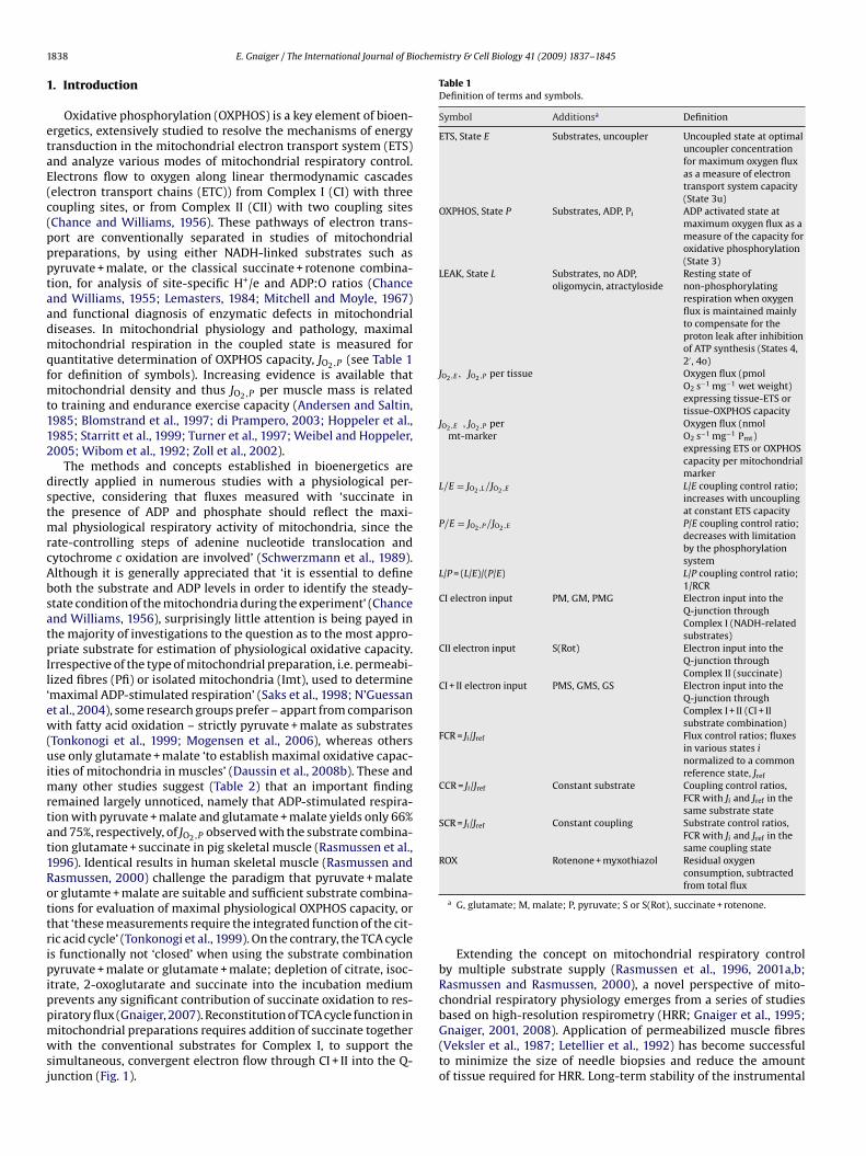
1. Introduction
Oxidative phosphorylation (OXPHOS) is a key element of bioen-
ergetics, extensively studied to resolve the mechanisms of energy
transduction in the mitochondrial electron transport system (ETS)
and analyze various modes of mitochondrial respiratory control.
Electrons flow to oxygen along linear thermodynamic cascades
(electron transport chains (ETC)) from Complex I (CI) with three
coupling sites, or from Complex II (CII) with two coupling sites
(Chance and Williams, 1956). These pathways of electron trans-
port are conventionally separated in studies of mitochondrial
preparations, by using either NADH-linked substrates such as
pyruvate + malate, or the classical succinate + rotenone combina-
tion, for analysis of site-specific H+/e and ADP:O ratios (Chance
and Williams, 1955; Lemasters, 1984; Mitchell and Moyle, 1967)
and functional diagnosis of enzymatic defects in mitochondrial
diseases. In mitochondrial physiology and pathology, maximal
mitochondrial respiration in the coupled state is measured for
quantitative determination of OXPHOS capacity, JO2,P (see Table 1
for definition of symbols). Increasing evidence is available that
mitochondrial density and thus JO2,P per muscle mass is related
to training and endurance exercise capacity (Andersen and Saltin,
1985; Blomstrand et al., 1997; di Prampero, 2003; Hoppeler et al.,
1985; Starritt et al., 1999; Turner et al., 1997; Weibel and Hoppeler,
2005; Wibom et al., 1992; Zoll et al., 2002).
The methods and concepts established in bioenergetics are
directly applied in numerous studies with a physiological per-
spective, considering that fluxes measured with ‘succinate in
the presence of ADP and phosphate should reflect the maxi-
mal physiological respiratory activity of mitochondria, since the
rate-controlling steps of adenine nucleotide translocation and
cytochrome c oxidation are involved’ (Schwerzmann et al., 1989).
Although it is generally appreciated that ‘it is essential to define
both the substrate and ADP levels in order to identify the steady-
state condition of the mitochondria during the experiment’ (Chance
and Williams, 1956), surprisingly little attention is being payed in
the majority of investigations to the question as to the most appro-
priate substrate for estimation of physiological oxidative capacity.
Irrespective of the type of mitochondrial preparation, i.e. permeabi-
lized fibres (Pfi) or isolated mitochondria (Imt), used to determine
‘maximal ADP-stimulated respiration’ (Saks et al., 1998; N’Guessan
et al., 2004), some research groups prefer – appart from comparison
with fatty acid oxidation – strictly pyruvate + malate as substrates
(Tonkonogi et al., 1999; Mogensen et al., 2006), whereas others
use only glutamate + malate ‘to establish maximal oxidative capac-
ities of mitochondria in muscles’ (Daussin et al., 2008b). These and
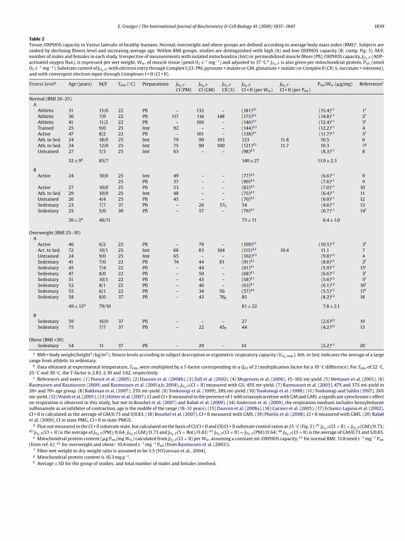
many other studies suggest (Table 2) that an important finding
remained largely unnoticed, namely that ADP-stimulated respira-
tion with pyruvate + malate and glutamate + malate yields only 66%
and 75%, respectively, of JO2,P observed with the substrate combina-
tion glutamate + succinate in pig skeletal muscle (Rasmussen et al.,
1996). Identical results in human skeletal muscle (Rasmussen and
Rasmussen, 2000) challenge the paradigm that pyruvate + malate
or glutamte + malate are suitable and sufficient substrate combina-
tions for evaluation of maximal physiological OXPHOS capacity, or
that ‘these measurements require the integrated function of the cit-
ric acid cycle’ (Tonkonogi et al., 1999). On the contrary, the TCA cycle
is functionally not ‘closed’ when using the substrate combination
pyruvate + malate or glutamate + malate; depletion of citrate, isoc-
itrate, 2-oxoglutarate and succinate into the incubation medium
prevents any significant contribution of succinate oxidation to res-
piratory flux (Gnaiger, 2007). Reconstitution of TCA cycle function in
mitochondrial preparations requires addition of succinate together
with the conventional substrates for Complex I, to support the
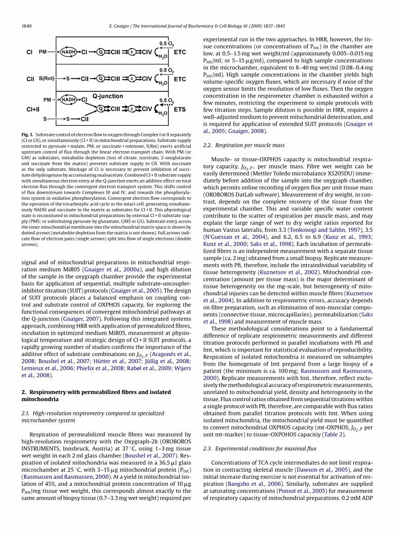
simultaneous, convergent electron flow through CI + II into the Q-
junction (Fig. 1).
Table 1
Definition of terms and symbols.
Symbol Additionsa Definition
ETS, State E Substrates, uncoupler Uncoupled state at optimal
uncoupler concentration
for maximum oxygen flux
as a measure of electron
transport system capacity
(State 3u)
OXPHOS, State P Substrates, ADP, Pi ADP activated state at
maximum oxygen flux as a
measure of the capacity for
oxidative phosphorylation
(State 3)
LEAK, State L Substrates, no ADP,
oligomycin, atractyloside
Resting state of
non-phosphorylating
respiration when oxygen
flux is maintained mainly
to compensate for the
proton leak after inhibition
of ATP synthesis (States 4,
2′ , 4o)
JO2,E, JO2,P per tissue Oxygen flux (pmol
O2 s−1 mg−1 wet weight)
expressing tissue-ETS or
tissue-OXPHOS capacity
JO2,E , JO2,P per
mt-marker
Oxygen flux (nmol
O2 s−1 mg−1 Pmt)
expressing ETS or OXPHOS
capacity per mitochondrial
marker
L/E = JO2,L/JO2,E L/E coupling control ratio;
increases with uncoupling
at constant ETS capacity
P/E = JO2,P /JO2,E P/E coupling control ratio;
decreases with limitation
by the phosphorylation
system
L/P = (L/E)/(P/E) L/P coupling control ratio;
1/RCR
CI electron input PM, GM, PMG Electron input into the
Q-junction through
Complex I (NADH-related
substrates)
CII electron input S(Rot) Electron input into the
Q-junction through
Complex II (succinate)
CI + II electron input PMS, GMS, GS Electron input into the
Q-junction through
Complex I + II (CI + II
substrate combination)
FCR = Ji/Jref Flux control ratios; fluxes
in various states i
normalized to a common
reference state, Jref
CCR = Ji/Jref Constant substrate Coupling control ratios,
FCR with Ji and Jref in the
same substrate state
SCR = Ji/Jref Constant coupling Substrate control ratios,
FCR with Ji and Jref in the
same coupling state
ROX Rotenone + myxothiazol Residual oxygen
consumption, subtracted
from total flux
a G, glutamate; M, malate; P, pyruvate; S or S(Rot), succinate + rotenone.
Extending the concept on mitochondrial respiratory control
by multiple substrate supply (Rasmussen et al., 1996, 2001a,b;
Rasmussen and Rasmussen, 2000), a novel perspective of mito-
chondrial respiratory physiology emerges from a series of studies
based on high-resolution respirometry (HRR; Gnaiger et al., 1995;
Gnaiger, 2001, 2008). Application of permeabilized muscle fibres
(Veksler et al., 1987; Letellier et al., 1992) has become successful
to minimize the size of needle biopsies and reduce the amount
of tissue required for HRR. Long-term stability of the instrumental
E. Gnaiger / The International Journal of Biochemistry & Cell Biology 41 (2009) 1837–1845 1839
Table 2
Tissue-OXPHOS capacity in Vastus lateralis of healthy humans. Normal, overweight and obese groups are defined according to average body mass index (BMI)a . Subjects are
ranked by declining fitness level and increasing average age. Within BMI groups, studies are distinguished with high (A) and low OXPHOS capacity (B; comp. Fig. 3). M/F,
number of males and females in each study. Irrespective of measurements with isolated mitochondria (Imt) or permeabilized muscle fibres (Pfi), OXPHOS capacity, JO2,P (ADP-
activated oxygen flux), is expressed per wet weight, Ww , of muscle tissue (pmol O2 s−1 mg−1) and adjusted to 37 ◦C.b JO2,P is also given per mitochondrial protein, Pmt (nmol
O2 s−1 mg−1). Substrate control of JO2,P , with electron entry through Complex I (CI; PM, pyruvate + malate or GM, glutamate + malate) or Complex II (CII; S, succinate + rotenone),
and with convergent electron input through Complexes I + II (CI + II).
Fitness levela Age (years) M/F Texp (◦C) Preparations JO2,P JO2,P JO2,P JO2,P JO2,P Pmt/Ww (�g/mg) Referencesc
CI (PM) CI (GM) CII (S) CI + II (per Ww) CI + II (per Pmt)
Normal (BMI 20–25)
A
Athletic 31 15/0 22 Pfi – 132 – (181)d1 (15.4)e1 1f
Athletic 36 7/0 22 Pfi 117 116 148 (173)d2 (14.8)e1 2f
Athletic 41 11/2 22 Pfi – 106 – (146)d1 (12.4)e1 3f
Trained 25 9/0 25 Imt 92 – – (144)d3 (12.2)e1 4
Active 47 8/2 22 Pfi – 101 – (138)d1 (11.7)e1 5f
Ath. to Sed. 24 18/0 25 Imt 79 90 103 123 11.8 10.5 6
Ath. to Sed. 24 12/0 25 Imt 75 90 100 (121)d2 11.7 10.3 7g
Untrained 27 5/3 25 Imt 63 – – (98)d3 (8.3)e1 8
32 ± 9h 85/7 140 ± 27 11.9 ± 2.3
B
Active 24 10/0 25 Imt 49 – – (77)d3 (6.6)e1 9
25 Pfi 57 – – (90)d3 (7.6)e1 9
Active 27 10/0 25 Pfi 53 – – (83)d3 (7.0)e1 10
Ath. to Sed. 29 10/0 25 Imt 48 – – (75)d3 (6.4)e1 11
Untrained 26 4/4 25 Pfi 45 – – (70)d3 (6.0)e1 12
Sedentary 23 7/7 37 Pfi – 26 57E 54 (4.6)e1 13
Sedentary 25 5/0 30 Pfi – 57 – (79)d1 (6.7)e1 14f
26 ± 2h 46/11 75 ± 11 6.4 ± 1.0
Overweight (BMI 25–30)
A
Active 46 6/2 22 Pfi – 79 – (109)d1 (10.5)e2 3f
Act. to Sed. 72 10/1 25 Imt 68 83 104 (115)d2 10.4 11.1 7
Untrained 24 9/0 25 Imt 65 – – (102)d3 (9.8)e2 4
Sedentary 41 7/0 22 Pfi 74 44 81 (91)d2 (8.8)e2 2f
Sedentary 45 7/4 22 Pfi – 44 – (61)d1 (5.9)e2 15f
Sedentary 47 8/0 22 Pfi – 50 – (68)d1 (6.6)e2 3f
Sedentary 51 10/1 22 Pfi – 43 – (58)d1 (5.6)e2 5f
Sedentary 52 8/1 22 Pfi – 46 – (63)d1 (6.1)e2 16f
Sedentary 55 6/1 22 Pfi – 34 56 (57)d4 (5.5)e2 17f
Sedentary 58 8/0 37 Pfi – 43 76E 85 (8.2)e2 18
49 ± 12h 79/10 81 ± 22 7.8 ± 2.1
B
Sedentary 59 16/0 37 Pfi – – – 27 (2.6)d2 19
Sedentary 75 7/7 37 Pfi – 22 45E 44 (4.2)d2 13
Obese (BMI >30)
Sedentary 54 11 37 Pfi – 29 – 61 (5.2)e2 20
a BMI = body weight/height2 (kg/m2); fitness levels according to subject description or ergometric respiratory capacity (V̇O2,max), Ath. to Sed. indicates the average of a large
range from athletic to sedentary.b Data obtained at experimental temperature, Texp , were multiplied by a T-factor corresponding to a Q10 of 2 (multiplication factor for a 10 ◦C difference). For Texp of 22 ◦C,
25 ◦C and 30 ◦C, the T-factor is 2.83, 2.30 and 1.62, respectively.c References and notes: (1) Ponsot et al. (2005). (2) Daussin et al. (2008b). (3) Zoll et al. (2002). (4) Mogensen et al. (2006), 15–16% mt-yield. (5) Mettauer et al. (2001). (6)
Rasmussen and Rasmussen (2000) and Rasmussen et al. (2001a,b, 2004), JO2,P (CI + II) measured with GS; 45% mt-yield. (7) Rasmussen et al. (2003), 47% and 37% mt-yield in
20+ and 70+ age group. (8) Bakkman et al. (2007); 23% mt-yield. (9) Tonkonogi et al. (1999), 28% mt-yield. (10) Tonkonogi et al. (1998). (11) Tonkonogi and Sahlin (1997), 26%
mt-yield. (12) Walsh et al. (2001). (13) Hütter et al. (2007), CI and CI + II measured in the presence of 1 mM octanoylcarnitine with GM and GMS, a significant cytochrome c effect
on respiration is observed in this study, but not in Boushel et al. (2007) and Rabøl et al. (2009). (14) Anderson et al. (2009), the respiration medium includes benzyltoluene
sulfonamide as an inhibitor of contraction, age is the middle of the range (18–31 years). (15) Daussin et al. (2008a). (16) Garnier et al. (2005). (17) Echaniz-Laguna et al. (2002),
CI + II is calculated as the average of GM/0.73 and S/0.83. (18) Boushel et al. (2007), CI + II measured with GMS. (19) Phielix et al. (2008), CI + II measured with GMS. (20) Rabøl
et al. (2009), CI in state PMG, CI + II in state PMGS.d Flux not measured in the CI + II substrate state, but calculated on the basis of CI/CI + II and CII/CI + II substrate control ratios at 25 ◦C (Fig. 2). d1 JO2,P (CI + II) = JO2,P (GM)/0.73;
d2 JO2,P (CI + II) is the average of JO2,P (PM)/0.64, JO2,P (GM)/0.73 and JO2,P (S + Rot)/0.83; d3 JO2,P (CI + II) = JO2,P (PM)/0.64; d4 JO2,P (CI + II) is the average of GM/0.73 and S/0.83.e Mitochondrial protein content (�g Pmt/mg Ww) calculated from JO2,P (CI + II) per Ww , assuming a constant mt-OXPHOS capacity, e1 for normal BMI: 11.8 nmol s−1 mg−1 Pmt
(from ref. 6); e2 for overweight and obese: 10.4 nmol s−1 mg−1 Pmt (from Rasmussen et al. (2003)).f Fibre wet weight to dry weight ratio is assumed to be 3.5 (N’Guessan et al., 2004).g Mitochondrial protein content is 10.3 mg g−1 .h Average ± SD for the group of studies, and total number of males and females involved.
1840 E. Gnaiger / The International Journal of Biochemistry & Cell Biology 41 (2009) 1837–1845
Fig. 1. Substrate control of electron flow to oxygen through Complex I or II separately
(CI or CII), or simultaneously (CI + II) in mitochondrial preparations. Substrate supply
restricted to pyruvate + malate, PM, or succinate + rotenone, S(Rot) exerts artificial
upstream control of flux through the linear electron transport chain. With PM (or
GM) as substrates, metabolite depletion (loss of citrate, isocitrate, 2-oxoglutarate
and succinate from the matrix) prevents substrate supply to CII. With succinate
as the only substrate, blockage of CI is necessary to prevent inhibition of succi-
nate dehydrogenase by accumulating oxaloacetate. Combined CI + II substrate supply
with simultaneous electron entry at the Q-junction exerts an additive effect on total
electron flux through the convergent electron transport system. This shifts control
of flux downstream towards Complexes III and IV, and towards the phosphoryla-
tion system in oxidative phosphorylation. Convergent electron flow corresponds to
the operation of the tricarboxylic acid cycle in the intact cell, generating simultane-
ously NADH and succinate in the matrix as substrates for CI + II. This physiological
state is reconsituted in mitochondrial preparations by external CI + II substrate sup-
ply (PMS; or substituting pyruvate by glutamate, GMS or GS). Substrate entry across
the inner mitochondrial membrane into the mitochondrial matrix space is shown by
dotted arrows (metabolite depletion from the matrix is not shown). Full arrows indi-
cate flow of electron pairs (single arrows) split into flow of single electrons (double
arrows).
signal and of mitochondrial preparations in mitochondrial respi-
ration medium MiR05 (Gnaiger et al., 2000a), and high dilution
of the sample in the oxygraph chamber provide the experimental
basis for application of sequential, multiple substrate-uncoupler-
inhibitor titration (SUIT) protocols (Gnaiger et al., 2005). The design
of SUIT protocols places a balanced emphasis on coupling con-
trol and substrate control of OXPHOS capacity, for exploring the
functional consequences of convergent mitochondrial pathways at
the Q-junction (Gnaiger, 2007). Following this integrated systems
approach, combining HRR with application of permeabilized fibres,
incubation in optimized medium MiR05, measurement at physio-
logical temperature and strategic design of CI + II SUIT protocols, a
rapidly growing number of studies confirms the importance of the
additive effect of substrate combinations on JO2,P (Aragonés et al.,
2008; Boushel et al., 2007; Hütter et al., 2007; Jüllig et al., 2008;
Lemieux et al., 2006; Phielix et al., 2008; Rabøl et al., 2009; Wijers
et al., 2008).
2. Respirometry with permeabilized fibres and isolated
mitochondria
2.1. High-resolution respirometry compared to specialized
microchamber system
Respiration of permeabilized muscle fibres was measured by
high-resolution respirometry with the Oxygraph-2k (OROBOROS
INSTRUMENTS, Innsbruck, Austria) at 37 ◦C, using 1–3 mg tissue
wet weight in each 2 ml glass chamber (Boushel et al., 2007). Res-
piration of isolated mitochondria was measured in a 36.5 �l glass
microchamber at 25 ◦C, with 3–15 �g mitochondrial protein (Pmt)
(Rasmussen and Rasmussen, 2000). At a yield in mitochondrial iso-
lation of 45%, and a mitochondrial protein concentration of 10 �g
Pmt/mg tissue wet weight, this corresponds almost exactly to the
same amount of biopsy tissue (0.7–3.3 mg wet weight) required per
experimental run in the two approaches. In HRR, however, the tis-
sue concentrations (or concentrations of Pmt) in the chamber are
low, at 0.5–1.5 mg wet weight/ml (approximately 0.005–0.015 mg
Pmt/ml; or 5–15 �g/ml), compared to high sample concentrations
in the microchamber, equivalent to 8–40 mg wet/ml (0.08–0.4 mg
Pmt/ml). High sample concentrations in the chamber yields high
volume-specific oxygen fluxes, which are necessary if noise of the
oxygen sensor limits the resolution of low fluxes. Then the oxygen
concentration in the respirometer chamber is exhausted within a
few minutes, restricting the experiment to simple protocols with
few titration steps. Sample dilution is possible in HRR, requires a
well-adjusted medium to prevent mitochondrial deterioration, and
is required for application of extended SUIT protocols (Gnaiger et
al., 2005; Gnaiger, 2008).
2.2. Respiration per muscle mass
Muscle- or tissue-OXPHOS capacity is mitochondrial respira-
tory capacity, JO2,P, per muscle mass. Fibre wet weight can be
easily determined (Mettler Toledo microbalance XS205DU) imme-
diately before addition of the sample into the oxygraph chamber,
which permits online recording of oxygen flux per unit tissue mass
(OROBOROS DatLab software). Measurement of dry weight, in con-
trast, depends on the complete recovery of the tissue from the
experimental chamber. This and variable specific water content
contribute to the scatter of respiration per muscle mass, and may
explain the large range of wet to dry weight ratios reported for
human Vastus lateralis, from 3.3 (Tonkonogi and Sahlin, 1997), 3.5
(N’Guessan et al., 2004), and 6.2, 6.5 to 6.9 (Kunz et al., 1993;
Kunz et al., 2000; Saks et al., 1998). Each incubation of permeabi-
lized fibres is an independent measurement with a separate tissue
sample (ca. 2 mg) obtained from a small biopsy. Replicate measure-
ments with Pfi, therefore, include the intraindividual variability of
tissue heterogeneity (Kuznetsov et al., 2002). Mitochondrial con-
centration (amount per tissue mass) is the major determinant of
tissue heterogeneity on the mg-scale, but heterogeneity of mito-
chondrial injuries can be detected within muscle fibres (Kuznetsov
et al., 2004). In addition to respirometric errors, accuracy depends
on fibre preparation, such as elimination of non-muscular compo-
nents (connective tissue, microcapillaries), permeabilization (Saks
et al., 1998) and measurement of muscle mass.
These methodological considerations point to a fundamental
difference of replicate respirometric measurements and different
titration protocols performed in parallel incubations with Pfi and
Imt, which is important for statistical evaluation of reproducibility.
Respiration of isolated mitochondria is measured on subsamples
from the homogenate of Imt prepared from a large biopsy of a
patient (the minimum is ca. 100 mg; Rasmussen and Rasmussen,
2000). Replicate measurements with Imt, therefore, reflect exclu-
sively the methodological accuracy of respirometric measurements,
unrelated to mitochondrial yield, density and heterogeneity in the
tissue. Flux control ratios obtained from sequential titrations within
a single protocol with Pfi, therefore, are comparable with flux ratios
obtained from parallel titration protocols with Imt. When using
isolated mitochondria, the mitochondrial yield must be quantified
to convert mitochondrial OXPHOS capacity (mt-OXPHOS, JO2,P per
unit mt-marker) to tissue-OXPOHOS capacitiy (Table 2).
2.3. Experimental conditions for maximal flux
Concentrations of TCA cycle intermediates do not limit respira-
tion in contracting skeletal muscle (Dawson et al., 2005), and the
initial increase during exercise is not essential for activation of res-
piration (Bangsbo et al., 2006). Similarly, substrates are supplied
at saturating concentrations (Ponsot et al., 2005) for measurement
of respiratory capacity of mitochondrial preparations. 0.2 mM ADP
E. Gnaiger / The International Journal of Biochemistry & Cell Biology 41 (2009) 1837–1845 1841
does not fully saturate flux in isolated mitochondria (Puchowicz
et al., 2004; Gnaiger, 2001), and high [ADP] concentrations are
required particularly in permeabilized fibres to overcome limita-
tions by diffusion and by the tubulin-regulated conductance of the
outer mitochondrial membrane (Rostovtseva et al., 2008). In per-
meabilized muscle fibre bundles of high respiratory capacity, the
apparent Km for ADP increases up to 0.5 mM (Saks et al., 1998). This
implies that >90% saturation is reached only at >5 mM ADP, yet few
studies use such high ADP concentrations in permeabilized muscle
fibres, and lack of stimulation at 2.5 mM ADP by increasing [ADP]
provides evidence for lower K′m,ADP. Maximal aerobic capacity in
vivo is limited by oxygen supply (di Prampero, 2003; Mitchell and
Saltin, 2003; Richardson et al., 1999), but respiratory capacity of
mitochondrial preparations must be evaluated at kinetic oxygen
saturation, comparable to the application of saturating ADP con-
centrations for measuring JO2,P . Even at 20 �M, oxygen does not
limit respiration of isolated mitochondria and small cells (20–50-
fold above the c50 or K ′
m,O2; Gnaiger et al., 1998, 2000b; Scandurra
and Gnaiger, 2009). In permeabilized muscle fibre bundles, how-
ever, diffusion restriction increases the sensitivity to oxygen supply
100-fold (rat soleus and rat heart; Gnaiger, 2003). Oxygen limitation
of respiration in permeabilized fibres is prevented by maintaining
oxygen levels in the respirometer above air saturation in the range
of 500–200 �M (Boushel et al., 2007; Aragonés et al., 2008). Lack of
attention to experimental oxygen limitation of Pfi, however, partly
explains apparent low specific activities (Table 2).
3. Convergent CI + II electron input and OXPHOS capacity
3.1. Reconstitution of TCA cycle function
In addition to coupling and substrate supply, metabolite deple-
tion of TCA cycle intermediates plays a fundamental role in OXPHOS
flux control in isolated mitochondria or permeabilized cells. Using
classical NADH-related substrates such as pyruvate, malate and
glutamate, the TCA cycle metabolites citrate and 2-oxoglutarate
are exchanged rapidly for malate by the tricarboxylate and 2-
oxoglutarate carrier, and succinate is lost from the matrix space
in exchange for inorganic phosphate catalyzed by the dicarboxy-
late carrier (Gnaiger, 2007). Evaluation of the functional design of
the OXPHOS system requires a conceptual transition from analyses
of the electron transport chain to a perspective of the convergent
structure of electron flow to the Q-junction in the mitochondrial
electron transport system (Fig. 1). The convergent structure of
the ETS is functionally related to substrate compartmentation and
mitochondrial network organization (Benard et al., 2007, 2008).
Molar ratios of redox components of the ETS cannot be understood
in terms of a linear chain (Fig. 1; ETC CI and CII). Instead, the sum of
redox components upstream of the Q-cycle (the convergent input
branches) is related to the linear sequence of redox components
downstream of the Q-junction (Fig. 2; ETS CI + II). Consequently, the
excess capacity of Complexes III and IV is high when related to CI or
CII electron input (Benard et al., 2008; Gnaiger et al., 1998; Rossignol
et al., 2003). These apparent excess capacities are in part explained
functionally, as a necessary requirement for higher capacities of the
Q-cycle and respiratory complexes downstream of Q, which have to
match the sum of the partial capacities of all contributory branches
converging at the Q-junction (Fig. 2; CI + II).
A ‘substrate cocktail’ (NADH + glutamate + malate + succinate)
activates in mitochondria of fungi not only convergent CI + II
electron input, but in addition the alternative NADH-ubiquinone
oxidoreductase which together support a high oxygen flux, whereas
flux with NADH, pyruvate or malate alone is 0.85, 0.14 and 0.17 of
flux with the substrate cocktail (Tudella et al., 2003). No conclu-
sions can be drawn on the additive effect of convergent pathway
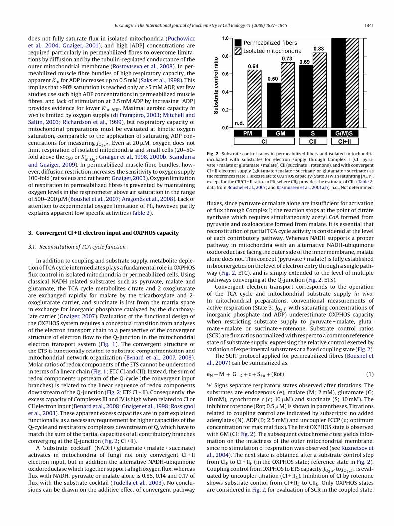
Fig. 2. Substrate control ratios in permeabilized fibers and isolated mitochondria
incubated with substrates for electron supply through Complex I (CI; pyru-
vate + malate or glutamate + malate), CII (succinate + rotenone), and with convergent
CI + II electron supply (glutamate + malate + succinate or glutamate + succinate) as
the references state. Fluxes relate to OXPHOS capacity (State 3) with saturating [ADP],
except for the CII/CI + II ratios in Pfi, where CIIE provides the estimate of CIIP (Table 2;
data from Boushel et al., 2007; and Rasmussen et al., 2001a,b). n.d., Not determined.
fluxes, since pyruvate or malate alone are insufficient for activation
of flux through Complex I; the reaction stops at the point of citrate
synthase which requires simultaneously acetyl CoA formed from
pyruvate and oxaloacetate formed from malate. It is essential that
reconstitution of partial TCA cycle activity is considered at the level
of each contributory pathway. Whereas NADH supports a proper
pathway in mitochondria with an alternative NADH-ubiquinone
oxidoreductase facing the outer side of the inner membrane, malate
alone does not. This concept (pyruvate + malate) is fully established
in bioenergetics on the level of electron entry through a single path-
way (Fig. 2, ETC), and is simply extended to the level of multiple
pathways converging at the Q-junction (Fig. 2, ETS).
Convergent electron transport corresponds to the operation
of the TCA cycle and mitochondrial substrate supply in vivo.
In mitochondrial preparations, conventional measurements of
active respiration (State 3; JO2,P with saturating concentrations of
inorganic phosphate and ADP) underestimate OXPHOS capacity
when restricting substrate supply to pyruvate + malate, gluta-
mate + malate or succinate + rotenone. Substrate control ratios
(SCR) are flux ratios normalized with respect to a common reference
state of substrate supply, expressing the relative control exerted by
variation of experimental substrates at a fixed coupling state (Fig. 2).
The SUIT protocol applied for permeabilized fibres (Boushel et
al., 2007) can be summarized as,
eN + M + G+D + c + S+u + (Rot) (1)
‘+’ Signs separate respiratory states observed after titrations. The
substrates are endogenous (e), malate (M; 2 mM), glutamate (G;
10 mM), cytochrome c (c; 10 �M) and succinate (S; 10 mM). The
inhibitor rotenone (Rot; 0.5 �M) is shown in parentheses. Titrations
related to coupling control are indicated by subscripts: no added
adenylates (N), ADP (D; 2.5 mM) and uncoupler FCCP (u; optimum
concentration for maximal flux). The first OXPHOS state is observed
with GM (CI; Fig. 2). The subsequent cytochrome c test yields infor-
mation on the intactness of the outer mitochondrial membrane,
since no stimulation of respiration was observed (see Kuznetsov et
al., 2004). The next state is obtained after a substrate control step
from CIP to CI + IIP (in the OXPHOS state; reference state in Fig. 2).
Coupling control from OXPHOS to ETS capacity, JO2,P to JO2,E, is eval-
uated by uncoupler titration (CI + IIE). Inhibition of CI by rotenone
shows substrate control from CI + IIE to CIIE. Only OXPHOS states
are considered in Fig. 2, for evaluation of SCR in the coupled state,
1842 E. Gnaiger / The International Journal of Biochemistry & Cell Biology 41 (2009) 1837–1845
except for CIIP which has not been measured (protocol 1) but is
estimated on the basis of CIIE (see Table 1 for definition of terms).
3.2. Oxidative phosphorylation versus electron transport system:
coupling and substrate control ratios
Importantly, the SCR for OXPHOS capacity (Fig. 2; State P) dif-
fer from the SCR for the same substrate in the uncoupled State
E (ETS capacity), since the phosphorylation system exerts control
over coupled respiration and limits JO2,P relative to JO2,E. This is
the case in human skeletal muscle (the JO2,P/JO2,E or P/E coupling
control ratio is 0.7–0.8 with CI + II; Boushel et al., 2007; Rasmussen
et al., 2001a), rat skeletal muscle respiring with pyruvate + malate
(P/E = 0.56; Benard et al., 2008; 0.88; Johnson et al., 2006; Scheibye-
Knudsen and Quistorff, 2009), but not in mouse skeletal muscle
mitochondria (P/E = 1 with CI + II substrates; Aragonés et al., 2008).
Although the phosphorylation system (adenine nucleotide translo-
case, phosphate transporter, ATP synthase) limits JO2,P(CI) and
particularly JO2,P(CI + II) in human muscle mitochondria, this is not
necessarily the case with succinate + rotenone. The phosphoryla-
tion flux is much lower with succinate, due to the lower ADP:O
ratio with CII compared to CI and lower JO2,P(CII) compared to
JO2,P(CI + II). Then the capacity of the phosphorylation system is
sufficient to maintain the P/E ratio close to (0.93; Rasmussen et al.,
2001b; 25 ◦C) or not significantly different from 1.0 (Gnaiger et al.,
2005; 30 ◦C) with succinate + rotenone in human skeletal muscle
mitochondria.
Substrate control ratios shown in Fig. 2 express the additive
effect of combined CI + II substrate supply. The general flux control
pattern is identical in Pfi and Imt (Fig. 2). At present it is impossible
to identify the causes that may be responsible for the lower SCR for
GM/GMS and S/GMS in Pfi (0.50 and 0.69) compared to the SCR for
GM/GS and S/GS in Imt (0.73 and 0.83; Fig. 2). These variations may
result from different experimental conditions (respiration media;
37 ◦C versus 25 ◦C) and sample preparation, or the SCR reflect actual
differences of mitochondrial quality in the two study groups (fitness
levels, age; Table 2).
3.3. Permeabilized fibres and isolated mitochondria
In human Vastus lateralis, rat skeletal muscle and rat heart, JO2,P
of permeabilized fibres is 0.86, 0.92 and 0.87 of flux in isolated mito-
chondria (Kunz et al., 1993; Veksler et al., 1987). In other studies,
a 1.16- and 1.22-fold higher respiration is obtained in Pfi com-
pared to Imt in human Vastus lateralis (Tonkonogi et al., 1999) and
rat skeletal muscle (Scheibye-Knudsen and Quistorff, 2009). Taken
together, both types of mitochondrial preparation (Pfi and Imt) yield
within experimental error comparable estimates of OXPHOS capac-
ity when normalized to tissue mass or Pmt. This conclusion is further
supported by the data summarized in Table 2. The quantitative
agreement of mitochondrial respiratory capacity derived from Pfi
and Imt establishes a close link between studies of Pfi and Imt, in
contrast to the separation of proponents of Pfi versus Imt. (i) Despite
low mitochondrial yields in the range of 15–45% (Table 2), the frac-
tion of mitochondria obtained in Imt is representative of the entire
mitochondrial population retained in Pfi. (ii) The additive effect of
convergent CI + II electron flow applies equally to Pfi and Imt. In
general, therefore, the apparent excess capacity of cytochrome c
oxidase (CIV) is lower with reference to the higher flux in the CI + II
substrate state (GMS) compared to a CI substrate state (GM; Table 2).
This has been interpreted as a salient feature of respiratory capac-
ity in permeabilized fibres versus isolated mitochondria (Kunz et
al., 2000; in contrast to Kunz et al., 1993). The apparent CIV excess
capacity (comparable to SCR in general) depends on the reference
state (Gnaiger et al., 1998; Rossignol et al., 2003). When CI or CII
substrate supply is taken as the reference state with an artificially
limited OXPHOS capacity, then the apparent CIV excess capacity is
increased, whereas it declines relative to the high CI + II OXPHOS
capacity, which is independent of Pfi or Imt as shown by the statis-
tical identity of absolute fluxes (Table 2) and similarity of substrate
control ratios (Fig. 2).
4. Tissue-OXPHOS capacity in human permeabilized muscle
fibres and isolated mitochondria
Few studies on human muscle mitochondria apply physiolog-
ical conditions for estimating mitochondrial OXPHOS capacity. In
Table 2, a physiological reference state is defined with CI + II sub-
strate supply (Fig. 2) at experimental temperature close to body
temperature, for quantitative evaluation of JO2,P in mammalian
tissues. The further the experimental conditions differ from the
physiological reference state, the larger the error becomes which
may result from adjustment to the CI + II substrate state and 37 ◦C,
applying the substrate control ratios from Fig. 2 and a commonly
assumed temperature coefficient (Table 2). Among 20 publications,
five report JO2,P with convergent electron transport based on CI + II
substrate supply, and the most frequently chosen experimental
temperatures are 25 ◦C or even 22 ◦C (Table 2). To summarize results
obtained under such a wide and inhomogenous range of exper-
imental conditions, only healthy control groups are considered
where anthropometric information is available to assess fitness
levels (Table 2).
Any diagnostic evaluation of OXPHOS capacity in patients
requires comparison with healthy controls who are carfully
matched ‘not only for age, but also for physical activity using
cyclo-ergometric incremental exercise tests with measure of oxy-
gen uptake to determine fitness level of patients and controls’
(Echaniz-Laguna et al., 2002). To the same extent, comparison of
different studies on healthy subjects requires a matching of fit-
ness levels. If the technically more involved ergometric tests are
not available, the body mass index provides a generally acces-
sible anthropometric index of fitness. Accordingly, subjects were
grouped into normal, overweight and obese (Table 2). Highest JO2,P
of 150–180 pmol O2 s−1 mg−1 wet weight were calculated for ath-
letes. Within the group of normal average body mass index, the
average tissue-OXPHOS capacity for a wide range of athletic to
sedentary fitness levels (123 pmol O2 s−1 mg−1; Rasmussen et al.,
2001a,b, 2004; Rasmussen and Rasmussen, 2000) is in good agree-
ment with 138 and 144 pmol O2 s−1 mg−1 in the active or trained
groups studied by Mettauer et al. (2001) and Mogensen et al. (2006),
comparing results with Imt and Pfi adjusted from different temper-
atures and protocols (Table 2). The active and trained groups have
higher tissue-OXPHOS capacity than the untrained (Table 2, Normal
– A).
Variations of fitness level explain to a large extent the range
of tissue-OXPHOS capacities in healthy controls (Table 2). A direct
relationship between aerobic exercise capacity and JO2,P has been
reported by several authors (Rasmussen et al., 2001b; Tonkonogi
and Sahlin, 1997; Mettauer et al., 2001; Zoll et al., 2002; Daussin
et al., 2008b). Comparison of tissue-OXPHOS capacity adjusted
to a common physiological reference state and maximal whole
body oxygen uptake reveal quantitative agreement between sev-
eral study groups, but also show discrepancies which may identify
some cases of low mitochondrial respiratory capacity as pos-
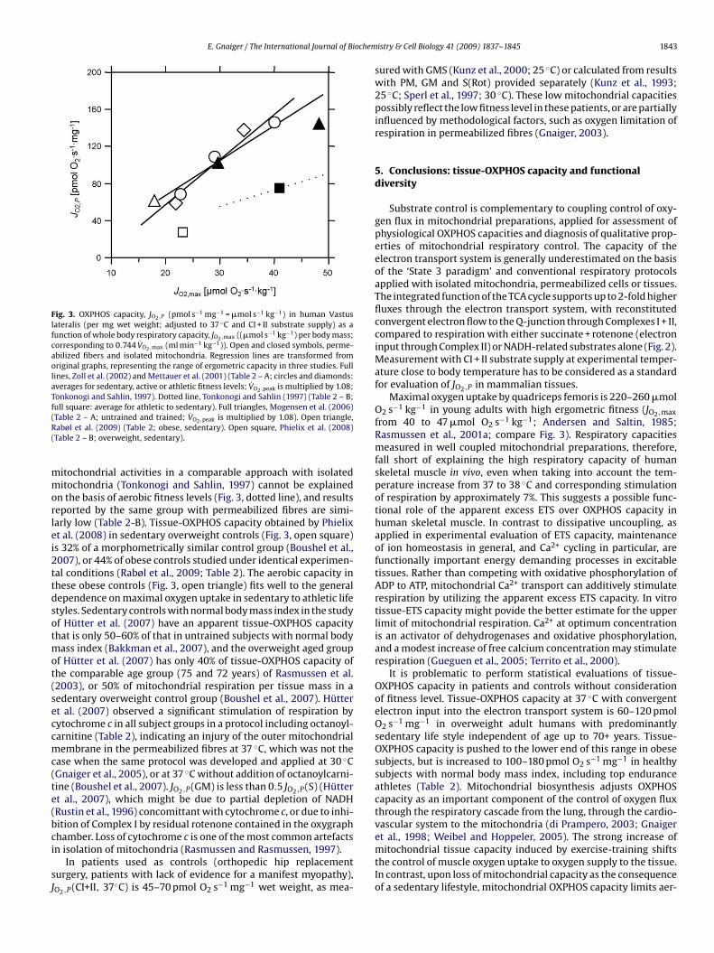
sible outliers (Fig. 3). Numerous potential artefacts are known
to reduce mitochondrial respiration (Rasmussen and Rasmussen,
1997), whereas systematic errors rarely cause an overestimation of
oxygen flux. High tissue-OXPHOS capacities and an identical depen-
dence on maximal whole-body oxygen uptake are obtained with
permeabilized fibres (Mettauer et al., 2001; Zoll et al., 2002) and
isolated mitochondria (Mogensen et al., 2006) (Fig. 3). The low
E. Gnaiger / The International Journal of Biochemistry & Cell Biology 41 (2009) 1837–1845 1843
Fig. 3. OXPHOS capacity, JO2,P (pmol s−1 mg−1 = �mol s−1 kg−1) in human Vastus
lateralis (per mg wet weight; adjusted to 37 ◦C and CI + II substrate supply) as a
function of whole body respiratory capacity, JO2,max ((�mol s−1 kg−1) per body mass;
corresponding to 0.744 V̇O2,max (ml min−1 kg−1)). Open and closed symbols, perme-
abilized fibers and isolated mitochondria. Regression lines are transformed from
original graphs, representing the range of ergometric capacity in three studies. Full
lines, Zoll et al. (2002) and Mettauer et al. (2001) (Table 2 – A; circles and diamonds:
averages for sedentary, active or athletic fitness levels; V̇O2,peak is multiplied by 1.08;
Tonkonogi and Sahlin, 1997). Dotted line, Tonkonogi and Sahlin (1997) (Table 2 – B;
full square: average for athletic to sedentary). Full triangles, Mogensen et al. (2006)
(Table 2 – A; untrained and trained; V̇O2,peak is multiplied by 1.08). Open triangle,
Rabøl et al. (2009) (Table 2; obese, sedentary). Open square, Phielix et al. (2008)
(Table 2 – B; overweight, sedentary).
mitochondrial activities in a comparable approach with isolated
mitochondria (Tonkonogi and Sahlin, 1997) cannot be explained
on the basis of aerobic fitness levels (Fig. 3, dotted line), and results
reported by the same group with permeabilized fibres are simi-
larly low (Table 2-B). Tissue-OXPHOS capacity obtained by Phielix
et al. (2008) in sedentary overweight controls (Fig. 3, open square)
is 32% of a morphometrically similar control group (Boushel et al.,
2007), or 44% of obese controls studied under identical experimen-
tal conditions (Rabøl et al., 2009; Table 2). The aerobic capacity in
these obese controls (Fig. 3, open triangle) fits well to the general
dependence on maximal oxygen uptake in sedentary to athletic life
styles. Sedentary controls with normal body mass index in the study
of Hütter et al. (2007) have an apparent tissue-OXPHOS capacity
that is only 50–60% of that in untrained subjects with normal body
mass index (Bakkman et al., 2007), and the overweight aged group
of Hütter et al. (2007) has only 40% of tissue-OXPHOS capacity of
the comparable age group (75 and 72 years) of Rasmussen et al.
(2003), or 50% of mitochondrial respiration per tissue mass in a
sedentary overweight control group (Boushel et al., 2007). Hütter
et al. (2007) observed a significant stimulation of respiration by
cytochrome c in all subject groups in a protocol including octanoyl-
carnitine (Table 2), indicating an injury of the outer mitochondrial
membrane in the permeabilized fibres at 37 ◦C, which was not the
case when the same protocol was developed and applied at 30 ◦C
(Gnaiger et al., 2005), or at 37 ◦C without addition of octanoylcarni-
tine (Boushel et al., 2007). JO2,P(GM) is less than 0.5 JO2,P(S) (Hütter
et al., 2007), which might be due to partial depletion of NADH
(Rustin et al., 1996) concomittant with cytochrome c, or due to inhi-
bition of Complex I by residual rotenone contained in the oxygraph
chamber. Loss of cytochrome c is one of the most common artefacts
in isolation of mitochondria (Rasmussen and Rasmussen, 1997).
In patients used as controls (orthopedic hip replacement
surgery, patients with lack of evidence for a manifest myopathy),
JO2,P(CI+II, 37◦C) is 45–70 pmol O2 s−1 mg−1 wet weight, as mea-
sured with GMS (Kunz et al., 2000; 25 ◦C) or calculated from results
with PM, GM and S(Rot) provided separately (Kunz et al., 1993;
25 ◦C; Sperl et al., 1997; 30 ◦C). These low mitochondrial capacities
possibly reflect the low fitness level in these patients, or are partially
influenced by methodological factors, such as oxygen limitation of
respiration in permeabilized fibres (Gnaiger, 2003).
5. Conclusions: tissue-OXPHOS capacity and functional
diversity
Substrate control is complementary to coupling control of oxy-
gen flux in mitochondrial preparations, applied for assessment of
physiological OXPHOS capacities and diagnosis of qualitative prop-
erties of mitochondrial respiratory control. The capacity of the
electron transport system is generally underestimated on the basis
of the ‘State 3 paradigm’ and conventional respiratory protocols
applied with isolated mitochondria, permeabilized cells or tissues.
The integrated function of the TCA cycle supports up to 2-fold higher
fluxes through the electron transport system, with reconstituted
convergent electron flow to the Q-junction through Complexes I + II,
compared to respiration with either succinate + rotenone (electron
input through Complex II) or NADH-related substrates alone (Fig. 2).
Measurement with CI + II substrate supply at experimental temper-
ature close to body temperature has to be considered as a standard
for evaluation of JO2,P in mammalian tissues.
Maximal oxygen uptake by quadriceps femoris is 220–260 �mol
O2 s−1 kg−1 in young adults with high ergometric fitness (JO2,max
from 40 to 47 �mol O2 s−1 kg−1; Andersen and Saltin, 1985;
Rasmussen et al., 2001a; compare Fig. 3). Respiratory capacities
measured in well coupled mitochondrial preparations, therefore,
fall short of explaining the high respiratory capacity of human
skeletal muscle in vivo, even when taking into account the tem-
perature increase from 37 to 38 ◦C and corresponding stimulation
of respiration by approximately 7%. This suggests a possible func-
tional role of the apparent excess ETS over OXPHOS capacity in
human skeletal muscle. In contrast to dissipative uncoupling, as
applied in experimental evaluation of ETS capacity, maintenance
of ion homeostasis in general, and Ca2+ cycling in particular, are
functionally important energy demanding processes in excitable
tissues. Rather than competing with oxidative phosphorylation of
ADP to ATP, mitochondrial Ca2+ transport can additively stimulate
respiration by utilizing the apparent excess ETS capacity. In vitro
tissue-ETS capacity might povide the better estimate for the upper
limit of mitochondrial respiration. Ca2+ at optimum concentration
is an activator of dehydrogenases and oxidative phosphorylation,
and a modest increase of free calcium concentration may stimulate
respiration (Gueguen et al., 2005; Territo et al., 2000).
It is problematic to perform statistical evaluations of tissue-
OXPHOS capacity in patients and controls without consideration
of fitness level. Tissue-OXPHOS capacity at 37 ◦C with convergent
electron input into the electron transport system is 60–120 pmol
O2 s−1 mg−1 in overweight adult humans with predominantly
sedentary life style independent of age up to 70+ years. Tissue-
OXPHOS capacity is pushed to the lower end of this range in obese
subjects, but is increased to 100–180 pmol O2 s−1 mg−1 in healthy
subjects with normal body mass index, including top endurance
athletes (Table 2). Mitochondrial biosynthesis adjusts OXPHOS
capacity as an important component of the control of oxygen flux
through the respiratory cascade from the lung, through the cardio-
vascular system to the mitochondria (di Prampero, 2003; Gnaiger
et al., 1998; Weibel and Hoppeler, 2005). The strong increase of
mitochondrial tissue capacity induced by exercise-training shifts
the control of muscle oxygen uptake to oxygen supply to the tissue.
In contrast, upon loss of mitochondrial capacity as the consequence
of a sedentary lifestyle, mitochondrial OXPHOS capacity limits aer-
1844 E. Gnaiger / The International Journal of Biochemistry & Cell Biology 41 (2009) 1837–1845
obic exercise capacity in untrained young individuals (Haseler et al.,
2004) and contributes to the loss of aerobic power with advancing
age in sedentary people (Short et al., 2005). Upregulation of proin-
flammatory cytokines such as TNF-� is associated with obesity
as a state of chronic low-grade inflammation, disrupting the sig-
nalling cascade for mitochondrial biogenesis (Valerio et al., 2006).
This molecular mechanism contributes, therefore, to the decline of
tissue-OXPHOS capacity in the progression towards obesity (Fig. 3).
Considering the range from 60 to 180 pmol O2 s−1 mg−1, the
tissue-OXPHOS capacity increases 3-fold from sedentary to athletic
humans, comparable to the 2.7-fold difference of mitochondrial
density between types I and IIb skeletal muscle (rabbit soleus and
gracilis; Jackman and Willis, 1996). Mitochondrial volume per body
mass and maximal oxygen consumption per body mass increase
2.5-fold when comparing athletic/sedentary animals (dog/goat;
pony/calf; horse/steer; Weibel et al., 1991). This co-variation of
functional capacity and mitochondrial density has established the
paradigm that a higher respiratory capacity in skeletal muscle of
mammalian species – from the 30 g mouse to the 500 kg horse – is
achieved by simply building ‘more mitochondria of the same kind’
(Weibel et al., 1991; Weibel and Hoppeler, 2005). Comparison of
JO2,P versus JO2,E (P/E coupling control ratios) and substrate control
in mitochondria of human and mouse skeletal muscle, however,
provides direct evidence for the fact that mitochondrial plasticity
does not only involve a change in structure (mitochondrial density),
but qualitative differences are highly significant in mitochondrial
physiology of mouse (Aragonés et al., 2008) and man (Boushel et al.,
2007). Further investigations of a wide range of species, tissues and
fitness levels are required for describing the qualitative differences
of mitochondria in terms of substrate control and coupling control.
Without a proper comparative foundation of mitochondrial physi-
ology, we cannot at present understand the functional implications
of mitochondrial diversity from mouse to man, nor can we evaluate
properly different tissue-OXPHOS and ETS capacities in health and
disease.
Acknowledgements
I thank Robert Boushel, Hélène Lemieux, Steven Hand, Kathrin
Renner, Dominique Votion, Bengt Kayser, Richard Haas, Charles
Hoppel, Rodrigue Rossignol, Guy Brown and many unnamed col-
leagues for sharing their views on mitochondrial physiology during
joint projects, numerous courses on high-resolution respirometry
and Mitochondrial Physiology Conferences.
References
Andersen P, Saltin B. Maximal perfusion of skeletal muscle in man. J Physiol1985;366:233–49.
Anderson EJ, Lustig ME, Boyle KE, Woodlief TL, Kane DA, Lin C-T, et al. MitochondrialH2O2 emission and cellular redox state link excess fat intake to insulin resistancein both rodents and humans. J Clin Invest 2009;119:573–81.
Aragonés J, Schneider M, Van Geyte K, Fraisl P, Dresselaers T, Mazzone M, et al.Deficiency or inhibition of oxygen sensor Phd1 induces hypoxia tolerance byreprogramming basal metabolism. Nat Genetics 2008;40:170–80.
Bakkman L, Sahlin K, Holmberg HC, Tonkonogi M. Quantitative and qualita-tive adaptation of human skeletal muscle mitochondria to hypoxic comparedwith normoxic training at the same relative work rate. Acta Physiol (Oxford)2007;190:243–51.
Bangsbo J, Gibala MJ, Howarth KR, Krustrup P. Tricarboxylic acid cycle intermediatesaccumulate at the onset of intense exercise in man but are not essential for theincrease in muscle oxygen uptake. Pflugers Arch 2006;452:737–43.
Benard G, Bellance N, James D, Parrone P, Fernandez H, Letellier T, et al. Mitochondrialbioenergetics and structural network organization. J Cell Sci 2007;120:838–48.
Benard G, Faustin B, Galinier A, Rocher C, Bellance N, Smolkova K, et al. Functionaldynamic compartmentalization of respiratory chain intermediate substrates:implications for the control of energy production and mitochondrdial diseases.Intl J Biochem Cell Biol 2008;40:1543–54.
Blomstrand E, Raedegran G, Saltin B. Maximum rate of oxygen uptake by humanskeletal muscle in relation to maximal activities of enzymes in the Krebs cycle.J Physiol 1997;501:455–60.
Boushel R, Gnaiger E, Schjerling P, Skovbro M, Kraunsoe R, Flemming D. Patientswith Type 2 diabetes have normal mitochondrial function in skeletal muscle.Diabetologia 2007;50:790–6.
Chance B, Williams GR. Respiratory enzymes in oxidative phosphorylation. IV. Therespiratory chain. J Biol Chem 1955;217:429–38.
Chance B, Williams GR. The respiratory chain and oxidative phosphorylation. AdvEnzymol 1956;17:65–134.
Daussin FN, Zoll J, Dufour SP, Ponsot E, Lonsdorfer-Wolf E, Doutreleau S, et al. Effectof interval versus continuous training on cardiorespiratory and mitochondrialfunctions: relationship to aerobic performance improvements in sedentary sub-jects. Am J Physiol Regul Integr Comp Physiol 2008a;295:R264–72.
Daussin FN, Zoll J, Ponsot E, Dufour SP, Doutreleau S, Lonsdorfer E, et al. Trainingat high exercise intensity promotes qualitative adaptations of mitochondrialfunction in human skeletal muscle. J Appl Physiol 2008b;104:1436–41.
Dawson KD, Baker DJ, Greenhaff PL, Gibala MJ. An accute decrease in TCA cycleintermediates does not affect aerobic energy delivery in contracting rat skeletalmuscle. J Physiol 2005;565:637–43.
di Prampero PE. Factors limiting maximal performance in humans. Eur J Appl Physiol2003;90:420–9.
Echaniz-Laguna A, Zoll J, Ribera F, Tranchant C, Warter JM, Lonsdorfer J, et al. Mito-chondrial respiratory chain function in skeletal muscle of ALS patients. AnnNeurol 2002;52:623–7.
Garnier A, Fortin D, Zoll J, N’guessan B, Mettauer B, Lampert E, et al. Cooredi-nated changes in mitochondrial function and biogenesis in healthy and diseasedhuman skeletal muscle. FASEB J 2005;19:43–52.
Gnaiger E. Bioenergetics at low oxygen: dependence of respiration and phos-phorylation on oxygen and adenosine diphosphate supply. Respir Physiol2001;128:277–97.
Gnaiger E. Oxygen conformance of cellular respiration. A perspective of mitochon-drial physiology. Adv Exp Med Biol 2003;543:39–55.
Gnaiger E. (Ed.), Mitochondrial pathways and respiratory control. 1st ed. Innsbruck:OROBOROS MiPNet Publications; 2007. Electronic ISBN 978-3-9502399-0-4.www.oroboros.at.
Gnaiger E. Polarographic oxygen sensors the oxygraph and high-resolutionrespirometry to assess mitochondrial function. In: Dykens JA, Will Y, edi-tors. Mitochondrial dysfunction in drug-induced toxicity. John Wiley; 2008. p.327–52.
Gnaiger E, Kuznetsov AV, Schneeberger S, Seiler R, Brandacher G, Steurer W, et al.Mitochondria in the cold. In: Heldmaier G, Klingenspor M, editors. Life in thecold. Berlin, New York: Heidelberg Springer; 2000a. p. 431–42.
Gnaiger E, Lassnig B, Kuznetsov AV, Rieger G, Margreiter R. Mitochondrial oxygenaffinity, respiratory flux control, and excess capacity of cytochrome c oxidase. JExp Biol 1998;201:1129–39.
Gnaiger E, Méndez G, Hand SC. High phosphorylation efficiency and depression ofuncoupled respiration in mitochondria under hypoxia. Proc Natl Acad Sci USA2000b;97:11080–5.
Gnaiger E, Steinlechner-Maran R, Méndez G, Eberl T, Margreiter R. Controlof mitochondrial and cellular respiration by oxygen. J Bioenerg Biomembr1995;27:583–96.
Gnaiger E, Wright-Paradis C, Sondergaard H, Lundby C, Calbet JA, Saltin B, et al.High-resolution respirometry in small biopsies of human muscle: correlationswith body mass index and age. Mitochondr Physiol Network 2005;10(9):14–5,www.mitophysiology.org/index.php?gnaigere.
Gueguen N, Lefaucheur L, Ecolan P, Fillaut M, Herpin P. Ca2+-activated myosin-ATPases, creatine and adenylate kinases regulate mitochondrial functionaccording to myofibre type in rabbit. J Physiol 2005;564:723–35.
Haseler LJ, Lin AP, Richardson RS. Skeletal muscle oxidative metabolism in seden-tary humans: 31P-MRS assessment of O2 supply and demand limitations. J ApplPhysiol 2004;97:1077–81.
Hoppeler H, Howald H, Conley K, Lindstedt SL, Claassen H, Vock P, et al. Endurancetraining in humans: aerobic capacity and structure of skeletal muscle. J ApplPhysiol 1985;59:320–7.
Hütter E, Skovbro M, Lener B, Prats C, Rabol R, Dela F, et al. Oxidative stress andmitochondrial impairment can be separated from lipofuscin accumulation inaged human skeletal muscle. Aging Cell 2007;6:245–56.
Jackman MR, Willis WT. Characteristics of mitochondria isolated from type I andtype IIb skeletal muscle. Am J Physiol 1996;270:C673–678.
Johnson G, Roussel D, Dumas JF, Douay O, Malthièry Y, Simard G, et al. Influ-ence of intensity of food restriction on skeletal muscle mitochondrial energymetabolism in rats. Am J Physiol Endocrinol Metab 2006;291:E460–7.
Jüllig M, Hickey AJ, Chai CC, Skea GL, Middleditch MJ, Costa S, et al. Is the failingheart out of fuel or a worn engine running rich? A study of mitochondria in oldspontaneously hypertensive rats. Proteomics 2008;8:2556–72.
Kunz WS, Kudin A, Vielhaber S, Elger CE, Attardi G, Villani G. Flux control ofcytochrome c oxidase in human skeletal muscle. J Biol Chem 2000;275:27741–5.
Kunz WS, Kuznetsov AV, Schulze W, Eichhorn K, Schild L, Striggow F, et al.Functional characterization of mitochondrial oxidative phosphorylation insaponin-skinned human muscle fibers. Biochim Biophys Acta 1993;1144:46–53.
Kuznetsov AV, Schneeberger S, Seiler R, Brandacher G, Mark W, Steurer W, et al.Mitochondrial defects and heterogeneous cytochrome c release after cardiac coldischemia and reperfusion. Am J Physiol Heart Circ Physiol 2004;286:H1633–41.
Kuznetsov AV, Strobl D, Ruttmann E, Königsrainer A, Margreiter R, Gnaiger E. Eval-uation of mitochondrial respiratory function in small biopsies of liver. AnalBiochem 2002;305:186–94.
Lemasters JJ. The ATP-to-oxygen stoichiometries of oxidative phosphorylation by ratliver mitochondria. J Biol Chem 1984;259:13123–30.
E. Gnaiger / The International Journal of Biochemistry & Cell Biology 41 (2009) 1837–1845 1845
Lemieux H, Garedew A, Blier PU, Tardif J-C, Gnaiger E. Temperature effects on thecontrol and capacity of mitochondrial respiration in permeabilized fibers of themouse heart. Biochim Biophys Acta 2006;14:201–2, EBEC Short Reports Supple-ment.
Letellier T, Malgat M, Coquet M, Moretto B, Parrot-Roulaud F, Mazat JP. Mitochondrialmyopathy studies on permeabilized muscle fibers. Pediatr Res 1992;32:17–22.
Mettauer B, Zoll J, Sanchez H, Lampert E, Ribera F, Veksler V, et al. Oxidative capacityof skeletal muscle in heart failure patients versus sedentary or active controlsubjects. J Am Coll Cardiol 2001;38:947–54.
Mitchell JH, Saltin B. The oxygen transport system and maximal oxygen uptake. In:Exercise physiology. Oxford Univ. Press; 2003.
Mitchell P, Moyle J. Respiration-driven proton translocation in rat liver mitochondria.Biochem J 1967;105:1147–62.
Mogensen M, Bagger M, Pedersen PK, Fernström M, Sahlin K. Cycling efficiency inhumans is related to low UCP3 content and to type I fibres but not to mitochon-drial efficiency. J Physiol 2006;571:669–81.
N’Guessan B, Zoll J, Ribera F, Ponsot E, Lampert E, Ventura-Clapier R, et al. Evalua-tion of quantitative and qualitative aspects of mitochondrial function in humanskeletal and cardiac muscles. Mol Cell Biochem 2004;256–257:267–80.
Phielix E, Schrauwen-Hinderling VB, Mensink M, Lenaers E, Meex R, Hoeks J, etal. Lower intrinsic ADP-stimulated mitochondrial respiration underlies in vivomitochondrial dysfunction in muscle of male type 2 diabetic patients. Diabetes2008;57:2943–9.
Ponsot E, Zoll J, N’guessan B, Ribera F, Lampert F, Richard E, et al. Mitochondrialtissue specificity of substrates utilization in rat cardiac and skeletal muscles. JCell Physiol 2005;203:479–86.
Puchowicz MA, Varnes ME, Cohen BH, Friedman NR, Kerr DS, Hoppel CL. Oxidativephosphorylation analysis: assessing the integrated functional activity of humanskeletal muscle mitochondria – case studies. Mitochondrion 2004;4:377–85.
Rabøl R, Højberg PMV, Almdal T, Boushel R, Haugaard SB, Madsbad S, Dela F. Effect ofhyperglycemia on mitochondrial respiration in type 2 diabetes. J Clin EndocrinolMetab 2009;94:1372–8.
Rasmussen HN, Rasmussen UF. Small scale preparation of skeletal muscle mitochon-dria, criteria for integrity, and assays with reference to tissue function. Mol CellBiochem 1997;174:55–60.
Rasmussen UF, Rasmussen HN. Human quadriceps muscle mitochondria: a func-tional characterization. Mol Cell Biochem 2000;208:37–44.
Rasmussen UF, Rasmussen HN, Andersen AJ, Fogd Jorgensen P, Quistorff B. Char-acterization of mitochondria from pig muscle: higher activity of exo-NADHoxidase in animals suffering from malignant hyperthermia. Biochem J 1996;315:659–63.
Rasmussen UF, Krustrup P, Bangbo J, Rasmussen HN. The effect of high-intensityexhaustive exercise studied in isolated mitochondria from human skeletal mus-cle. Eur J Physiol 2001a;443:180–7.
Rasmussen UF, Krustrup P, Kjaer M, Rasmussen HN. Experimental evidence aginastthe mitochondrial theory of aging: a study of isolated human skeletal musclemitochondria. Exp Gerontol 2003;38:877–86.
Rasmussen UF, Rasmussen HN, Krustrup P, Quistorff B, Saltin B, Bangsbo J. Aerobicmetabolism of human quadriceps muscle: in vivo data parallel measurementson isolated mitochondria. Am J Physiol Endocrinol Metab 2001b;280:E301–7.
Rasmussen UF, Vielwerth SE, Rasmussen HN. Skeletal muscle bioenergetics: a com-parative study of mitochondria isolated from pigeon pectoralis, rat soleus, ratbiceps brachii, pig biceps femoris and human quadriceps. Comp Biochem PhysiolA Mol Integr Physiol 2004;137:435–46.
Richardson RS, Leigh JS, Wagner PD, Noyszewski EA. Cellular PO2 as a determinantof maximal mitochondrial O2 consumption in trained human skeletal muscle. JAppl Physiol 1999;87:325–31.
Rossignol R, Faustin B, Rocher C, Malgat M, Mazat JP, Letellier T. Mitochondrial thresh-old effects. Biochem J 2003;370:751–62.
Rostovtseva TK, Sheldon KL, Hassanzadeh E, Monge C, Saks V, Bezrukov SM, et al.Tubulin binding blocks mitochondrial voltage-dependent anion channel andregulates respiration. Proc Natl Acad Sci USA 2008;105:18746–51.
Rustin P, Parfait B, Chretien D, Bourgeron T, Djouadi F, Bastin J, et al. Fluxes of nicoti-namide adenine dinucleotides through mitochondrial membranes in humancultured cells. J Biol Chem 1996;271:14785–90.
Saks VA, Veksler VI, Kuznetsov AV, Kay L, Sikk P, Tiivel T, et al. Permeabilised cell andskinned fiber techniques in studies of mitochondrial function in vivo. Mol CellBiochem 1998;184:81–100.
Scandurra FM, Gnaiger E. Cell respiration under hypoxia: facts and artefacts in mito-chondrial oxygen kinetics. Adv Exp Med Biol 2009.
Scheibye-Knudsen M, Quistorff B. Regulation of mitochondrial respiration byinorganic phosphate; comparing permeabilized muscle fibers and isolated mito-chondria prepared from type-1 and type-2 rat skeletal muscle. Eur J Appl Physiol2009;105:279–87.
Schwerzmann K, Hoppeler H, Kayar SR, Weibel ER. Oxidative capacity of muscleand mitochondria: Correlation of physiological, biochemical and morphometriccharacteristics. Proc Natl Acad Sci USA 1989;86:1583–7.
Short KR, Bigelow ML, Kahl J, Singh R, Coenen-Schimke J, Raghavakaimal S, et al.Decline in skeletal muscle mitochondrial function with aging in humans. ProcNatl Acad Sci USA 2005;102:5618–23.
Sperl W, Skladal D, Gnaiger E, Wyss M, Mayr U, Hager J, et al. High resolutionrespirometry of premeabilized skeletal muscle fibers in the diagnosis of neu-romuscular disorders. Mol Cell Biochem 1997;174:71–8.
Starritt EC, Angus D, Hargreaves M. Effect of short-term training on mitochondrialATP production rate in human skeletal muscle. J Appl Physiol 1999;86:450–4.
Territo PR, Mootha VK, French SA, Balaban RS. Ca2+ activation of heart mitochondrialoxidative phosphorylation: role of the F0/F1-ATPase. Am J Phyiol Cell Physiol2000;278:C423–35.
Tonkonogi M, Harris B, Sahlin K. Mitochondrial oxidative function in human saponin-skinned muscle fibres: effects of prolonged exercise. J Physiol 1998;510:279–86.
Tonkonogi M, Sahlin K. Rate of oxidative phosphorylation in isolated mitochon-dria from human skeletal muscle: effect of training status. Acta Physiol Scand1997;161:345–53.
Tonkonogi M, Walsh B, Tiivel T, Saks V, Sahlin K. Mitochondrial function inhuman skeletal muscle is not impaired by high intensity exercise. Eur J Physiol1999;437:562–8.
Tudella VG, Curti C, Soriani FM, Santos AC, Uyemura SA. In situ evidence of an alter-native oxidase and an uncoupling protein in the respiratory chain of Aspergillusfumigatus. Intl J Biochem Cell Biol 2003;36:162–72.
Turner DL, Hoppeler H, Claassen H, Vock P, Kayser B, Schena F, et al. Effects ofendurance training on oxidative capacity and structural composition of humanarm and leg muscles. Acta Physiol Scand 1997;161:459–64.
Valerio A, Cardile A, Cozzi V, Bracale R, Tedesco L, Pisconti A, et al. TNF-� downregu-lates eNOS expression and mitochondrial biogenesis in fat and muscle of obeserodents. J Clin Invest 2006;116:2791–8.
Veksler VI, Kuznetsov AV, Sharov VG, Kapelko VI, Saks VA. Mitochondrial respiratoryparameters in cardiac tissue: a novel method of assessment by using saponin-skinned fibers. Biochim Biophys Acta 1987;892:191–6.
Walsh B, Tonkonogi M, Sahlin K. Effect of endurance training on oxidative andantoxidiative function in human permeabilized muscle fibres. Pflügers Arch2001;442:420–5.
Weibel ER, Hoppeler H. Exercise-induced maximal metabolic rates scales with mus-cle aerobic capacity. J Exp Biol 2005;208:1635–44.
Weibel ER, Taylor CR, Hoppeler H. The concept of symmorphosis: a testable hypothe-sis of structure-function relationship. Proc Natl Acad Sci USA 1991;88:10357–61.
Wibom R, Hultman E, Johansson M, Matherei K, Constantin-Teodosiu D, SchantzPG. Adaptation of mitochondrial ATP production in human skeletal muscle toendurance training and detraining. J Appl Physiol 1992;73:2004–10.
Wijers SL, Schrauwen P, Saris WH, van Marken Lichtenbelt WD. Human skeletalmuscle mitochondrial uncoupling is associated with cold induced adaptive ther-mogenesis. PLoS ONE 2008;3(3):e1777.
Zoll J, Sanchez H, N’Guessan B, Ribera F, Lampert E, Bigard X, et al. Physical activitychanges the regulation of mitochondrial respiration in human skeletal muscle. JPhysiol 2002;543:191–200.


MitoFit in health and protective medicine
Physical inactivity is one of the greatest drivers of metabolic dysregulation
and preventable diseases in modern societies and has an impact on ageing populations in particular. It is becoming increasingly apparent that
high and robust mitochondrial capacity is central to sustain metabolic health throughout life. An active lifestyle increases mitochondrial capacity
and, therefore, reduces the risk of degenerative diseases.
High and efficient mitochondrial performance prevents metabolic diseases,
promotes health and improves quality of life in ageing populations. Surprisingly, however, there is currently no regimented, quantitative
system or database dedicated to routinely test, compare and monitor mitochondrial function in individuals and populations.
MitoFit develops novel laboratory standards and diagnostic monitoring of a mitochondrial fitness score. MitoFit provides a signature for high-end
health tourism, introducing a scientific perspective on the benefits of mitochondrial fitness.
The OROBOROS O2k is the only instrument with sufficient stability and reliability for quantitative high-resolution respirometry (HRR). This world-
leading technology introduced the quality control tools required for calibrated and standardised in vitro diagnostics – with the final aim to
maintain mitochondria fit and well - to live a healthier, longer life.
The project MitoFit is
funded by the Land Tirol
within the program
K-Regio of Standort-
agentur Tirol.
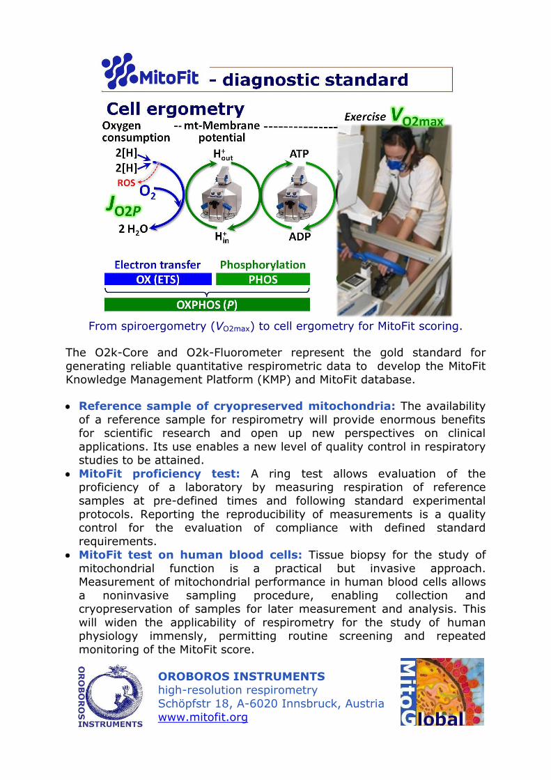
From spiroergometry (VO2max) to cell ergometry for MitoFit scoring.
The O2k-Core and O2k-Fluorometer represent the gold standard for
generating reliable quantitative respirometric data to develop the MitoFit Knowledge Management Platform (KMP) and MitoFit database.
Reference sample of cryopreserved mitochondria: The availability
of a reference sample for respirometry will provide enormous benefits for scientific research and open up new perspectives on clinical
applications. Its use enables a new level of quality control in respiratory studies to be attained.
MitoFit proficiency test: A ring test allows evaluation of the proficiency of a laboratory by measuring respiration of reference
samples at pre-defined times and following standard experimental
protocols. Reporting the reproducibility of measurements is a quality control for the evaluation of compliance with defined standard
requirements. MitoFit test on human blood cells: Tissue biopsy for the study of
mitochondrial function is a practical but invasive approach. Measurement of mitochondrial performance in human blood cells allows
a noninvasive sampling procedure, enabling collection and cryopreservation of samples for later measurement and analysis. This
will widen the applicability of respirometry for the study of human physiology immensly, permitting routine screening and repeated
monitoring of the MitoFit score.
OROBOROS INSTRUMENTS high-resolution respirometry
Schöpfstr 18, A-6020 Innsbruck, Austria
www.mitofit.org