CONTACT TRANSFER OF VX FROM CONTAMINATED … · thank dr. bruce king ... contact transfer of vx...
30
CONTACT TRANSFER OF VX FROM CONTAMINATED GRASS ONTO ARMY COMBAT UNIFORM ECBC-TR-1429 Mark V. Haley Ronald T. Checkai Michael Simini Richard J. Lawrence RESEARCH AND TECHNOLOGY DIRECTORATE Michael W. Busch EXCET, INC. Springfield, VA 22150-2519 January 2017 Approved for public release; distribution unlimited.
Transcript of CONTACT TRANSFER OF VX FROM CONTAMINATED … · thank dr. bruce king ... contact transfer of vx...

CONTACT TRANSFER OF VX FROM CONTAMINATED GRASS ONTO ARMY COMBAT UNIFORM
ECBC-TR-1429
Mark V. Haley Ronald T. Checkai
Michael Simini Richard J. Lawrence
RESEARCH AND TECHNOLOGY DIRECTORATE
Michael W. Busch
EXCET, INC.
Springfield, VA 22150-2519
January 2017
Approved for public release; distribution unlimited.

Disclaimer
The findings in this report are not to be construed as an official Department of the Army position unless so designated by other authorizing documents.
REPORT DOCUMENTATION PAGE Form Approved
OMB No. 0704-0188 Public reporting burden for this collection of information is estimated to average 1 h per response, including the time for reviewing instructions, searching existing data sources, gathering and maintaining the data needed, and completing and reviewing this collection of information. Send comments regarding this burden estimate or any other aspect of this collection of information, including suggestions for reducing this burden to Department of Defense, Washington Headquarters Services, Directorate for Information Operations and Reports (0704-0188), 1215 Jefferson Davis Highway, Suite 1204, Arlington, VA 22202-4302. Respondents should be aware that notwithstanding any other provision of law, no person shall be subject to any penalty for failing to comply with a collection of information if it does not display a currently valid OMB control number. PLEASE DO NOT RETURN YOUR FORM TO THE ABOVE ADDRESS.
1. REPORT DATE (DD-MM-YYYY)
XX-01-2017 2. REPORT TYPE
Final 3. DATES COVERED (From - To)
May 2014 – Sep 2015 4. TITLE AND SUBTITLE
Contact Transfer of VX from Contaminated Grass onto Army Combat Uniform 5a. CONTRACT NUMBER
5b. GRANT NUMBER
5c. PROGRAM ELEMENT NUMBER
6. AUTHOR(S)
Haley, Mark V.; Checkai, Ronald T.; Simini, Michael; Lawrence, Richard J.
(ECBC); and Busch, Michael W. (Excet)
5d. PROJECT NUMBER
WBS R.0013813.81.4 5e. TASK NUMBER
5f. WORK UNIT NUMBER
7. PERFORMING ORGANIZATION NAME(S) AND ADDRESS(ES)
Director, ECBC, ATTN: RDCB-DRT-M, APG, MD 21010-5424
Excet, Inc., 6225 Brandon Avenue, Suite 360, Springfield, VA 22150-2519
8. PERFORMING ORGANIZATION REPORT NUMBER
ECBC-TR-1429
9. SPONSORING / MONITORING AGENCY NAME(S) AND ADDRESS(ES)
Defense Threat Reduction Agency, 8725 John J. Kingman Road, MSC 6201,
Fort Belvoir, VA 22060-6201
10. SPONSOR/MONITOR’S ACRONYM(S)
DTRA 11. SPONSOR/MONITOR’S REPORT NUMBER(S)
12. DISTRIBUTION / AVAILABILITY STATEMENT
Approved for public release; distribution unlimited.
13. SUPPLEMENTARY NOTES 14. ABSTRACT:
Toxicological investigations have shown that exposure to surfaces contaminated with chemical warfare agents (CWAs) can
present a contact hazard. Previously, we developed standardized protocols for determining contact transfer (exposure) of agent
from contaminated soils onto Army Combat Boot soles and Army Combat Uniforms (ACUs). We adapted those protocols to
determine the direct contact transfer of CWA from contaminated leaf surfaces onto ACU swatches. Grass leaves (Echinochloa
crus-galli) from intact, living plants were individually contaminated with 1 μL of O-ethyl-S-(2-diisopropylaminoethyl) methyl
phosphonothioate (VX). Post-dissemination, leaves were removed, and three layers of ACU were placed atop each
contaminated leaf, so that the bottom ACU layer was in direct contact with the VX-contaminated leaf surface. The ACU layers
were covered with a Plexiglas disk (0.6 cm thick × 9.8 cm diameter) to equally distribute the force resulting from central
placement of a standard mass atop the disk. Total proportions of VX transferred from contaminated leaves to ACU at
0.017 (1 min), 0.25, 0.5, 1, and 4 h post-dissemination were approximately 71, 5, 0.8, 0.3, and 0.1%, respectively, of the VX
disseminated per leaf. Trace amounts of VX were detected in the third layers of ACU at times 0.017 and 0.25 h post-
dissemination.
15. SUBJECT TERMS
Contact transfer Foliage
Chemical warfare agent (CWA) Barnyard grass
Army Combat Uniform (ACU) Echinochloa crus-galli
O-Ethyl-S-(2-diisopropylaminoethyl) methyl phosphonothiolate (VX)
16. SECURITY CLASSIFICATION OF:
17. LIMITATION OF ABSTRACT
UU
18. NUMBER OF PAGES
30
19a. NAME OF RESPONSIBLE PERSON
Renu B. Rastogi a. REPORT
U
b. ABSTRACT
U
c. THIS PAGE
U
19b. TELEPHONE NUMBER (include area code)
(410) 436-7545 Standard Form 298 (Rev. 8-98)
Prescribed by ANSI Std. Z39.18

ii
Blank

iii
PREFACE
The work described in this report was authorized under WBS R.0013813.81.4. The
work was started in May 2014 and completed in September 2015.
The use of either trade or manufacturers’ names in this report does not constitute
an official endorsement of any commercial products. This report may not be cited for purposes of
advertisement.
This report has been approved for public release.
Acknowledgments
The authors acknowledge Dr. David J. McGarvey (U.S. Army Edgewood
Chemical Biological Center; Aberdeen Proving Ground, MD [ECBC]) for conducting VX purity
determinations by nuclear magnetic resonance spectroscopy. The authors would also like to
thank Dr. Bruce King (ECBC) and Dr. Morgan Minyard (Defense Threat Reduction Agency;
Fort Belvoir, VA [DTRA]) for consultation throughout the duration of the project. Funding for
this project was provided by DTRA.

iv
Blank

v
CONTENTS
1. INTRODUCTION ...................................................................................................1
2. METHODS ..............................................................................................................2
2.1 Chemical ............................................................................................................2
2.2 Plant Selection and Culture ................................................................................2
2.3 Transfer Material ...............................................................................................2
2.4 Dissemination of Agent Droplets onto Leaves ..................................................3
2.5 Contact Transfer Procedure ...............................................................................4
2.6 Analytical Determinations .................................................................................5
2.7 Determination of Appropriate Mass to Apply ...................................................7
2.8 Determination of Appropriate ACU Contact Time
on Agent-Contaminated Leaves .........................................................................7
2.9 Statistical Analyses ............................................................................................7
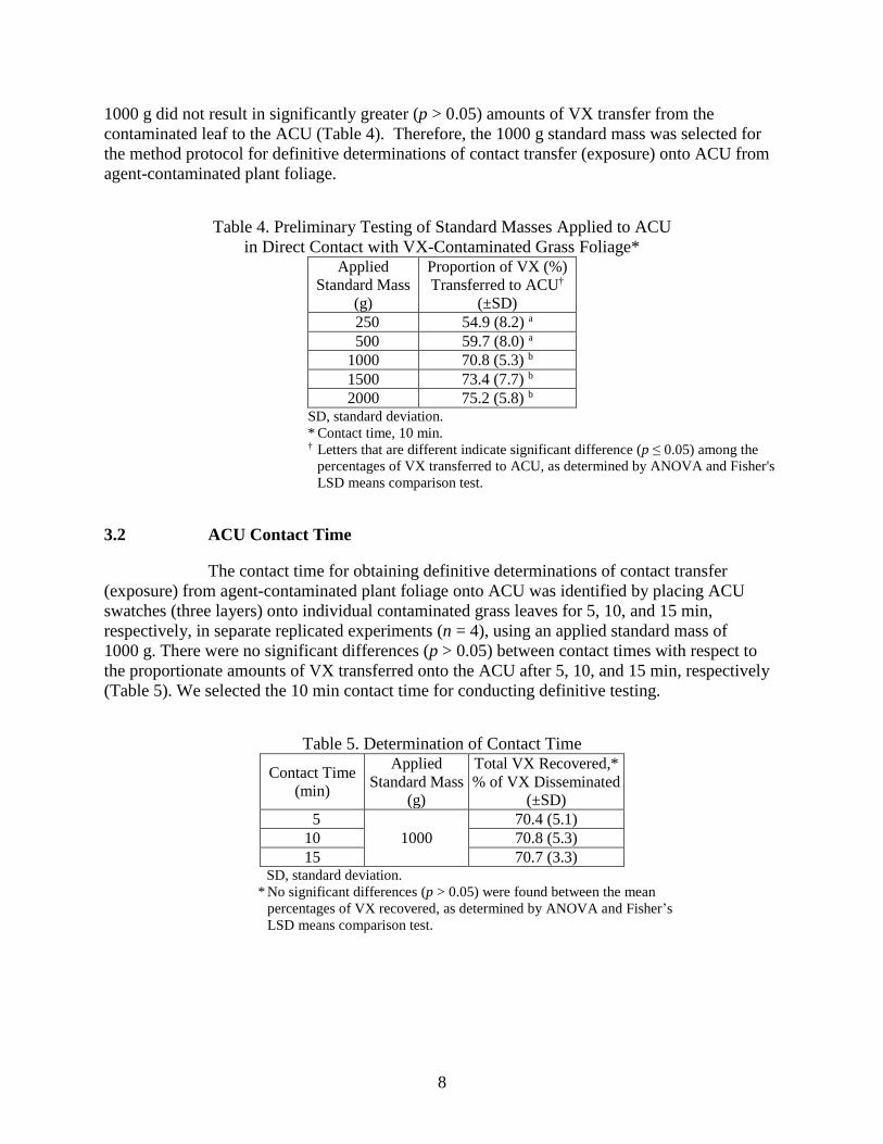
3. RESULTS ................................................................................................................7
3.1 Applied Mass .....................................................................................................7
3.2 ACU Contact Time ............................................................................................8
3.3 Agent Transferred to ACU.................................................................................9
4. DISCUSSION ........................................................................................................10
5. CONCLUSIONS....................................................................................................13
LITERATURE CITED ..........................................................................................15
ACRONYMS AND ABBREVIATIONS ..............................................................17
APPENDIX: PHYSICAL AND CHEMICAL PROPERTIES
OF MEDIUM SAND ......................................................................19

vi
FIGURES
1. E. crus-galli plants with plant stands in the surety hood .........................................3
2. Clear plastic tape was used to secure E. crus-galli leaves in a
horizontal position on the plant stands.....................................................................4
3. (A) leaf with midrib vein positioned between the M8 indicator
papers; (B) leaf with applied ACU and Plexiglas disk; (C) standard
mass (1000 g) applied to the Plexiglas disk, initiating the contact
transfer process ........................................................................................................5
TABLES
1. ACU Material Specifications ...................................................................................3
2. HPLC Gradient for VX Quantitation .......................................................................6
3 MRM Mass Transitions ...........................................................................................6
4. Preliminary Testing of Standard Masses Applied to ACU in Direct
Contact with VX-Contaminated Grass Foliage .......................................................8
5. Determination of Contact Time ...............................................................................8
6. Contact Transfer of VX from Grass Foliage onto ACU ........................................10

1
Contact Transfer of VX from Contaminated Grass Onto Army Combat Uniform
CONTACT TRANSFER OF VX FROM CONTAMINATED GRASS
ONTO ARMY COMBAT UNIFORM
1. INTRODUCTION
Little scientific information exists to describe the hazard to Soldiers that is
associated with chemical agent–plant interactions. Without a more complete understanding of
these interactions, it is difficult to predict the persistence of the potential hazard posed by agents
in the environment, specifically, that arising from the contact transfer of chemical warfare agent
(CWA) from contaminated plant foliage. Toxicological investigations have shown that exposure
to surfaces contaminated with chemical agent presents a contact hazard (Manthei et al., 1986,
1988). Because the chemical agent O-ethyl-S-(2-diisopropylaminoethyl) methyl
phosphonothiolate (VX) has a low vapor pressure, contact with contaminated surfaces is
considered to be a primary route of exposure for Soldiers.
Contact transfer (exposure) testing measures the amount of contamination on a
surface that transfers, under standardized, representative conditions, from a contaminated surface
onto a transfer material of interest. In conjunction with toxicity data, contact transfer (exposure)
results can predict contact hazards from the transmission or migration of toxic materials to skin.
Various materials, such as acrylic, glass, stainless steel, painted steel, concrete, cloth, and dental
dam, have been used by researchers as transfer materials to determine residual toxicity (Manthei
et al., 1986, 1988). In those studies, transfer materials were contaminated with agent and then
decontaminated by rinsing. The resulting transfer materials were placed directly onto rabbit skin.
Studies have also been performed whereby transfer materials were placed on rabbits before the
outer surface of the transfer material was contaminated (D’Onofrio, 2013). For all of these
studies, the objective was to use the rabbits’ toxic responses to determine the amount of chemical
agent and time (post-dissemination) that are required for the agent to penetrate the material.
In research conducted in the late 1950s (Reich, 1959a, 1959b), field plots
composed of mostly grasses were contaminated with VX. A roller covered with material was
then traversed through the field (simulating a crawling Soldier) to estimate the contact transfer
from a VX-contaminated field to a Soldier. Blotting samplers were also used to estimate the
amount of VX transferred through contact. However, in these studies, variables that are
inherently unstable in ambient field conditions were not adequately controlled. Ground
temperatures ranged from 12 to 28 °C and relative humidity ranged from 38 to 81%, thereby
resulting in highly variable data. Other studies have shown that weather conditions can affect the
persistence of agent in the environment (Reich, 1960).
In previous studies, we developed procedures to reliably determine contact
transfer of chemical agents from soil directly onto Army Combat Uniform (ACU). Standard
mass (× gravity) was used as the force to produce a standard measure of exposure potential. For
the work reported here, we adapted these methods to study the contact transfer of chemical
agents that were disseminated onto living, intact plants within chemical surety hoods under
controlled conditions, from grass foliage to ACU.

2
We investigated the amount of VX transferred from contaminated Echinochloa
crus-galli grass (commonly referred to as barnyard grass) directly onto ACU under standardized,
controlled conditions over a range of representative times post-dissemination. The information
gained from this study, in conjunction with measurements from mammalian toxicity testing, will
be useful in understanding and modeling the hazards associated with contacting VX-
contaminated foliage.
2. METHODS
2.1 Chemical
The CWA used in this study was VX (93% purity; Chemical Agent Standard
Analytical Reference Material [CASARM] grade; Chemical Abstracts Service [CAS] no. 50782-
69-9). VX was stabilized with diisopropylcarbodiimide (5% by weight; CAS no. 693-13-0;
Sigma-Aldrich; St. Louis, MO). Reagent-grade isopropyl alcohol (IPA; CAS no. 67-63-0;
Sigma-Aldrich) was used in solvent extraction.
2.2 Plant Selection and Culture
We selected the grass species E. crus-galli for this investigation. E. crus-galli is
one of the most prevalent natural grass species worldwide. It is tolerant of dry and wet natural
habitats, and it provides forage for grazing animals as well as food and habitat for wildlife
(USDA NRCS 2016). Methods were developed to enable E. crus-galli plant growth within a
chemical agent surety hood, as described in detail by Simini et al. (2016).
2.3 Transfer Material
We used ACU as our transfer material (Table 1), which was obtained from the
U.S. Army Natick Soldier Research, Development and Engineering Center (Natick, MA). To
replicate battlefield condition, the ACU material was laundered four times in accordance with
American Association of Textile Chemists and Colorists (AATCC; Research Triangle Park, NC)
Test Method 135. Laundering included the use of AATCC Standard Reference Detergent 124, a
140 °F wash temperature, an 80 °F rinse temperature, and automatic drying on the permanent
press setting. Swatches were cut from the laundered ACU to use as transfer material. ACU
swatches (2.5 × 3.2 cm) were cut using an Olfa 60 mm Quick Change rotary cutter (model
RTY3/NS; Olfa Corporation; Osaka, Japan).

3
Table 1. ACU Material Specifications* In accordance with MIL-DTL-44436A, Class 8
Wind-resistant poplin
50%–50% nylon–cotton ripstop blend
Wrinkle-resistant finish
Desert camouflage pattern * From MIL-DTL-44436A, Detail Specification: Cloth, Camouflage
Pattern, Wind Resistant Poplin, Nylon/Cotton Blend.
2.4 Dissemination of Agent Droplets onto Leaves
Plant stands were constructed to hold the pots in fixed positions, and a Petri dish
was placed under each pot (Figure 1). Each pot was secured to a ring stand with an adjustable
ring clamp. Individual E. crus-galli leaves were secured in a horizontal position before VX
dissemination to prevent uncontrolled deposition of agent during and after dissemination. Plant
leaves were laid horizontally across a ring near the top of the plant canopy and secured to the
ring by lengths of clear plastic (cellulose acetate) tape folded onto itself and placed across the
leaf surface, thus preventing the sticky adhesive from coming into contact with the leaf surface.
The ends of the folded tape were secured to the ring with additional tape while maintaining slight
pressure on the leaf surface (Figure 2). This method of securing individual leaves in a horizontal
position prevented any possible leaf surface damage caused by tape removal. It also ensured that
the disseminated agent droplets contacted the leaf surface at the intended point, and that those
locations could be easily identified for further investigation.
Figure 1. E. crus-galli plants with plant stands in the surety hood.
4
Figure 2. Clear plastic tape was used to secure E. crus-galli leaves
in a horizontal position on the plant stands.
A Hamilton Gastight syringe (Hamilton Company; Reno, NV) was used to apply
a single 1 µL VX droplet (for all experiments, average droplet size was 0.9059 ± 0.0335 mg)
onto each secured plant leaf. One droplet was applied to each single leaf to prevent the merging
of droplets (which might confound subsequent measurements). The leaves remained attached to
the plants until predetermined times after agent dissemination, when each leaf was removed and
used for the contact transfer procedures. During each experiment, at intervals during the
dissemination of agent onto foliage, individual replicate reference droplets of VX were placed in
sample vials. The amount of VX in each replicate droplet was analytically determined for quality
assurance and control purposes. The mean VX values for the respective reference droplets were
used in calculations to determine the percentages of VX that transferred from the foliage onto the
ACU. The surety hood was equipped with two LumiBar LED light strips (LumiGrow, Inc.;
Emeryville, CA). The canopy light intensity was 300 to 350 µmol s–1 m–2, as measured with an
MQ-200 Quantum sensor equipped with an AM-310 sensor wand (Apogee Instruments; Logan,
UT). The temperature within the surety hood was maintained at 22 ± 2 °C, and the relative
humidity was maintained at 50 ± 10%. The average airflow through the hood was
1.50 ± 0.09 mph, as measured at the face of the hood using an AirData multimeter (ADM-870C;
Shortridge Instruments, Inc.; Scottsdale, AZ).
2.5 Contact Transfer Procedure
In separate experiments, single 1 µL VX droplets were disseminated onto
individual plant leaves. The VX droplets were allowed to equilibrate on the leaves for 0.017,
0.25, 0.5, 1, and 4 h before individual leaf replicates were removed from the grass plants and
immediately subjected to contact transfer experiments.
When the leaf replicates were placed on a solid surface, the midrib veins on the
ventral sides of the leaves were large enough to prevent the even distribution of applied force
during the contact transfer experiments. Therefore, instead of a solid surface, we used a sand
media bed to conduct the experiments on. When the ventral sides of the leaves were pressed into

5
the sand, the applied force on the adaxial (upper) side could be uniformly distributed across the
leaf. Air-dried medium sand (as described in the appendix) was placed in glass Petri dishes
(10.0 × 2.0 cm), and the sand was settled by tapping the Petri dishes on the benchtop. A stainless
steel screed was applied to remove excess sand and level the sand surface with the top edge of
the dish (a 2.0 cm depth).
To determine whether the disseminated VX droplet was soaking through the leaf,
two pieces of M8 indicator paper were placed on the sand bed under the leaf. A space of
approximately 5 mm was left between the two M8 strips, which was sufficient to allow the veins
to depress into the sand. At a predetermined time post-dissemination, the VX-contaminated leaf
was removed from the plant by using forceps to hold the leaf approximately 3 in. (7.5 cm) from
the end closest to the stem. The leaf was then cut next to the forceps (on the side of the forceps
nearer the stem). The leaf was placed onto the sand bed, and the central midrib vein was
positioned in the space between the M8 indicator papers (Figure 3A). Layers of ACU (three
swatches, each 2.5 × 3.2 cm) were placed over the contaminated area of the leaf, and a Plexiglas
disk (0.6 cm thick × 9.8 cm diameter; Laird Plastics; Baltimore, MD) was placed atop the ACU
material (Figure 3B). A standard mass (1000 g) was centrally placed atop the Plexiglas disk
(Figure 3C) to equally distribute the force resulting from the standard mass × gravity. The stack
of ACU swatches remained in contact with the contaminated leaf for 10 min and was then
removed. Each swatch was placed in a separate sample bottle and extracted with 2 mL of IPA for
a minimum of 1 h. We used three layers of ACU to determine whether VX would penetrate
beyond the first ACU layer when in direct contact with a leaf contaminated with 1 µL of VX,
and also to ensure that the VX on the contaminated leaf would not transfer through multiple
layers onto the Plexiglas disk, and therefore, not be included in the analytical determination of
the total amount of VX transferred.
Figure 3. (A) leaf with midrib vein positioned between the M8 indicator papers; (B) leaf with
applied ACU and Plexiglas disk; (C) standard mass (1000 g) applied to the Plexiglas disk,
initiating the contact transfer process.
2.6 Analytical Determinations
Quantitative analytical VX determinations were conducted using an Agilent 6890
gas chromatography (GC) system equipped with a flame photometric detector (Agilent
Technologies; Santa Clara, CA). Quantification was achieved using an Agilent DB-5 fused silica
column (30 m × 0.32 mm, 0.5 mm film thickness). A 1 µL sample volume was injected into the
GC system using an Agilent 7683B series autosampler. The sample inlet temperature was
maintained at 225 °C in splitless mode. The initial oven temperature was 80 °C, with a
A B C

6
temperature ramp rate of 45 °C, to a final temperature of 300 °C. A nine-point calibration curve
(0.014, 0.072, 0.14, 0.73, 1.45, 3.91, 5.81, 11.62, and 23.82 ng/µL) was used to determine VX
concentrations in ACU extracts. The coefficient of determination (r2) for linear regression of the
standard curve throughout these studies was r2 = 0.9995 (±0.0003). The instrument limit of
detection was 0.005 ng/µL, based on peak-to-peak background noise for this method.
Quantitative analytical determinations of VX, for low levels of VX and
confirmation of GC results, were conducted using high-performance liquid chromatography
linked with tandem mass spectrometry (HPLC–MS/MS) with an Agilent 1260 liquid
chromatography (LC) triple-quadrupole mass spectrometer and MassHunter data acquisition and
analysis software (Agilent). The HPLC system was fitted with an Agilent Eclipse XDB-C18
column (5 µm, 4.6 × 150 mm). Sample injections were 1 µL. A 13 min separation method was
used; the composition of mobile phase A was 0.1% formic acid (v/v) in water, and mobile
phase B was 0.1% formic acid (v/v) in methanol. The gradient conditions used for LC separation
are presented in Table 2.
Table 2. HPLC Gradient for VX Quantitation
Time
(min)
Mobile Phase
(%)
A B
0 99.9 0.1
2 99.9 0.1
7 5 95
8 5 95
11 99.9 0.1
13 99.9 0.1
The HPLC column eluent was delivered to an electrospray ionization source
maintained in positive ion mode. MS/MS discrimination was performed via the multiple reaction
monitoring (MRM) technique, incorporating isotope dilution (VX-d5), using three mass
transitions: VX quantitation, VX confirmation, and VX-d5 internal standard (Table 3).
Table 3. MRM Mass Transitions
Analyte
Mass
(Da)
Precursor Product
VX-d5 internal standard 273 128
VX quantitation 268 128
VX confirmation 268 86
Calibration was conducted by plotting the relative responses of VX and VX-d5 as
functions of concentration. An 11-point calibration curve (5.0–5000 pg/µL of VX, each with
50 pg/µL of VX-d5) was used to construct a linear calibration curve (1/x weighting). All
analyzed samples were prepared to contain 50 pg/µL of VX-d5 (internal standard), and reported

7
VX concentrations were calculated by application of the equation of fit and dilution factors, as
applicable. The instrument limit of detection was 0.5 pg/µL, based on the peak-to-peak
background noise for this method.
2.7 Determination of Appropriate Mass to Apply
The standard flat-headed, stackable masses that were used in developing the
protocols for this method were obtained from Fisher Scientific (Suwanee, GA) and Central
Carolina Scale (Sanford, NC). The efficacy of a net standard mass applied to ACU was
determined to produce standardized measures of exposure. As described in Section 2.5, a 1 µL
droplet of VX was disseminated onto an intact grass leaf, and at 0.017 h (1 min) post-
dissemination, the leaf was removed from the plant and placed onto sand-bed media. ACU
swatches (three layers) were placed over the contaminated site and covered with a Plexiglas disk,
and a standard mass was applied. The net standard masses tested were 250, 500, 1000, 1500, and
2000 g, with four replicate samples (n = 4) determined per net standard mass tested. The three
swatches remained in contact with the contaminated leaf for 10 min. The swatches were then
removed, separated, and extracted individually for a minimum of 1 h with 2 mL of IPA, to
determine the relative efficacy of each net standard mass in the contact transfer process.
2.8 Determination of Appropriate ACU Contact Time on Agent-Contaminated
Leaves
We determined the appropriate amount of exposure time that the ACU swatches
should remain in contact with the contaminated leaf. As described in Section 2.5, after a 1 µL
droplet was disseminated onto a leaf and allowed to equilibrate for 0.017 h (1 min), the leaf was
removed from the plant and placed on a sand bed, three layers of ACU were placed over the VX
droplet site, a Plexiglas disk was applied to the ACU, and a 1000 g standard mass was placed
atop the Plexiglas disk. The contact times of 5, 10, and 15 min were independently tested, and
each was replicated a minimum of four times (n ≥ 4). At the conclusion of each contact time, the
ACU swatches were removed, and each was extracted separately for a minimum of 1 h with
2 mL of IPA.
2.9 Statistical Analyses
The 95% confidence level was used in all calculated determinations of statistical
significance. Analysis of variance (ANOVA) and Fisher’s least-significant difference (LSD)
means comparison test were conducted on untransformed data. Systat, version 11 (Systat
Software, Inc.; San Jose, CA) was used to perform the statistical calculations.
3. RESULTS
3.1 Applied Mass
The amount of VX transferred from a contaminated leaf (1 µL VX droplet) by
contact resulting from a 1000 g standard mass, applied to the Plexiglas disk covering the ACU
swatches, was significantly greater (probability [p] ≤ 0.05) than that from the respective forces
resulting from either the 250 or 500 g standard masses × gravity. Standard masses greater than
8
1000 g did not result in significantly greater (p > 0.05) amounts of VX transfer from the
contaminated leaf to the ACU (Table 4). Therefore, the 1000 g standard mass was selected for
the method protocol for definitive determinations of contact transfer (exposure) onto ACU from
agent-contaminated plant foliage.
Table 4. Preliminary Testing of Standard Masses Applied to ACU
in Direct Contact with VX-Contaminated Grass Foliage* Applied
Standard Mass
(g)
Proportion of VX (%)
Transferred to ACU†
(±SD)
250 54.9 (8.2) a
500 59.7 (8.0) a
1000 70.8 (5.3) b
1500 73.4 (7.7) b
2000 75.2 (5.8) b SD, standard deviation.
* Contact time, 10 min. † Letters that are different indicate significant difference (p ≤ 0.05) among the
percentages of VX transferred to ACU, as determined by ANOVA and Fisher's
LSD means comparison test.
3.2 ACU Contact Time
The contact time for obtaining definitive determinations of contact transfer
(exposure) from agent-contaminated plant foliage onto ACU was identified by placing ACU
swatches (three layers) onto individual contaminated grass leaves for 5, 10, and 15 min,
respectively, in separate replicated experiments (n = 4), using an applied standard mass of
1000 g. There were no significant differences (p > 0.05) between contact times with respect to
the proportionate amounts of VX transferred onto the ACU after 5, 10, and 15 min, respectively
(Table 5). We selected the 10 min contact time for conducting definitive testing.
Table 5. Determination of Contact Time
Contact Time
(min)
Applied
Standard Mass
(g)
Total VX Recovered,*
% of VX Disseminated
(±SD)
5
1000
70.4 (5.1)
10 70.8 (5.3)
15 70.7 (3.3) SD, standard deviation.
* No significant differences (p > 0.05) were found between the mean
percentages of VX recovered, as determined by ANOVA and Fisher’s
LSD means comparison test.

9
3.3 Agent Transferred to ACU
The average mass of VX disseminated as a single 1 µL droplet onto individual
grass leaves throughout all contact transfer testing was 0.9059 ± 0.0335 mg. Results from
replicated definitive testing indicated that the proportion of VX transferred per leaf onto the
ACU swatches from contaminated grass leaves at 1 min (0.017 h) post-dissemination was
approximately 71% of the VX disseminated (70.8 ± 5.3%; 0.605 ± 0.045 mg) (Table 6). At
0.25 h post-dissemination, the VX transfer by direct contact was approximately 5% of the VX
disseminated (5.1 ± 2.3%; 0.045 ± 0.021 mg). At 0.5 h post-dissemination, the VX transfer by
direct contact was approximately 1% of the VX disseminated, and at later times post-
dissemination (1 and 4 h), the VX transfer by direct contact was substantially <1% of the VX
disseminated. The respective amounts of VX that were absorbed into the second layers of ACU
at time points of 0.017, 0.25, 0.5, 1, and 4 h post-dissemination were <1% of the total VX
disseminated. Trace amounts of VX were detected in the third layers of ACU at time points
0.017, 0.25, and 0.5 h post-dissemination (Table 6).
Under field conditions, leaf tissues can be damaged by troop movement over
contaminated areas, and this could potentially cause the release of additional agent from
contaminated leaves. To test this hypothesis, after completion of the direct contact transfer
process, at 4 h post-dissemination, each leaf (n = 4) was placed into a glass Petri dish, and one
swatch of ACU was placed over the droplet site. A new sample vial was then used to apply
pressure to each ACU swatch, which remained in contact with the contaminated leaf, in a back-
and-forth, leaf-grinding motion. The additional amount of VX transferred onto the fresh ACU
swatch averaged approximately 0.015 mg, which represents an additional 1.6% of the amount of
VX that was initially disseminated (Table 6).
10
Table 6. Contact Transfer of VX from Grass Foliage onto ACU
Time Post-
Dissemination of
Agent onto Leaf
(h)
VX Transferred by Direct Contact with Grass
(mg)
Total VX
Transferred (mg)
by Contact from
Foliage onto
ACU
(±SD)
Total Proportion
of VX
Transferred (%)
from Foliage by
Contact*
(±SD)
Onto First
Layer of ACU
Onto Second
Layer of ACU
Onto Third
Layer of ACU
0.017 0.6005 4.6 × 10⁻³ 4.5 × 10⁻⁵ i 0.605 (0.045) 70.8 (5.3) a
0.25 0.0443 7.59 × 10⁻⁴ 4.76 × 10⁻⁵ ii 0.045 (0.021) 5.1 (2.3) b
0.5 0.0075 8.13 × 10⁻⁵ 3.2 × 10⁻⁵ iii 0.008 (0.005) 0.8 (0.6) c
1 0.0025 1.14 × 10⁻⁵ iv BDL 0.002 (0.001) 0.3 (0.1) c
4 0.0007 BDL BDL 0.0007 (0.0007) 0.1 (0.1) c
4 v 0.0147 NC NC 0.0147 (0.004) 1.6 (0.4) b Notes: A single 1 µL droplet of neat VX was disseminated onto an intact, live grass leaf and allowed to equilibrate
on the leaf for a predetermined time post-dissemination. Three layers of ACU were used as the transfer material,
with the first layer of ACU in direct contact with the contaminated leaf. A 1000 g standard mass was applied for
10 min exposure. Means shown for VX transferred are averages for four replicates (n = 4).
BDL, below detection limit.
NC, not conducted.
SD, standard deviation.
* Same letter at respective values indicates no significant difference (p > 0.05) among the percentages of VX
transferred to ACU, as determined by ANOVA and Fisher’s LSD means comparison test. Percentages were
calculated using the means for VX transferred from individual contaminated leaves post-dissemination versus the
means of the VX reference droplets for those individual leaves. i VX penetrated into the third layer of ACU (two out of four replicates). ii VX penetrated into the third layer of ACU (four out of four replicates). iii VX penetrated into the third layer of ACU (one out of four replicates). iv VX penetrated into the second layer of ACU (two out of four replicates). v Leaf replicates individually crushed after contact transfer was completed, 4 h post-dissemination, then contact
transfer was again conducted using a fresh ACU swatch.
4. DISCUSSION
We determined the amount of agent that was transferred at various times post-
dissemination onto ACU swatches in contact with an E. crus-galli grass leaf that had been
contaminated with a 1 µL droplet of VX (0.9059 ± 0.0335 mg). The amount of VX transferred to
the ACU swatches at 1 min post-dissemination was 0.605 mg, which corresponds to a contact
transfer of 71% of the VX that had been disseminated onto the grass. At 1 h post-dissemination,
the amount of VX transferred to the ACU swatches was 0.002 mg, which corresponds to a
contact transfer of approximately 0.3% of the VX disseminated. Exposure to VX-contaminated
grass at 1 h post-dissemination, rather than 1 min post-dissemination, resulted in a 99.6%
reduction in the proportion of agent that was transferred by direct contact. At 4 h post-
dissemination or later, ≥99.8% reduction occurred in the net proportion of agent transferred by
contact with contaminated grass, as compared with the proportion of agent that was transferred at
1 min post-dissemination.
Similar trends were observed during open-air field studies conducted on Carroll
Island, MD, in 1958 (Reich, 1959a), where VX was aerially disseminated onto sod plots (at a
concentration of 2.2–13.9 g/m2) and then monitored for persistence. Persistence was examined

11
by blotting the contaminated areas at 1, 24, and 48 h post-dissemination. The blotting material
was extracted with IPA, and the extracts were analyzed. Results showed that the proportionate
amount of VX transferred to the blotter at 1 h post-dissemination was approximately 20%
(19.3 ± 5.5%) of the initial VX concentration. At 24 and 48 h post-dissemination, the
proportionate amounts of VX transferred had decreased to approximately 1% (±0.2%) and 0.07%
(±0.02%), respectively, of the initial VX concentration. However, the authors noted that
precipitation occurred post-dissemination, during both the 1–24 h and the 24–48 h intervals.
Reich conducted additional studies on Carroll Island in 1959, wherein open-air grass plots were
contaminated with VX and then traversed with a roller covered in cotton material (Reich,
1959b). Results of those studies showed that at 24 h post-dissemination, the amount of VX
transferred to the roller was approximately 97% less than the amounts that were picked up when
the grass was traversed within 1 h post-dissemination. In all of these Carroll Island studies,
although variables such as temperature, relative humidity, and precipitation could not be
controlled and replication was minimal, those results exhibited a trend that was similar to the
results we obtained from the studies reported herein, which were conducted under controlled,
well-focused laboratory conditions.
Ballard et al. (1968) immersed the stems and leaves of 10 day old oat seedlings in
an aqueous solution of VX (10 mg/mL) for 10 s. They then monitored the amount of VX that
was removed (as a function of time post-exposure) by rinsing the leaves with petroleum ether,
which is a solvent for waxes, oils, and fats (CDC, 1977). They found that after 4 h post-exposure,
the amount of VX residue removed from the plant leaves by the solvent wash was reduced by
approximately 97%, as compared with the amount removed by solvent immediately after
immersion.
The U.S. Environmental Protection Agency (USEPA) recommends reentry
intervals for agricultural workers who use pesticides. The reentry intervals are based on the
available toxicity data, concentrations of chemicals used, environmental conditions, crops being
treated, and frequency of treatment (Watson et al., 1992). The USEPA recommends a minimum
reentry interval of 48 h for workers using some of the more toxic organophosphate pesticides.
State regulators are free to set more stringent intervals. Watson suggested using the Rapid
Screening Hazard (RASH) method to determine the reentry interval for individuals working with
VX (Watson et al., 1992). The RASH method is based on the determination of a relative potency
factor (RPF), which is derived from the ratio of toxicity data from a reference chemical with an
established reentry interval to toxicity data of a chemical with an undetermined reentry interval.
The respective RPF values can then be used to establish a reentry interval. Using the RASH
method, the reentry interval for VX would be 60 to 90 days.
Based on the results presented in this report, the RASH method that uses RPF
values for pesticide exposure of agricultural workers appears to be unrealistic for extrapolating to
the exposure of Warfighters under battlefield conditions. In our studies, multiple layers of ACU
were used in definitive, direct, standardized contact transfer testing to determine whether VX
would penetrate beyond the layer of ACU that was in direct contact with a grass leaf
contaminated with a 1 µL VX droplet. At 0.017 and 0.5 h post-dissemination, contacting the
contaminated leaf for 10 min produced VX penetration into all three ACU layers; however, at
4 h post-dissemination, VX was only detected in the ACU layer that was in direct contact with

12
the contaminated leaf (approximately 0.1% of the VX initially applied). In a study by Simini et
al. (2016), a 1 µL droplet of VX visually appeared to be fully absorbed into a grass leaf by 4 h
post-dissemination. On the basis of these two results for VX, we anticipated that full absorption
into leaves should reduce the proportion of contaminating agent that is immediately available for
direct contact transfer. Results from the present study confirmed that when VX droplets
(1 µL) equilibrate on grass leaves for ≥4 h, compared with equilibrating for 1 min, direct contact
transfer of VX onto ACU is reduced by 99.9%. However, we also found that if additional
pressure is applied with a twisting force to the same contaminated leaf, emulating direct contact
in the field by a Soldier’s knee or palm of hand, additional VX is released from plant leaf tissues
even 4 h post-dissemination, thereby increasing the potential hazard. This type of rugged contact
with VX-contaminated foliage at 4 h post-dissemination increased the average proportion of VX
that was transferred onto the ACU by more than an order of magnitude. The effect of rugged
contact or contact with damaged VX-contaminated foliage yielded exposure that approximately
corresponded with that expected from direct, standardized contact transfer as early as 0.25 h
(15 min) post-dissemination (95% statistical confidence; p > 0.05).
Based on a 70 kg solider, the percutaneous dose that is lethal to 50% of test
subjects (the LD50 value) for VX is 3 mg (Safety Data Sheet, 2015). Potential exposure by
standardized, direct contact transfer to a Soldier’s ACU at 1 min post-dissemination from one
grass leaf contaminated with one 1 μL droplet of VX was approximately fivefold less than the
estimated human percutaneous LD50 value. However, when an otherwise unprotected ACU
comes into direct contact with contaminated foliage that bears the equivalent of five or more
1 μL droplets of VX at 1 min post-dissemination, the resulting exposure is expected to be greater
than the estimated human percutaneous LD50 value, based on the results of the standardized
testing reported herein. Such equivalent exposure may occur as a result of contact with a greater
number of grass leaves contaminated at this level (1 μL droplet of VX per grass leaf) or simply
by contact with grass contaminated at greater levels of VX (e.g., more than one 1 μL droplet per
grass leaf, or droplets that are >1 μL in size on foliage). At 0.25 or 0.5 h post-dissemination,
direct exposure that is equivalent to approximately 67 or 375 droplets (1 μL), respectively, of VX
on foliage is expected to exceed the estimated human percutaneous LD50 value. If the VX has
equilibrated on E. crus-galli grass foliage for ≥1 h, then direct contact via an otherwise-
unprotected ACU that is equivalent to 1500 or more 1 μL droplets would be needed to equal or
exceed the estimated percutaneous LD50 value. Thus, the extent of the VX-contaminated area
that must be traversed should be considered by commanders making decisions regarding hazards
related to Warfighter exposure to agent on the battlefield. However, the passage of time post-
dissemination of VX relatively quickly reduces potential imminent hazard. Additionally,
persistence of CWA in the field can be influenced by the type of foliage (Sanyal et al., 2006),
and it is also a function of the CWA properties, the environmental conditions, and the
epicuticular waxes and cuticles of the contaminated plants (Deseret Test Center, 1980;
Gorzkowska-Sobas, 2013; Simini et al., 2016).

13
5. CONCLUSIONS
The standardized contact transfer method for determining agent transfer from
direct contact with CWA-contaminated foliage was adapted from methods and protocols for
contaminated soil. Use of this method allows for standardized comparisons of chemical agents
on plant foliage and the respective corresponding hazards. The results of these focused
standardized contact transfer tests, conducted under controlled environmental conditions,
comport with the trends and range of results reported from open-air field studies that were
conducted decades ago.
From this work, the following conclusions can be drawn:
The proportionate amount of agent available for contact transfer from
VX-contaminated E. crus-galli grass leaves 1 min post-dissemination was
approximately 71% of the total amount of VX disseminated onto the foliage.
When VX had equilibrated on grass foliage for 1 h or more, the proportionate
amount of directly transferable VX was reduced to <1% of the initial amount
of VX disseminated onto the foliage.
At 0.017 (1 min) and 0.5 h post-dissemination, direct contact with a grass leaf
contaminated by a single 1 μL droplet of VX resulted in the penetration of VX
into three layers of ACU.
At 4 h post-dissemination, direct contact with a grass leaf contaminated with a
single 1 μL droplet of VX resulted in transfer that was detected in only the
first layer of ACU.
When VX had equilibrated on E. crus-galli grass foliage for 1 to 4 h, the
proportionate amount of directly transferable VX was reduced to 0.4 to 0.1%
of the respective initial amount of VX that was disseminated onto the foliage.
Rugged contact with contaminated grass foliage 4 h post-dissemination
increased the proportion of VX transferred onto ACU by more than an order
of magnitude, compared with the proportion that was measured from direct,
standardized contact transfer, thereby increasing imminent hazard.

14
Blank

15
LITERATURE CITED
Ballard, T.A.; Siegsmund, K.; Owens, E.J. The Absorption of VX by the Leaves
and Stems of Oat Seedlings; EA-TM-1134; U.S. Army Edgewood Arsenal: Aberdeen Proving
Ground, MD, 1968; UNCLASSIFIED Report (AD0836843).
Centers for Disease Control and Prevention (CDC). Criteria for a Recommended
Standard: Occupational Exposure to Refined Petroleum Solvents. III. Biologic Effects of
Exposure; DHHS (NIOSH) Publication 77-192 (77-192c); National Institute for Occupational
Safety and Health, Centers for Disease Control and Prevention: Atlanta, GA, 1977.
Detail Specification: Cloth, Camouflage Pattern, Wind Resistant Poplin,
Nylon/Cotton Blend; MIL-DTL-44436A; Defense Supply Center Philadelphia, Clothing and
Textiles Directorate; Philadelphia, PA, 19 April 2005; UNCLASSIFIED Specification.
Dimensional Changes of Fabrics after Home Laundering; AATCC TM-135;
American Association of Textile Chemists and Colorists: Research Triangle Park, NC, 2004.
D’Onofrio, T.G. Development of a Contact Permeation Test Fixture and Method;
ECBC-TR-1141; U.S. Army Edgewood Chemical Biological Center: Aberdeen Proving Ground,
MD, 2013; UNCLASSIFIED Report (ADA583691).
Environmental Impact Statement for the Study of the Toxicity and Fate of Agent
and Residues in Vegetation and Soil; DTC Test CT-1 and CT-2; Deseret Test Center: Fort
Douglas, UT, 1970; UNCLASSIFIED Report (CBRNIAC-CB-104377).
Gorzkowska-Sobas, A.A. Chemical Warfare Agents and Their Interactions with
Solid Surfaces; FFI-rapport 2013/00574; Norwegian Defence Research Establishment (FFI):
Kjeller, Norway, 2013.
Manthei, J.H.; Heitkamp, D.H.; Dorsey, R.W.; Starke, W.C.; Bona, D.M.; Moore,
R.D.; Cameron, K.P. Mustard Contact Hazard, Correlation of Effects in Skin with
Contamination Levels Recovered from Dental Dam and Painted Steel Surfaces. I. Animal and
Chemical Data; CRDEC-TR-88142; U.S. Army Chemical Research, Development and
Engineering Center: Aberdeen Proving Ground, MD, 1988; UNCLASSIFIED Report
(ADB126615).
Manthei, J.H.; Heitkamp, D.H.; Dorsey, R.W.; Starke, W.C.; Braun, R.A.; Bona,
D.M.; Moore, R.D.; Cameron, K.P.; Klein, J.M. Toxic Hazard to the Rabbit from Direct and
Vapor Contact with HD-Contaminated Plexiglas, Concrete, orXM40 Nylon Carrier Material;
CRDEC-TR-86072; U.S. Army Chemical Research, Development and Engineering Center:
Aberdeen Proving Ground, MD,1986; UNCLASSIFIED Report (ADB105323).

16
Reich, N. Interim Report: CWL Traversal Program Phase A—Persistence; CWL-
TM-33-18; U.S. Army Chemical Warfare Laboratories: Army Chemical Center, MD, 1959a;
UNCLASSIFIED Report (AD306321).
Reich, N. Interim Report: CWL Traversal Program, Phase B—Pick-Up; CWL-
TM-33-19; U.S. Army Chemical Warfare Laboratories: Army Chemical Center, MD, 1959b;
UNCLASSIFIED Report (AD0306322).
Reich, N. Interim Report: CWL Traversal Program, Phase B—Pick-Up (Effects of
Ground Moisture); CWL-TM-33-26; U.S. Army Chemical Warfare Laboratories: Army
Chemical Center, MD, 1960; UNCLASSIFIED Report (AD0318492).
Safety Data Sheet: Lethal Nerve Agent (VX); U.S. Army Edgewood Chemical
Biological Center, ATTN: RDCB-DPC-RH: Aberdeen Proving Ground, MD, 2015.
Sanyal, D.; Bhowmik, P.C.; Reddy, K.N. Influence of Leaf Surface
Micromorphology, Wax Content, and Surfactant on Primisulfuron Droplet Spread on
Barnyardgrass (Echinochloa crus-galli) and Green Foxtail (Setaria viridis). Weed Science 2006,
54 (4), 627–633.
Simini, M.; Checkai, R.T.; Haley, M.V. Visual Characterization of VX Droplets
on Plant Foliage; ECBC-TR-1362; U.S. Army Edgewood Chemical Biological Center:
Aberdeen Proving Ground, MD, 2016; UNCLASSIFIED Report.
The PLANTS Database; U.S. Department of Agriculture, Natural Resource
Conservation Service (USDA NRCS) National Plant Data Team: Greensboro, NC.
http://plants.usda.gov (accessed November 2016).
Watson, A.P.; Jones, T.D.; Adams, J.D. Relative Potency Estimates of Acceptable
Residues and Reentry Intervals after Nerve Agent Release. Ecotoxicology and Environmental
Safety 1992, 23, 328–342.
17
ACRONYMS AND ABBREVIATIONS
ACU Army Combat Uniform
ANOVA analysis of variance
CAS Chemical Abstracts Service
CWA chemical warfare agent
GC gas chromatography
HPLC high-performance liquid chromatography
IPA isopropyl alcohol
LC liquid chromatography
LD50 dose that is lethal to 50% of test subjects
LSD least-significant difference
MRM multiple reaction monitoring
MS/MS tandem mass spectrometry
p probability
r2 coefficient of determination
RASH Rapid Screening Hazard
RPF relative potency factor
SD standard deviation
USDA U.S. Department of Agriculture
USEPA U.S. Environmental Protection Agency
VX O-ethyl-S-(2-diisopropylaminoethyl) methyl phosphonothiolate

18
Blank

19
APPENDIX
PHYSICAL AND CHEMICAL PROPERTIES OF MEDIUM SAND
Table A1. Properties of AFS-50 Medium Sand*
Property Mean Standard
Error
pH 6.05 0.15
Organic matter (%) 0.0 0.00
Sand (%) 98.9 0.20
Silt (%) 0.85 0.25
Clay (%) 0.25 0.05
Texture Sand n/a
Particle size range (mm) 0.25–0.50 n/a
BET surface area (m2/g) 0.234 0.002
Cation exchange capacity (cmol/kg) 2.25 0.05
K (mg/kg) 3.5 0.5
Mg (mg/kg) 3.5 0.5
Ca (mg/kg) 42.5 0.5
% Saturation of
Cation Exchange
Capacity
K 0.4 0.1
Mg 1.3 0.2
Ca 9.4 0.1
P (mg/kg) 2.0 0.0
Cu (mg/kg) 0.3 0.0
Zn (mg/kg) 0.25 0.05
Acidity (cmol/100 g) 2.0 0.0
Conductivity (mmhos/cm) 0.065 0.005
Water-holding capacity (%) 5.3 0.3
* Obtained from Warmwell Quarry, Bardon Aggregates, Southern Warmwell, Dorchester, U.K.
Notes: All values were based on soil-test results (n = 2) analytically determined by Agricultural
Analytical Services Laboratory, Penn State University, University Park, PA; except particle
size range was supplied by the manufacturer; surface area (n = 2) was determined by
Micromeritics, Inc., Northcross, GA; and water-holding capacity (n = 3) was determined by
ECBC Environmental Toxicology Branch.
BET, Brunauer–Emmett–Teller.
n/a, not applicable.


DISTRIBUTION LIST
The following individuals and organizations were provided with one Adobe
portable document format (pdf) electronic version of this report:
U.S. Army Edgewood Chemical
Biological Center (ECBC)
RDCB-DRT-M
ATTN: Haley, M.
Checkai, R.
Simini, M.
Lawrence, R.
Kristovich, R.
Defense Threat Reduction Agency
J9-CBS
ATTN: Graziano, A.
Department of Homeland Security
RDCB-PI-CSAC
ATTN: Negron, A.
DHS-S&T-RDP-CSAC
ATTN: Strang, P.
Defense Technical Information Center
ATTN: DTIC OA
G-3 History Office
U.S. Army RDECOM
ATTN: Smart, J.
Office of the Chief Counsel
AMSRD-CC
ATTN: Upchurch, V.
ECBC Rock Island
RDCB-DES
ATTN: Lee, K.
RDCB-DEM
ATTN: Grodecki, J.
ECBC Technical Library
RDCB-DRB-BL
ATTN: Foppiano, S.
Stein, J.