CONSUMER TALCUMS AND POWDERS: MINERAL AND - Mount Sinai.pdf · Phlogopite, a magnesium mica, and...
30
CONSUMER TALCUMS AND POWDERS: MINERAL AND CHEMICAL CHARACTERIZATION A. N. Rohl, A. M. Langer, I. J. Selikoff, A. Tordini, R. Klimentidis Environmental Sciences Laboratory, Mount Sinai School of Medicine of the City University of New York New York, New York D. R. Bowes, D. L. Skinner Department of Geology, The University of Glasgow, Glasgow, Scotland Represent ative consumer talcums and powders, including 20 bod y powders, bab y powders, f aci al talcums, and also one phormaccutlcol talc, were anal yzed to determine their mi neralogi cal and chemi cal compositi on. Where known, all were formulated prior to 1973. Of the 20 products TO cont ai ned detectable amount s of tremoll te and ont hophylllte, prlncl pa/l y asbestlt orm, while some also contai ned f ragmented f orms of t hese ml nerat s. T he amount s r anged f rom t ent hs of a percent to over 1496 b y wei ght , · two contai ned detectable amount s of chr ysoti/e asbestos f iber. Ei ght contained quartz, seven ranging f rom 2 to 596 , wi th one as hi gh as 35/16. The anal yses showed that the consumer product s examined were rarel y t he pure mi neral tal c, but rat her were mi xtures of vari ous mi neral s; some sampl e s consi st ed of t hree to f ive mi neral s , onl y one of which wa s talc. Other common mi neral phases Included chlorlte, pl at y serpentine, pyrophyttlte, mica, and carbonate mineral s. Kaoli n additive was identif ied In two prod uct s. The si ngle pharmaceuti cal talc examined contai ned onl y a trace amount of quart z. T he chem/ ca/ composition of these pr oduct s, includi ng both ma j or oxide and trace el ement content , correlated wit h thei r mi neral component s. F our samples contai ned subst anti al concent rati ons of nickel , cobalt , and chromi um, suggesti ng l atti ce subst itu- tion or the presence of trace mi neral phases. Geologlcal provenance of the talcs ma y be ascertained on the basi s of chemi str y. Possible adverse health eff ects from intermittent use of these product s, espec/ a/l y those t hat contai n asbestl i orm and f ragmented ant hophy/ / it e and t remol it e, chr ysot tl e , quartz , and trace metal s , ore presentl y unknown and warrant evaluation. INTRODUCTION Consumer talcums and powders are considered by the general public to be talc, an impression that is conveyed and strengthened by the product This study was supported in part by grant ES 00928 from the National Institute of Environmental Health Sciences. One of us (AML) received support under Career Scientist Award ES 44812, National Institute of Environmental Health Sciences. Requests for reprints should be sent to A. N. Rohl, Environmental Sciences Laboratory, Mount Sinai School of Medicine of the City University of New York, New York, New York 10029. 255 Journal of Toxicology and Environmental Health, 2:25S-284, 1976 Copyright© 1976 by Hemisphere Publishing Corporation EXHlfT /Cf : ~~: E:~ /cf C. Campbell, RDR CRR CSR #13921
Transcript of CONSUMER TALCUMS AND POWDERS: MINERAL AND - Mount Sinai.pdf · Phlogopite, a magnesium mica, and...

CONSUMER TALCUMS AND POWDERS: MINERAL AND CHEMICAL CHARACTERIZATION
A. N. Rohl, A. M. Langer, I. J. Selikoff, A. Tordini, R. Klimentidis Environmental Sciences Laboratory, Mount Sinai School of Medicine of the City University of New York New York, New York
D. R. Bowes, D. L. Skinner Department of Geology, The University of Glasgow, Glasgow, Scotland
Representative consumer talcums and powders, including 20 body powders, baby powders, facial talcums, and also one phormaccutlcol talc, were analyzed to determine their mineralogical and chemical composition. Where known, all were formulated prior to 1973. Of the 20 products TO contained detectable amounts of tremollte and onthophylllte, prlnclpa/ly asbestltorm, while some also contained fragmented forms of these mlnerats. The amounts ranged from tenths of a percent to over 1496 by weight,· two contained detectable amounts of chrysoti/e asbestos fiber. Eight contained quartz, seven ranging from 2 to 596, with one as high as 35/16. The analyses showed that the consumer products examined were rarely the pure mineral talc, but rather were mixtures of various minerals; some samples consisted of three to five minerals, only one of which was talc. Other common mineral phases Included chlorlte, platy serpentine, pyrophyttlte, mica, and carbonate minerals. Kaolin additive was identified In two products. The single pharmaceutical talc examined contained only a trace amount of quartz. The chem/ca/ composition of these products, including both major oxide and trace element content, correlated with their mineral components. Four samples contained substantial concentrations of nickel, cobalt, and chromium, suggesting lattice substitu tion or the presence of trace mineral phases. Geologlcal provenance of the talcs may be ascertained on the basis of chemistry. Possible adverse health effects from intermittent use of these products, espec/a/ly those that contain asbestliorm and fragmented anthophy//ite and tremolite, chrysottle, quartz, and trace metals, ore presently unknown and warrant evaluation.
INTRODUCTION Consumer talcums and powders are considered by the general public to
be talc, an impression that is conveyed and strengthened by the product
This study was supported in part by grant ES 00928 from the National Institute of Environmental Health Sciences. One of us (AML) received support under Career Scientist Award ES 44812, National Institute of Environmental Health Sciences.
Requests for reprints should be sent to A. N. Rohl, Environmental Sciences Laboratory, Mount Sinai School of Medicine of the City University of New York, New York, New York 10029.
255
Journal of Toxicology and Environmental Health, 2:25S-284, 1976 Copyright© 1976 by Hemisphere Publishing Corporation
EXHlfT /Cf :~~:E:~/cf C. Campbell, RDR CRR CSR #13921

256 A. N. ROHL ET AL.
names and ingredients listed on the container labels. However, knowledge of the geological occurrence and mineralogical character of source materials suggests the nature of talc to be highly variable and complex. Twenty-one consumer talcums and powders (Table 1) were mineralogically and chemically analyzed to determine whether these products are actually talc mineral.
Talc is a defined mineral entity, based on specific chemical, crystalline, and physical properties ( Ford, 1957). The empirical chemical formula, Mg3Si4010(0H)2, is seldom observed in nature as a result of cation substitution. For example, magnesium is frequently replaced by iron, nickel, chromium, or manganese in the crystal structure. Talc is a sheet silicate, with a structural unit consisting of three layers; a sheet of octahedrally coordinated magnesium hydroxide groups is sandwiched between two layers of tetrahedrally linked silica layers. The Van der Waals bond between the talc sheets are of low energy accounting for the ease with which talc as well as other sheet silicate minerals (micas, clays) cleave
TABLE 1. Designation of Contents in Brand Name or Label
Sample no. Date of formulation
Products designated as talcs 3 Not available 8 Not available
12 Approximately 1972 7 September 1972
17 Not available 6 April 16, 1973
19 May 1973 21 Not available
Products designated as powders (talc on label) 4 December 1970
18 Not available 20 December 1970 5 February 1973
16 Not available
Products designated as powders (or dust) 1 Between January 1968 and July 1970
14 October 3, 1970 15 Between October 1970 and March 1973 9 February or March 1973
11 July 1969 13 July 16, 1970 2 Not available
10 Approximately1972
CONSUMER TALCUMS AND POWDERS 257
or break into platy fragments. This facile cleavage, with resultant high surface area, and its softness, small particle size, and whiteness confer upon talc its usefulness as a cosmetic material.
GEOLOGICAL OCCURRENCE OF TALC
Talc rocks (including those commercially worked) are formed by several complex geological processes reacting upon many possible, chem ically diverse preexisting rock types. Hydrothermal alteration of magnesia and silica-rich ultramafic rocks, under a range of low-to-moderate tempera tures and pressures, may produce talc. Thermal metamorphism of silica rich dolomite [ CaMg(C03 }2) will produce talc as well. These processes, however, also commonly result in the formation of a number of other coexisting mineral phases, predominantly hydrous magnesium silicates. Some of these, for example, anthophyllite, tremolite, and serpentine minerals (including chrysotile), occur as microscopic intergrowths with talc, as macroscopic nodules, or even as discrete zones within or adjacent to talc (Table 2). Talc rock is therefore generally not monominerallic but is often a mixture of minerals that may vary widely with respect to kind and quantity. Phlogopite, a magnesium mica, and chlorite, a group of minerals related to the micas, are also commonly associated with talc. Some of these associated mineral phases are asbestiform arnphiboles and chrysotile (see discussion of the terms asbestos and asbestiform in Appendix A). Conversely, talc has been described as a common accessory mineral in commercial asbestos deposits (Hurlbut and Williams, 1935).
Talc deposits may be zoned, with different mineral assemblages physically changing in occurrence and proportions over extremely variable
TABLE 2. Minerals that Commonly Occur in Talc Deposits
Mineral group Phase Formula
Carbonates
Amphiboles
Serpentine
Others
Calcite Dolomite Magnesite
Tremolite0
Anthophy!lite0
Antigorite Chrysotile (uncommon) Lizardite (uncommon)
Quartz Mica, e.g., phlogopite Chlorite, e.g., penninite Pyrophyllite
csco, CaMg(C03 )1 MgC03
Ca, Mg5 Sig On (OH)2 ( FeMg)7 Sig 021 (OH)2
Mg3 Si, 05 (OH)4 Mg3Si205(0H)4 Mg3 Si105 (OH)4 Si02
K2(Mg,Fe)6 [Si6Al,020] (OH)4 (Mg,Al,Fe}i, [(Si,Al)g 010 J (OH)i6 A14 [(Si8010) (OH)4
0occurring as fibrous and nonfibrous forms. Other trace mineral phases are often present but are not included.

258 A. N. ROHL ET AL.
distances, ranging from centimeters to tens of meters. Mineral phases in such deposits may include talc plates and fibers, tremolite and anthophyl lite fibers, intergrowths of amphibole and talc, serpentine minerals (which may include chrysotile), and free silica (quartz) (Ross et al., 1968). The fiber intergrowth is often such that even extensive beneficiation may not yield a pure product. Thus, where fine-grained intergrowths of talc and tremolite occur, the processed product will likely contain residual tremo lite. Further details concerning the crystal chemistry, structure, synthesis, and geological occurrence of talc are found in Appendix B.
INDUSTRIAL AND COSMETIC GRADE TALCS It is generally recognized that various commercial grades of talc are
marketed in the United States (Appendix C). Hildick-Smith (1976) has stated that a talc suitable for pharmaceutical purposes, used in cosmetic and toiletry products, contains at least 90% talc mineral and no detectable asbestos. Such stated compositional restrictions are not placed on indus trial grade talcs. One study demonstrated that a number of industrial talcs contained substantial quantities of tremolite, up to 87% by weight of the sample (Schulz and Williams, 1942).
In 1968, a study (Cralley et al., 1968) of 22 cosmetic talcum products demonstrated fiber contents ranging
from 8 to 30% by count with an average of 19%. The fibrous material was predominately talc but probably contained minor amounts of tremolite, anthophyllite and chrysotile as these are often present in fibrous talc mineral deposits.
With the exception of 4 of the 22 cosmetic talcum products analyzed, the levels of free silica, cobalt, nickel, chromium, and manganese were generally of a low magnitude and within a narrow range .... The levels of silica, chromium, and nickel in these four products are sufficiently high, however, to be of concern in their potential to cause disease.
Thus, as late as 1968 some consumer talcum products marketed in the United States contained asbestiform minerals, free silica, and trace metals.
HUMAN DISEASE ASSOCIATED WITH TALC EXPOSURE
For nearly half a century a number of reports have shown that occupational exposure to talc dust is associated with a fine diffuse interstitial lung scarring known as talcosis. Fibrous talcs appeared to be more pathogenic than platy talcs, producing in addition to talcosls, increased risk of malignant tumors in exposed workers ( Kleinfeld and

CONSUMER TALCUMS AND POWDERS 259
Messite, 1960; Kleinfeld et al., 1967). Studies concerning the biological consequences associated with talc dust exposure, including cancer, are referred to in Appendix D.
OBJECTIVES OF THE PRESENT STUDY
Twenty-one samples of consumer talcums and powders, including baby powders, body powders, facial powders, and a pharmaceutical talcum, were obtained at retail stores in the New York City area. These samples were acquired and studied during the period 1971-1975 (Table 1 ). The major purpose of the study was to determine the mineralogical and chemical composition, with particular emphasis on the quantitative determination of tremolite, anthophyllite, serpentine minerals, and quartz. Another objective was to establish a base line for consumer talcums and powders, based on a sampling of products available during the period 1971-1975. This base line provides an index for evaluating possible changes in subsequent formu lations.
METHODOLOGY AND RESULTS OF MINERAL AND CHEMICAL CHARACTERIZATION
The analytical techniques employed for mineral identification and quantification included optical microscopy, transmission electron micro scopy with selected area electron diffraction, X-ray diffraction, and scanning electron microscopy with X-ray analysis capabilities. Chemical determinations (bulk chemistry and trace metals) were made with a number of standard instruments and geochemical techniques, including spectrophotometry [ Si02, Ti02, Al2 03, total Fe ( Fe2 03 by difference from FeO), P205 J; atomic absorption (MnO, MgO, CaO); flame photo metry (Na20, K20); wet chemical assay (FeO); weight loss, volatiles (H2 0, CO2, organics). X-ray fluorescence was used for all trace metals (Bowes and Langer, 1974).
Optical Microscopy Optical microscopy is a conventional technique for the identification
of minerals and for the study of mineral relationships. A microscope equipped with bright field illumination and polarized light optics was used to analyze the cosmetic powders. Approximately 0.5 mg of powder was placed on a precleaned glass slide and immersed in index oils of known refractive indices. These were checked on a refractometer. The information obtained on particles with this method included most of the measurable optical properties, including indices of refraction, extinction angles of fibers, general morphology, and size characteristics of mineral phases (Fig. 1 ). In coarse-grained powders, fibers could be identified (tremo!ite, anthophyllite, talc). Two samples contained cornstarch, easily recognized
FIGURE 1. Optical photomicrographs of cosmetic talcums and powders grossly contaminated with asbestiform minerals. Photos obtained in plane polarized light (A-F) and between crossed polars (G). Scale in (A) for all photos. Fibers shown have optical properties consistent with tremolite (Tr) and anthophyllite (An). Quartz grains (Qu) and calcite fragments (Ca) are also shown. Most tremolite fibers tend to possess smaller length-to-width ratios than anthophyllite. Tremolite is observed as fragments (C). Photo obtained between crossed polars (G) also demonstrates the presence of fibers in the matrix material. Asbestiform length-to-width ratios measured up to 20:1 (F).
260

CONSUMER TALCUMS AND POWDERS 261
by morphological and optical characteristics. One product consisted entirely of cornstarch.
In most samples, however, the powders were too fine grained, with particle dimensions significantly less than 1.0 µm, for the technique to be useful. The limiting factor for determination of optical constants, and hence for identification of particles, is the resolving power of the microscope. The presence of talc fibers, which may have indices of refraction similar to amphibole (tremolite, anthophyllite) fibers, also confounds analysis. Therefore, although this technique is an excellent diagnostic instrument in some instances these restrictions limited its usefulness.
Other investigators have experienced similar difficulties. For example, in a study of the asbestos content of talc (Stanley and Norwood, 1973), there was difficulty in applying optical microscopy to the problem. The authors concluded that optical microscopy
while it works well on pure samples of fairly massive fiber length from 3 to 5 microns, our observations by transmission electron microscopy have shown that naturally occurring asbesti form minerals often lie below the working resolution capabilities of the light microscope and furthermore while massive fiber bundles can often be observed by either light or electron microscopy the observation of individual fibers smaller than 0.5 by 0.2 micrometers often will require the high resolution capability of the transmission electron microscope (emphasis added}.
It is further stated that "light microscopy was helpful only in screening samples with large particles and high concentrations of objectionable fibers."
By comparing the results of optical microscopy with those of quantita tive X-ray diffraction and electron microscopy, we observed that large numbers of fibers go undetected. In addition to the restraints of resolution imposed by light microscopy, another major drawback relates to the strong tendency of asbestiform minerals to cleave or break along planes of weakness when they are crushed, producing large numbers of small fibers. For example, light microscopic examination of a talc sample (no. 8), which contains over 7% tremolite (see Table 4), demonstrates the presence of mineral fragments that are primarily not asbestiform (Fig. le). How ever, electron microscopic examination of the same sample demonstrates that many of the submicroscopic tremolite particles are fibrous (see Fig. 3E). The problem involving the determination of the relative proportions of each of these morphological phases in the same sample (mass vs. number) is presently unresolved. Basically, the large fibers are broken during milling yielding a new size distribution in the submicroscopic range.
262 A. N. ROHL ET AL.
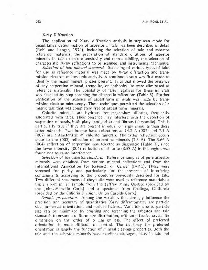
X-ray Diffraction The application of X-ray diffraction analysis in step-scan mode for
quantitative determination of asbestos in talc has been described in detail (Rohl and Langer, 1974), including the selection of talc and asbestos reference materials, the preparation of standard dilutions of asbestos minerals in talc to ensure sensitivity and reproducibility, the selection of characteristic X-ray reflections to be scanned, and instrumental technique.
Selection of talc mineral standard. Screening of various types of talcs for use as reference material was made by X-ray diffraction and trans mission electron microscopic analysis. A continuous scan was first made to identify the major mineral phases present. Talcs that showed the presence of any serpentine mineral, tremolite, or anthophyllite were eliminated as reference materials. The possibility of false negatives for these minerals was checked by step scanning the diagnostic reflections (Table 3). Further verification of the absence of asbestiform minerals was made by trans mission electron microscopy. These techniques permitted the selection of a matrix talc that was completely free of asbestiform minerals.
Chlorite minerals are hydrous iron-magnesium silicates, frequently associated with talcs. Their presence may interfere with the detection of serpentine minerals, both platy (antigorite) and fibrous {chrysotile). This is particularly true if they are present in equal or larger amounts than these latter minerals. Two intense basal reflections at 14.2 A (001) and 7.1 A (002) are characteristic of chlorite minerals. The latter reflection occurs close to the (002) reflection of serpentine minerals (7.3 A). The 3.66 A (004) reflection of serpentine was selected as diagnostic (Table 3), since the lower intensity {004) reflection of chlorite (3.53 A) in this region was found not to cause interference.
Selection of the asbestos standard. Reference samples of pure asbestos minerals were obtained from various mineral collections and from the International Association for Research on Cancer (IARC). These were screened for purity and particularly for the presence of interfering contaminants according to the procedures previously described for talc. Two different specimens of chrysotile were used as reference materials: a triple air-jet milled sample from the Jeffrey Mine, Quebec (provided by the Johns-Manville Corp.) and a specimen from Coalinga, California (provided by the Calidria Division, Union Carbide Corp.).
Sample preparation. Among the variables that strongly influence the precision and accuracy of quantitative X-ray diffractometry are particle size, preferred orientation, and surface flatness. Variation due to particle size can be minimized by crushing and screening the asbestos and talc standards to ensure a uniform size distribution, with an effective crystallite dimension on the order of 5 µm or less. The effect of preferred orientation is more difficult to control. The tendency for preferred orientation is largely the function of mineral cleavage properties. Both the talc and the asbestos minerals have excellent cleavages, platy in talc and
CONSUMER TALCUMS AND POWDERS 263
TABLE 3. Calibration Curve Data for the Determination of Asbestiform Minerals and Quartz in Talc by X-ray Anatvsis"
Mineral phase
Parameter Anthophyllite Chrvsotile Quartz Tremolite
Miller index of diagnostic reflection (210) (004) (211)b ( 110) Corresponding d·spacing (A) 8.26 3.66 1.54 8.38 Relative intensity 55 80 15 100 Step-scan interval (2 theta) 10.0-11.0 23.5-25.0 59.5-60.5 10.0-11.0
Percent mineral in talc and corresponding area of reflection
Anthophyllite Chrysotile Quartz
% in.2C % in. 2 % in. 2
5.0 0.92 0.25 0.01 5.0 2.75 10.0 2.35 0.5 0.03 10.0 3.75 15.0 3.99 1.0 0.08 20.0 7.11 20.0 4.65 2.0 0.22 25.0 9.30 25.0 5.12 5.0 0.94 30.0 13.20 30.0 7.22 7.0 1.52 40.0 16.90 35.0 9.92 8.5 2.21
8.9 2.34
Tremolite
% in.2
0.1 0.13 0.5 0.30 2.2 0.43 2.7 0.67 4.3 1.25 5.0 1.65 7.0 2.08
10.0 2.80
Detection limit
Wt% Anthophyllite == Area (210) = 0.27 (%An)- 0.52 R2 == 0.95 Wt% Chrysotile == Area (004) = 0.27 (%Ch) -0.50 R2 == 0.98 Wt% Quartz == Area ( 211) = 0.43 (%Q) - 0.60 R 2 == 0.96 Wt% Tremolite == Area (110) = 0.28 (%Tr) - 0.04 R2 == 0.98
2.0% 0.7% 1.4% 0.1%
0Jnstrumental settings: target/filter, Cu/Ni, 45 kV/20 mA; scintillation counter, 1450 VDC; monochromator, graphite; pulse-time analyzer, 20 V, 5 V (width, level); continuous scan, 1° 2 0/min; step-scan, 0.02° 2 0 at 2,000 counts fixed. bRhombohedral index. 'Repeated measurements of areas under curves with a polar planimeter indicated average
deviation of ± 0.02-0.05 in. 2•
fibrous in the case of tremolite, anthophyllite, and chrysotile (see Rohl and Langer, 1974, Fig. 2). In attempting to reduce or eliminate the effects of preferred orientation in X-ray analysis, a number of sample preparation and instrumental techniques have been developed (Bragg, 1967; Brindley and Kurtossy, 1961; Cullity, 1956; Klug and Alexander, 1954). In the present study these techniques were tested, but none was found to provide adequate precision (reproducibility). Accordingly, a sample preparation technique was developed that was successfully used, in conjunction with X-ray diffraction in the step-scan mode, to detect diagnostic reflections of these minerals in a talc matrix over a range of concentrations (Table 3).

264 A. N. ROHL ET AL.
The reproducibility of reflection intensities was also greater than other preparation techniques tested.
Binary dilution standards of chrysotile, anthophyllite, and tremolite in talc were prepared gravimetrically. Asbestos fiber concentrations were prepared initially at 5.0, 4.0, 2.0, 1.0, 0.5, 0.2, and 0.1%. Fifty milligrams of the talc-asbestos mixtures were homogenized in 10 ml filtered water utilizing ultrasonic energy. This slurry was poured into a 30 cc syringe and filtered through a 0.22 µm pore size membrane filter. To prevent stratification due to differential particle size and density effects, the syringe is held in a horizontal position, rotated, and shaken during filtration. The residue forms a flat cake of about 0.5 mm uniform thickness on the membrane filter. When dried, the sample is affixed to a glass slide for X-ray diffraction analysis (Rohl and Langer, 1974).
Selection of X-ray reflections. Because of crystal structure similarities in the minerals being studied (i.e., tremolite and anthophyllite), consider able overlapping and interference of X-ray reflections occur. The low symmetry and consequent complex X-ray diffractograms of such minerals as talc, chlorite, and mica, as well as possible interferences from admixed phases such as kaolinite, make it necessary to select a reflection or set of reflections for each mineral component that could be used as an index of the amount of that mineral in a mixture. Such diagnostic reflections were selected by referring to standard X-ray powder diffraction data. These diagnostic reflections were step-scanned at 0.02° 2 theta in a fixed count mode (2 X 10³ counts). Precise positions and profiles of the diagnostic reflections were determined. In the fixed count mode, each of the angular intervals selected are scanned with equal accuracy. Thus weak reflections can be determined with equal precision as high intensity reflections. The statistical accuracy depends only on the total number of counts recorded, and the counting rate selected gives a percentage probable error of about 2%. Profiles of the diagnostic reflections, plotted as a function of number of counts vs. 2 theta, are measured with a compensating polar planimeter. The intensity of a reflection is proportional to, but not necessarily a linear function of, its concentration. Other factors that may influence reflection intensities include instrumental conditions, particle size, degree of pre ferred orientation, sample thickness and flatness, and absorption character istics (Klug and Alexander, 1954; Rohl and Langer, 1974).
Figure 2 shows calibration curves obtained for chrysotile, anthophyl- 1 ite, tremolite, and quartz using the step-scan technique. Measured areas of diagnostic reflections are plotted against percent dilution in talc. As indicated in Table 3 tremolite may be determined at levels as low as 0.10% by weight, chrysotile from 0.25 to 0.50%, and anthophyll ite, as low as 2% in talc. It is important to note that the limits of detection given in Table 3 are higher and based on a best fit regression analysis. For example, regression analysis indicates that the detection limit for chryso tile is 0. 7%, whereas from 0.25 to 0.5% can be actually detected,

CONSUMER TALCUMS AND POWDERS 265
Tremolite
2
" C:
'° Cl)
~
20
N C:
15 '° Cl)
~
10
0 Anthophyllite 5
1 2 3 4 5 6 7 8 9 10 5 10 15 20 25 30 35 40 45 Percent asbestiform minerals in talc Percent asbestiform amphibole/quartz in talc
FIGURE 2. Calibration of asbestiform amphiboles and quartz in talc.
depending on particle size, degree of crystallinity, etc. The changes in slope at the lower end of the curves are not reflected so that axial intercepts are exaggerated on the high end of the abscissa.
By using X-ray diffraction in the step-scan mode, Stanley and Nor wood (1973} were able to detect a minimum of 0.25% tremo/ite in talc and a minimum of 0.5% chrysotile and the other asbestiform minerals. However, such low levels of chrysotile were not detected when chlorite was present.
Step scanning. The contents of the containers were thoroughly mixed with a sample splitter to avoid stratification effects. Aliquots of each, weighing 50 mg, were prepared using the identical methodology described for preparation of the dilution standards. The filter-mounted samples were then step scanned over the goniometric intervals diagnostic for the standard asbestiform minerals and quartz. Instrument operating conditions were identical with those used for analyzing the dilution standards. Profiles plotted for the diagnostic intervals and reflection areas after peak stripping were measured by polar planimetry. The weight percents of anthophyllite, tremolite, and quartz contents were estimated by referring to the appropriate regression curve. The quantities of asbestiform materials in the 21 talcums and powders found by this technique are shown in Table 4. The results show that 10 of the 21 samples contain amphibole
TABLE 4. Summary of Mineralogical Composition of 21 Consumer Talcums and Powders
Sample no.
Minerals 1 2 3 4 5 6 7 8 9 10 11 12 13 14 15 16 17 18 19 20 21
Talc Ma - M M M M M p M M M M M M M M M M M M M Chlo rite p - - p Tr p p - p p p p p p p - p p p p p Phlogopitc - - - - Tr - - pb - - p p Calcite - - - - - p - Tr - p p p p p p - - - - - p
N 0\ Dolomite - - - - - - - - - - - - - - - - - - p 0\
Quartz 5.5 - 1.6 - - 1.9 - 35.1 - - - 4.0 - 3.0 - 2.0 - 1.6 2.0 Kaolin - - - - - - Tr p Trcmolite 2.7 - - - - 0.5 0.4 7.4 - - 10.3 - - - 0.1 3.0 0.1 - - - 2.4 Anthophyllite 11.4 - - - - - - 4.9 - - - 4.6 - - 2.4 5.2 2.1 - - - 6.5 Chrysotile - - - - - - - - - - - <0.50 - - <0.50 Pyrophyllite - - - - - - - p - - - p Ru tile - - - - - - Tr
0M = major; P = present; Tr = trace. 6High K20 and Na20 suggests that this phase is a mica (muscovite/biotitc).

CONSUMER TALCUMS AND POWDERS 267
minerals, ranging in amounts from a few tenths of a percent to over 14%. Tremolite was the most commonly found (9 of the 21), and anthophyllite occurred with tremolite in 6 of the 21. A serpentine mineral phase was indicated in two samples, in amounts at or near the lower limits of detection. Verification of the serpentine phase as chrysotile in the two samples in amounts corresponding to the observed concentrations was made by electron microscopy.
Continuous scanning. In order to study the presence of all mineralog ical (and possibly other crystalline) components the samples were scanned from 5° to 70° 2 theta at a scanning rate of 1 ° 2 theta per minute. This technique, as expected, proved satisfactory for the identification of major components, but it was generally found to be incapable of detecting tremolite, anthophyllite, or serpentine minerals except in cases of gross contamination. The high noise level (low peak-to-background ratio) often prevents the detection of quantities on the order of 4-6% or less and also excludes this technique for quantitative analysis. As a result of high noise level three false positives, as indicated by continuous scanning, were subsequently shown to be negative for amphibole by step scanning and electron microscopy. In addition, continuous scanning may not generally detect serpentine minerals in the presence of chlorite or kaolinite.
Electron Microscopy and Electron Diffraction The transmission electron microscope has been shown to possess the
sensitivity required for fiber identification and for determination of particle size distribution of submicroscopic particles (Langer and Pooley, 1973; Langer et al., 1973).
Accordingly, aliquots of talcum samples were prepared for electron microscopic analysis by a technique that disperses particles in a drop of nitrocellulose solution on a glass slide. A second glass slide is placed on the first and the two are drawn lightly apart, leaving a film. This technique is intended to minimize the alteration of particle size distribu tion. The film is mounted on electron microscope grids and scanned at magnifications of X20,000.
Morphologically, amphibole minerals are generally quite dissimilar from other silicate minerals. Both anthophyllite and tremolite are rectilinear, often with amphibole-type step cleavage or, infrequently, with prismatic terminations. Tremolite fibers tend to be electron dense and shorter than anthophyllite ( Fig. 3), while the latter has a tendency to be electron translucent and to show diffraction contrast figures (Langer and Pooley, 1973). Sheet silicate minerals (talc, chlorite, micas) tend to be equi dimensional in shape, often with pseudohexagonal outlines (Fig. 4). Curled talc plates or talc fibers on edge may superficially resemble asbestiform minerals, but selected area electron diffraction patterns easily distinguish between the two (compare Fig. SA, C, and D).
Electron microscopy, in combination with selected area diffraction,

-- .,_,,-;;,-, - . . ~ ......... "
•
A I um B ..
C ·lµm
•
- p;-:..·.,
0.5µm
' E l urn
FIGURE 3. Transmission electron micrographs showing range in morphological characteristics of asbestiform trernolite and anthophyllite in talc. The entire range of morphological variations observed for these minerals is observed in the asbestos standards: rectilinear fibers with parallel ends and edges (A); step-cleavage ends (B); unit fibrils protruding from fiber body, (C); curvilinear fiber with amphibole cleavage end (D); high length-to-width ratio fibers (E); fibers protruding from interiors of talc plates (F). All of these morphological variations and forms (A-E) have been described in anthophylfite and tremolite asebestos samples. The amphibole structure was confirmed in all cases by selected area electron diffraction characterization. Scale as marked. Micrographs obtained on a J EOL J EM 120 U with an accelerating voltage at 120 kV.
268
CONSUMER TALCUMS AND POWDERS 269
- '
.
' I C J ... ,
·# . f. ,.,,,~,°' I.
I,
,-~ .. . .. . , a. ., •' "'·~ I • \ .. -
~· ·•. 1( t f#t..,,. ~ ·--i· .· · . •. ~· ·:!"·' ~,. ·."' k. ~..... . t; ;~ ... • •
.,. ... ~ . ~ .
J
~&1 ....... ' - ...... ~ f. \,
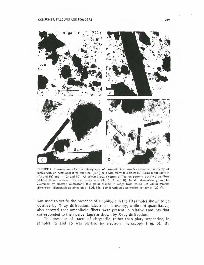
FIGURE 4. Transmission electron micrographs of cosmetic talc samples composed primarily of plates with an occasional large talc fiber (B, C); talc with many talc fibers (D); Scale is the same in (A) and (B) and in (C) and (D). All selected area electron diffraction patterns obtained on fibers yielded those consistent for talc plates (see Fig. 5, A and B}. In all talc-containing samples examined by electron microscopy talc grains tended to range from 25 to 0.2 µm in greatest dimension. Micrograph obtained on a J EOL J EM 120 U with an acceleration voltage at 120 kV.
was used to verify the presence of amphibole in the 10 samples shown to be positive by X-ray diffraction. Electron microscopy, while not quantitative, also showed that amphibole fibers were present in relative amounts that corresponded to their percentages as shown by X-ray diffraction.
The presence of traces of chrysotile, rather than platy serpentine, in samples 12 and 15 was verified by electron microscopy (Fig. 6). By

~-· TP r
i.
5µm
A
' 1· ' . -
: ·.-B
·'·71 .. .-- ,.,
ti I ·•
FIGURE 5. Transmission electron rnicrographs and accompanying selected area electron diffraction (SAED) patterns for cosmetic talc samples. The talc plate (TP) in (A) shows typical polygonal cleavage and diffraction contrast contours for the mineral species. The accompanying SAED pattern displays the characteristic reciprocal ab* plane pseudohexagonal symmetry for talc. Talc a* and b* directions indicated on (A). Measurement of pattern indicates a 5.3 A repeat along a* and a 9.1 A repeat for b* (measured at [110) ). Talc fiber (TF) in (B) displays irregular ends and nonrectilinear edges. The SAED pattern is also pseudohexagonal, but some reflection intensities [e.g., the (060), (0.12.0)] are more pronounced. This may be due to both orientation and structural effects. The curled talc plate (CT) in (C) displays an incipient Debye-Scherrer ring pattern (the effects of both folding over of talc and small associated grains). The amphibole fiber (D) was diffracted only on one of the protruding unit fibrils. The c* axis is shown, with repeat measured at 5.3 A. Areas where diffraction patterns were obtained arc indicated by location circles; particles were photo· graphed at the SAE D magnification X 26,500. Scale is the same in A-C; scale in D as marked. Micrographs obtained on a J EOL J EM 120U with an accelerating voltage at 120 kV.
270

0.25.um
A
0.25µm
0.3 C JJm
FIGURE 6. Transmission electron micrographs of asbestiform minerals in cosmetic talc, other than amphibole. (A and B) Two chrysotile fibers with morphological characteristics induced by electron beam damage. No diffraction pattern was obtained on either fiber. Arrow markers (A and B) indicate areas where these beam-damaged features are most prominent. Both fiber bundles appear to rest on talc plate substrates. Free chrysotile fibers and fibrils (C) were found in a sample found negative for asbestos by all other techniques. Scale as marked. Micro graphs obtained on a J EOL J EM 120 U with an accelerating voltage at 120 kV.
271

272 A. N. ROHL ET AL.
comparison with known dilution levels of chrysotile in talc observed by electron microscopy, the levels of contamination of chrysotile in the two samples correspond to about 0.25-0.5% chrysotile, which was suggested by the X-ray diffraction results. The chrysotile fibers were all shorter than 2 µm and the diameters less than 0.2 µm, explaining why they were not visible by optical microscopy.
Chemistry of Consumer Talcums and Powders The bulk chemistry (Table 5) and mineral contents (Table 4) of the
talcums and powders complement each other in that one data set implies limits for the other. For example, analysis of sample 1 shows the presence of FeO and CaO (Table 5). Recalculation of these oxides into values for the empirical formulas for trernolite and anthophyllite indicates that sufficient quantities are present to account for the presence of these minerals (Table 4). An appreciable decrease in Si02, which should normally occur, was not observed, because of the occurrence of over 5% quartz content. The Al2 03, Na2 0, and K2 0 are reflected by the presence of chlorite (probably the penninite phase). Variations in oxides were also observed in the other samples that contain amphibole minerals (6-8, 11, 12, 15-17, and 21). In these samples, a number of other factors account for the wide variations in oxide percentages: in 6, the presence of chlorite
TABLE 5. Major Oxide Content of 21 Consumer Talcum and Powders"
Sample no.
Major oxide 2b 3c 4 5 6 7 8 9 10 11
Si02 61.99 0.00 62.68 58.72 61.94 52.95 59.67 71.93 58.68 51.65 49.48 Ti02 0.10 0.00 0.14 0.04 0.03 0.17 1.40 0.19 0.08 0.71 0.22 Al203 0.82 1.30 0.37 0.24 0.45 1.16 0.87 15.73 0.58 1.45 2.32 Fe203 0.00 0.00 0.08 0.04 0.03 0.02 0.18 0.10 0.12 0.09 1.23 FeO 1.51 0.00 0.32 3.03 0.51 0.86 0.84 0.34 3.12 0.34 0.07 MnO 0.00 0.00 0.00 0.00 0.01 0.00 0.00 0.00 0.00 0.00 0.00 MgO 29.60 0.00 29.83 28.25 30.19 29.02 29.23 2.95 29.23 27.90 25.67 CaO 0.40 0.00 0.04 0.10 0.04 3.46 0.90 2.37 0.17 4.75 1.61 Na10 0.07 0.00 0.03 0.03 o.os 0.00 0.05 0.48 0.00 0.03 0.42 K20 0.02 0.00 0.02 0.02 0.02 0.05 0.05 1.37 0.00 0.00 0.19 P2 Os 0.01 0.00 0.03 O.Q3 0.01 0.13 0.14 0.05 0.00 0.05 0.01 Volatilel 5.36 98.70 4.66 5.38 5.51 10.32 5.64 5.25 5.34 9.72 10.41
Total 99.88 100.00 98.20 95.88 98.79 98.14 98.97 100. 76 97.32 96.69 91.63
0weight percent, recalculated as oxides, following standard petrochemical procedures. The bulk analysis of the powders reflects the combined mineral content after additives were extracted using water, dilute HCI, acetone, benzene, and ether. b Analysis of 2: 98. 7% = starch + organics and volatiles; 1.3% = Al203 (aluminum chlorhydrate?). c Standard talc used as matrix for fiber standard dilutions.

CONSUMER :rALCUMS AND POWDERS 273
sharply reduces the Si02 content and elevates the Al2 03 content. The presence of the carbonate mineral calcite increases the expected CaO and volatile contents (the latter includes CO2); in 7, the high Ti02 is reflected by the presence of the mineral rutile (Ti02); in 11, the high CaO, moderately high Al2 03, and low Si02 contents reflect the presence of calcite, chlorite, and tremolite.
Several of the above samples (numbers 8, 12, and 15) require special evaluation. Sample 8 is extremely high in Si02, Al2 03, Na2 0, and K2 0 and extremely low in MgO content. The chemistry indicates that this material was not derived from a talc rock, but rather from one rich in alumina and silica. The mineralogy reflects this, as do the trace metals (see trace metal section). The amphibole minerals in sample 8 are associated with both pyrophyllite [Al4Si8 022 (OH)4] and quartz, both present in substantial quantities. The mica phase is not phlogopite, but muscovite, accounting for the presence of substantial quantities of K2 0 and Na2 0. A plagioclase feldspar was also detected in the mineral phase. Sample 12 is very high in Al2 03 but extremely low in Si02, apparently the result of high chlorite content as well as substantial amounts of pyrophyllite. Sample 15 is low in Si02 and extremely high in volatile content, reflecting the presence of both carbonate phases and organic additives.
The trace element analyses (Table 6) show distributions that are in accordance with the known behavior of trace elements in minerals. With
TABLE 5 (continued) Major Oxide Content of 21 Consumer Talcum and Powders?
Sample no.
12 13 14 15 16 17 18 19 20 21 Ad ae
47.32 53.83 57.47 44.83 62.26 54.45 59.93 58.54 62.19 56.34 57.34 61.49 0.18 0.11 0.11 0.07 0.06 0.10 0.10 0.18 0.08 0.12 0.21 0.01 9.34 1. 74 1.65 0.69 0.45 4.26 0.79 1.11 0.69 1.35 2.30 1.20 0.05 0.03 0.02 0.00 0.18 0.04 0.00 0.00 0.00 0.02 0.11 0.38 1.22 0.70 0.65 0.62 1.04 1.41 0.84 1.38 0.81 1.39 1.05 1.07 0.00 0.00 0.00 0.00 0.00 0.00 0.00 0.01 0.00 0.00 0.00 0.00
29.83 27.14 26.98 23.88 30.00 30.79 30.40 29.83 30.19 27.90 27.44 30.54 0.69 5.49 5.52 3.67 0.13 1.13 0.50 1.1 3 0.43 1.53 1.69 0.46 0.03 0.07 0.05 0.35 0.09 0.09 0.09 0.00 0.07 0.07 0.10 0.05 0.00 0.02 0.07 0.05 0.02 0.02 0.00 0.00 0.00 0.10 0.21 0.14 0.13 0.10 0.01 0.04 0.13 0.00 .0.13 0.02 0.70
10.26 10.81 10.41 21.34 5.92 7.54 5.65 5.94 5.14 9.99 8.03 5.00
99.18 100.06 98.01 95.62 100.19 99.87 98.45 98.21 99.73 98.73 98.20 100.11
d Average of 20 talc samples. e Average of 8 talc analyses in Deer et al. (1962). 1Volatiles are lost on ignition (total H2 0, CO2, organics, and other volatiles).

TABLE 6. Trace Element Content of 21 Consumer Talcum and Powders"
Sample no. Trace metals 1 2 3 4 5 6 7 8 9 10 11 12 13 14 15 16 17 18 19 20 21
Ba <10 - <10 <10 <10 <10 <10 so <10 990 <10 <10 <10 <10 <10 <10 <10 <10 <10 <10 <10 Ce <10 - <10 <10 <10 <10 <10 10 <10 <10 <10 <10 <10 <10 <10 <10 <10 <10 <10 <10 <10 Cl 210 - 130 135 185 200 140 130 115 125 115 120 430 435 410 165 140 130 110 120 130 Co 3S - <3 85 <3 <3 <3 <3 88 <3 <3 <3 <3 <3 <3 21 <3 4 <3 <3 <3 Cr 310 - 28 600 16 23 24 <15 820 25 18 24 38 49 41 340 32 30 28 25 42 Cu <5 - <5 <5 8 <5 <5 9 <5 <5 <5 <5 5 8 10 13 6 7 6 6 13
N Ga <1 - 4 <1 <1 1 <1 20 <1 1 3 13 2 1 2 2 7 3 2 3 <1 -..i La <10 - <10 <10 <10 <10 <10 40 <10 <10 <10 <10 <10 <10 30 <10 <10 10 <10 <10 <10 .I>,
Nb 7 - 8 8 11 7 9 14 5 7 6 18 7 7 5 5 9 8 9 9 7 Ni 710 - 17 172 27 13 10 <4 2210 34 4 11 10 17 10 460 14 20 28 19 42 Pb 8 - 8 <5 <5 <5 <5 49 7 <5 17 5 7 12 8 6 9 7 8 5 16 Rb 5 - 5 <S <5 <5 s 45 <5 <5 <S <5 <5 5 5 5 <5 5 5 5 <5 s 235 - 130 120 2230 440 150 305 155 425 110 280 485 595 535 320 140 160 105 155 1070 Sr 10 - 10 10 10 20 30 160 <10 25 100 <10 15 20 20 <10 15 10 10 10 15 Th <S - <5 <5 <5 <5 <5 6 <5 <5 <5 <S <5 <5 <5 <5 <5 <5 <5 <5 <5 Zn )> - 85 37 18 12 15 20 37 > > > 35 11 20 73 27 8 11 3 > Tr <10 - <10 <10 <10 90 10 190 <10 10 10 30 <10 <10 <10 <10 50 <10 <10 <10 <10
0values in parts per million. The symbol < indicates the limit of detection of the analytical method. The symbol > indicates concentrations of Zn are greater than 1,000 ppm.

CONSUMER TALCUMS AND POWDERS 275
the crystal lattice of a mineral acting as a sorting mechanism for cations, the cations can enter a crystal structure providing they have appropriate size and charge. These phenomena apply to major as well as minor elements. Thus, barium is present in large amounts in sample 8, which has a high K2 0 content (1.37%). Since barium and potassium have similar ionic radii, barium is easily admitted into potassium minerals, such as micas and feldspars {both found in 8). Rubidium and strontium are also enriched in sample 8, since these metals also easily substitute for potassium. Gallium is found in large amounts in samples 8, 12, and 17. These samples are also very high in Al203• Gallium has the same ionic charge and radius as aluminum and, in fact, is found only in aluminum bearing minerals.
In four samples (1, 4, 9, and 16) there are significantly higher concentrations of cobalt, chromium, and nickel than found in the other samples. These four samples also have high contents of FeO (Table 5). The association of these four transition metals has been observed before in certain geochemical environments, particularly in ultramafic rocks. Since talcs derived from the metamorphism of serpentines and peridotites (ultramafic rocks) are considerably enriched in FeO (Deer et al., 1962), it is likely that the divalent cations are substituting for iron in the brucite layer of the talc.
DISCUSSION AND CONCLUSIONS
Talc used in the United States represents a wide range of mineralogical substances. Industrial grade talcs are obtained from different rock types of highly variable mineral composition with the result that the mineral talc may actually be a minor constituent. However, it has been stated that consumer talcum products should contain at least 90% of the mineral of the same name and no asbestos fiber (Hildick-Smith, 1976). Review of the literature suggests that at least until 1968, materials that were marketed as cosmetic talcum products did not necessarily conform to these criteria.
Talc mineral may occur in a platy form or in a fibrous form. Talc fiber may occur as a small proportion of the mineral desposit or as a major constituent. lntergrowths of talc with other mineral phases are common. These phases may be simply macroscopic zones adjoining talc mineral or may occur as microscopic intergrowths within the talc. Of the many minerals that may coexist with talc, a number of asbestiform phases commonly occur: tremolite, anthophyllite, and chrysotile have been identified in these deposits. In addition, free silica (quartz) is a frequent constituent. The trace metal content may include elevated levels of nickel, chromium, and cobalt.
There is general agreement between the mineral composition and the major and trace element content of the consumer talcum products. On the
276 A. N. ROHL ET AL.
basis of mineral and chemical contents, the type of geological provenance may be ascertained.
Methodology has been developed for quantitative X-ray diffraction determination of anthophyllite, tremolite, serpentine, and quartz in con sumer talcums and powders. Important factors in the calibration standard development include selection of talc and reference minerals and the selection of diagnostic X-ray reflections. The sample preparation technique is sensitive and reproducible. Dilution standards are step scanned over diagnostic reflection areas, peak areas are measured, and a set of standard calibration curves is developed by regression analysis. Samples of consumer talcums and powders are prepared and analyzed under identical conditions and compared with the calibration curves, permitting quantitative analysis of these minerals. X-ray diffraction alone cannot distinguish between asbestiform and fragmented forms of anthophyllite and tremolite nor between asbestiform and platy serpentine varieties. Electron microscopic analysis was used to distinguish between these forms.
Mineralogical characterization of 21 consumer talcums and powders showed that 10 contained measurable concentrations of asbestiform tremo lite and anthophyllite, and some also contained fragmented forms of these minerals. Two samples contained trace quantities of chrysotile {0.25-0.5%). These observations were confirmed by transmission electron microscopy. The amphibole phases present in these talcum products ranged in amounts from several tenths of a percent to over 14% by weight. Quartz was present in eight consumer talcs in amounts ranging from 1.6 to 35.1 % by weight. ·
Consumer talcum products are for the most part complex mineral assemblages, which confer X-ray sorbing and fluorescing effects that are not equivalent to, and are usually greater than, those of the binary systems used in preparing the dilution standards. In consumer talcum pro ducts minerals such as talc, micas, chlorite, calcite, dolomite, and others tend to diminish reflection intensities of asbestiform minerals by sorbing X-rays or by contributing to background noise. Also, repeat runs on some selected specimens have demonstrated greater peak areas due to slight modifications in instrumental settings (e.g., increase in receiving slit width). Therefore, the values for weight percent concentrations given in this report are conservative.
Examination of the same consumer talcum products by both optical and transmission electron microscopy indicates that not all of the mater ials fall within the definition of fiber or asbestiform. For example, one consumer talcum product that contained more than 7% tremolite was observed to contain both fragmented tremolite grains by optical micro scopy and asbestiform fiber with 3: 1 or greater length-to-width ratio by transmission electron microscopy. Optical microscopy may provide useful information. However, more complete characterization can be obtained by electron microscopy and selected area electron diffraction. Using electron
CONSUMER TALCUMS ANO POWDERS 277
microscopy, for example, several samples of consumer talcum products exhibited both free amphibole fiber, discrete from talc grains, and, in addition, numerous small amphibole fibers were visible, apparently inter layered between talc or chlorite plates (see Fig. 6A).
Preliminary examination of the asbestiform amphiboles by an electron microprobe technique has demonstrated that individual fiber chemistry is identical to those fibers encountered in the IARC Asbestos Standards {Timbrell and Rendall, 1971).
On the basis of the mineralogical and chemical characterization of these products, all formulated prior to June 1973, we conclude that cosmetic grade talc was not used exclusively. The presence in these products of asbestiform anthophyllite and tremolite, chrysotile, and quartz indicates the need for a regulatory standard for cosmetic talc. This standard should be cognizant of talc complexities, mineralogical and chemical in nature, and should provide for adequate analytical protocols to ensure monitoring. We also recommend that evaluation be made to determine possible health hazards associated with the use of these products.
APPENDIX A: DEFINITIONS OF TERMS USED IN TEXT
Asbestos "A name applied to a group of naturally fibrous minerals" {chrysotile, amosite, crocidolite, tremolite, anthophyllite cited by name) (Bureau of Mines, 1968). The term asbestos has also been applied to commercially exploited fibrous clays, including attapulgite and palygorskite (Whittaker, 1968).
Asbestos implies current or possible exploitation, based on the presence of special physical and chemical properties, determined on the bulk sample level. For example, high fiber tensile strength, flexibility, low heat conductivity, high electrical resistance, and chem ical inertness are properties of asbestos. Noncommercial varieties of the same mineral may not possess the same qualities on the bulk level. For example, amosite has been considered to be the economically exploited variety of grunerite (Deer et al., 1962). If so, and even this is contested among mineralogists today, large macrocrystals are signifi cantly different physically and structurally. Grunerite fiber is rigid, amosite fiber is flexible; grunerite yields well-defined single X-ray reflections with nonrotational film techniques, arnosite yields multiple reflections as if rotated in the X-ray beam; grunerite appears to be a single crystal, amosite splays as if composed of strands. However, when both substances are pulverized, the resultant powder yields submicro scopic fibers, many of which are virtually indistinguishable on the basis of morphology, structure (determined by selected area electron diffraction), and chemistry (determined by an electron probe

278 A. N. ROHL ET AL.
technique). Amosite may be considered as an aggregate of unoriented, discrete, grunerite crystals with only the c axis in common alignment.
Comminution of such aggregates produces fibers with character istics identical to those of single crystals of grunerite that have been similarly pulverized. Some workers have suggested that mechanical size reduction of amosite yields fibers with crystal growth surfaces rather than cleavage surfaces. Since amphibole cleavage tends to parallel prominent crystal face planes, such distinctions on the submicroscopic level may disappear. Th is appears to be the case for tremol ite and anthophyllite as well. However, because no methods exist to distin guish between possible differences in fiber surface, we do not refer to anthophyllite and tremolite fibers in these talcums as asbestos. Instead they are referred to as asbestiform. It should be stressed, however, that evidence does. not exist that would indicate that fibers with crystal growth surfaces or .cleavage surfaces possess lesser or greater biological potential than fibers from commercial asbestos deposits.
Asbestiform "Formed like or resembling asbestos; fibrous; .... (Bureau of Mines, 1968). The term is used herein for amphiboles (anthophyllite and tremolite) seen on both light and submicroscopic examination, which resemble comminuted asbestos varieties, on the basis of morphology. Essentially, when these fibers are derived from commer cial deposits we term them "asbestos" and when analytically identical fibers are found as noncommercial intrusions with the mineral talc, we term them "asbestiform." The use of two terms does not imply differences that can be analytically determined.
Fiber "The smallest single strand of asbestos or other fibrous materials" (Bureau of Mines, 1968). We use this term in a broader sense. For example, chrysotile fibers are called fibrils, possessing unit diameters of about 200-400 A. Coherent bundles of fibrils are also called fibers. Fiber in the present text is used to denote any elongated single mineral unit visible on the light or electron microscopic level. The Occupational Safety and Health Administration has applied a 3: 1 length-to-width ratio to distinguish fiber from mineral fragment.
APPENDIX B: CRYSTAL CHEMISTRY, CRYSTAL STRUCTURE, AND GEOLOGICAL OCCURRENCE OF TALC
Chemistry of Talc
The empirical chemical formula of talc is Mg3 Si4 010 (OH}i, but ferrous and ferric oxides, alumina, titania, soda, lime, and oxides of manganese have been reported in quantities up to several percents by

CONSUMER TALCUMS AND POWDERS 279
weight. Titanium and aluminum appear to substitute for silicon, whereas iron, nickel, and manganese substitute for magnesium. Alkali metals are not readily accommodated in the structure and evidently occur as inter layer ions or as components of mineral impurities. For example, excess calcium may reflect the presence of the interlayer mineral phase tremolite (Deer et al., 1962; Stemple and Brindley, 1960). One major talc deposit in the eastern United States contains substantial amounts of nickel, as much as 0.2%. Notwithstanding these minor components, talc is essentially (by weight) 32% MgO, 63% Si02, and 5% structurally bound water.
Talc Structure and Crystal Habit The three-layered crystal structure comprises a sheet of octahedrally
coordinated Mg{OH)2 groups (the brucite layer) sandwiched between two planes of tetrahedrally linked Si04 groups (silica layers). Apical oxygens of the silica sheets are directed toward the brucite layer and in part re placed by hydroxyl groups, which form a portion of the inner structural unit. Valence balance is accomplished within the structure, so that there is a net zero charge on juxtaposed unit layers at the silica base interfaces.
The basic unit of the talc structure was determined over 40 yr ago (Gruner, 1934; Hendricks, 1938), yet the repeated cell geometry and space group were only recently resolved. X-ray single crystal patterns now indicate talc to be triclinic ( Rayner and Brown, 1966; Ross et al., 1968).
In addition to chemical and structural complexities, talc occurs with both plate and fiber habits (Ford, 1957). The development of the fibrous cyrstal form, with an elongated crystallographic a-axis, may be a mani festation of ionic substitution since its refractive index is higher than platy talc ( Fleischer and Osborn, 1957; Gruner, 1944). Talc that contains substantial amounts of these elongated forms is referred to in the mineralogical literature as fibrous talc. Similar observations with regard to the mineral brucite have been reported (Liebling and Langer, 1972) in which high iron content in the normally platy brucite is associated with the development of a fibrous habit. It is of interest to note that minnesotaite, considered by some to be an iron-rich form of talc, always occurs with a fibrous, or even a needle-like, habit (Gruner, 1944).
Formation of Talc in the Laboratory of Nature In closely controlled experimental systems, talc has been synthesized
(Bowen and Tuttle, 1949; Yoder, 1952}. Bulk chemistry, water fugacity, temperature, and pressure parameters are defined within extremely restrict ed limits and ranges to produce relatively pure crystallization products. However, in laboratory synthesis, just as in nature, coexisting mineral phases are produced if slight variations in any of the parameters are introduced. These phases include anthophyllite, serpentine, and in some instances tremolite.

280 A. N. ROHL ET AL.
Nature of Talc Plates Electron microscopic examination of talc minerals demonstrates that
single talc grains consist of contiguous single crystals, mosaics of dis oriented crystallites, and intergrowths with other mineral phases, particu larly tremolite (Kleinfeld et al., 1973; Stemple and Brindley, 1960; Wright, 1960). Selected area electron diffraction patterns obtained on these objects display, in order, single crystal arrays, Debye-Scherrer rings, and superimposed complex patterns characteristic of intergrown single crystal phases.
Talc and Mineral lntergrowths
Tremolite is one common intergrowth in talc, and it requires relatively little energy thermodynamically to occur. Replacement of magnesium by calcium in the brucite layer may lead to structural as well as chemical modification (Bragg and Claringbull, 1965}. Rotation of unit tetrahedra in talc forms double chains from sheets, readily accomplished by substitution of Mg{OH)2 by Ca{OH}2• The bulk chemistry is thereby changed from Mg6Si8020}(0H)4 to Ca2Mg5Si8022(0Hh. The final structural array is remarkably similar in both materials; the crystallographic a-axis of talc is approximately 5.26 A, which corresponds with the c-crystallographic axis of tremolite (approximately 5.24 A); the b-axis for talc is approximately 9.10 A, which is equal to the b/2-axis of tremolite; the c-axis of talc, approximately 18.8 A, is about equal to twice the a-dimension of tremolite (approximately 18.2 A). The monoclinic stacking angles, the beta-angle, are within a few degrees of each other.
lntergrowths may form in which arnphibole formation is not complete so that a mixed phase exists, referred to mineralogically as "talcboles." These are not as rare as once believed and may even be common in the more complex talc deposits.
APPENDIX C: INDUSTRIAL AND COSMETIC GRADE TALCS Studies have demonstrated that industrial talc often consists of a
variety of minerals, the utilization of which is based on physical properties rather than mineral composition (Hogue and Mallette, 1949; Schulz and Williams, 1942; Thompson, 1974; Wells, 1965).
Early analyses of cosmetic talc also showed a wide range in mineral composition. Of six such products examined in one study, only 6-47% by weight of the inorganic material that constituted the product was the mineral talc; 14-51 %, serpentine minerals; 5-77% carbonate minerals; 0-trace, quartz; 0-trace, tremolite; 3-12%, other minerals (Schulz and Williams, 1942).
CONSUMER TALCUMS AND POWDERS 281
APPENDIX D: BIOLOGICAL HAZARDS ASSOCIATED WITH TALC EXPOSURE A fine, diffuse, bilateral, progressive fibrosis was observed among
miners and millers of tremolite talc in Georgia ( Dreessen, 1933; Dreessen and Dalla Valle, 1935). Siegal et al. (1943) studied a population of workers mining and milling tremolite and anthophvllite-bearing talc deposits in New York state. Jn addition to the bilateral fibrosis, pleural plaques, similar to those encountered in asbestos workers, were observed. Review of postmortem material in this study indicated that asbestos bodies were present in lung tissue. These findings were also reported in cases of severe pneumoconiosis in tremolite millers by Daymon ( 1946), and by Porro and Levine (1946).
Millman (1947) reported that exposure to cosmetic-grade talc pro duced nodular fibrosis in workers. No quartz was detected in the dust. The author concluded that talc itself was capable of producing scarring. The observation was supported in studies by Reichman (1944) and by Wyers (1949) and in a study of talc miners and millers in Italy where exposure to pure talc produced a 10% incidence of pneumoconiosis in workers (Parmeggiani, 1948). Excess deaths attributed to pneumoconiosis have been reported among workers in northern Italy mining talc con sidered to be free of asbestiform fibers (Rubino et al., 1976).
Some investigators have held that fibrous talcs (not differentiated as talc or asbestos fiber) are biologically more hazardous than platy talcs. For example, in a review of the literature by Porro et al. (1942), Gloyne and Gardner are referred to as considering that the clinical, radiological, and pathological disease states of asbestosis and talcosis are very similar. There are several reports of the occurrence of asbestos bodies in the lung tissue of workers exposed to talc (Daymon, 1946; Hobbs, 1950; Kleinfeld et al., 1973; McLaughlin et al., 1949; Porro et al., 1942).
Several studies suggest that fibrous talcs are more dangerous as a result of the included asbestos fiber. For example, McLaughlin et al. (1949) compared fibers in talc with the proportion of fibers recovered from the lung tissue of an exposed worker. A larger concentration of fibers was found in the tissues as compared with the raw talc. Talc pneurnoconiosis was reaffirmed by Kleinfeld and Messite (1960) in their study of the New York state talc workers.
In a study by Kleinfeld et al. (1967) it was demonstrated that talc pneumoconiosis accounted for almost 30% of excess deaths among the talc miners and millers. Most of these were due to the complication of pneumoconiosis, cor pulmonale. However, 21 % of the 91 deaths recorded were due to malignant tumors: lung carcinoma, pleural fibrosarcoma, and stomach, colon, rectum, and pancreatic cancers. A peritoneal rneso thelioma was reported as well. In addition to these tumors, retroperitoneal sarcoma, hepatoma, and leukemia were also found. Statistical evaluation of

282 A. N. ROHL ET AL.
these data indicated that a 3- to 4-fold excess of cancers existed in this group, as compared to a matched control population.
The biological activity of both tremolite and anthophyllite fibers has been known for some time, and both have been cited as asbestos minerals by Merewether (1930) and Noro (1946). Asbestos disease among workers (and others exposed to anthophyllite and tremolite) has been reported [Burilkov and Badajov, 1970; Kiviluoto, 1960; Meurman, 1968; Meurman et al., 1974; Schepers, 1965; Wegelius, 1947; Weiss and Boettner, 1967).
Recent experimental data also indicate that tremolite fibers are biologically active (Graham and Graham, 1967). Some investigators have suggested that inorganic fiber fibrogenicity and carcinogenicity is limited only by its ability to reach the alveolar space (Holt et al., 1965; Pott and Friedrichs, 1972; Pott et al., 1974; Robock and Klosterkotter, 1976; Stanton and Wrench, 1972).
Wagner et al. (1975) reported lung scarring in Wistar rats with pure talc, exposed by inhalation. The severity and extent of the lung scarring was comparable to that produced by chrysotile asbestos under identical experimental conditions. In addition to lung scarring, ingestion of talc was reported to be associated with leiomyosarcoma of the stomach as well as one adenoma and several sarcomas of the uterus. However, the exposure levels were high and the numbers of observed tumors small, so that statistical validation of the carcinogenic potential of pure talc and its relevance to human exposures were not achieved.
There are also extensive data concerning hazards associated with exposure to silica or trace metals, particularly nickel and chromium (National Research Council, 1975). Analytical data are presented here that suggest possible disease potential and the need for investigation in these areas.
REFERENCES Bowen, N. L. and Tuttle, 0. F. 1949. The system Mg0-Si02 -H2 0: Bull. Geo/. Soc. Am.
60:439-460. Bowes, D. R. and Langer, A. M. 1974. Petrochemistry of the Manhattan Formation. Kristalinikum
10:39-52. Bragg, R. H. 1967. Quantitative analysis by powder diffraction. In Handbook of X-rays. New York:
Mc.Graw-Hill. Bragg, L. and Claringbull, G. F. 1965. The crystal structure of minerals. London: Bell and Sons. Brindley, G. W. and Kurtossy, S. S. 1961. Quantitative determination of kaolinite by x-ray
diffraction. Am. Mineral. 46: 1205-1215. Bureau of Mines. 1968. Dictionary of mining, mineral and related terms, ed. P. W. Thrush.
Washington, D.C.: U.S. Government Printing Office. Burilkov, T. and Badajov, L. 1970. Ein Beltrag zum endemischen Auftreten doppelseitiger
Pleuraverkalkungen. Prax, Pneumal. 24:433-438. Cralley, L., Key, M. M., Groth, D. H., Lainhart, W. S. and Ugo R. M. 1968. Fibrous and mineral
content of cosmetic talcum products. Am. Ind. Hyg. Assoc. J. 29:350-354. Cullity, B. O. 1956. Elements of X-ray diffraction. Reading, Mass.: Addison-Wesley. Daymon, H. 1946. Latent silicosis and tuberculosis. Am. Rev. Tuberculosis 53 :554-559.

CONSUMER TALCUMS AND POWDERS 283
Deer, W. A., Howie, R. A. and Zussman, J. 1962. Rock-forming minerals, vol. 3, Sheet silicates, pp. 203-374. New York: Wiley.
Dreessen, W. C. 1933. Effects of certain silicate dusts in the lungs. J. Jndust. Hyg. 15 :66-78. Dreessen, W. C. and DaHa Valle, J. M. 1935. The effects of exposure to dust in two Georgia talc
mills and mines. Pub!. Health Rep ts. 50: 1405-1415. Fleischer, S. S. and Osborn, E. F. 1957. Studies of the system iron oxide-silica-water at low
oxygen partial pressures. Econ. Geo/. 52:923-943. Ford, W. E. 1957. Dana's textbook of mineralogy. New York: Wiley. Graham, J. and Graham, R. 1967. Ovarian cancer and asbestos. Environ. Res. 1 :115-128. Gruner, J. W. 1934. The crystal structure of talc and pyrophyllite. Zeit. Krist. 88 :412-419. Gruner, J. W. 1944. The composition and structure of minnesotaite, a common iron silicate in iron
formations. Am. Mineral. 29:363-372. Hendricks, S. B. 1938. On the crystal structure of talc and pvrophyllite, Zeit. Krist. 99:264-274. Hildick-Smith, G. 1976. Talc: Review of epidemiologic studies. Proc. Br. Occup. Health Soc.,
Edinburgh, Sept. 1975. In press. Hobbs, A. A. 1950. A type of pneumoconiosis. Am. J. Roentgenol. Radiol. Therop, 5 8:488-497. Hogue, W. L and Mallette, F. S. 1949. A study of workers exposed to talc and other dusting
compounds in the rubber industry.}. lndust. Hyg. Toxicot, 31 :359-364. Holt, P. F., Mills, J. and Young, D. K. 1965. Experimental asbestosis with four types of fibers:
Importance of small particles. Ann. N. Y. Acad. Sci. 132:87-98. Hurlbut, C. S., Jr. and Williams, 0. R. 1935. The mineralogy of asbestos dust. }. lndust. Hyg.
17:289-293. Kiviluoto, R. 1960. Pleural calcification as a roentgenologic sign of non-occupational endemic
anthophyllite asbestosis: Acta Rad. Scand. 194:1-67. Kleinfeld, M. and Messite, J. 1960. Problem areas in pneumoconiosis. Arch. Environ. Health
5:428-437. Kleinfeld, M., Messite, J ., Kooyman, 0. and Zaki, M. H. 1967. Mortality among talc miners and
millers in New York State. Arch. Environ. Health 14:663-667. Kleinfeld, M., Messite, J. and Langer, A. M. 1973. A study of workers exposed to asbestiform
minerals in commercial talc manufacture. Environ. Res. 6: 132-143. Klug, H. P. and Alexander, L. E. 1954. X-ray diffraction procedures. New York: Wiley. Langer, A. M. and Pooley, F. D. 1973. Identification of single asbestos fibers in human tissues. In
Proceedings on the biological effects of asbestos, ed. Bogovsky et al., pp. 119-125. Lyon: IARC.
Langer, A. M., et al. 1973. Identification of asbestos in human tissues. /. Occup. Med. 15(3):287-295.
liebling, R. S. and Langer, A. M. 1972. Optical properties of fibrous brucite from Asbestos, Quebec. Am. Mineral. 57:857-864.
McLaughlin, A., Rogers, E. and Dunham, K. C. 1949. Talc pneumoconiosis. Br. J. lndust. Med. 6:184-194.
Merewether, E. R. A. 1930. The occurrence of pulmonary fibrosis and other pulmonary affections in asbestos workers.}. Ind. Hyg. 12:198-222, 239-257.
Meurman, L. 0. 1968. Pleural fibrocalcific plaques and asbestos exposure. Environ. Res. 2:30-46. Meurman, L. 0., Kiviluoto, R. and Hakama, M. 1974. Mortality and morbidity among working
populations of anthophyllite asbestos miners in Finland. Br. J. lndust. Med. 31: 105-112. Millman, N. 1974. Pneumoconiosis due to talc in the cosmetic industry. Occup. Med. 4:391-394. National Research Council. 1975. Nickel. Washington, D.C.: National Academy of Sciences. Noro, L. 1946. On the history of asbestosis. Acta Pathol. Microbial. Scana, 23 :53-59. Parmeggiani, L. 1948. Le pneumoconiosi dei minatori e dei mugnai del talco nel Pinerolese. Ross.
Med. Ind. 17:16-17. Porro, F. W. and Levine, N. M. 1946. Pathology of talc pneurnoconiosis with report of an autopsy.
North. N. Y. State Med. /. 3:23-25.

284 A. N. ROHL ET AL.
Porro, F. W., Patton, J. R. and Hobbs, A. A. 1942. Pneumoconiosis in the talc industry. Am. /. Roentqenol. 47:507-524.
Pott, F. and Friedrichs, K. H. 1972. Tumoren der Ratte nach i.p. lnjektion faserformiger Staube. Naturwissenschaften 59: 318.
Pott, F., Huth, F. and Friedrichs, K. H. 1974. Tumorigenic effects of fibrous dust in experimental animals. Environ. Health Persp, 9:313-31 S.
Rayner, J. H. and Brown, G. 1966. Triclinic form of talc. Nature 212:1352-1353. Reichman, V. 1944. Uber Talkumstaublunge. Arch. Gewerbepathol. Gewerbehyg. 12:319-322. Robock, K. and Klosterkotter, W. 1976. The biological effect of dusts of asbestos and asbestos
cement products. Proc. Br. Occup. Health Soc., Edinburgh, Sept. 1975. In press. Rohl, A. N. and Langer, A. M. 1974. Identification and quantitation of asbestos in talc. Environ.
Health Persp. 9:95-109. Ross, M., Smith, W. L. and Ashton, W. H. 1968. Triclinic talc and associated amphiboles from
Gouverneur Mining District, New York. Am. Mineral. 53 :751-769. Rubino, G. F., Scansetti, G., Piolatto, G. and Romano, C. A. 1976. Mortality study of talc miners
and millers./. occuo. Med. 18:186-193. Schepers, G. W. H. 1965. Discussion. Epidemiology of mesothelial tumors in the London area. Ann.
N. Y. Acad. Sci. 132:579-602. Schulz, R. Z. and Williams, C. R. 1942. Commercial talc, animal and mineral studies./. Ind. Hyg.
24:75-82. Siegal, W., Smith, A. R. and Greenburg, L. 1943. The dust hazard in trernolite talc mining,
including roentgenological findings in talc workers. Am. }. Roentgenol. 4: 11-29. Stanley, H. D. and Norwood, R. E. 1973. The detection and identification of asbestos and
asbestiform materials in talc. Unpublished report for Pfizer, Inc. Stanton, M. F. and Wrench, C. 1972. Mechanisms of mesothelioma induction with asbestos and
fibrous glass. /. Natl. Cancer Inst. 48:797-821. Stemple, I. S. and Brindley, G. W. 1960. Structural study of talc and talc-trernollte relations. /.
Am. Ceramic Soc. 43:34-42. Thompson, C. S. 1974. Discussion of the mineralogy of industrial talcs. U.S. Bur. Mines Circ.
1 C-863, 22-44. Timrell, V. and Rendall, R. E. G. 1971. Preparation of the UICC (!ARC) standard reference
samples of asbestos. Powder Technol. ~:279-287. Wagner, J. C., Berry, G., Cooke, T. )., Hill, R. )., Pooley, F. D. and Skidmore, J. W. 1975. Animal
experiments with talc. Proc. Br. Occup. Health Soc., Edinburgh, Sept. 19 75. In press. Wegelius, C. 1947. Changes in the lungs in 126 cases of asbestosis observed in Finland. Acta Radio/.
28: 139-15 2. Weiss, B. and Boettner, E. 1967. Commercial talc and talcosis. Arch. Environ. Health 14:304-308. Wells, J. R. 1965. Talc, soapstone and pyrophyllite. In Mineral facts and problems. Washington,
D.C.: Government Printing Office. Whittaker, E. J. W. 1968. The crystal chemistry of the arnphiboles. Acta Crystal. 13:291-298. Wright, H. D. 1960. Optical study of talc-trernolite relations./. Am. Ceramic Soc. 43:42-43. Wyers, H. 1949. Asbestos. Postqrad. Med. }. 631-638. Yoder, H. S. 195 2. The MgO-All 03 -Si02 -H2 0 system and related metamorphic facies. Am. }.
Sci., Bowen Mem. Vol. 569-627.
Received April 26, 1976 Accepted August 13, 19 76