Constraint Induced Movement Therapy - DiVA portal144461/FULLTEXT01.pdf · UMEÅ UNIVERSITY MEDICAL...
46
UMEÅ UNIVERSITY MEDICAL DISSERTATIONS NEW SERIES NO 1025 – ISBN 91-7264-071-5 – ISSN 0346-6612 Constraint Induced Movement Therapy influence of restraint and type of training on performance and on brain plasticity Christina Brogårdh Department of Community Medicine and Rehabilitation, Rehabilitation Medicine, Umeå University, Umeå 2006
Transcript of Constraint Induced Movement Therapy - DiVA portal144461/FULLTEXT01.pdf · UMEÅ UNIVERSITY MEDICAL...
UMEÅ UNIVERSITY MEDICAL DISSERTATIONS NEW SERIES NO 1025 – ISBN 91-7264-071-5 – ISSN 0346-6612
Constraint Induced Movement Therapy influence of restraint and type of training on performance and on brain plasticity
Christina Brogårdh
Department of Community Medicine and Rehabilitation,
Rehabilitation Medicine, Umeå University, Umeå 2006
2
Department of Community Medicine and Rehabilitation, Rehabilitation Medicine, Umeå University, SE-901 87 Sweden Copyright © Christina Brogårdh Cover Illustration reprinted by permission of Lydia Kibiuk ISSN 0346-6612 ISBN 91-7264-071-5 Printed by Print & Media, Umeå University, Sweden 2006

3
To my family
4
CONTENTS ABSTRACT ............................................................................6 ABBREVIATIONS ................................................................7 ORIGINAL PAPERS.............................................................8 PREFACE ...............................................................................9 INTRODUCTION ................................................................10 Stroke epidemiology.................................................................... 10 Impairments and functional recovery after stroke.................. 10 Theories of motor control........................................................... 10 Modern knowledge about motor control of the hand.............. 11 Constraint Induced Movement Therapy .................................. 12 The learned non-use phenomenon ....................................... 12 Patients eligible for CIT....................................................... 14 Modes of hand training in CIT............................................. 15 Limitations of traditional CIT.............................................. 15 Modifications of CIT ........................................................... 15 Effects of CIT....................................................................... 16 Brain plasticity ............................................................................ 16 Mechanisms of plasticity ..................................................... 16 How to measure brain plasticity .......................................... 17 Methodological considerations................................................... 18 AIMS......................................................................................19 METHODS............................................................................20 Patients and subjects................................................................... 20 Inclusion criteria ......................................................................... 20 Interventions................................................................................ 20 Test procedure paper II.............................................................. 21 Outcome measures ...................................................................... 22 Transcranial Magnetic Stimulation ......................................... 22 Statistics ....................................................................................... 23 Ethics ............................................................................................ 24 SUMMARY OF RESULTS.................................................25 Effectiveness of modified forms of CIT (papers I and III)...... 25 Effects of mitt use (papers I and III) ......................................... 25 Effects on somatosensory function (papers I and III) ............. 25 Effects on dexterity and brain plasticity after shaping and general activity training (paper IV) .......................................... 26
5
The usefulness of the Sollerman hand function test in patients with stroke (paper II) ................................................... 26 GENERAL DISCUSSION...................................................27 Is there a learned non-use phenomenon in humans after stroke?.......................................................................................... 27 Which component in the CIT concept seems to be most beneficial? .................................................................................... 28 Context dependent cortical plasticity........................................ 29 The clinical relevance of CIT ..................................................... 30 The benefit of CIT for subgroups of patients....................... 31 Side-effects associated with CIT.......................................... 32 Concluding remarks ................................................................... 32 SAMMANFATTNING PÅ SVENSKA ..............................33 ACKNOWLEDGEMENTS.................................................35 REFERENCES .....................................................................37 PAPERS I-IV ........................................................................46
6
ABSTRACT Partial paralysis of the hand is one of the main impairments after stroke. Constraint Induced Movement Therapy (CIT) is a new treatment technique that appears to improve upper extremity function after stroke. CIT consists of 6 hours of training/day for the affected arm (mainly with shaping exercises) and of restraint (mitt) of the non affected arm for two weeks. There are concerns about the practicality and resource issues in carrying out CIT according to the original model. In this thesis the benefit of modifications of CIT, of an assessment tool and of two common types of hand training have been evaluated. CIT (n=16) administered in groups for two weeks (paper I) seems to be a feasible alternative to improve upper limb motor function after chronic stroke. The arm/hand motor performance improved significantly on Motor Assessment Scale (MAS; p= 0.003) and on Sollerman hand function test (p= 0.037). The median self reported motor ability (MAL) also improved (p < 0.001). No additional effect was seen from wearing a mitt for an extended period of three months. The reliability of the Sollerman hand function test (paper II) was studied in patients with chronic stroke. Three examiners observed 24 patients at three experimental sessions. There was agreement (kappa ≥ 0.4) between the examiners for 15/20 subtests. Using total sum scores, the agreement within the examiners was higher than 0.96 (for Spearman’s rhos and ICCs) and agreement between the examiners was higher than 0.96 (Spearman’s rhos) and 0.92 (ICCs), respectively. In a cohort of 24 patients with subacute stroke (paper III) forced use therapy (FUT; mitt use and 3 hours of training/day for 2 weeks) improved arm/hand function, but not more than regular arm therapy given to the control group. Significant improvements in arm/hand motor performance were found in the FUT group (n=12) as well as in the control group (n=12) on the Sollerman hand function test (p= 0.001), on MAS (p< 0.05) and on MAL (p < 0.05). No significant differences were seen between the groups pre- or post training or at three months follow up, demonstrating that the mitt had limited importance. In a separate study on 30 healthy subjects (paper IV), employing transcranial magnetic brain stimulation (TMS), we found that shaping exercises but not general activity training increased dexterity (p<0.001; Purdue peg board test) of the trained non dominant hand. After shaping exercises the cortical motor map shifted forwardly into the premotor area but did not expand. After general activity training the cortical motor map expanded significantly (p=0.03) in the posterior (sensory) direction. Shift of location of active TMS positions rather than their numbers might therefore be a critical factor for the interpretation of cortical plasticity. In conclusion, the studies in this thesis have shown that less resource consuming modifications of CIT may be feasible to improve upper limb motor function after stroke. The type and amount of training for the more affected arm seems to be an important factor rather than the mitt use in itself. Shaping exercises, at least in healthy people, are effective in improving dexterity and the Sollerman hand function test reliable to evaluate arm/hand function after stroke. Key words: Constraint induced movement therapy, hand, stroke, group practise, reliability, mode of training, dexterity, brain plasticity
7
ABBREVIATIONS APB Abductor Pollicis Brevis muscle ANOVA Analysis of variance CI Confidence interval CIT Constraint induced movement therapy EMG Electromyography GABA γ-amino-butyric acid ICC Intraclass correlation coefficient LTP Long-term potentiation MAL Motor Activity Log MAS Motor Assessment Scale MCA Middle Cerebral Artery MEP Motor Evoked Potential 2-PD Two-point discrimination test SD Standard deviation SPSS Statistical Package for the Social Sciences TMS Transcranial magnetic stimulation
8
ORIGINAL PAPERS This thesis is based on the following papers, which will be referred to by their Roman numbers: I. Brogårdh C, Sjölund BH. Constraint Induced Movement Therapy in patients with stroke: A pilot study on effects of small group training and of extended mitt use. Clin Rehabil 2006; 20: 218-227. II. Brogårdh C, Persson AL, Sjölund BH. Intra- and inter-rater reliability of the Sollerman hand function test in patients with chronic stroke. Accepted for publication in Disability & Rehabilitation 2006. III. Brogårdh C, Vestling M, Sjölund BH. Forced use therapy in patients with subacute stroke: The mitt can be thrown! A randomised controlled study with blinded observers. Submitted. IV. Brogårdh C, Johansson FW, Nygren F, Sjölund BH. Mode of hand training determines cortical reorganisation. A randomized controlled study in healthy adults. Submitted.
9
PREFACE In my daily work as a physiotherapist with a focus on rehabilitation in patients with stroke, I often see patients that regain their walking ability but are unable to reuse their hand in different activities. The tradition of research in rehabilitation is rather young, and it has often been discussed which of the physiotherapeutic approaches in a rehabilitation program that is most beneficial. So-called constraint induced movement therapy (CIT) became very popular in Sweden at the end of the nineties without much scientific proof of benefit. Since there is a need to find effective treatment techniques for upper extremity functioning in stroke rehabilitation, the interest of evaluating CIT arouse to me and became the subject for my thesis.
10
INTRODUCTION Stroke epidemiology Stroke is a major global health problem. In Sweden approximately 25 000-30 000 people are diagnosed with stroke every year. It is the third most common cause of death in Sweden and risk factors for stroke onset are high blood pressure, smoking, diabetes, heart failure, carotid artery stenosis and hyperlipidemia (SBU 1992; Gresham et al. 1995). Approximately 85% of all stroke cases are ischemic, and most ischemic strokes affect one of the cerebral hemispheres by occlusion of the middle cerebral artery (MCA). In the acute stage, mechanisms such as oxygen depletion, necrosis, brain edema, excitotoxicity and inflammatory processes are at play. After the acute stage there is a phase of regeneration with neuronal plasticity and (partial) functional recovery (Dahlquist 2003). Impairments and functional recovery after stroke Many stroke survivors experience impairments such as hemiparesis, spasticity, sensory/perceptual disorders, hemianopsia, dysphasia or cognitive impairments (Gresham et al. 1995). Most patients regain their walking ability (Jorgensen et al. 1995; Dobkin 2005), but between 30 and 60% (Wade et al. 1983; Heller et al. 1987; Dobkin 2005) are no longer able to use their more affected hand after 3-6 months. Only 11% (Kwakkel et al. 2003) to 18% (Nakayama et al. 1994) of those sustaining a severe post stroke upper extremity paresis achieve full upper extremity function. The inability to reach, to grasp and to manipulate objects limits activities and causes particular difficulties to perform daily personal care. Perceived loss of arm function has been reported as a major problem in approximately 65% of patients with stroke (Broeks et al. 1999). Thus, there is a strong need to develop effective arm-hand treatment methods in stroke rehabilitation. Theories of motor control Various neurofacilitation approaches have been applied in stroke rehabilitation during the last decades, for example Bobath’s neurodevelopment technique (Bobath 1970), Brunnström’s movement therapy (Brunnström 1970) and proprioceptive neuromuscular facilitation (PNF) developed by Knott and Voss (Knott and Voss 1968). These treatment techniques have been developed in response to reflex/hierarchic models of motor control (Shumway-Cook and Woollacott 2001; Wolf et al. 2002) as well as on clinical observations. They are based on theories that sensory input is a prerequisite for motor control in normal
11
movement, resulting from cerebral and cerebellar control over spinal-level reflexes. Each approach focuses on retraining motor control through techniques designed to facilitate and/or inhibit various movement patterns to achieve normal movements. Compensatory movement strategies are often used in addition to traditional rehabilitation, even if components of training are used for the more affected limb. Thus, the traditional approaches still dominate the way clinicians manage patients with neurological deficits even if the effectiveness has been shown to be minor to moderate (Butefisch et al. 1995; Duncan 1997). In addition, due to present reductions in rehabilitation length of stay, therapists are often forced to use compensatory strategies and to focus on independence training rather than on restitution of motor function to achieve independence of daily activities (Ostendorf and Wolf 1981). A more recent theory on motor control refers to system’s theory, in which a dynamic interplay between perception, cognition, and action systems results in organized and normal movements. In this approach impairments are treated most effectively through repetitive, task-oriented and goal-directed activities (Shumway-Cook and Woollacott 2001; Wolf et al. 2002). Modern knowledge about motor control of the hand Key elements in normal reach, grasp and manipulation skills require (i) the ability to locate the target (coordination of eye-head movements), (ii) the ability to transport the arm and the hand in space as well as postural support, (iii) the ability to grasp (including grip formation and release), and (iv) hand manipulation skills (Shumway-Cook and Woollacott 2001). Motor execution (movements) is planned using sensory frames of reference. Different motor commands must result in predictable and effective sensory states to be able to achieve a task or goal (Johansson and Cole 1994; Johansson 1996b; Johansson 1998). The vision and eye movements have been shown to play an important role in goal directed movements (Land et al. 1999; Johansson et al. 2001; Battaglia-Mayer et al. 2003), and help us to locate, to identify and to reach objects in space. Once the hand comes into contact with the object, critical tactile and pressure sensors of the skin give us information to identify the texture and the density of the object for developing proper grip and load forces when moving the object (Johansson et al. 1992; Carr and Shepherd 2000). Earlier experience (internal sensorimotor memory) influences the prediction of an adequate grip and load force (Flanagan et al. 2001; Flanagan et al. 2003), and the posterior parietal cortex seems to be particularly important in this process of prediction (Ehrsson et al. 2003; Crawford et al. 2004). Manipulation of objects require the execution of motor programs in a feed forward manner on the basis of sensory feedback from cutaneous, proprioceptive and visual afferents, as well as from activation of spinal reflexes
12
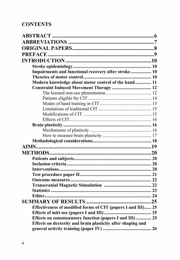
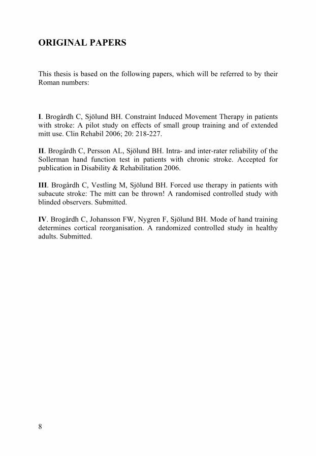
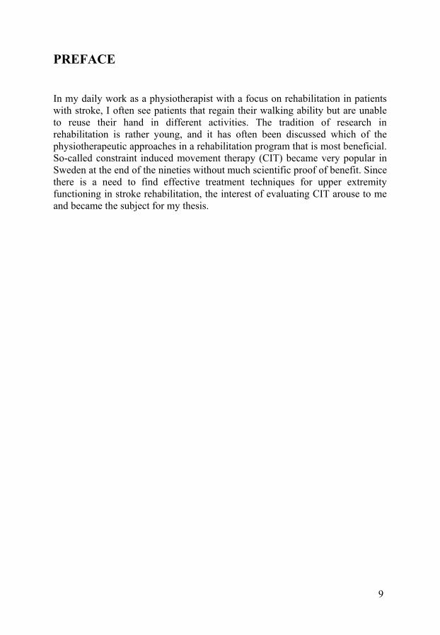
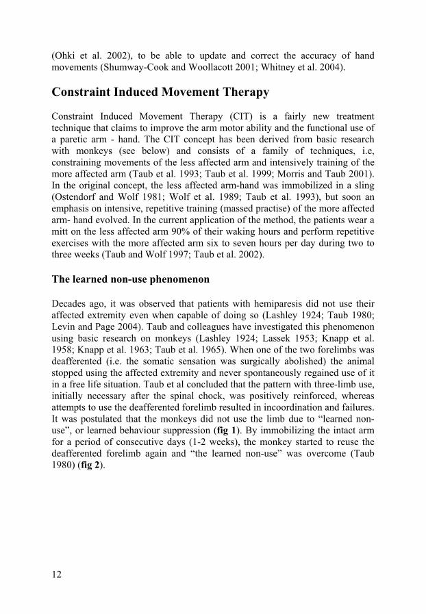
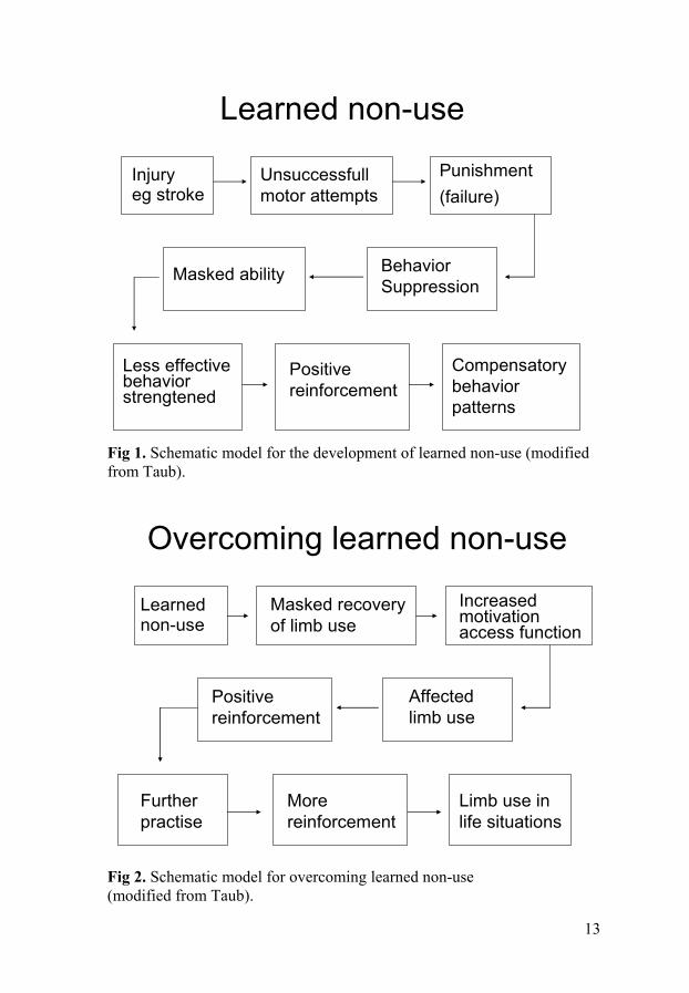
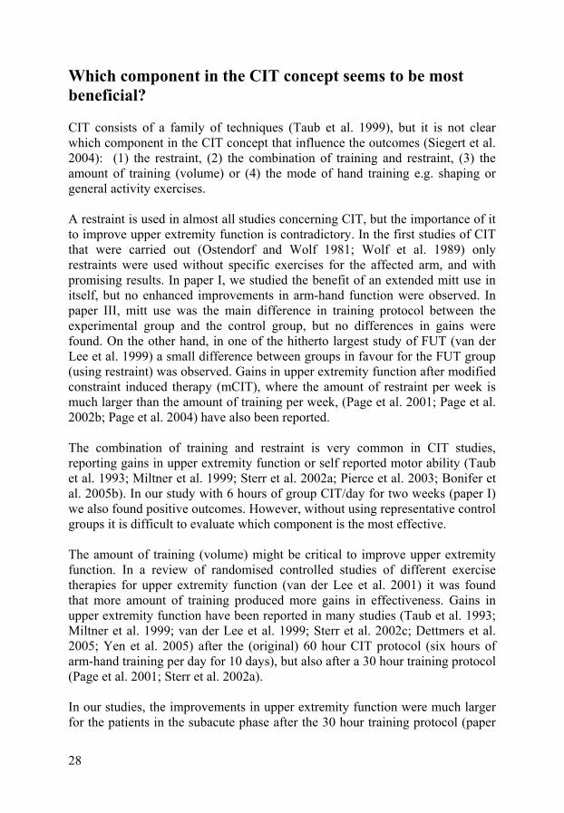
(Ohki et al. 2002), to be able to update and correct the accuracy of hand movements (Shumway-Cook and Woollacott 2001; Whitney et al. 2004). Constraint Induced Movement Therapy Constraint Induced Movement Therapy (CIT) is a fairly new treatment technique that claims to improve the arm motor ability and the functional use of a paretic arm - hand. The CIT concept has been derived from basic research with monkeys (see below) and consists of a family of techniques, i.e, constraining movements of the less affected arm and intensively training of the more affected arm (Taub et al. 1993; Taub et al. 1999; Morris and Taub 2001). In the original concept, the less affected arm-hand was immobilized in a sling (Ostendorf and Wolf 1981; Wolf et al. 1989; Taub et al. 1993), but soon an emphasis on intensive, repetitive training (massed practise) of the more affected arm- hand evolved. In the current application of the method, the patients wear a mitt on the less affected arm 90% of their waking hours and perform repetitive exercises with the more affected arm six to seven hours per day during two to three weeks (Taub and Wolf 1997; Taub et al. 2002). The learned non-use phenomenon Decades ago, it was observed that patients with hemiparesis did not use their affected extremity even when capable of doing so (Lashley 1924; Taub 1980; Levin and Page 2004). Taub and colleagues have investigated this phenomenon using basic research on monkeys (Lashley 1924; Lassek 1953; Knapp et al. 1958; Knapp et al. 1963; Taub et al. 1965). When one of the two forelimbs was deafferented (i.e. the somatic sensation was surgically abolished) the animal stopped using the affected extremity and never spontaneously regained use of it in a free life situation. Taub et al concluded that the pattern with three-limb use, initially necessary after the spinal chock, was positively reinforced, whereas attempts to use the deafferented forelimb resulted in incoordination and failures. It was postulated that the monkeys did not use the limb due to “learned non-use”, or learned behaviour suppression (fig 1). By immobilizing the intact arm for a period of consecutive days (1-2 weeks), the monkey started to reuse the deafferented forelimb again and “the learned non-use” was overcome (Taub 1980) (fig 2).
13
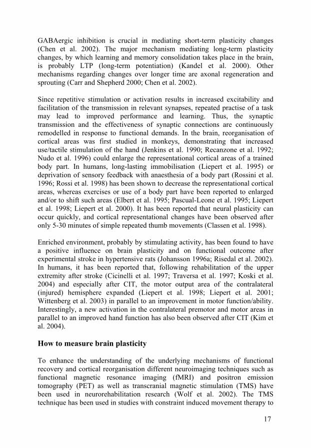
Learned non-use
Injuryeg stroke
Unsuccessfullmotor attempts
Punishment(failure)
BehaviorSuppression
Masked ability
Less effectivebehaviorstrengtened
Positive reinforcement
Compensatorybehaviorpatterns
Overcoming learned non-use
Learnednon-use
Masked recoveryof limb use
Increasedmotivation access function
Affectedlimb use
Positive reinforcement
Furtherpractise
Morereinforcement
Limb use in life situations
Fig 1. Schematic model for the development of learned non-use (modified from Taub).
Fig 2. Schematic model for overcoming learned non-use (modified from Taub).
14
The mechanism of learned non-use was also thought to apply to humans who suffer from hemiparesis after brain injury (Taub 1980; Wolf et al. 1989; Taub and Wolf 1997). Although motor function may gradually return as the result of a combination of spontaneous recovery and rehabilitation, the actual use of the more affected arm often seems to be less than its potential use (Andrews and Stewart 1979; Taub 1980; Sterr et al. 2002b) . Patients eligible for CIT Constraint Induced Movement Therapy has been applied in humans after different diagnoses, for example after stroke (Taub et al. 1993; Kunkel et al. 1999; Miltner et al. 1999; van der Lee et al. 1999; Dromerick et al. 2000; Page et al. 2002b; Sterr et al. 2002a; Atteya 2004; Page et al. 2004; Dettmers et al. 2005; Page et al. 2005; Taub et al. 2005; Brogårdh and Sjölund 2006; Taub et al. 2006), after traumatic brain injury (Wolf et al. 1989; Sterr et al. 2002a; Page and Levine 2003) and in children with hemiparesis (Glover et al. 2002) and cerebral palsy (Taub et al. 2004; Eliasson et al. 2005). Inclusion criteria for patients with stroke to participate in CIT interventions are usually some limitation of hand function, high motivation, minimal cognitive dysfunctions, adequate balance and walking ability when wearing the restraint (Taub et al. 1993; Miltner et al. 1999; van der Lee et al. 1999; Brogårdh and Sjölund 2006). According to Taub, the patient’s hand function can be divided into quartiles: (1) the first quartile includes ability to extend the wrist at least 20°, ability to extend the fingers at least 10° and to abduct the thumb at least 10°; (2) the second quartile includes ability to extend the wrist at least 10°, ability to extend two fingers at least 10° and to abduct the thumb at least 10°; (3) the third quartile includes ability to grasp and release a tennis ball or washrag; and (4) the fourth quartile implies little or no finger function (Taub et al. 1999). CIT or modifications of CIT have been applied mainly in chronic stroke patients, when the patients are supposed to have reached a plateau in recovery (Wolf et al. 1989; Taub et al. 1993; Morris et al. 1997; Kunkel et al. 1999; Miltner et al. 1999; Sterr et al. 2002a; Page et al. 2004; Dettmers et al. 2005; Taub et al. 2005; Brogårdh and Sjölund 2006; Taub et al. 2006). The technique has also been applied after subacute (Blanton and Wolf 1999; Page et al. 2002b; Atteya 2004) and after acute stroke (Dromerick et al. 2000; Grotta et al. 2004; Ploughman and Corbett 2004; Page et al. 2005). Doubts regarding intensive training early in the acute phase after a cerebrovascular incident have been reported from studies on animals. Thus, “forced overuse” of the affected limb, including restraint of the non affected limb, within the first seven days after a sensory motor cortex lesion, has been
15
shown to impede motor recovery and to enlarge the lesion volume (Humm et al. 1998; Bland et al. 2000). This assumption has so far not been confirmed in humans in the acute phase after stroke (Grotta et al. 2004). Modes of hand training in CIT Different approaches of hand training, such as general task practise (i.e., eating a meal, making coffee, dialling phone numbers and so on) as well as focussed “shaping” exercises are used in CIT (Taub et al. 2006). It has been suggested that shaping exercises is of outmost importance for improving upper limb motor function and to transfer arm-hand use to real life situations (Taub et al. 1994; Taub et al. 2002). The shaping technique is defined as a behavioural technique in which movements are approached in small steps of progressively increasing difficulties including verbal feedback. The shaping exercises are similar to ordinary training techniques used by physiotherapists and occupational therapists, but a shaping task is broken down to subtasks, is quantifiable and is repeated many times so small improvements in performance can be measured and systematically made apparent to the patient (Morris et al. 1997; Taub and Wolf 1997; Wolf et al. 2002; Taub et al. 2006). The benefit of the shaping exercises for improving upper motor function after stroke has been confirmed by independent authors (Sterr and Freivogel 2003; Yen et al. 2005). On the other hand, it has also been reported that forced use therapy (FUT) without shaping exercises may improve upper extremity function after stroke (Liepert et al. 2001; Ploughman and Corbett 2004). Limitations of traditional CIT Traditional CIT with one patient per therapist requires a lot of resources and the six hours training protocol may be strenuous for the patients. According to Page et al (Page et al. 2002a), CIT is not widely viewed by clinicians as a useful therapeutic intervention and considered unfeasible due to patients’ concerns about the intensive schedule of treatment. In addition, therapists are concerned about patients’ compliance, about safety issues and about clinical resources. Modifications of CIT To make CIT clinically adaptable with limited resources as regards therapists, various modifications of the original concept have been attempted, for example shortened CIT (Sterr et al. 2002a) (i.e 3 hours of training/day for two weeks); forced use therapy (FUT; restraint of the less affected arm but without specific shaping exercises for the affected arm) (van der Lee et al. 1999; Ploughman and Corbett 2004), modified CIT (Page et al. 2001; Levin and Page 2004) (consisting of 3 hours of training per week for 10 weeks with the intact arm in
16
restraint 5 hours/day for 5 days/week), automated delivery of constraint induced therapy (AutoCITE) i.e., a computerized form of CIT (Taub et al. 2005), distributed CIT (3 hours of training per day distributed for 20 days) (Dettmers et al. 2005), and group CIT (with 2-3 patients per therapist) (Brogårdh and Sjölund 2006). Effects of CIT Gains in upper extremity function after constraint induced therapy have been reported in all stages after the onset of stroke (Wolf et al. 2002; Hakkennes and Keating 2005). Two possible mechanisms for the observed effects are believed to be (1) overcoming the learned non-use of the more affected arm (i.e, increased use of the more affected arm) and (2) use dependent cortical reorganisation (Taub et al. 1999; Liepert et al. 2000; Morris and Taub 2001; Taub et al. 2002; Wolf et al. 2002). Brain Plasticity Decades ago it was widely believed by neuroscientists that the human brain was a rather static organ and that no new neural connections could be formed in the adult brain (Kandel et al. 2000). It was assumed that once neural connections had been established in the early foetal stage of life, they would hardly change later in life. This assumption has radically been changed since research in animals and humans has established that the nervous system can modify its organization and function continuously throughout the individual’s lifespan, depending on internal and environmental factors, a phenomenon called brain plasticity (Carr and Shepherd 2000; Kandel et al. 2000; Chen et al. 2002). Mechanisms of plasticity Mechanisms of brain plasticity include the capacity for neurochemical, neuroreceptor and neuronal structural changes. Normally, many connections between the periphery and the cortex as well as intracortical connections are probably physiologically silent because of inhibitory influences (Wall 1977). There seems to be a wide overlap in cortical neuronal networks targeting different body parts (Carr and Shepherd 2000). Both strengthening and weakening of synaptic connexions have been proposed as learning mechanisms (Schweighofer et al. 2001; Jörntell and Ekeroth 2003). The mechanism of plasticity probably differs depending on the time course (Chen et al. 2002). Rapid changes (within minutes) in motor representation are likely due to unmasking of silent synapses involving mediation by gamma-aminobutyric acid (GABA) (Jones 1993). GABA seems to be the most important inhibitory neurotransmitter in the brain and evidence is strong that a reduction of
17
GABAergic inhibition is crucial in mediating short-term plasticity changes (Chen et al. 2002). The major mechanism mediating long-term plasticity changes, by which learning and memory consolidation takes place in the brain, is probably LTP (long-term potentiation) (Kandel et al. 2000). Other mechanisms regarding changes over longer time are axonal regeneration and sprouting (Carr and Shepherd 2000; Chen et al. 2002). Since repetitive stimulation or activation results in increased excitability and facilitation of the transmission in relevant synapses, repeated practise of a task may lead to improved performance and learning. Thus, the synaptic transmission and the effectiveness of synaptic connections are continuously remodelled in response to functional demands. In the brain, reorganisation of cortical areas was first studied in monkeys, demonstrating that increased use/tactile stimulation of the hand (Jenkins et al. 1990; Recanzone et al. 1992; Nudo et al. 1996) could enlarge the representational cortical areas of a trained body part. In humans, long-lasting immobilisation (Liepert et al. 1995) or deprivation of sensory feedback with anaesthesia of a body part (Rossini et al. 1996; Rossi et al. 1998) has been shown to decrease the representational cortical areas, whereas exercises or use of a body part have been reported to enlarged and/or to shift such areas (Elbert et al. 1995; Pascual-Leone et al. 1995; Liepert et al. 1998; Liepert et al. 2000). It has been reported that neural plasticity can occur quickly, and cortical representational changes have been observed after only 5-30 minutes of simple repeated thumb movements (Classen et al. 1998). Enriched environment, probably by stimulating activity, has been found to have a positive influence on brain plasticity and on functional outcome after experimental stroke in hypertensive rats (Johansson 1996a; Risedal et al. 2002). In humans, it has been reported that, following rehabilitation of the upper extremity after stroke (Cicinelli et al. 1997; Traversa et al. 1997; Koski et al. 2004) and especially after CIT, the motor output area of the contralateral (injured) hemisphere expanded (Liepert et al. 1998; Liepert et al. 2001; Wittenberg et al. 2003) in parallel to an improvement in motor function/ability. Interestingly, a new activation in the contralateral premotor and motor areas in parallel to an improved hand function has also been observed after CIT (Kim et al. 2004). How to measure brain plasticity To enhance the understanding of the underlying mechanisms of functional recovery and cortical reorganisation different neuroimaging techniques such as functional magnetic resonance imaging (fMRI) and positron emission tomography (PET) as well as transcranial magnetic stimulation (TMS) have been used in neurorehabilitation research (Wolf et al. 2002). The TMS technique has been used in studies with constraint induced movement therapy to
18
evaluate the cortical organisation (Liepert et al. 1998; Liepert et al. 2000; Liepert et al. 2001; Wolf et al. 2002, Wittenberg et al. 2003), and has been reported to be a safe, relatively painless and a reliable technique (Mortifee et al. 1994). TMS is based on the principle of electromagnetic induction. A strong electrical current pulse is directed through a hand-held coil. When held over the scalp, a rapidly changing magnetic field is able to induce a small electrical current in the underlying brain tissue. By stimulating the motor cortex the muscle responses can be recorded as motor evoked potentials (MEP), and monitored by electro-myography (EMG) (Ellaway et al. 1999; Wolf et al. 2002). Methodological considerations There are two ways of approaching hand function assessment, either by (a) instruments for observation by independent observers or by (b) instruments for self assessment. Commonly used observation tests for assessing the upper extremity function in the literature are the Fugl-Meyer Assessment of Physical Performance (Fugl-Meyer et al. 1975), the Action Research Arm Test (ARA test) (Lyle RC 1981), the Wolf Motor Function Test (Wolf et al. 1989) and the Arm Motor Ability Test (Kopp et al. 1997). Some of these tests are used to assess the patient’s ability to perform a specific activity of daily living (ADL), but lack either in the assessment of quality of movement (Wolf et al. 1989) or in the assessment of performance time (Fugl-Meyer et al. 1975; Lyle RC 1981). Only one of these tests has been translated into Swedish and has been tested for validity and reliability (Fugl-Meyer et al. 1975). The Sollerman hand function test (Sollerman and Ejeskar 1995) is often used in clinical rehabilitation settings in Scandinavia. The test is standardised and includes 20 subtests reflecting daily activities of the hand (see methods). It has been developed to evaluate the patient’s hand function before and after hand surgery (Sollerman and Ejeskar 1995). Despite being utilized in patients’ after injury to the central nervous system, it has not been evaluated for reliability in patients with stroke. A commonly used self-assessment tool in CIT is the Motor Activity Log test (MAL) (Taub et al. 1993). The MAL is a self reported questionnaire that asks how often (amount of use [AOU]) and how well (quality of movement [QOM]) the affected hand is used for the daily activities (see methods). The improvements in arm motor ability after CIT have, however, often been reported to be much larger in the self reported MAL than in more objective observation tests.
19
AIMS The overall aims of this thesis were to elucidate the effectiveness of modified forms of Constraint Induced Movement Therapy for arm-hand function and to evaluate the effectiveness of wearing restraint as well as the benefit of two different types of hand training. The specific aims were:
- (i) to evaluate the effectiveness of constraint induced group therapy for chronic stroke patients (paper I).
- (ii) to explore if extended mitt use may enhance the treatment effect (paper I).
- (iii) to evaluate whether the Sollerman hand function test is reliable for patients with chronic stroke (paper II).
- (iv) to investigate the effectiveness of a three hour forced use protocol for two weeks in patients with subacute stroke (paper III).
- (v) to explore if the mitt use may enhance the treatment effect (paper III).
- (vi) to examine how shaping versus activity training influences the dexterity of the non dominant hand and the plasticity of the contralateral motor cortex hand area in normal adults (paper IV).
20
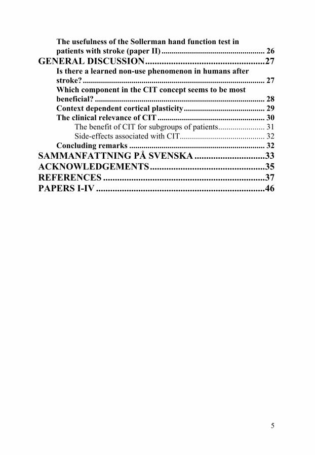
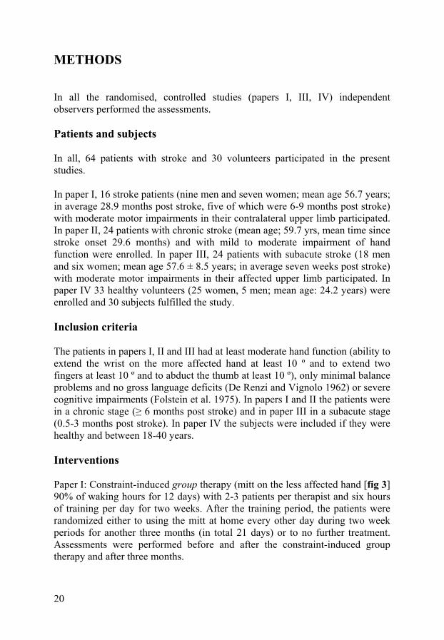
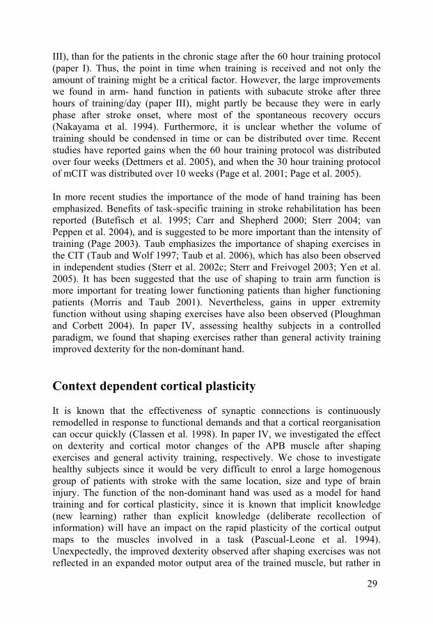
METHODS In all the randomised, controlled studies (papers I, III, IV) independent observers performed the assessments. Patients and subjects In all, 64 patients with stroke and 30 volunteers participated in the present studies. In paper I, 16 stroke patients (nine men and seven women; mean age 56.7 years; in average 28.9 months post stroke, five of which were 6-9 months post stroke) with moderate motor impairments in their contralateral upper limb participated. In paper II, 24 patients with chronic stroke (mean age; 59.7 yrs, mean time since stroke onset 29.6 months) and with mild to moderate impairment of hand function were enrolled. In paper III, 24 patients with subacute stroke (18 men and six women; mean age 57.6 ± 8.5 years; in average seven weeks post stroke) with moderate motor impairments in their affected upper limb participated. In paper IV 33 healthy volunteers (25 women, 5 men; mean age: 24.2 years) were enrolled and 30 subjects fulfilled the study. Inclusion criteria The patients in papers I, II and III had at least moderate hand function (ability to extend the wrist on the more affected hand at least 10 º and to extend two fingers at least 10 º and to abduct the thumb at least 10 º), only minimal balance problems and no gross language deficits (De Renzi and Vignolo 1962) or severe cognitive impairments (Folstein et al. 1975). In papers I and II the patients were in a chronic stage (≥ 6 months post stroke) and in paper III in a subacute stage (0.5-3 months post stroke). In paper IV the subjects were included if they were healthy and between 18-40 years. Interventions Paper I: Constraint-induced group therapy (mitt on the less affected hand [fig 3] 90% of waking hours for 12 days) with 2-3 patients per therapist and six hours of training per day for two weeks. After the training period, the patients were randomized either to using the mitt at home every other day during two week periods for another three months (in total 21 days) or to no further treatment. Assessments were performed before and after the constraint-induced group therapy and after three months.
21
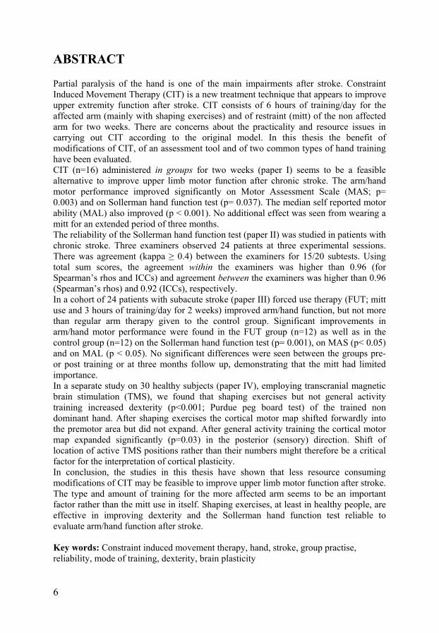
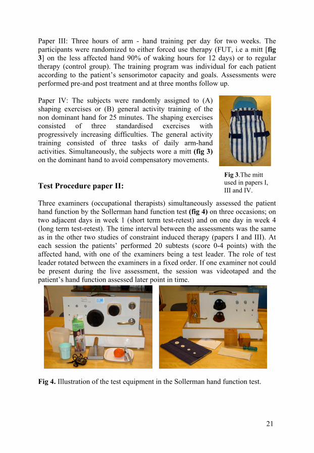
Paper III: Three hours of arm - hand training per day for two weeks. The participants were randomized to either forced use therapy (FUT, i.e a mitt [fig 3] on the less affected hand 90% of waking hours for 12 days) or to regular therapy (control group). The training program was individual for each patient according to the patient’s sensorimotor capacity and goals. Assessments were performed pre-and post treatment and at three months follow up. Paper IV: The subjects were randomly assigned to (A) shaping exercises or (B) general activity training of the non dominant hand for 25 minutes. The shaping exercises consisted of three standardised exercises with progressively increasing difficulties. The general activity training consisted of three tasks of daily arm-hand activities. Simultaneously, the subjects wore a mitt (fig 3) on the dominant hand to avoid compensatory movements. Test Procedure paper II: Three examiners (occupational therapists) simultaneously assessed the patient hand function by the Sollerman hand function test (fig 4) on three occasions; on two adjacent days in week 1 (short term test-retest) and on one day in week 4 (long term test-retest). The time interval between the assessments was the same as in the other two studies of constraint induced therapy (papers I and III). At each session the patients’ performed 20 subtests (score 0-4 points) with the affected hand, with one of the examiners being a test leader. The role of test leader rotated between the examiners in a fixed order. If one examiner not could be present during the live assessment, the session was videotaped and the patient’s hand function assessed later point in time.
Fig 4. Illustration of the test equipment in the Sollerman hand function test.
Fig 3.The mitt used in papers I, III and IV.
22
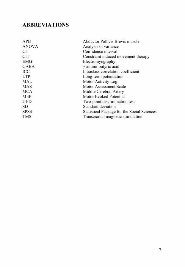
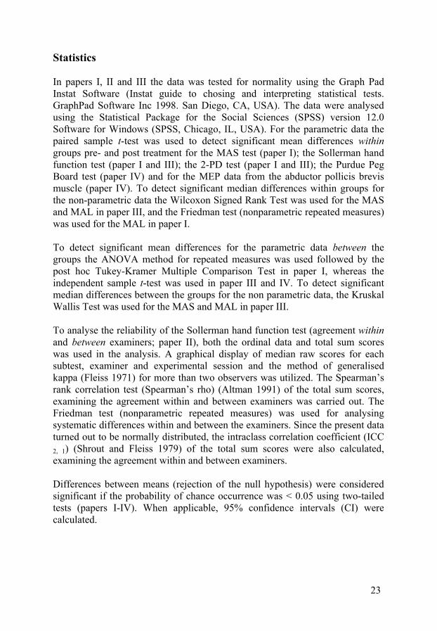
Outcome measures The instruments used for assessments in papers I and III were the modified version of the Motor Assessment Scale (MAS) (Carr et al. 1985; Arnell et al. 1996; Barkelius et al. 1997), the Sollerman Hand Function Test (Sollerman and Ejeskar 1995), the 2-Point Discrimination Test (2-PD) (Moberg 1958) and the Motor Activity Log (MAL) (Taub et al. 1993). The modified Motor Assessment Scale for upper extremity has been tested for validity and reliability (Arnell et al. 1996; Barkelius et al. 1997) and consists of 15 tasks from gross arm to fine finger movements in a 0-5 point scale, where the observer assesses the quality of movement and the speed of performance of each task. The Sollerman Hand Function test has been tested for validity and reliability in some diagnosis (Sollerman and Ejeskar 1995) and consists of 20-subtests reflecting daily hand activities. The type of grasp, the quality of movement and the speed of performance for the more affected arm is assessed in a 0-4 point scale. The 2-PD test measures the tactile somatosensation in a 0-10 point scale. The MAL (tested for validity and reliability) (van der Lee et al. 2004; Uswatte et al. 2005) is a 30-item self reported questionnaire and asks how often (amount of use [AOU]) and how well (quality of movement [QOM]) the affected hand is used in daily activities in a 0-5 point scale. In paper II the Sollerman test was used only to assess the reproducibility of the test. In paper IV the subjects went through dexterity testing and cortical mapping before and after 25 minutes of hand training. The dexterity was evaluated by the Purdue Peg Board test (Tiffin and Ascher 1948) which includes four tasks: place as many pins as possibly with the right hand, with the left hand, and with both hand in 30 seconds as well as an assembly task (within 60 seconds). Transcranial Magnetic Stimulation The cortical motor maps of the abductor pollicis brevis (APB) muscle were obtained employing transcranial magnetic stimulation (TMS) of the non-dominant hemisphere and surface electromyographic (EMG) recording of motor evoked potentials (MEPs) (fig 5).
Fig 5. (A) the transcranial magnetic stimulator and (B) the coil for stimulating the cortical motor area of the abductor pollicis brevis muscle. A B
23
Statistics In papers I, II and III the data was tested for normality using the Graph Pad Instat Software (Instat guide to chosing and interpreting statistical tests. GraphPad Software Inc 1998. San Diego, CA, USA). The data were analysed using the Statistical Package for the Social Sciences (SPSS) version 12.0 Software for Windows (SPSS, Chicago, IL, USA). For the parametric data the paired sample t-test was used to detect significant mean differences within groups pre- and post treatment for the MAS test (paper I); the Sollerman hand function test (paper I and III); the 2-PD test (paper I and III); the Purdue Peg Board test (paper IV) and for the MEP data from the abductor pollicis brevis muscle (paper IV). To detect significant median differences within groups for the non-parametric data the Wilcoxon Signed Rank Test was used for the MAS and MAL in paper III, and the Friedman test (nonparametric repeated measures) was used for the MAL in paper I. To detect significant mean differences for the parametric data between the groups the ANOVA method for repeated measures was used followed by the post hoc Tukey-Kramer Multiple Comparison Test in paper I, whereas the independent sample t-test was used in paper III and IV. To detect significant median differences between the groups for the non parametric data, the Kruskal Wallis Test was used for the MAS and MAL in paper III. To analyse the reliability of the Sollerman hand function test (agreement within and between examiners; paper II), both the ordinal data and total sum scores was used in the analysis. A graphical display of median raw scores for each subtest, examiner and experimental session and the method of generalised kappa (Fleiss 1971) for more than two observers was utilized. The Spearman’s rank correlation test (Spearman’s rho) (Altman 1991) of the total sum scores, examining the agreement within and between examiners was carried out. The Friedman test (nonparametric repeated measures) was used for analysing systematic differences within and between the examiners. Since the present data turned out to be normally distributed, the intraclass correlation coefficient (ICC 2, 1) (Shrout and Fleiss 1979) of the total sum scores were also calculated, examining the agreement within and between examiners. Differences between means (rejection of the null hypothesis) were considered significant if the probability of chance occurrence was < 0.05 using two-tailed tests (papers I-IV). When applicable, 95% confidence intervals (CI) were calculated.
24
Ethics The research protocols in paper I, II and III were approved by the Medical Ethics Committee of Lund University and the research protocol in paper IV was approved by the Medical Ethics Committee of Umeå University.
25
SUMMARY OF RESULTS Effectiveness of modified forms of CIT (papers I and III) Constraint induced therapy was beneficial even when limiting the therapist availability through training in small groups. Gains in arm- hand function were seen in chronic stroke patients after two weeks of group CIT (paper I) on the MAS (p= 0.003) and on the Sollerman hand function test (p= 0.037). The changes in self reported use of the more affected arm on Motor Activity Log scores were also significant (p < 0.001). Also in patients with subacute stroke (paper III), gains in upper extremity function were found after three hours of arm therapy per day during two weeks, but the forced use therapy (FUT) was not more effective than regular training. The arm/hand motor performance improved significantly within both groups after two weeks of therapy on the Sollerman hand function test (p= 0.001), on MAS (p < 0.05) and on self reported motor ability (MAL; p < 0.05). After three months some additional improvements in arm motor performance and self reported motor ability were seen mainly in the FUT group. However, no significant differences between the groups were found in any measures or point in times. Effects of mitt use (papers I and III) In paper I no additional effects of extended mitt use for three months were seen in any of the assessments. The hand function scores measured by the Sollerman hand function test seemed to improve slightly for the participants with extended mitt use, but the mean difference was not significant. In addition, patients randomized to extended mitt use reported some difficulties in compliance. In paper III no significant differences in arm-hand function were found between the patients wearing a mitt 90% of the time during waking hours and the control group in any of the measures or point in time, despite satisfactory mitt compliance in the FUT group. Effects on somatosensory function (papers I and III) No significant difference in sensory discrimination with the 2-PD test was seen in any of the intervention studies on patients with stroke.
26
Effects on dexterity and brain plasticity after shaping and general activity training (paper IV) In a group of young healthy adults, shaping exercises of the non-dominant hand increased the dexterity significantly (p<0.005) for both hands, mostly so in the non dominant hand. After the general activity training, no significant improvement in dexterity of the non dominant hand was found. The cortical motor map (number of excitable positions) of the APB muscle upon TMS shifted forward into the premotor area after the shaping exercises but did not expand, whereas after general activity training the cortical motor map of the non dominant APB muscle expanded significantly in the posterior (sensory) direction (p=0.03). The usefulness of the Sollerman hand function test in patients with stroke (paper II) The Sollerman test seems to be a reliable test to evaluate a patient’s hand function pre-and post treatment after stroke. When analysing each subtest the median scores of each examiner generally agreed for all of the 20 subtests in all three sessions (paper II, fig 2). In the kappa analysis the agreement between the examiners was very strong (kappa > 0.9) for one subtest. For seven other subtests the kappa values were between 0.6 - 0.9, and for another seven subtests the kappa values were between 0.4 - 0.6. Only for five subtests were the kappa values lower than 0.4 in some of the sessions. When analysing the total sum scores the agreement within each examiner was ≥ 0.96 (Spearman’s rhos and ICCs, respectively) for short- as well as long term test-retests. The agreement between the three examiners was also high at all three sessions (Spearman’s rhos ≥ 0.96; ICCs ≥ 0.92). However, statistically systematic differences (Friedman’s test: p< 0.05) were found between examiners A-B and A-C for all three sessions, indicating that the same assessor should perform pre- and post treatment evaluations. Video assessments were found to be a good alternative to live assessments, since no systematically differences in total sum scores were found within any of the examiners regarding live versus video assessments.
27
GENERAL DISCUSSION This thesis has focused on evaluating the benefit of modified forms of constraint induced movement therapy, and to elucidate which mode of hand training is most effective for improving dexterity, and also to evaluate the usefulness of a hand function test after stroke. Is there a learned non-use phenomenon in humans after stroke? It has been reported that constraint induced movement therapy can improve hand function through overcoming the learned non-use phenomenon in parallel to a use dependent cortical reorganisation in the brain (Liepert et al. 1998; Taub et al. 2002; Wolf et al. 2002; Sterr 2004). However, it has been questioned whether the “learned non-use” phenomenon from basic research in monkeys with secondary lesions in spinal cord after deafferentation of a single fore-limb, can be transferred into humans after stroke with lesions in the brain (Siegert et al. 2004; Sunderland and Tuke 2005). Our data strongly indicate that the restraint is an unimportant factor in regaining hand function (papers I and III), speaking against the relevance of a learned non-use phenomenon in patients with stroke. However, we cannot exclude that the results are at least partly due to that all patients included had accepted the possibility of being restrained from the general information given at study entry. It can be hypothesised that the patients in the control group had developed a “cognitive mitt”, i.e, were aware of the importance of preferably using the affected arm in daily activities. The notion of a “cognitive mitt” that might influence the results in human studies, but does not occur in animals has not previously been described. Furthermore, it has been argued that the large improvements in self-reported motor ability (as measured by MAL) that have frequently been reported after only one week of CIT (and also was observed in our study of group CIT; paper I), as well as the cortical changes measured by TMS, reflect normal adaptive learning rather than restoration of representation lost due to brain injury or non-use of the limb (Sunderland and Tuke 2005). According to Taub, the main effect of CIT is an increased arm-hand use in real life situations (Morris and Taub 2001), i e improved activity. However, there are reports of decreases in impairment such as improved grip strength, reduced spasticity (Dettmers et al. 2005), improved force control in dexterous function (Alberts et al. 2004) as well as increased finger coordination after shaping exercises (Sterr et al. 2002c).
28
Which component in the CIT concept seems to be most beneficial? CIT consists of a family of techniques (Taub et al. 1999), but it is not clear which component in the CIT concept that influence the outcomes (Siegert et al. 2004): (1) the restraint, (2) the combination of training and restraint, (3) the amount of training (volume) or (4) the mode of hand training e.g. shaping or general activity exercises. A restraint is used in almost all studies concerning CIT, but the importance of it to improve upper extremity function is contradictory. In the first studies of CIT that were carried out (Ostendorf and Wolf 1981; Wolf et al. 1989) only restraints were used without specific exercises for the affected arm, and with promising results. In paper I, we studied the benefit of an extended mitt use in itself, but no enhanced improvements in arm-hand function were observed. In paper III, mitt use was the main difference in training protocol between the experimental group and the control group, but no differences in gains were found. On the other hand, in one of the hitherto largest study of FUT (van der Lee et al. 1999) a small difference between groups in favour for the FUT group (using restraint) was observed. Gains in upper extremity function after modified constraint induced therapy (mCIT), where the amount of restraint per week is much larger than the amount of training per week, (Page et al. 2001; Page et al. 2002b; Page et al. 2004) have also been reported. The combination of training and restraint is very common in CIT studies, reporting gains in upper extremity function or self reported motor ability (Taub et al. 1993; Miltner et al. 1999; Sterr et al. 2002a; Pierce et al. 2003; Bonifer et al. 2005b). In our study with 6 hours of group CIT/day for two weeks (paper I) we also found positive outcomes. However, without using representative control groups it is difficult to evaluate which component is the most effective. The amount of training (volume) might be critical to improve upper extremity function. In a review of randomised controlled studies of different exercise therapies for upper extremity function (van der Lee et al. 2001) it was found that more amount of training produced more gains in effectiveness. Gains in upper extremity function have been reported in many studies (Taub et al. 1993; Miltner et al. 1999; van der Lee et al. 1999; Sterr et al. 2002c; Dettmers et al. 2005; Yen et al. 2005) after the (original) 60 hour CIT protocol (six hours of arm-hand training per day for 10 days), but also after a 30 hour training protocol (Page et al. 2001; Sterr et al. 2002a). In our studies, the improvements in upper extremity function were much larger for the patients in the subacute phase after the 30 hour training protocol (paper
29
III), than for the patients in the chronic stage after the 60 hour training protocol (paper I). Thus, the point in time when training is received and not only the amount of training might be a critical factor. However, the large improvements we found in arm- hand function in patients with subacute stroke after three hours of training/day (paper III), might partly be because they were in early phase after stroke onset, where most of the spontaneous recovery occurs (Nakayama et al. 1994). Furthermore, it is unclear whether the volume of training should be condensed in time or can be distributed over time. Recent studies have reported gains when the 60 hour training protocol was distributed over four weeks (Dettmers et al. 2005), and when the 30 hour training protocol of mCIT was distributed over 10 weeks (Page et al. 2001; Page et al. 2005). In more recent studies the importance of the mode of hand training has been emphasized. Benefits of task-specific training in stroke rehabilitation has been reported (Butefisch et al. 1995; Carr and Shepherd 2000; Sterr 2004; van Peppen et al. 2004), and is suggested to be more important than the intensity of training (Page 2003). Taub emphasizes the importance of shaping exercises in the CIT (Taub and Wolf 1997; Taub et al. 2006), which has also been observed in independent studies (Sterr et al. 2002c; Sterr and Freivogel 2003; Yen et al. 2005). It has been suggested that the use of shaping to train arm function is more important for treating lower functioning patients than higher functioning patients (Morris and Taub 2001). Nevertheless, gains in upper extremity function without using shaping exercises have also been observed (Ploughman and Corbett 2004). In paper IV, assessing healthy subjects in a controlled paradigm, we found that shaping exercises rather than general activity training improved dexterity for the non-dominant hand. Context dependent cortical plasticity It is known that the effectiveness of synaptic connections is continuously remodelled in response to functional demands and that a cortical reorganisation can occur quickly (Classen et al. 1998). In paper IV, we investigated the effect on dexterity and cortical motor changes of the APB muscle after shaping exercises and general activity training, respectively. We chose to investigate healthy subjects since it would be very difficult to enrol a large homogenous group of patients with stroke with the same location, size and type of brain injury. The function of the non-dominant hand was used as a model for hand training and for cortical plasticity, since it is known that implicit knowledge (new learning) rather than explicit knowledge (deliberate recollection of information) will have an impact on the rapid plasticity of the cortical output maps to the muscles involved in a task (Pascual-Leone et al. 1994). Unexpectedly, the improved dexterity observed after shaping exercises was not reflected in an expanded motor output area of the trained muscle, but rather in
30
shifts of its location. An enlargement of the motor output area in parallel to improvements is the more commonly reported change after CIT (Liepert et al. 1998; Liepert et al. 2000; Liepert et al. 2001; Wittenberg et al. 2003) but some studies have reported minor shifts in location after repetitive movements (Liepert et al. 2000; Pearce et al. 2000; Kim et al. 2004). Hence, a shift of location rather than the size of a motor area may be a critical factor in evaluating cortical plasticity in humans after common behavioural tasks. Furthermore, in our subjects the improved dexterity was paralleled by a shift into the premotor area, known to be active in motor planning. General activity training, on the other hand, expanded and extended the area in the posterior (and inferior) direction. It is tempting to assume that this may be related to spatial coordination necessary for global activities. The clinical relevance of CIT To be able to evaluate a possible treatment effect it is important to use reliable test instruments. We therefore examined the Sollerman hand functions test (paper II) that was found to be a useful and reliable test for stroke patients with mild to moderate impairments of hand function. It would be interesting to know what “cut off level” in the total Sollerman scores that indicates a patient’s independence to perform hand –related activities in daily life, but such data are so far anecdotal. Statistically significant changes in outcome measures cannot automatically be interpreted as clinically relevant. However, in our clinical studies (papers I and III) we consistently found that the improvements in observation tests were in accordance with the patients’ opinion, expressed by spontaneous comments of improved motor ability as well as reports in MAL. Only one study has evaluated the impact on the patients’ quality of life after CIT (measured by the Stroke Impact Scale), and improvements in domains such as physical function, social participation and communication were reported either post treatment or at six months follow-up (Dettmers et al. 2005). The MAL test has most frequently been used to measure the patients’ opinion of changes in arm use and quality of arm movement after CIT. The questionnaire has been found to have sufficient internal consistency and reliability, but the construct validity has been questioned (van der Lee et al. 2004). In one study, the quality of movement scale (QOM) but not the amount of use scale (AOU) was found to be reliable (Uswatte et al. 2005). Most studies regarding CIT have been carried out in patients with a considerable residual functional capacity of the hand (referred as to the first quartile) (Taub et al. 1993; Kunkel et al. 1999; Miltner et al. 1999; Taub et al. 1999; van der Lee et al. 1999; Sterr et al. 2002a; Page et al. 2004; Dettmers et al. 2005), in which also the gains seem to be the largest (Taub et al. 2002),
31
probably due to a strong potential for improved hand use (Sunderland and Tuke 2005). In papers I and III we chose to include patients with mild to moderate impairment of hand function (representing the first and second quartiles), partly because of difficulties in recruiting patients. Other recent studies have included patients with lower hand function (referred to as second quartile) (Taub et al. 1998; Bonifer et al. 2005b), as well as minimal hand function (representing the third quartile) (Bonifer et al. 2005a) with promising treatment effects. Also patients using wheelchairs for transportation have been reported to be able to participate in CIT (Tarkka et al. 2005) and it is conceivable that CIT can be applied in a larger part of the stroke population than was originally thought (Morris and Taub 2001). CIT has, in a recent review article (van Peppen et al. 2004), been reported to be beneficial to improve upper extremity function as compared to traditional neurological treatment approaches, biofeedback, neuromuscular electrical stimulation etc. Few studies have, however investigated the long-term clinical benefits of CIT (Wolf et al. 1989; Taub et al. 1993; van der Lee et al. 1999) and more research is warranted (Sterr 2004). To gain acceptance in clinical settings, modifications of the original CIT concept is probably needed. In accordance with the reports of others (Page et al. 2001; Sterr et al. 2002a; Dettmers et al. 2005), our modifications of CIT (paper I and III) were found to be feasible and beneficial. Our findings of large improvements for the control group in the subacute study (paper III) contrast to other CIT studies (Page et al. 2002b, Page, 2001 #16; Atteya 2004). However, differences in established practises of stroke rehabilitation programs between countries, both regarding the amount of training per day and regarding the length of stay for inpatient stroke rehabilitation could be one explanation for this observation (Dobkin 2005). The benefit of CIT for subgroups of patients One of the hitherto largest studies of CIT after stroke, including 66 patients in the chronic phase, has shown that CIT was only clinically relevant for patients with sensory disorders and hemineglect (van der Lee et al. 1999). In our studies (papers I and III) we could not confirm this, perhaps due to smaller sample sizes. In another study, the influence of individual factors, such as sensory loss, spasticity, age, gender, amount of rehabilitation after stroke onset or affected hemisphere, have not, however, been shown to impact the outcome of CIT (Rijntjes et al. 2005).
32
Side-effects associated with CIT Few studies have reported harms associated with CIT, such as burns, minor skin lesions and muscle soreness (stiffness and discomfort) in the affected upper extremity (Taub et al. 1993; van der Lee et al. 1999; Hakkennes and Keating 2005). Shoulder pain in the acute phase after stroke has not been shown to increase after wearing a constraint (Ploughman and Corbett 2004). Concluding remarks The studies in this thesis indicate that modifications of traditional constraint induced movement therapy into group therapy or with less hours of training per day, are both feasible and beneficial. Wearing a restraint, frequently considered to be an integral part of CIT, seems to be of minor importance for improving upper extremity function, both in patients with chronic and with subacute stroke. Shaping exercises rather than general activity training improved dexterity for the trained non dominant hand and was interestingly coupled to a shift rather than to an expansion of the involved motor output brain area. Additionally, the Sollerman hand function test seems to be reliable and useful in patients with stroke. Although many studies have reported gains in upper extremity function after CIT, conclusive evidence is still limited. To clarify the benefit of CIT, randomised controlled studies with large sample sizes and with long-terms follow up are required. There is a need to elucidate the optimal CIT protocol (or the modification of it) to assess what mode of hand training is most beneficial, the impact of quality of life, the costs associated with the intervention, and also the clinical relevance for subgroups of patients.
33
SAMMANFATTNING PÅ SVENSKA Kvarstående nedsättning av arm –hand funktionen är ett vanligt problem efter insjuknande i stroke. En ny träningsmetod kallad Constraint Induced Movement Therapy (CIT eller CI-terapi) tycks kunna förbättra arm-hand funktionen lång tid efter insjuknandet. CI-terapi är en intensiv och forcerad träningsmetod, som innebär restriktion att använda den normala handen (genom att bära en vante eller slynga) 90 % av vaken tid i förening med 6 timmars träning/dag för den försvagade handen. Eftersom CI-terapin är mycket resurskrävande, finns det ett behov av att modifiera träningsmetoden. Inom ramen för denna avhandling har följande utvärderats: effekten av modifieringar av CI-terapin, vilken typ av hand träning som tycks förbättra fingerfärdigheten och dess inverkan på hjärnbarkens organisation samt tillförlitligheten av ett test för bedömning av handfunktionen efter stroke, Sollerman handfunktions test.
Två veckors CI-terapi i grupp (n=16) (delarbete I) tycks kunna förbättra arm- hand funktionen hos strokepatienter i kroniskt skede. En signifikant förbättring av arm- hand funktionen noterades enligt utvärderingsinstrumenten Motor Assessment Scale (MAS; p= 0.003) och Sollerman handfunktion test (p= 0.037). Handens känsel var dock oförändrad (mätt med 2- punktsurskiljande). Patienternas subjektiva förmåga att använda handen med nedsatt funktion i vardagliga aktiviteter förbättrades också signifikant enligt Motor Activity Log (MAL; p <0.001). En längre tids användning av vante i hemmet (under tre månader) gav dock inte någon ytterligare förbättring av arm- handfunktionen.
Forcerad arm/handträning (FUT; 3 timmars träning/dag i 2 veckor samt vante på icke paretisk hand 90% av vaken tid) i subakut skede efter stroke (delarbete III), förbättrade arm-hand funktionen signifikant, men inte mer än traditionell träning utan vante. I både FUT gruppen (n=12) och kontrollgruppen (n=12) noterades förbättringar mätt med Sollerman hand funktions test (p= 0.001), MAS (p < 0.05) och i självskattad förmåga att använda armen med nedsatt funktion (MAL; p < 0.05). Inga signifikanta skillnader mellan grupperna noterades.
Sollerman handfunktionstest tycks vara ett tillförlitligt och reproducerbart bedömnings-instrument efter stroke (delarbete II). Testet består av 20 uppgifter som avspeglar dagliga hand aktiviteter (skala 0-4). Tjugofyra patienter med mer än 6 månader sedan insjuknandet deltog i studien. Patientens handfunktion bedömdes tre gånger; två gånger v 1 (test 1 och 2) samt en gång v 4 (test 3) av tre erfarna arbetsterapeuter (AT) samtidigt. En AT utsågs till testledare enligt ett rullande schema. För 15/20 uppgifter fanns det en tillfredsställande överensstämmelse (kappa ≥ 0.4) mellan bedömarna. Vid beräkning av total
34
summan var pålitligheten hög för varje bedömare vid upprepade bedömningar (Spearman’s rho/ICC > 0.96) såväl mellan test 1 och 2 som mellan test 2 och 3. Även mellan bedömarna var reproducerbarheten hög: > 0.96 (Spearman’s rho) och 0.92 (ICCs). Vi fann dock systematiska skillnader (p< 0.05) mellan bedömarna, där en AT genomgående skattade patienternas handfunktion lägre än de två andra bedömarna.
I en separat studie med 30 friska försökspersoner (delarbete IV) fann vi att sk shaping-övningar (med gradvis ökande svårighetsgrad och återkopplande kommentarer av behandlaren) förbättrade fingerfärdigheten (p<0.001; Purdue peg board test) jämfört med aktivitetsbaserad handträning (hushållsgöromål, rituppgifter) av den tränade icke-dominanta handen. Efter shaping-övningar noterades en förskjutning av den kortikala motoriska kartan av den tränade muskeln (abductor pollicis brevis muskeln) mot områdena framför den motoriska hjärnbarken (mätt med transkraniell magnetstimulering; TMS). Efter aktivitets-baserad handträning expanderade däremot den kortikala motoriska arean signifikant (p=0.03) bakåt. En förskjutning av antalet aktiva TMS punkter, snarare än en utökning av antalet tycks vara en viktig faktor i utvärderingen av hjärnbarkens förändringsförmåga (plasticitet). Sammanfattningsvis har studierna i denna avhandling visat att mindre resurskrävande modifieringar av CI-terapi tycks vara möjliga med bibehållen förbättring av arm –hand funktionen hos patienter med stroke. Typen av handträningsövningar samt mängden träning för den påverkade handen verkar vara viktiga komponenter och inte användningen av själva vanten i sig. Shaping-övningar tycks förbättra fingerfärdigheten och Sollerman hand funktions test tycks vara ett tillförlitligt bedömningsinstrument för att utvärdera arm-hand funktionen hos patienter efter stroke.
35
ACKNOWLEDGEMENTS I would like to deeply express my sincere gratitude to all those who have helped and supported me throughout the work with this thesis. In particular, I would like to thank: All patients and subjects who participated in the study. Professor Bengt H Sjölund, my supervisor and co-author, for support and encouragement, for excellent scientific discussions and for providing me with grants and thereby good working conditions. Ann Persson, RPT, PhD, my collegue and co-author in paper II, being a kind of mentor to me, with creative scientific discussions but also discussions about the big and little things in life. Monika Vestling, Reg.OT, Med.Lic, my co-author in paper III, for introducing me to and sharing with me your interest of CI-therapy and for your help with the designs of the study and with some of the training exercises. Fredrik W Johansson and Frida Nygren, medical students at Umeå University and co-authors in paper IV, for your cooperation and your invaluable assistance in recruiting volunteers and with independent assessments. Caroline Thern, Siv Karlsson and Anna Näslund occupational therapists at the Department of Rehabilitation, Lund University Hospital, for your time and interest spent on the assessments in paper II. Maria Bylund, for always being very helpful with secretarial assistance, helping me with computer work, illustrations and the layout of this thesis. Ann Bandenius, RPT, Anders Pålsson, RPT and Lisbeth Emanuelsson, Reg.OT for all time you have spent on independent assessments. Per-Erik Isberg, for statistical guidance in paper II. Bertil Tufvesson MD, PhD, my line-manager at the Department of Rehabilitation, Lund University Hospital, for assisting in recruiting patients and showing interest in the studies. My colleagues and physiotherapists at the Department of Rehabilitation, Lund University Hospital, and especially Håkan Carlsson and Susanne Hansen, for your help with recruiting patients and with assistance of the training.
36
The staff and assistant nurses at the Department of Rehabilitation, Lund University Hospital, for helping me with some of the training sessions and with practical arrangements. The staff at the Department of Rehabilitation Medicine, Umeå University, for your kindness and support throughout my time as a doctoral student. Dr Edward Taub, for your hospitality when I visited your laboratory at the University of Alabama in Birmingham, USA. Emily Granström, Reg.OT, for revision of the English language. And last but not least my husband Mats and our beloved children Axel, Emil and Filip for giving me love and support, and for having patience when I sometimes have spent more time with the computer and research work than with the family. Mats, because of your love and care of our children it has been possible for me to be a doctoral student at Umeå University.
37
REFERENCES Alberts JL, Butler AJ and Wolf SL (2004). The effect of constraint-induced
therapy on precision grip: a preliminary study. NeuroRehabil Neural Repair (18) 250-258.
Altman DG (1991). Practical statistics for medical research. Chapman & Hall London.
Andrews K and Stewart J (1979). Stroke recovery: he can but does he? Rheumatol Rehabil (18) 43-48.
Arnell M, Sigge L, Westlin C and Lindmark B (1996). Vidareutveckling och reliabilitetsprovning av modifierad Motor Assessment Scale enligt Uppsala Akademiska Sjukhus (Swedish). Sjukgymnasten (12) 32-37.
Atteya A (2004). Effects of modified constraint induced therapy on upper limb function in subacute stroke patients. Neurosciences (9) 24-29.
Barkelius K, Johansson A, Korm K and Lindmark B (1997). Reliabilitets- och validitetsprovning av Modifierad Motor Assessment Scale enligt Uppsala Akademiska sjukhus-95 (Swedish). Nordisk Fysioterapi (1) 121-126.
Battaglia-Mayer A, Caminiti R, Lacquaniti F and Zago M (2003). Multiple levels of representation of reaching in the parieto-frontal network. Cereb Cortex (13) 1009-1022.
Bland ST, Schallert T, Strong R, Aronowski J and Grotta JC (2000). Early exclusive use of the affected forelimb after moderate transient focal ischemia in rats. Functional and anatomic outcome. Stroke (31) 1144-1152.
Blanton S and Wolf SL (1999). An application of upper-extremity constraint-induced movement therapy in a patient with subacute stroke. Phys Ther (79) 847-853.
Bobath B (1970). Adult Hemiplegia: Evaluation and Treatment. Wm Heinemann Medical Books. London.
Bonifer NM, Anderson KM and Arciniegas DB (2005a). Constraint-induced movement therapy after stroke: efficacy for patients with minimal upper-extremity motor ability. Arch Phys Med Rehabil (86) 1867-1873.
Bonifer NM, Anderson KM and Arciniegas DB (2005b). Constraint-induced therapy for moderate chronic upper extremity impairment after stroke. Brain Injury (19) 323-330.
Broeks JG, Lankhorst GJ, Rumping K and Prevo AJH (1999). The long-term outcome of arm function after stroke: results of a follow up study. Disability and Rehabilitation (21) 357-364.
Brogårdh C and Sjölund BH (2006). Constraint induced movement therapy in patients with stroke: a pilot study on effects of small group training and of extended mitt use. Clin Rehabil (20) 218-227.
Brunnström S (1970). Movement therapy in hemiplegia: a neurophysiological approach. Harper and Row, New York.
38
Butefisch C, Hummelsheim H, Denzler P and Mauritz KH (1995). Repetitive training of isolated movements improves the outcome of motor rehabilitation of the centrally paretic hand. J Neurol Sci (130) 59-68.
Carr J and Shepherd R (2000). Neurological Rehabilitation-Optimizing motor performance. Butterworth-Heinemann, Oxford.
Carr JH, Shepherd RB, Nordholm L and Lynne D (1985). Investigation of a new Motor Assessment Scale for stroke patients. Phys Ther (65) 175-180.
Chen R, Cohen LG and Hallett M (2002). Nervous system reorganization following injury. Neuroscience (111) 761-773.
Cicinelli P, Traversa R and Rossini PM (1997). Post-stroke reorganization of brain motor output to the hand: a 2-4 month follow-up with focal magnetic transcranial stimulation. Electroencephalogr Clin Neurophysiol (105) 438-450.
Classen J, Liepert J, Wise SP, Hallett M and Cohen LG (1998). Rapid plasticity of human cortical movement representation induced by practice. J Neurophysiol (79) 1117-23.
Crawford JD, Medendorp WP and Marotta JJ (2004). Spatial transformations for eye-hand coordination. J Neurophysiol (92) 10-19.
Dahlquist P. Environmental enrichment and stroke recovery. Thesis. Dept of Public Health and Clinical Medicine, Umeå University, Umeå, 2003.
De Renzi E and Vignolo LA (1962). The Token test: A sensitive test to detect receptive disturbances in aphasics. Brain (85) 665-678.
Dettmers C, Teske U, Hamzei F, Uswatte G, Taub E and Weiller C (2005). Distributed form of constraint-induced movement therapy improves functional outcome and quality of life after stroke. Arch Phys Med Rehabil (86) 204-209.
Dobkin BH (2005). Rehabilitation after stroke. N Engl J Med (352) 1677-1684. Dromerick AW, Edwards DF and Hahn M (2000). Does the application of
constraint-induced movement therapy during acute rehabilitation reduce arm impairment after ischemic stroke? Stroke (31) 2984-2988.
Duncan PW (1997). Synthesis of intervention trials to improve motor recovery following stroke. Top Stroke Rehabil (3) 1-20.
Ehrsson HH, Fagergren A, Johansson RS and Forssberg H (2003). Evidence for the involvement of the posterior parietal cortex in coordination of fingertip forces for grasp stability in manipulation. J Neurophysiol (90) 2978-2986.
Elbert T, Pantev C, Wienbruch C, Rockstroh B and Taub E (1995). Increased cortical representation of the fingers of the left hand in string players. Science (270) 305-307.
Eliasson A-C, Krumlinde-Sundholm L, Shaw K and Wang C (2005). Effects of constraint-induced movement therapy in young children with hemiplegic cerebral palsy: an adapted model. Developmental Medicine & Child Neurology (47) 266-275.
39
Ellaway PH, Davey NJ and Ljubisavljevic M (1999). Magnetic stimulation of the nervous system. In: U Windhorst and H Johansson (Eds.); Modern techniques in neuroscience research, pp. 869-891.
Flanagan JR, King S, Wolpert DM and Johansson RS (2001). Sensorimotor prediction and memory in object manipulation. Can J Exp Psychol (55) 87-95.
Flanagan JR, Vetter P, Johansson RS and Wolpert DM (2003). Prediction precedes control in motor learning. Curr Biol (13) 146-150.
Fleiss JL (1971). Measuring nominal scale agreement among many raters. Psychol Bull (76) 378-382.
Folstein MF, Folstein SE and McHugh PR (1975). "Mini-Mental State" A practical method for grading the cognitive state for patients for the clinician. J Psychiatr Res (12) 189-198.
Fugl-Meyer AR, Jääskö L, Leyman I, Olsson S and Steglind S (1975). The post-stroke hemiplegic patient. I. A method for evaluation of physical performance. Scand J Rehab Med (7) 13-31.
Glover JE, Mateer CA, Yoell C and Speed S (2002). The effectiveness of constraint induced movement therapy in two young children with hemiplegia. Pediatric Rehabilitation (5) 125-131.
Gresham GE, Duncan PW and Stason WB (1995). Post-stroke rehabilitation; Clinical practise guidline. Vol. 16 AHCPR.
Grotta JC, Noser EA, Ro T, Boake C, Levin H, Aronowski J and Schallert T (2004). Constraint-induced movement therapy. Stroke (35 Suppl I) 2699-2701.
Hakkennes S and Keating JL (2005). Constraint-induced movement therapy following stroke: A systematic review of randomised controlls trials. Australian Journal of Physiotherapy (51) 221-231.
Heller A, Wade DT, Wood VA, Sunderland A, Langton-Hewer R and Ward E (1987). Arm function after stroke: measurement and recovery over the first three months. J Neurol Neurosurg Psychiatry (50) 714-719.
Humm JL, Kozlowski D, James DC, Gotts JE and Schallert T (1998). Use-dependent exacerbation of brain damage occurs during an early post-lesion vulnerable period. Brain Research (783) 286-292.
Jenkins WM, Merzenich MM, Ochs MT, Allard T and Guic-Robles E (1990). Functional reorganization of primary somatosensory cortex in adult owl monkeys after behaviorally controlled tactile stimulation. J Neurophysiol (63) 82-104.
Johansson BB (1996a). Functional outcome in rats transferred to an enriched environment 15 days after focal brain ischemia. Stroke (27) 324-326.
Johansson RS (1996b). Sensory control of dexterous manipulation in humans. In: Hand and brain: the neurophysiology and psychology of hand movements. (Wing AM, Haggard P, Flanagan JR eds) pp 381-414. Academics, San Diego.
40
Johansson RS (1998). Sensory inputs and control of grip. Novartris Found Symp (218) 45-59.
Johansson RS and Cole KJ (1994). Grasp stability during manipulative actions. Can J Physiol Pharmacol (72) 511-524.
Johansson RS, Riso R, Häger-Ross C and Bäckström L (1992). Somatosensory control of precision grip during unpredictable pulling loads. Exp Brain Res (89) 181-191.
Johansson RS, Westling G, Backstrom A and Flanagan JR (2001). Eye-hand coordination in object manipulation. J Neurosci (21) 6917-6932.
Jones EG (1993). GABAergic neurons and their role in cortical plasticity in primates. Cereb Cortex (3) 361-372.
Jorgensen HS, Nakayama H, Raaschou HO and Olsen TS (1995). Recovery of walking function in stroke patients: the Copenhagen stroke study. Arch Phys Med Rehabil (76) 27-32.
Jörntell H and Ekeroth C-F (2003). Receptive field plasticity profoundly alters the cutaneous parallel fiber synaptic input to cerebellar interneurons in vivo. J Neurosci (23) 9620-9631.
Kandel ER, Schwartz JH and Jessell TM (2000). Principles of neural science. McGraw-Hill.
Kim YH, Park JW, Ko MH, Jang SH and Lee PK (2004). Plastic changes of motor network after constraint-induced movement therapy. Yonsei Med J (45) 241-246.
Knapp HD, Taub E and Berman AJ (1958). Effect of deafferentation on a conditioned avoidance response. Science (128) 842-843.
Knapp HD, Taub E and Berman AJ (1963). Movements in monkeys with deafferented forelimbs. Exp Neurol (7) 305-315.
Knott M and Voss DE (1968). Proprioceptive Neuromuscular Facilitation. Harper and Row New York.
Kopp B, Kunkel A, Flor H, Platz T, Rose U, Mauritz KH, Gresser K, McCulloch KL and Taub E (1997). The Arm Motor Ability Test: reliability, validity, and sensitivity to change of an instrument for assessing disabilities in activities of daily living. Arch Phys Med Rehabil (78) 615-620.
Koski L, Mernar TJ and Dobkin BH (2004). Immediate and long-term changes in corticomotor output in response to rehabilitation: correlation with functional improvements in chronic stroke. Neurorehabil Neural Repair (18) 230-249.
Kunkel A, Kopp B, Muller G, Villringer K, Villringer A and Taub E (1999). Constraint-induced movement therapy for motor recovery in chronic stroke patients. Arch Phys Med Rehabil (80) 624-628.
Kwakkel G, Kollen BJ, van der Grond J and Prevo AJH (2003). Probability of regaining dexterity in the flaccid upper extremity limb. Stroke (34) 2181-2186.
41
Land M, Mennie N and Rusted J (1999). The roles of vision and eye movements in the control of activities of daily living. Perception (28) 1311-1328.
Lashley KS (1924). Studies of cerebral function in learning: V. The retention of motor areas in primates. Arch Neurol Psychiatr (12) 249-276.
Lassek AM (1953). Inactivation of voluntary motor function following rhizotomy. J Neuropath Exper Studies (2) 83-87.
Levin P and Page SJ (2004). Modified constraint-induced therapy: a promising restorative outpatient therapy. Top Stroke Rehabil (11) 1-10.
Liepert J, Bauder H, Wolfgang HR, Miltner WH, Taub E and Weiller C (2000). Treatment-induced cortical reorganization after stroke in humans. Stroke (31) 1210-1216.
Liepert J, Miltner WH, Bauder H, Sommer M, Dettmers C, Taub E and Weiller C (1998). Motor cortex plasticity during constraint-induced movement therapy in stroke patients. Neurosci Lett (250) 5-8.
Liepert J, Tegenthoff M and Malin JP (1995). Changes of cortical motor area size during immobilization. Electroencephalogr Clin Neurophysiol (97) 382-386.
Liepert J, Uhde I, Graf S, Leidner O and Weiller C (2001). Motor cortex plasticity during forced-use therapy in stroke patients: a preliminary study. J Neurol (248) 315-321.
Lyle RC (1981). A performance test for assessment of upper limb function in physical rehabilitation treatment and research. Int J Rehab Research (4) 483-492.
Miltner WHR, Bauder H, Sommer M, Dettmers C and Taub E (1999). Effects of constraint-induced movement therapy on patients with chronic motor deficits after stroke. Stroke (30) 586-592.
Moberg E (1958). Objective methods for determining the functional value of sensibility in the hand. J Bone Joint Surg (40B) 454-476.
Morris DM, Crago JE, DeLuca SC, Pidikiti RD and Taub E (1997). Constraint-induced movement therapy for motor recovery after stroke. NeuroRehabilitation (9) 29-43.
Morris DM and Taub E (2001). Constraint-induced therapy approach to restoring function after neurological injury. Top Stroke Rehabil (8) 16-30.
Mortifee P, Stewart H, Schulzer M and Eisen A (1994). Reliability of transcranial magnetic stimulation for mapping the human motor cortex. Electroencephalogr Clin Neurophysiol (93) 131-137.
Nakayama H, Jorgensen HS, Raaschou HO and Olsen TS (1994). Recovery of upper extremity function in stroke patients: the Copenhagen stroke study. Arch Phys Med Rehabil (75) 394-398.
Nudo RJ, Milliken GW, Jenkins WM and Merzenich MM (1996). Use-dependent alterations of movement representations in primary motor cortex of adult squirrel monkeys. J Neurosci (16) 785-807.
42
Ohki Y, Edin BB and Johansson RS (2002). Predictions specify reactive control of individual digits in manipulation. J Neurosci (22) 600-610.
Ostendorf C and Wolf SL (1981). Effect of forced use of the upper extremity of a hemiplegic patient on changes in function. Physical Therapy (61) 1022-1028.
Page S (2003). Intensity versus task-specificity after stroke. How important is intensity? Am J Phys Med Rehabil (82) 730-732.
Page S and Levine P (2003). Forced use after TBI: promoting plasticity and function through practice. Brain Injury (17) 675-684.
Page S, Levine P and Leonard AC (2005). Modified constraint-induced therapy in acute stroke: a randomized controlled pilot study. Neurorehabil Neural Repair (19) 27-32.
Page S, Levine P, Sisto SA, Bond Q and Johnston MV (2002a). Stroke patients´ and therapists´ opinions of constraint-induced movement therapy. Clin Rehabil (16) 55-60.
Page S, Sisto SA, Johnston MV and Levine P (2002b). Modified constraint-induced therapy after subacute stroke: a preliminary study. NeuroRehabil Neural Repair (16) 290-295.
Page S, Sisto SA, Levine P, Johnston MV and Hughes M (2001). Modified constraint induced therapy: a randomized feasibility and efficacy study. J Rehabil Res Dev (38) 583-590.
Page SJ, Sisto SA, Levine P and McGrath RE (2004). Efficacy of modified constraint-induced movement therapy in chronic stroke: a single-blinded randomized controlled trial. Arch Phys Med Rehabil (85) 14-18.
Pascual-Leone A, Grafman J and Hallett M (1994). Modulation of cortical motor output maps during development of implicit and explicit knowledge. Science (263) 1287-1289.
Pascual-Leone A, Wassermann EM, Sadato N and Hallett M (1995). The role of reading activity on the modulation of motor cortical outputs to the reading hand in Braille readers. Ann Neurol (38) 910-915.
Pearce AJ, Thickbroom GW, Byrnes ML and Mastaglia FL (2000). Functional reorganisation of the corticomotor projection to the hand in skilled raquet players. Exp Brain Res (130) 238-243.
Pierce SR, Gallagher KG, Schaumburg SW, Gershkoff AM, Gaughan JP and Shutter L (2003). Home forced use in an outpatient rehabilitation program for adults with hemiplegia: a pilot study. NeuroRehabil Neural Repair (17) 214-219.
Ploughman M and Corbett D (2004). Can forced-use therapy be clinically applied after stroke? An exploratory randomized controlled trial. Arch Phys Med Rehabil (85) 1417-1423.
43
Recanzone GH, Merzenich MM, Jenkins WM, Grajski KA and Dinse HR (1992). Topographic reorganization of the hand representation in cortical area 3b owl monkeys trained in a frequency-discrimination task. J Neurophysiol (67) 1031-1056.
Rijntjes M, Hobbeling V, Hamzei F, Dohse S, Ketels G, Liepert J and Weiller C (2005). Individual factors in constraint-induced movement therapy after stroke. NeuroRehabil Neural Repair (19) 238-249.
Risedal A, Mattsson B, Dahlqvist P, Nordborg C, Olsson T and Johansson BB (2002). Environmental influences on functional outcome after a cortical infarct in the rat. Brain Res Bull (58) 315-321.
Rossi S, Pasqualetti P, Tecchio F, Sabato A and Rossini PM (1998). Modulation of corticospinal output to human hand muscles following deprivation of sensory feedback. Neuroimage (8) 163-175.
Rossini PM, Rossi S, Tecchio F, Pasqualetti P, Finazzi-Agro A and Sabato A (1996). Focal brain stimulation in healthy humans: motor maps changes following partial hand sensory deprivation. Neurosci Lett (214) 191-195.
SBU (1992). Slaganfall (Swedish). Norstedts tryckeri AB, Stockholm. Schweighofer N, Doya K and Lay F (2001). Unsupervised learning of granule
cell sparse codes enhances cerebellar adaptive control. Neuroscience (103) 35-50.
Shrout PE and Fleiss JL (1979). Intraclass correlations: uses in assessing rater reliability. Psychol Bull (86) 420-428.
Shumway-Cook A and Woollacott MH (2001). Motor control. Theory and practical applications. Lippincott Williams & Wilkins
Siegert RJ, Lord S and Porter K (2004). Constraint-induced movement therapy: time for a little restraint? Clin Rehabil (18) 110-114.
Sollerman C and Ejeskar A (1995). Sollerman hand function test. A standardised method and its use in tetraplegic patients. Scand J Plast Reconstr Surg Hand Surg (29) 167-176.
Sterr A (2004). Training-based interventions in motor rehabilitation efter stroke: theoretical and clinical considerations. Behavioural Neurology (15) 55-63.
Sterr A, Elbert T, Berthold I, Kölbel S, Rockstroh B and Taub E (2002a). Longer versus shorter daily constraint-induced movement therapy of chronic hemiparesis: an exploratory study. Arch Phys Med Rehabil (83) 1374-1377.
Sterr A and Freivogel S (2003). Motor-improvement following intensiv training in low-functioning chronic hemiparesis. Neurology (61) 842-844.
Sterr A, Freivogel S and Schmalohr D (2002b). Neurobehavioral aspects of recovery: assessment of the learned nonuse phenomenon in hemiparetic adolescent. Arch Phys Med Rehabil (83) 1726-1731.
44
Sterr A, Freivogel S and Voss A (2002c). Exploring a repetitive training regime for upper limb hemiparesis in an in-patient setting: a report on three case studies. Brain Injury (16) 1093-1107.
Sunderland A and Tuke A (2005). Neuroplasticity, learning and recovery after stroke: A critical evaluation of constraint-induced therapy. Neuropsychological Rehabilitation (15) 81-96.
Tarkka IM, Pitkänen K and Sivenius J (2005). Paretic hand rehabilitation with constraint-induced movement therapy after stroke. Am J Phys Med Rehabil (84) 501-505.
Taub E (1980). Somatosensory deafferentation research with monkeys: implications for rehabilitation medicine. In Ince LP, ed. Behavioral Psychology in Rehabilitation Medicine, Clinical Applications, Baltimore: William and Wilkins 371-401.
Taub E, Bacon RC and Berman AJ (1965). Acquisition of a trace-conditioned avoidance response after deafferentation of the responding limb. J Comp Physiol Psychol (58) 275-279.
Taub E, Crago JE, Burgio LD, Groomes TE, Cook EW, 3rd, DeLuca SC and Miller NE (1994). An operant approach to rehabilitation medicine: overcoming learned nonuse by shaping. J Exp Anal Behav (61) 281-293.
Taub E, Lum PS, Hardin P, Mark VW and Uswatte G (2005). Automated delivery of CI therapy with reduced effort by therapists. Stroke (36) 1301-1304.
Taub E, Miller NE, Novack TA, Cook EW, Fleming WC and Nepomuceno CS (1993). Technique to improve chronic motor deficit after stroke. Arch Phys Med Rehabil (74) 347-354.
Taub E, Pidikiti RD, Uswatte G, Shaw SE and Yakley S (1998). Constraint-induced (CI) movement therapy: Application to lower functioning stroke patients. Neurosci Abstracts (24) 1769.
Taub E, Ramey SL, DeLuca S and Echols K (2004). Efficacy of constraint-induced movement therapy for children with cerebral palsy with assymetric motor impairment. Pediatrics (113) 305-312.
Taub E, Uswatte G and Elbert T (2002). New treatments in neurorehabilitation founded on basic research. Nature Reviews Neuroscience (3) 226-236.
Taub E, Uswatte G, King DK, Morris D, Crago JE and Chatterjee A (2006). A placebo-controlled trial of constraint-induced movement therapy for upper extremity after stroke. Stroke (37) 1045-1049.
Taub E, Uswatte G and Pidikiti R (1999). Constraint-induced movement therapy: a new family of techniques with broad application to physical rehabilitation--A clinical review. J Rehabil Res Dev (36) 237-251.
Taub E and Wolf S (1997). Constraint induced movement techniques to facilitate upper extremity use in stroke patients. Top Stroke Rehabil (3) 38-61.
45
Tiffin J and Ascher EJ (1948). The Purdue pegboard: norms and studies of reliability and validity. J Appl Psychol (32) 234-247.
Traversa R, Cicinelli P, Bassi A, Rossini PM and Bernardi G (1997). Mapping of motor cortical reorganization after stroke. A brain stimulation study with focal magnetic pulses. Stroke (28) 110-117.
Uswatte G, Taub E, Morris D, Vignolo M and McCulloch K (2005). Reliability and validity of the upper-extremity Motor Activity Log-14 for measuring real-world arm use. Stroke (36) 2496-2499.
Wade DT, Langton-Hewer R, Wood VA, Skilbeck CE and Ismail HM (1983). The hemiplegic arm after stroke: measurement and recovery. J Neurol Neurosurg Psychiatry (46) 521-524.
Wall PD (1977). The presence of ineffective synapses and the circumstances which unmask them. Phil Trans R Soc Lond B (278) 361-372.
van der Lee JH, Beckerman H, Knol DL, de Vet HCW and Bouter LM (2004). Clinimetric properties of the Motor Activity Log for the assessment of arm use in hemiparetic patients. Stroke (35) 1410-1414.
van der Lee JH, Snels IAK, Beckerman H, Lankhorst GJ, Waagenar RC and Bouter LM (2001). Exercise therapy for arm function in stroke patients: a systematic review of randomized controlled trials. Clin Rehabil (15) 20-31.
van der Lee JH, Wagenaar RC, Lankhorst GJ, Vogelaar TW, Deville WL and Bouter LM (1999). Forced use of the upper extremity in chronic stroke patients. Stroke (30) 2369-2375.
van Peppen RPS, Kwakkel G, Wood-Dauphinee S, Hendriks HJM, van der Wees P and Dekker J (2004). The impact of physical therapy on functional outcomes after stroke: what 's the evidence? Clin Rehabil (18) 833-862.
Whitney AG, Wing A, Thonnard JL and Smith AM (2004). The cutaneous grip contribution to adaptive precision grip. Trends in Neurosciences (27) 638-643.
Wittenberg GF, Chen R, Ishii K, Bushara KO, Eckloff S, Croarkin E, Taub E, Gerber LH, Hallett M and Cohen LG (2003). Constraint-induced therapy in stroke: magnetic-stimulation motor maps and cerebral activation. Neurorehabil Neural Repair (17) 48-57.
Wolf S, Lecraw D, Barton L and Jann B (1989). Forced use of hemiplegic upper extremities to reverse the effect of learned nonuse among chronic stroke and head-injured patients. Exp Neurol (104) 125-132.
Wolf SL, Blanton S, Baer H, Breshears J and Butler AJ (2002). Repetitive task practise: a critical review of constraint-induced movement therapy in stroke. The Neurologist (8) 325-338.
Yen J-G, Wang R-Y, Chen H-H and Hong C-T (2005). Effectiveness of modified constraint -induced movement therapy on upper limb function in stroke subjects. Acta Neurologica Taiwanica (14) 16-20.