Consolidation
44
Consolidation Dr/ Hytham Nafady
-
Upload
hytham-nafady -
Category
Documents
-
view
158 -
download
1
Transcript of Consolidation
Consolidation
Dr/ Hytham Nafady
Replacement of alveolar air by
• fluid,
• blood,
• pus,
• or other materials.
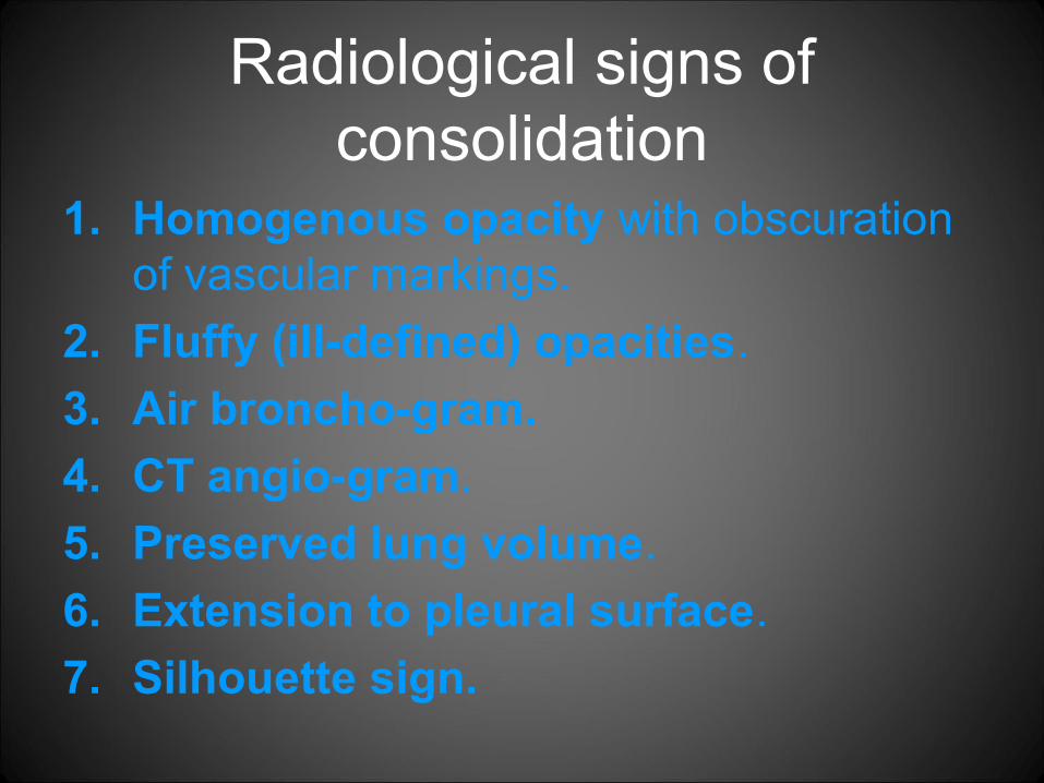
Radiological signs of consolidation
1. Homogenous opacity with obscuration of vascular markings.
2. Fluffy (ill-defined) opacities.
3. Air broncho-gram.
4. CT angio-gram.
5. Preserved lung volume.
6. Extension to pleural surface.
7. Silhouette sign.

Air bronchogram


CT angiogram sign

Causes of consolidation

Patterns of consolidation
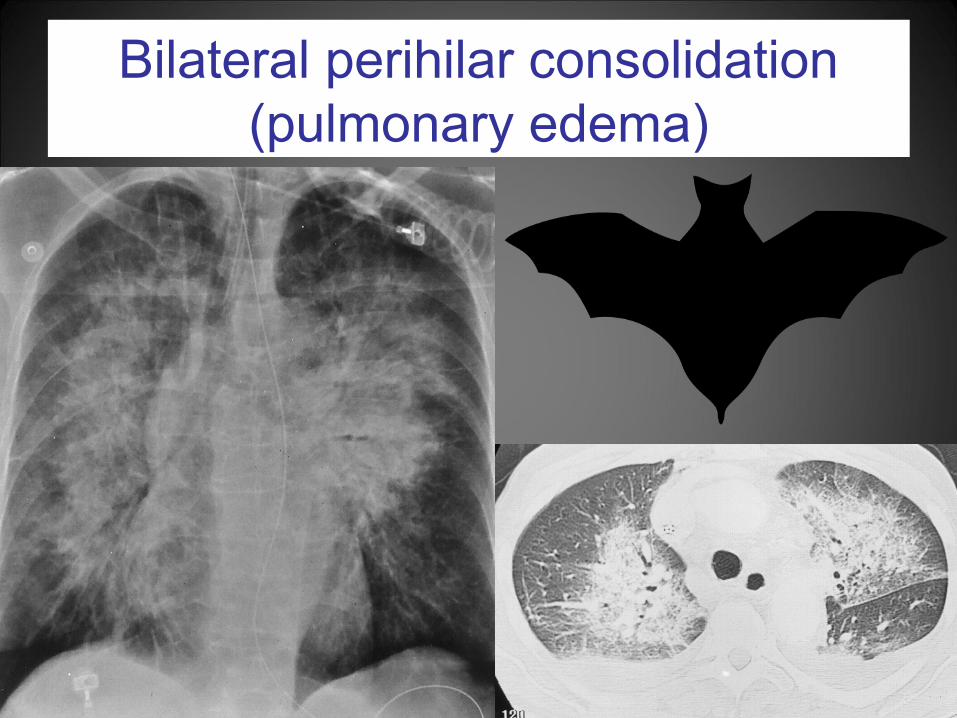
Bilateral perihilar consolidation(pulmonary edema)

Bilateral perihilar consolidation
• Pulmonary edema

Bilateral perihilar consolidation
• Cryptogenic organizing pneumonia.

DD of bilateral perihilar consolidation
• Pulmonary edema (especially cardiogenic)
• Pneumonia – aspiration pneumonia– pneumocystis pneumonia (PCP)– viral pneumonia– lipoid pneumonia– COP
• inhalation injury– noxious gas– liquid
• Pulmonary alveolar proteinosis.• Pulmonary hemorrhage (e.g. WG or Goodpasture syndrome)• lymphoma / leukaemia• Broncho-alveolar carcinoma.
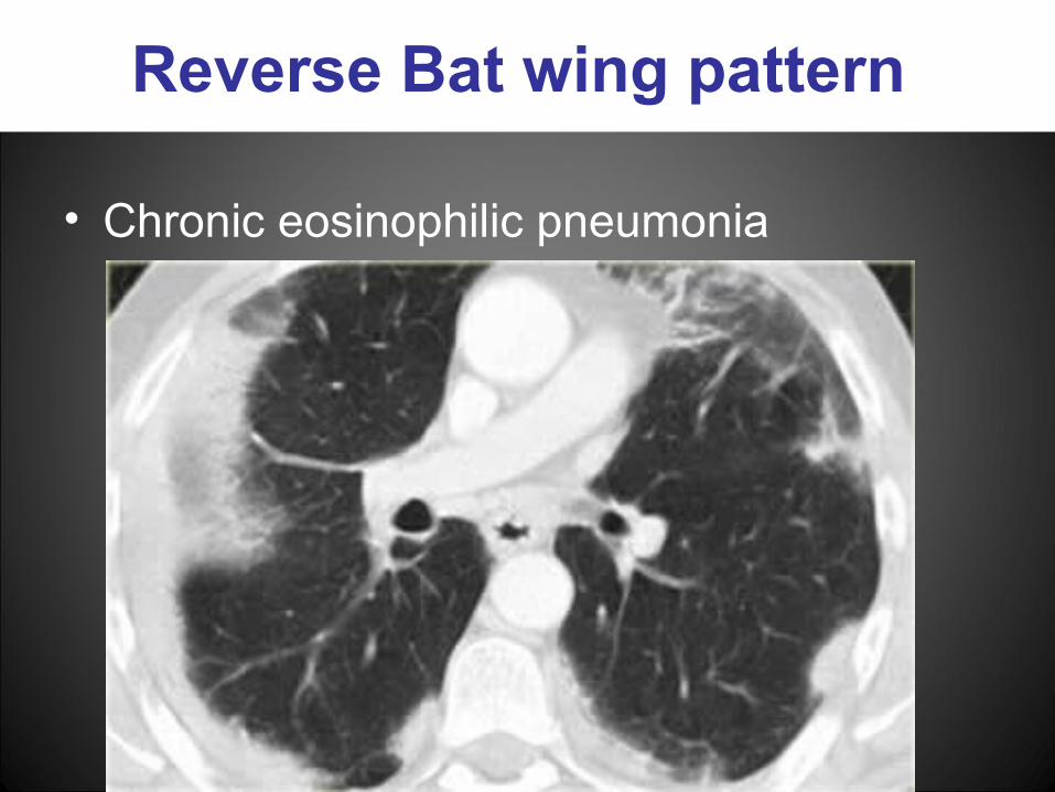
Reverse Bat wing pattern
• Chronic eosinophilic pneumonia
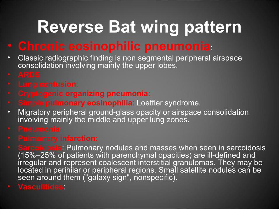
Reverse Bat wing pattern• Chronic eosinophilic pneumonia: • Classic radiographic finding is non segmental peripheral airspace
consolidation involving mainly the upper lobes.• ARDS• Lung contusion:• Cryptogenic organizing pneumonia:• Simple pulmonary eosinophilia: Loeffler syndrome. • Migratory peripheral ground-glass opacity or airspace consolidation
involving mainly the middle and upper lung zones. • Pneumonia:• Pulmonary infarction:• Sarcoidosis: Pulmonary nodules and masses when seen in sarcoidosis
(15%–25% of patients with parenchymal opacities) are ill-defined and irregular and represent coalescent interstitial granulomas. They may be located in perihilar or peripheral regions. Small satellite nodules can be seen around them ("galaxy sign", nonspecific).
• Vasculitides:

Multifocal patchy consolidation
• Bronchopneumonia

Multifocal patchy consolidation
Diffuse alveolar hemorrhage (Wegner granulomatosis)

Multifocal patchy consolidation
Acute causes:
• Bronchopneumonia.• Pulmonary hemorrhage.
• Pulmonary edema.
Chronic causes:
• Chronic pneumonia (eosinophilic pneumonia, COP)
• Neoplastic (BAC or lymphoma).
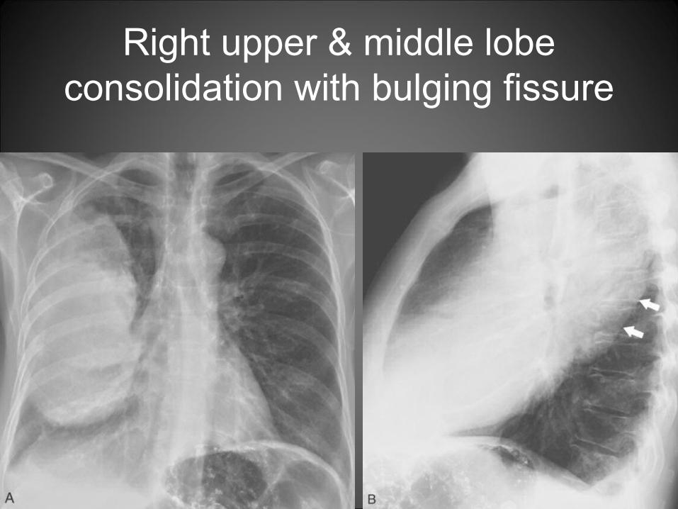
Right upper & middle lobe consolidation with bulging fissure

Round pneumonia
Antibiotic TTT
1 month

Bilateral diffuse consolidation
• ARDS


Atlas of consolidation
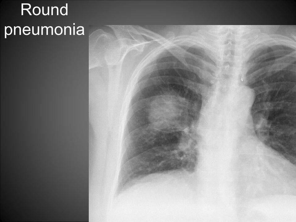
Round pneumonia

Round pneumonia

Lingular consolidation
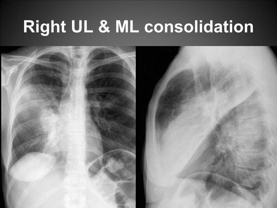
Right UL & ML consolidation

RML consolidation

Medial segment RML consolidation


Superior segment RLL pneumonia

Anteromedial segment LLL pneumonia

RUL pneumonia

RLL pneumonia