Consideration of Intercondylar Angles in Determining a ... · ship for an intraoral sleep appliance...
9
41 Sleep Diagnosis and Therapy Vol 7 No 3 2012 Articles Consideration of Intercondylar Angles in Determining a Maxillo-Mandibular Relationship for Intraoral Sleep Appliances Allen J. Moses D.D.S., Richard A. Bonato Ph.D. and Gloria L. Pacini R.D.H. Abstract The long axes of human condyles are angulated medially and slightly backward. A line through one condylar pole and projected to the midline will meet the line from the other side at approximately the anterior border of foramen magnum. The lateral and medial poles of the mandibular condyles are not in a linear plane. Mandibular movement based on translatory movement of the condyle against the disc and that of the disc in independent translatory movement against the glenoid fossa describes diarthrodial movement. In human beings, unique in the animal kingdom for their upright bipodal posture, the most important function of the back and neck is to balance the head on the spinal column. Because of the dual function of the oropharynx as foodway and airway, it is essential that human beings be able to breathe during mastication. Adult humans lack the ability of most mammals to breathe and swallow at the same time. The mechanical advantage of the diarthrodial anterior trans- latory movement of the TMJs is to keep the airway patent during mastication. Internal software in a 3D cone beam tomographic unit calculated each condylar angle relative to where each intra- condylar line intersected a line perpendicular to the defined midline. Sleep group mean intracondylar angle was 5.4 degrees; intracondylar angle in the control group was 5.18 degrees. There was no statistically significant difference in overall intracondylar angle between the sleep group and non- sleep group. Intracondylar asymmetry is shown in this study to be the normal state in human beings and this being the case, translatory jaw movement would almost never occur in a straight midline linear plane. Some devices which restrict mandibular protrusion to a midline linear plane to register the maxillomandibular relation- ship for an intraoral sleep appliance do not take into account the predominance of intercondylar asymmetry in humans. Their use would be biomechanically contraindicated. Polypro- pylene bite shims are shown that can be aligned so the mandible can freely slide in a protrusive path guided by muscle, ligaments, nerves and bony physical irregularities, rather than by an artificially imposed midline linear plane. TMJ Mechanics Temporomandibular joint (TMJ) movement in humans has been a controversial subject for over half a century. The ap- plication of proven biological and mechanical as well as 3-D computerized graphic representations to anatomic function has resulted in significant progress relative to understanding temporomandibular joint function. For many years it was thought that the human TMJ was capable of hinge function. 1,2,3,4 The reference in the older dental literature to the human TMJs as a ginglymoarthrodial (hinge-sliding) joint is testimony to this popular misconception. 5,6,7,8,9,10,11,12,13,14,15 The TMJs of many lower animals do indeed function as a pure hinge (see Figure 1). The TMJs of animals are bilateral, joined as a functional unit by the body of the mandible. The requisite condition for bilateral hinge function to occur is that the hinges must be in a linear plane. Door hinges are an example of multiple hinges functioning as a mechanical unit in a linear plane. There are many animals whose jaws are capable of pure hinge function and whose mandibular condyles are in the same linear plane. They do not assume an upright bipodal posture and they do not have a flexible, compliant airway. The long axes of human condyles are angulated medially and slightly backward (see Figure 2). A line through one condylar pole and projected to the midline will meet the line from the other side at approximately the anterior border of foramen magnum. 16 The lateral and medial poles of the man- dibular condyles are clearly not in a linear plane. Therefore, pure hinge movement of the human mandibular condyles is not mechanically possible (see Figures 3A & 3B). The mandibular condyle in humans is a convex bony surface from front to back that articulates with the temporomandibular disc. No articular surfaces are perfectly flat and the surface curvatures vary from point to point. The temporomandibular disc in turn articulates with the glenoid fossa of the temporal bone. Mandibular movement is based on translatory move- ment of the condyle against the disc and that of the disc in independent translatory movement against the glenoid fossa. “The articulation of each side of the jaw is a composite that encloses two joints within its single capsule, an upper joint between articular eminence and disc and a lower joint between disc and mandibular condyle. In essence then it can be said that the functional joint articulation is a double-double joint.” 17 This more accurately describes diarthrodial movement rather than ginglymoarthrodial movement. Biological Evidence for Pure Diarthrodial TMJ Movement The human tongue is unique among mammals. In humans the anterior 2/3 of the tongue is oriented horizontally in the mouth with the posterior 1/3 being oriented vertically in the Richard Bonato, Ph.D. is CEO and C-founder of BRAEBON Medical Corporation which sells medical devices for snoring and sleep apnea to sleep laboratories and dentists worldwide. He has no financial interest in bite shims or any bite registration products. Allen J. Moses, D.D.S. and Gloria L. Pacini, R.D.H. are co-inventors of the bite shims shown in figure 10. They have a financial interest in their sales. E.G. Sleep Diagnosis and Therapy Vol 7 No 3 p 41-49
Transcript of Consideration of Intercondylar Angles in Determining a ... · ship for an intraoral sleep appliance...

41Sleep Diagnosis and Therapy Vol 7 No 3 2012
Articles
Consideration of Intercondylar Angles in Determining a Maxillo-Mandibular Relationship for Intraoral Sleep Appliances
Allen J. Moses D.D.S., Richard A. Bonato Ph.D. and Gloria L. Pacini R.D.H.
AbstractThe long axes of human condyles are angulated medially and slightly backward. A line through one condylar pole and projected to the midline will meet the line from the other side at approximately the anterior border of foramen magnum. The lateral and medial poles of the mandibular condyles are not in a linear plane. Mandibular movement based on translatory movement of the condyle against the disc and that of the disc in independent translatory movement against the glenoid fossa describes diarthrodial movement.
In human beings, unique in the animal kingdom for their upright bipodal posture, the most important function of the back and neck is to balance the head on the spinal column. Because of the dual function of the oropharynx as foodway and airway, it is essential that human beings be able to breathe during mastication. Adult humans lack the ability of most mammals to breathe and swallow at the same time. The mechanical advantage of the diarthrodial anterior trans-latory movement of the TMJs is to keep the airway patent during mastication.
Internal software in a 3D cone beam tomographic unit calculated each condylar angle relative to where each intra-condylar line intersected a line perpendicular to the defined midline. Sleep group mean intracondylar angle was 5.4 degrees; intracondylar angle in the control group was 5.18 degrees. There was no statistically significant difference in overall intracondylar angle between the sleep group and non-sleep group. Intracondylar asymmetry is shown in this study to be the normal state in human beings and this being the case, translatory jaw movement would almost never occur in a straight midline linear plane.
Some devices which restrict mandibular protrusion to a midline linear plane to register the maxillomandibular relation-ship for an intraoral sleep appliance do not take into account the predominance of intercondylar asymmetry in humans. Their use would be biomechanically contraindicated. Polypro-pylene bite shims are shown that can be aligned so the mandible can freely slide in a protrusive path guided by muscle, ligaments, nerves and bony physical irregularities, rather than by an artificially imposed midline linear plane.
TMJ MechanicsTemporomandibular joint (TMJ) movement in humans has been a controversial subject for over half a century. The ap-plication of proven biological and mechanical as well as 3-D computerized graphic representations to anatomic function has resulted in significant progress relative to understanding temporomandibular joint function. For many years it was thought that the human TMJ was capable of hinge function.1,2,3,4 The reference in the older dental literature to the human TMJs as a ginglymoarthrodial (hinge-sliding) joint is testimony to this popular misconception.5,6,7,8,9,10,11,12,13,14,15 The TMJs of many lower animals do indeed function as a pure hinge (see Figure 1). The TMJs of animals are bilateral, joined as a functional unit by the body of the mandible. The requisite condition for bilateral hinge function to occur is that the hinges must be in a linear plane. Door hinges are an example of multiple hinges functioning as a mechanical unit in a linear plane. There are many animals whose jaws are capable of pure hinge function and whose mandibular condyles are in the same linear plane. They do not assume an upright bipodal posture and they do not have a flexible, compliant airway.
The long axes of human condyles are angulated medially and slightly backward (see Figure 2). A line through one condylar pole and projected to the midline will meet the line from the other side at approximately the anterior border of foramen magnum.16 The lateral and medial poles of the man-dibular condyles are clearly not in a linear plane. Therefore, pure hinge movement of the human mandibular condyles is not mechanically possible (see Figures 3A & 3B).
The mandibular condyle in humans is a convex bony surface from front to back that articulates with the temporomandibular disc. No articular surfaces are perfectly flat and the surface curvatures vary from point to point. The temporomandibular disc in turn articulates with the glenoid fossa of the temporal bone. Mandibular movement is based on translatory move-ment of the condyle against the disc and that of the disc in independent translatory movement against the glenoid fossa. “The articulation of each side of the jaw is a composite that encloses two joints within its single capsule, an upper joint between articular eminence and disc and a lower joint between disc and mandibular condyle. In essence then it can be said that the functional joint articulation is a double-double joint.”17 This more accurately describes diarthrodial movement rather than ginglymoarthrodial movement.
Biological Evidence for Pure Diarthrodial TMJ Movement The human tongue is unique among mammals. In humans the anterior 2/3 of the tongue is oriented horizontally in the mouth with the posterior 1/3 being oriented vertically in the
Richard Bonato, Ph.D. is CEO and C-founder of BRAEBON Medical Corporation which sells medical devices for snoring and sleep apnea to sleep laboratories and dentists worldwide. He has no financial interest in bite shims or any bite registration products.Allen J. Moses, D.D.S. and Gloria L. Pacini, R.D.H. are co-inventors of the bite shims shown in figure 10. They have a financial interest in their sales.
E.G. Sleep Diagnosis and Therapy Vol 7 No 3 p 41-49

42 Sleep Diagnosis and Therapy Vol 7 No 3 2012
Articles
Fig. 1. Mammalian carnivore mandible with condyles in a linear plane (yellow line) perpendicular to the midline. This animal is mechanically capable of pure hinge movement.
Fig. 2. Angulation of Human Condyles from Eisenberger19
Fig. 3. (3A) Hinges aligned in the same linear plane are capable of hinge function. (3B) Hinges, as shown above, not aligned in the same linear plane are not capable of hinge function. Human jaws are aligned relative to each other in a nonlinear plane at an angle similar to that illustrated above. Human jaws are not capable of hinge function.

43Sleep Diagnosis and Therapy Vol 7 No 3 2012
Articles
oropharynx, when the human is standing or sitting erect. In human beings, unique in the animal kingdom for their upright bipodal posture, the most important function of the back and neck is to balance the head on the spinal column.
The head however, has a mobile part, the mandible, that needs constant counterbalancing as it moves about in its normal functions of speech, mastication and swallowing. The human oropharynx serves the dual function of foodway as well as airway and humans are unique for the ability to articulate speech. The flexible airway from the soft palate to epiglottis was certainly a major evolutionary change that facilitated speech. Creation of a flexible airway and highly innervated tongue is the defining characteristic that enables vowel sound creation. The benefits of speech with a flexible airway/foodway also engender the downside risks of apnea, snoring and choking.
Because of the dual function of the oropharynx as foodway and airway, it is essential that human beings be able to breathe during mastication. Adult humans lack the ability of most mammals to breathe and swallow at the same time because of the flexible airway and the posterior 1/3 of the tongue being vertical. If rotational TMJ movement were possible, the result-ing masticatory hinge motion would close the airway during mastication. The mechanical advantage of the diarthrodial anterior translatory movement of the TMJs is to keep the airway patent during mastication. The anatomic evidence for pure diarthrodial movement is indeed very convincing.
Collapse of the tongue and/or soft palate on the airway during sleep explains the etiology of obstructive sleep disor-dered breathing (SDB). Design of an oral appliance for treatment of SDB involves finding a position of maximal airway patency during sleep. In assessing the maxillo-mandibular position of maximal airway patency, clinicians must consider the me-chanics of jaw, tongue and lip positions. The chief basics of appliance design:
1. Mandibular advancement2. Oral airway dilation3. Maximize room for the tongue in the mouth4. Prevent collapse of the tongue and/or soft palate on the
airway5. Optimize nasal breathing18
The Bony Anatomy of Condylar TranslationThe directions and limits of mandibular movements are controlled by muscles, ligaments and nerves, but also by biomechanical restraints in the temporomandibular joints.19 Biomechanical restraints can be in relation to shape or func-tion. Asymmetry of condylar angles could be a limiting factor to pure midline translatory movement of the mandible.
Eisenberger,20 Christiansen21 and Yale22 have measured the intercondylar angle (ICA). The mean intercondylar angle in these papers was 139°, 129° – 132°, and 153° respectively. Eisenberger demonstrated no significant difference in the in-tercondylar distance between the dysfunctional group, the control group and the children’s group. These papers only reported the one intercondylar angle. They did not study or factor in condylar asymmetries. From a purely mechanical perspective, symmetrical condyles should be able to translate in a straight midline trajectory. Conversely, it is not logical to assume that translatory movement by asymmetric condyles could protrude the mandible in a straight midline trajectory.
There are certain situations in clinical treatment of tempo-romandibular disorders involving disc displacement or SDB where it might be of significance to determine whether the condyles could or should be directed protrusively in a midpoint linear plane, or whether the anatomic evidence is stronger that movement is more appropriately guided in an eccentric plane. The hypothesis being tested is whether intercondylar symmetry or asymmetry is the normal condition. If intracon-dylar asymmetry is the normal condition, might the extent of the asymmetry contraindicate the use of devices designed to record the maxillo-mandibular relationship for an intraoral sleep appliance that limit protrusive movement to a midline cranial plane? This is germane to the bite registration techniques employed by dentists worldwide for the fabrication of custom therapeutic oral appliances for the treatment of SDB. Consist-ent with previous research by Eisenberger et al. (1999),23 it was hypothesized that no statistically significant difference in intracondylar angles would be found between a group of sleep apnea patients and a control group.
MethodSubjectsThe data being presented is based on a retrospective study from the dental records of one author (AJM). Axial tomograms of the 33 most recent consecutive TMD/Sleep referral patients were analyzed as described above and the 33 randomly chosen general dental patients were identically analyzed as a control group. Appropriate releases were signed by all patients.
Data from two groups of 33 patients were retrospectively analyzed. The first group was referred for treatment of sleep apnea and included 25 men (sleep group mean age for men = 45.5 years) and 8 women (sleep group mean age for women = 58.3 years). The range of age is 18-71. The second group is a control group of patients, 17 men (non-sleep group mean age for men = 34.3 years) and 16 women (non-sleep group mean age for women = 33.0 years), seen during routine dental visits and evaluations. The range of age is 19-59.
Materials/Equipment A three dimensional cone beam tomography unit (Imaging Sciences i-CAT® Next Generation Cone Beam 3D Dental Imag-ing) was utilized. Axial radiographic slices were located that revealed the maxillary dental midline, the superior surface of both condyles and the foramen magnum. These tomographic views were oriented on the monitor screen so that the midline was defined as passing through the dental midline (between teeth numbers 8 and 9) and the center of foramen magnum (see Figure 4). Lines were identified and drawn through the mesial and distal poles and the center point of each condyle (MB). Internal software (Anatomage Invivo 5 Anatomy Imaging Software) in the cone beam unit then calculated each condylar angle relative to where each intracondylar line intersected a line perpendicular to the defined midline (see Figure 5).
ResultsTo test the hypothesis that there would be no difference in intracondylar angles between the two groups, the left condylar angle was subtracted from the right condylar angle and an independent sample t-test was performed on the absolute value of the intracondylar angle differences. The sleep group

44 Sleep Diagnosis and Therapy Vol 7 No 3 2012
Articles
Fig. 4A and 4B. Coronal view of skulls oriented so midline is marked from maxillary dental midline through center of foramen magnum.
Fig. 5A and 5B. i-CAT® software calculates intercondylar angles based on a line drawn through the mesial and distal poles of each condyle relative to the dotted blue line perpendicular to the midline defined in Figure 4.

45Sleep Diagnosis and Therapy Vol 7 No 3 2012
Articles
mean intracondylar angle was 5.4 degrees (sd = 3.93) whereas the mean intracondylar angle in the control group was 5.18 degrees (sd = 3.41). For the sleep group intracondylar angles ranged from 0.2 degrees to 15.9 degrees. The control group intracondylar angles ranged from 0.0 degrees to 15.1 degrees. The t-test revealed no statistically significant difference in overall intracondylar angle between the sleep group and non-sleep group, t(64) = 0.24, p = 0.81 (see Figure 6).
Additional independent sample t-tests did not reveal any statistically significant difference in right condylar angle dif-ferences between the sleep group (mean=24.71, sd = 8.54) and non-sleep group (mean = 28.47, sd = 7.27), t(64) = –1.92, p = 0.06, or in left condylar angle differences between the sleep group (mean = 25.48, sd = 8.65) and non-sleep group (mean = 27.97, sd =7.13), t(64) = –1.27, p = 0.20 (see Figures 7 & 8).
DiscussionThere does not appear to be any statistical difference between the study group of TMD/Sleep Patients and the control group. Both groups demonstrated similar intercondylar asymmetry. In fact, condylar symmetry appears to be the exception rather than the expectation. The average intercondylar asymmetry of the two groups combined is 5.36 degrees. If one assumes that the mechanics of protrusion are the same in all humans, then the study subject with an intercondylar asymmetry of 15.9 degrees employs the same mechanics to achieve protrusive translation as the person with a symmetric condylar relation-ship. Since the mandible is a single bone, the joints of each side are coordinated so that each contributes to every movement. The craniomandibular connection is one operating unit com-posed of right and left joint complexes.
Patient (Sleep Group and Gender)
Condylar Angel Patient (Non-Sleep/ Control and Gender)
Condylar AngelDifferenceR L Difference R L
1. F 26.1 15.4 10.7 34. M 24.7 21.7 3.0
2. M 14.8 22.9 8.1 35. M 25.8 30.4 4.6
3 M 20.7 4.8 15.9 36. M 9.3 24.4 15.1
4. M 31.7 27.2 4.5 37. M 24.0 25.5 1.5
5. M 18.7 17.3 1.4 38. F 27.3 21.6 5.7
6. M 16.5 17.4 0.9 39. F 26.6 24.2 2.4
7. M 32.5 35.7 3.2 40. M 34.6 25.7 8.9
8. F 32.4 39.7 7.3 41. M 31.4 20.4 11
9. M 17.5 24.1 6.6 42. F 32.4 22.8 9.6
10. M 19.4 9.7 9.7 43. F 25.7 23.9 1.8
11. F 32.9 36.8 3.9 44. M 31.0 31.0 0
12. M 43.7 36.3 7.4 45. F 29.5 21.6 7.9
13. M 26.9 30.2 3.3 46. F 33.4 27.3 6.1
14. F 18 24.4 6.4 47. F 29.7 22.6 7.1
15. M 20.8 22.3 1.5 48. F 21.4 28.7 7.3
16. M 34.5 31.6 2.9 49. F 43.4 38.3 5.1
17. M 42.2 34.4 7.8 50. F 33.2 22.9 10.3
18. M 21.0 16.4 4.6 51. M 27.1 20.8 6.3
19. F 16.2 18.9 2.7 52. F 29.5 27.2 2.3
20. M 31.2 33.8 2.6 53. M 29.9 26.9 3.0
21. M 31.9 28.1 3.8 54. M 29.6 34.5 4.9
22. M 29.8 33.8 4.0 55. M 28.3 34.1 5.8
23. M 11.9 24.1 12.2 56. M 28.6 30.6 2.0
24. M 23.4 25.2 1.8 57. M 38.0 40.5 2.5
25. M 18.3 19.5 1.2 58. M 20.7 30.1 9.4
26. F 26.4 30.5 4.1 59. F 15.9 12.7 3.2
27. F 13.2 23.4 10.2 60. M 43.3 47.4 4.1
28. F 29.6 22.0 7.6 61. F 35.6 35.7 0.1
29. M 31.5 31.7 0.2 62. F 29.8 35.0 5.2
30. M 34.1 38.0 3.9 63. F 28.5 32.0 3.5
31. M 19.1 23.4 4.3 64. F 19.1 24.7 5.6
32. M 17.2 30.4 13.2 65. M 16.4 20.7 4.3
33. M 11.3 11.5 0.2 66. M 36.0 37.3 1.3
Average 24.71 25.43 5.55 28.17 27.67 5.18
42 male subjects, 24 female subjects

46 Sleep Diagnosis and Therapy Vol 7 No 3 2012
Articles
Fig. 6. Box and whisker plots of mean intracondylar angles for both sleep group and nonsleep (control) group.
Fig. 7. Box and whisker plots of mean right intracondylar angle for both sleep group and nonsleep (control) group.

47Sleep Diagnosis and Therapy Vol 7 No 3 2012
Articles
Fig. 8. Box and whisker plots of mean left intracondylar angle for both sleep group and nonsleep (control) group.
Fig. 9. Two examples of devices designed to record the maxillo-mandibular relationship that restrict protrusive movement to a midline cranial plane.
The translatory component of joint movement is completely dependent on the shape of the articular eminence, according to Sicher and DuBrul.24 It is predictable that asymmetry of intercondylar angles also relates to asymmetry of the ar-ticular eminences of the temporal bones. Condyles, discs and eminences are in close contact in all movements and in all positions. This means that all movements of condyles with their discs must follow exactly the surfaces of the articular eminentia.25 Intercondylar asymmetry is shown in this study to be the normal state in human beings and this being the case, trans-latory jaw movement would almost never occur in a straight midline linear plane.
On the basis of the measurements from this study it would seem logical to assume that devices which restrict mandibular protrusion to a midline linear plane (see Figure 9) do not take into account the predominance of intercondylar asymmetry in humans. By their limitation of protrusive translation to a midline linear plane their use would be biomechanically contraindicated.
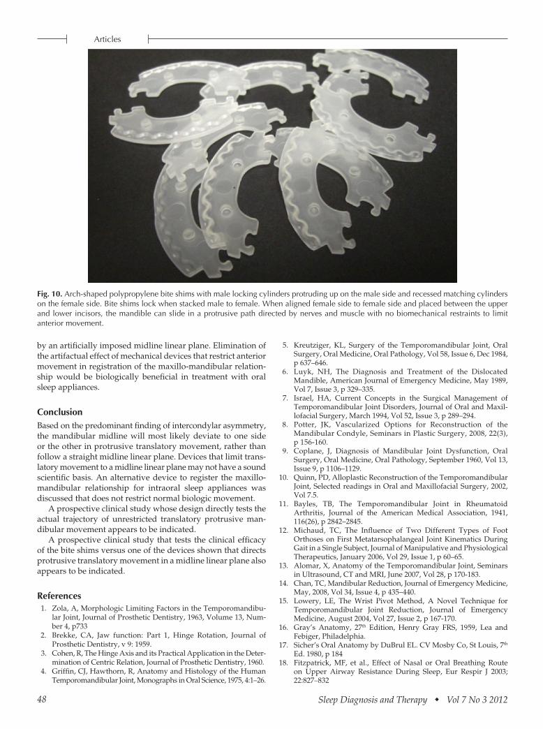
There are alternatives to devices that record the maxillo-mandibular relationship by physically guiding protrusive movement to a midline cranial plane. Polypropylene bite shims are shown (see Figure 10) that can be aligned so the mandible can freely slide in a protrusive path guided by muscle, ligaments, nerves and bony physical irregularities, rather than
48 Sleep Diagnosis and Therapy Vol 7 No 3 2012
Articles
Fig. 10. Arch-shaped polypropylene bite shims with male locking cylinders protruding up on the male side and recessed matching cylinders on the female side. Bite shims lock when stacked male to female. When aligned female side to female side and placed between the upper and lower incisors, the mandible can slide in a protrusive path directed by nerves and muscle with no biomechanical restraints to limit anterior movement.
by an artificially imposed midline linear plane. Elimination of the artifactual effect of mechanical devices that restrict anterior movement in registration of the maxillo-mandibular relation-ship would be biologically beneficial in treatment with oral sleep appliances.
ConclusionBased on the predominant finding of intercondylar asymmetry, the mandibular midline will most likely deviate to one side or the other in protrusive translatory movement, rather than follow a straight midline linear plane. Devices that limit trans-latory movement to a midline linear plane may not have a sound scientific basis. An alternative device to register the maxillo-mandibular relationship for intraoral sleep appliances was discussed that does not restrict normal biologic movement.
A prospective clinical study whose design directly tests the actual trajectory of unrestricted translatory protrusive man-dibular movement appears to be indicated.
A prospective clinical study that tests the clinical efficacy of the bite shims versus one of the devices shown that directs protrusive translatory movement in a midline linear plane also appears to be indicated.
References1. Zola, A, Morphologic Limiting Factors in the Temporomandibu-
lar Joint, Journal of Prosthetic Dentistry, 1963, Volume 13, Num-ber 4, p733
2. Brekke, CA, Jaw function: Part 1, Hinge Rotation, Journal of Prosthetic Dentistry, v 9: 1959.
3. Cohen, R, The Hinge Axis and its Practical Application in the Deter-mination of Centric Relation, Journal of Prosthetic Dentistry, 1960.
4. Griffin, CJ, Hawthorn, R, Anatomy and Histology of the Human Temporomandibular Joint, Monographs in Oral Science, 1975, 4:1–26.
5. Kreutziger, KL, Surgery of the Temporomandibular Joint, Oral Surgery, Oral Medicine, Oral Pathology, Vol 58, Issue 6, Dec 1984, p 637–646.
6. Luyk, NH, The Diagnosis and Treatment of the Dislocated Mandible, American Journal of Emergency Medicine, May 1989, Vol 7, Issue 3, p 329–335.
7. Israel, HA, Current Concepts in the Surgical Management of Temporomandibular Joint Disorders, Journal of Oral and Maxil-lofacial Surgery, March 1994, Vol 52, Issue 3, p 289–294.
8. Potter, JK, Vascularized Options for Reconstruction of the Mandibular Condyle, Seminars in Plastic Surgery, 2008, 22(3), p 156-160.
9. Coplane, J, Diagnosis of Mandibular Joint Dysfunction, Oral Surgery, Oral Medicine, Oral Pathology, September 1960, Vol 13, Issue 9, p 1106–1129.
10. Quinn, PD, Alloplastic Reconstruction of the Temporomandibular Joint, Selected readings in Oral and Maxillofacial Surgery, 2002, Vol 7.5.
11. Bayles, TB, The Temporomandibular Joint in Rheumatoid Arthritis, Journal of the American Medical Association, 1941, 116(26), p 2842–2845.
12. Michaud, TC, The Influence of Two Different Types of Foot Orthoses on First Metatarsophalangeal Joint Kinematics During Gait in a Single Subject, Journal of Manipulative and Physiological Therapeutics, January 2006, Vol 29, Issue 1, p 60–65.
13. Alomar, X, Anatomy of the Temporomandibular Joint, Seminars in Ultrasound, CT and MRI, June 2007, Vol 28, p 170-183.
14. Chan, TC, Mandibular Reduction, Journal of Emergency Medicine, May, 2008, Vol 34, Issue 4, p 435–440.
15. Lowery, LE, The Wrist Pivot Method, A Novel Technique for Temporomandibular Joint Reduction, Journal of Emergency Medicine, August 2004, Vol 27, Issue 2, p 167-170.
16. Gray’s Anatomy, 27th Edition, Henry Gray FRS, 1959, Lea and Febiger, Philadelphia.
17. Sicher’s Oral Anatomy by DuBrul EL. CV Mosby Co, St Louis, 7th Ed. 1980, p 184
18. Fitzpatrick, MF, et al., Effect of Nasal or Oral Breathing Route on Upper Airway Resistance During Sleep, Eur Respir J 2003; 22:827–832

49Sleep Diagnosis and Therapy Vol 7 No 3 2012
Articles
19. Osborn, JW, The disc of the human temporomandibular joint: design, function and failure. J Oral Rehabil. 1985, Jul 12(4):279-93
20. Eisenburger, M, The human mandibular intercondylar angle measured by computed tomography, Archives of Oral Biology, Vol 44, Issue 11, Nov 1999, p 947–951.
21. Christiansen, E., Intra- and inter-observer variability and accu-racy in the determination of linear and angular measurements in computed tomography: An in vitro and in situ study of human mandibles, Acta Odontologica Scandinavica, 1986, Vol. 44, No. 4, pp 221–229.
22. Yale, S, Laminagraphic cephalometry in the analysis of mandibular condyle morphology, Oral Surgery, Oral Medicine, Oral Pathology 14, 1961, pp 793–805.
23. Eisenburger, M, The human mandibular intercondylar angle measured by computed tomography, Archives of Oral Biology, Vol 44, Issue 11, Nov 1999, p 947–951.
24. Sicher’s Oral Anatomy by DuBrul EL. CV Mosby Co, St Louis, 7th Ed. 1980, p.192
25. Sicher’s Oral Anatomy by DuBrul EL. CV Mosby Co, St Louis, 7th Ed. 1980, p.192