Conservative Esthetic Rehabilitation of a Young Patient ...
9
OONTINUING EDUOATION 2 CONSERVATIVE RESTORATION Conservative Esthetic Rehabilitation of a Young Patient with Amelogenesis Imperfecta Aliasger Tunkiwala, BDS, MDS; and Danesh Vazifdar LEARNING OBJECTIVES Abstract; Conservative management of young adult patients vdth amelogenesis imperfecta using contemporary materials and techniques is needed in dentistry. These patients have malformed enamel that tends to wear down at a faster rate than normal and is prone to decay. Conventional management of such patients requires devitalization of all involved teeth, followed hy post cores and crown lengthening and preparing them to provide sufficient space to receive full-cov- erage restorations. This article outlines a minimally invasive method of manag- ing such cases. By increasing the vertical dimension of occlusion and using veiy minimal or no preparations and fabrication of lithium-disiiicate crowns to adhesively bond to the remaining tooth stinjcture, these teeth can be saved from being devitalized, as demonstrated in a case. This allows the structural integrity of the teeth to be maintained, along with their vitality. •describe the esthetic impact of ameiogenesis imperfecta (Ai) • discuss ciinicai and risk assessment in patients with AI •expiain why conservative approaches to treatment are especially important In patients with Ai A melogenesis imperfecta (AI) is an inherited disor- der of enamel with mutations in five genes—AMEL, ENAM, MMP20, KLK4, and FAM83H-and a wide range of clinical presentations. It affects the structure and appearance of the enamel of all teeth, both in the primary and secondary dentitions.' Teeth with this condition tend to be unusually small, discolored, pitted or grooved, and prone to rapid wear and breakage. With generalized compromise of enamel, there is loss of vertical dimension as well as lack of interproximal contacts, resulting in food lodgment and problems associated with it. In most cases the esthetic disability is striking. The high morbidity for such patients presents major restor- ative challenges during formula- tion of the treatment plan. Lack of mature enamel for bonding can be a formidable handicap when employing contemporaiy treatment protocols in adhesive dentistry. For patients suffering for conservative treatment options that save the existing tooth struc- ture while providing a durable treatment. The authors report on a 21-year-old patient who presented with severe discoloration and mild sensitivity of teeth (Figure 1). The teeth visible in his smile were very short, unsightly, and unbecom- ing for his age. from AI, there is a strong need ing smali, discolored, worn teeth. Clinical Assessment A thorough clinical examination and analysis were carried out to assess the esthetic and functional problems of the patient (Figure 2 through Figure 7). The medical histoiy was non-contributoiy, ex- cept for mild leukoderma. Tem- poromandibular joint (TMJ) function was within normal range. Mounted study casts were used to evaluate occlusion. The relevant findings were as fol- lows^: Facial analysis revealed a canted maxillary occlusal plane and canted dental midline. Fig 1. Preoperative frontai smiie of patient suffering from Ai, show- Dentolabial analysis showed that the maxillary incisors were not www.compendiumlive.com March 2014 COMPENDIUM 175
Transcript of Conservative Esthetic Rehabilitation of a Young Patient ...

OONTINUING EDUOATION 2
CONSERVATIVE RESTORATION
Conservative Esthetic Rehabilitation of a YoungPatient with Amelogenesis ImperfectaAliasger Tunkiwala, BDS, MDS; and Danesh Vazifdar
LEARNING OBJECTIVES
Abstract; Conservative management of young adult patients vdth amelogenesis
imperfecta using contemporary materials and techniques is needed in dentistry.
These patients have malformed enamel that tends to wear down at a faster rate
than normal and is prone to decay. Conventional management of such patients
requires devitalization of all involved teeth, followed hy post cores and crown
lengthening and preparing them to provide sufficient space to receive full-cov-
erage restorations. This article outlines a minimally invasive method of manag-
ing such cases. By increasing the vertical dimension of occlusion and using veiy
minimal or no preparations and fabrication of lithium-disiiicate crowns to adhesively bond to the remaining
tooth stinjcture, these teeth can be saved from being devitalized, as demonstrated in a case. This allows the
structural integrity of the teeth to be maintained, along with their vitality.
•describe the estheticimpact of ameiogenesisimperfecta (Ai)
• discuss ciinicai and riskassessment in patientswith AI
•expiain why conservativeapproaches to treatment
are especially important In
patients with Ai
Amelogenesis imperfecta (AI) is an inherited disor-der of enamel with mutations in five genes—AMEL,ENAM, MMP20, KLK4, and FAM83H-and awide range of clinical presentations. It affectsthe structure and appearance of the enamel of all
teeth, both in the primary and secondary dentitions.' Teeth withthis condition tend to be unusually small, discolored, pitted orgrooved, and prone to rapid wear and breakage. With generalizedcompromise of enamel, there is loss of vertical dimension as wellas lack of interproximal contacts, resulting in food lodgmentand problems associated withit. In most cases the estheticdisability is striking.
The high morbidity for suchpatients presents major restor-ative challenges during formula-tion of the treatment plan. Lackof mature enamel for bondingcan be a formidable handicapwhen employing contemporaiytreatment protocols in adhesivedentistry. For patients suffering
for conservative treatment options that save the existing tooth struc-ture while providing a durable treatment.
The authors report on a 21-year-old patient who presented withsevere discoloration and mild sensitivity of teeth (Figure 1). Theteeth visible in his smile were very short, unsightly, and unbecom-ing for his age.
from AI, there is a strong need ing smali, discolored, worn teeth.
Clinical AssessmentA thorough clinical examination and analysis were carried out toassess the esthetic and functional problems of the patient (Figure
2 through Figure 7). The medicalhistoiy was non-contributoiy, ex-cept for mild leukoderma. Tem-poromandibular joint (TMJ)function was within normal range.
Mounted study casts wereused to evaluate occlusion. Therelevant findings were as fol-lows : Facial analysis revealeda canted maxillary occlusalplane and canted dental midline.
Fig 1. Preoperative frontai smiie of patient suffering from Ai, show- Dentolabial analysis showed thatthe maxillary incisors were not
www.compendiumlive.com March 2014 COMPENDIUM 175

CONTINUING EDUCATION 2 I CONSERVATIVE RESTORATION
adequately visible during repose; it also revealed a reverse smileline, as well as a wide smile showing 12 teeth. Results from the pho-netic analysis were that "F" and "V" sounds revealed upper incisaishortening, "M" and "S" pronunciation disclosed a diminishedvertical dimension of occlusion (VDO), and "E" sounds showedsevere shortening of incisors. Dental analysis revealed thickbiotype,asymmetiy and inappropriate location of gingival levels and zenith,incorrect axial inclinations, a displeasing width-to-length ratio, andpitted surfaces on most teeth. Interproximal decay was evident onseveral posterior teeth, as revealed by radiographie analysis. Finally,occlusal examination indicated discrepancy between maximumintercuspation (MIP) and centric relation (CR) as well as a lackof anterior guidance/posterior disclusion.
Risk AssessmentRisk assessment strategies are used to gauge the potential difficultiesin treatment execution and understand the potential treatment out-come. A systematic approach was used for periodon tal, hiomechanical,functional, and dentofacial analysis.'' Periodontally, the patient waslow-risk, as there was minimal bone loss and no bleeding on probing,consistent with American Academy of Periodontology (AAP) classi-fication. Biomechanically the patient was deemed to be at moderaterisk, as there was inteiproximal decay, and, stmcturally, the lack ofenamel would make the teeth susceptible to lower bond strengthswith the restorations. Functionally, the patient was considered to beat moderate risk. Although the patient had acceptable fimction, theMIP was not coincident with CR, and there was occlusal wear on mostteeth. Since the occlusal surfaces of all teeth would need restorativetreatment to reassign the VDO, CR would be the starting point forreconstmction. Lastly, dentofacially the patient received a moderaterisk rating, as there was maximum tooth display and marginal gingivaldisplay due to the position of the lip line.
Comprehensive Treatment ConceptThe treatment plan was developed by identifying achievable clini-cal goals that included establishing: the correct VDO; dentofacialharmony, keeping in mind smile design principles; MIP in harmonywith CR; and eflFective anterior guidance to provide posterior disclu-sion in harmony with the envelope of function.
Aclinical obsei-vation in various cases of full-mouth rehabilitationand in cases of AI is that before post-core restorations, the clinicianmay devitalize the pulp while performing endodontic treatment formost or all teeth. In some cases, crown-lengthening surgeiy withbone removal may be necessary to attain sufficient tooth structureand retention form for restorations and to allow the clinician to pre-pare teeth as needed to gain space for restorative material. However,this approach also leads to weakening of the tooth structure.''
A contemporaiy approach in cases of AI is to adequately open theVDO within the physiologic limits of esthetics and phonetics and useadhesively retained crowns with minimal tooth preparation. Thisapproach allows teeth to retain their original strength and vitality.
Choice of Restorative Materials and DesignBecause the enamel is defective circumferentially aroimd all teeth in AI,full-coverage restoration is the ideal choice. Partial-bonded porcelainrestorations may not achieve sufficient bond strength in such cases andleave uncovered defective tooth structure prone to decay. Porcelain-fused-to-metal (PFM) restoration is known to have good sti'ength andreasonable esthetics. However, traditional PFMs require significanttooth reduction to achieve translucency and optical properties, thus ne-cessitating endodontic intervention in Al-affected teeth. Therefore, inthis case PFM restorations were not considered as a restorative option.
The restorative material of choice in such cases must havethe ability to provide excellent optical properties and estheticswith reasonably good strength. Lithium disilicate is an ideal
Fig 2. Preoperative 1:2 retracted frontal view. Fig 3. Preoperative 1;2 retracted right iateral view. Fig 4. Preoperative 1:2 retracted ieft iaterai view.Fig 5. Preoperative 1:1 retracted frontai view. Fig 6. Preoperative maxiilary occlusal view. Fig 7. Preoperative mandibular occlusai view.
176 COMPENDIUM March 2014 Volume 35. Numher 3
choice. (In this case, e.max" [IvoclarVivadent,www.ivoclarviva-dent.com] was used.) Improvements in formulations of lithiumdisilicate'^ have resulted in less tooth structure needing to beremoved while still providing good esthetics and strength inthinner layers. Moreover, if the underlying tooth color does notneed a drastic shade change, these restorations can be fabricatedwith supragingival or equigingival margins, thereby eliciting astable biologic response from the periodontium. With minimaloverall tooth preparations, the structural integrity of the toothis preserved, so as to resist crack propagation.'
Furthermore, using lithium disilicate in monolithic form forfull-contour posterior occlusal surfaces provides better strengththan layering a core of lithium disilicate with veneering porcelain,thereby eliminating concerns regarding chipping of the veneeringporcelain. In addition, its ability to adhesively bond to the underly-ing substrate eradicates the need for a retentive form in the toothpreparation. Bonding to well-formed enamel is clinically verifiedby the typical chalk-white enamel surface after phosphoric acid-etching. However, because AI patients sufi'er from a reduced enamellayer—especially in hj poplastic AI—bonding not only to enamelbut also to dentin is important. Short-etching with phosphoricacid (total-etch) or the application of self-etching primers is rec-ommended.' Recent clinical studies investigating enamel wear ofmonolithic lithium disilicate demonstrate that it seems to be withinthe range of normal enamel wear."
Porcelain-veneered zirconia crowns are another option that canbe exercised in AI cases. However, the high rate of fractiu-e (3% to25%) of veneering porcelain from underlying strong ceramic coressuch as zirconia has been shown in the literature.' Up to 90% ofthe porcelain-veneered zirconia crowns failed from veneer chip-offfracture in relatively fewer cycles and lesser force as compared withmonolithic lithium disilicate. " Additionally, the inherent opacity
of zirconia cores can make achieving good translucency more dif-ficult unless tooth preparation is aggressive and allows for ampleroom for veneering translucent porcelain.
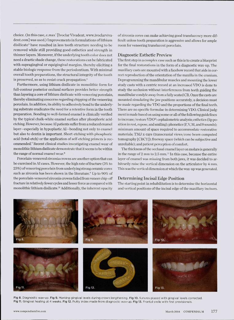
Diagnostic Esthetic PreviewThe first step in a complex case such as this is to create a blueprintfor the final restorations in the form of a diagnostic wax-up. Themaxillary casts are mounted with a facebow record that aids in cor-rect reproduction of the orientation of the maxilla in the cranium.Deprogramming the mandibular muscles and mounting the lowerstudy casts with a centric record at an increased VDO is done tostudy the occlusion without interferences from teeth guiding themandibular condyle away from a fully seated CR Once the casts aremounted simulating the jaw positions accurately, a decision mustbe made regarding the VDO and the proportions of the final teeth.There are no specific formulae in determining VDO. Clinical judg-ment is made based on using some or all of the following guidelinesto increase/restore VDO": cephalometrlc analysis; esthetics (lip po-sition in rest, repose, and smiling); phonetics (F, V, M, and S sounds);minimum amount of space required to accommodate restorativematerials; TMJ x-rays (transcranial views/cone beam computedtomography [CBCT]); freeway space (which can be subjective andunreliable); and patient perception of comfort.
The thickness of the occlusal enamel layer on molars is generallyin the range of 2 mm to 2.5 mm.' In this case, because the entirelayer of enamel was missing from both jaws, it was decided to ar-bitrarily raise the vertical dimension on the articulator by 4 mm.This was the vertical dimension at which the wax-up was generated.
Determining Incisai Edge PositionThe starting point in rehabilitation is to determine the horizontaland vertical positions of the incisai edge of the maxillaiy incisors.
Fig 8. Diagnostic wax-up. Fig 9. Marking gingival ieveis during crown iengthening. Fig 10. Sutures piaced with gingivai ieveis corrected.Fig n. Gingival healing at 4 weeks. Fig 12. Putty index made from diagnostic wax-up. Fig 13. Frontai smiie with first provisionais.
www.compendiumlive.com March 2014 COMPENDIUM 177

CONTINUING EDUCATION 2 j CONSERVATIVE RESTORATION
The vertical position of the incisai edge governs the length of cen-tral incisors and depends on several factors, including: envelopeof function; anterior guidance; upper lip positions in rest/repose/smiling; soft-tissue characteristics; facial proportion; culturalvariations; and phonetics (F, V, and E sounds). The horizontalposition, on the other hand, governs the incisai profile and itslabiolingual orientation.'^ The horizontal position of the incisaiedge must accommodate the patient's envelope of function.
A direct mock-up was carried out in the patient's mouth withlight-cured composite resin used to add length to the incisai edgesof the upper anterior teeth. The length and incisai profile was veri-fied for esthetics and phonetics. The lengths of teeth Nos. 8 and 9with reestablished incisai length were measured from the gingivalmargin to the incisai edge. All of this incisai edge position infor-mation was provided to the laboratory during wax-up (Figure 8).
The wax-up was to be made after correction of gingival levelsand zeniths of teeth on the stone models. The gingival levelsshould be decided on the basis of maintaining the width-to-length ratio of central incisors close to the ideal parameter of0.8.'* Once the gingival level of the centrals was determined, atangent drawn to that line onto the cuspids allowed visualiza-tion of the proposed gingival level for the cuspids. The greatestcorrections in gingival levels were needed in the upper cuspidand bicuspid regions.
Measurements were made from the incisai edge of the cuspidsto the proposed new gingival levels on stone casts. These mea-surements were used to mark the proposed gingival margin forthe cuspids in the patient's mouth. Similar markings were donefor all upper teeth, maintaining the principle of having gingivalmargins of cuspids and centrals at an equal level and havinggingival margins of laterals coronal to them.'^
Esthetic Repositioning of Gingival TissuesAfter administering local anesthesia, a periodontal probe was usedto soLind the bone on facial and interproximal aspeets of all upperteeth. With the upper cuspids and bicuspids, there was insufficientdistance between the proposed free gingival margin and the crestalbone (< 3 mm) (Figure 9). In such circumstances, merely trim-ming the gingival tissues without altering the bone could lead toa violation of biologic width, with its associated complications,one of which is the rebound of gingiva to its original level. Thus,for these teeth, an osteoplasty, along with gingival resection, wascarried out. On most other teeth, including the lower anterior andposterior teeth, a gingivectomy was sufficient to achieve correc-tion of gingival levels and proper width-to-length ratios of teeth.
An aspect of crown lengthening in cases of AI is to leave thegingival margins at the cementoenamel junction (CE J) of the teeth.This is done so that any defective and pitted enamel is exposedand tended to in the final restorative design. Failure to do thismay lead to recurrent gingival ill-health due to plaque accumula-tion in the pitted surfaces. In this case, however, upon raising themucoperiosteal fiap, it was found that the CEJ was missing as aresult of total absence of enamel. Thus, the gingival margins wereplaced at the desired esthetic levels and sutured with monoflla-ment sutures (Figure 10) and reevaluated after a healing periodof 4 weeks (Figure 11).
First Provisional Restoration/Mock-upThe wax-up at the desired empirical vertical dimension was final-ized and checked for all esthetic and functional parameters. Aputtysilicone index was made of the full-arch upper and lower wax-up.To record the finer details and texture of the wax-up, the puttyindex was relined with light-body elastomer (Figure 12).
Fig 14.1:2 retracted frontal view with provisionals in MIP. Fig 15. Final anterior tooth preparations. Fig 16. Anterior final restorations bonded.Fig 17. Posterior final tooth preparations: right side. Fig 18. Posterior final tooth preparations: left side. Fig 19. Postoperative frontal smile.
178 COMPENDIUM March 2014 Volume 35, Number 3

At 4 weeks after the esthetic repositioning of the gingival levels,the patient was scheduled for fabrication of the provisional restora-tion. The upper provisionals were fabricated first. All the teeth werecleaned with pumice slurry and etched with 37% phosphoric acidfor 3 seconds. After gentle air-drjdng and isolation, bonding agentwas applied to the teeth and light-cured for 20 seconds. The putty-wash index was loaded with bis-acryl composite (Protemp 4™, 3MESPE, www.3MESPE.com) and placed over the teeth with correctorientation that was verified by checking that the index was fullyseated. Undue pressure on any one side of the putty index must beavoided to achieve correct contours on the provisional.
After the material had set, the index was removed. A correctlyfabricated index will show a thin uniform flash of excess material,which, in this case, was carefully peeled off and cut with a sharp#12 blade. The provisionals on the lower jaw were fabricated in asimilar manner. Once the gross excess was removed, the patient wasguided to close in MIP, with the condyles guided to CR. The occlusalcontacts were then marked and adjusted until uniform contacts ofequal intensity were achieved on both sides and anterior guidancewas sufficient to disclude posteriors in all eccentric mandibularpositions (Figure 13 and Figure 14).
At this juncture, an evaluation of the esthetics, phonetics, VDO,and all other macro and micro elements'" of smile design should beverified. Any changes—additive or subtractive—can be carried outin the patient's mouth itself. The full-face smile photographs andclose-up views were taken and evaluated. The patient was askedto "test drive" the provisionals for a few weeks and report with anyfeedback so that necessaiy corrections could be carried out. At thistime, even the functional aspects can be verified, and a transcranialradiograph or a CBCT scan may be taken to verify the position ofcondyle in the glenoid fossa.
The patient was asked to use the provisionals for another 6 weeks.At the end of 12 weeks from the day of gingival surgery, the patientwas scheduled for the final impressions."*
Sequence of Fabrication of Final RestorationsThe sequencing of final restoration plays a role in simplifying thetreatment plan. One option is to fabricate all final restorationstogether. This requires precise records and use of a semi-adjust-able articulator that will allow the accurate simulation of man-dibular movements in protrusive and lateral excursions. It alsorequires the clinician to verify the anterior tooth lengths andcontours that will be needed to design the posterior tooth formso that the teeth disclude during eccentric mandibular move-ments. Preparing all teeth and taking accurate impressions allsimultaneously is a clinically demanding procedure.
Another sequencing option is to fabricate the upper and loweranterior teeth first, followed by all posteriors. This option requiresthe clinician to keep posterior provisionals in place while finalimpressions of the upper and lower anterior teeth are taken. Theseposterior provisionals will help maintain the desired VDO. In keep-ing with esthetic and phonetic guidelines, the anterior final restora-tions are designed to keep posteriors discluded during eccentricjaw movements. Once the anteriors are bonded, the posterior finalimpressions are taken, and these restorations are fabricated toconform to the established anterior guidance. This approach helpsin simplifying the treatment procedure, but it requires more timeand an extra seating for case completion.
Final RestorationsIt was decided to follow the lafter approach of making the anteriorfinals first. The approved provisional restorations were removed
Fig 20. Postoperative 1:2 retracted frontal view. Fig 21. Postoperative 1:2 retracted right lateral view. Fig 22. Postoperative 1:2 retracted left lateralview. Fig 23. Postoperative 1:1 retracted frontal view. Fig 24. Final restoration with mandibie in right lateral excursion. Fig 25. Final restorationwith mandible in left lateral excursion.
www.compendiLinilive.com March 2014 COMPENDIUM 179

CONTINUING EDUCATION 2 | CONSERVATIVE RESTORATION
Fig 26. Postoperative OPG; note the vitaiity of the anterior teeth.
by cutting them back withburs of a known diameter sothat a clearance of 0.5 mmto 1 mm was accomplishedon the labial surface and 1.5mm on the incisai. As thedepth cuts were made onthe anterior provisionals, itwas found that the underly-ing tooth structure hardlyneeded any preparation at all.Once all provisional materialwas stripped off, a mediumabrasive disc (Super-Snap*,Shofu, www.shofu.coni) wasused to smooth out the toothsurfaces (Figure 15). Usually,a chamfer margin is preparedconventionally in such restor-ative designs; however, since
there was no enamel present in the cervical areas, the authors pre-ferred to have a negligible-thickness, knife-edged margin placedequigingivally. The putty index from the provisionals was cut backand used to verify that sufficient clearance was present for the finalrestorations. The restorations were designed to replace the enamellayer on anterior teeth and also provide a definite CE J.
Margin placement and tissue management was carried out as perconservative protocols to control the restorative-periodontal inter-face.' A #000 retraction cord was packed in the healthy gingivalsulcus. The margins were modified to be 0.5 mm intracrevicular. Acustom tray was used to take the final impressions with polyether.
A stick bite record was made to orient the upper cast in the labo-ratory. Centric records were made by interposing warmed waxwafers between anterior teeth only and taking the records at thedesired VDO. New provisionals were fabricated using the indexfrom the wax-up.
The anterior restorations were fabricated with low-translucencylithium-disiiicate ingots and layered in the incisai half to providethe desired internal characteristics in accordance with the patient'sage. The final anterior restorations were then tried-in and verifiedfor marginal fit, esthetics, and phonetics. After the patient's ap-proval, they were bonded using dual-cured resin cement (Variolink"II, Ivoclar Vivadent) (Figure 16). Because the enamel in such casesis already compromised, bonding can become clinically unpredict-able. A self-etching primer was used on the teeth, followed by abonding agent (Clearfil™ ST Bond, Kuraray Dental, www.kura-raydental.com) that was light-cured for 20 seconds. The intaglio ofthe crowns was prepared by etching with 9% buffered hydrofiuoricacid for 60 seconds and silanating them to achieve optimum bondsto the resin cement."*
The posterior provisionals were then removed and final prepa-rations on posterior teeth were done (Figure 17 and Figure 18).Minimal occlusal preparation was needed, as the increase in VDOprovided the required occlusal clearance. Margin placementswere similar to the anterior teeth—knife-edged and equigingival.
After recording the final im-pressions, a facebow recordwas made to orient the up-per working cast with pos-terior tooth preparations.Centric record was madeat the desired VDO withanterior final restorationsproviding the vertical stopduring record making.
The final restorations werefabricated in the laboratoryusing monolithic lithium di-silicate, and then tried-in inthe patient's mouth and veri-fied for esthetics and function.They were then bonded withdual-cured resin cements fol-lowing tlie same protocol asoutlined for the anterior teeth.
The final restorations depicted restoration of form, function,and beauty, with good harmony of restoration and the periodon-tium (Figure 19 through Figure 25). MIP was in harmony with CR,and right and left lateral excursion discluded all posteriors withanterior group function. The postoperative orthopantomogram(OPG) (Figure 26) shows the treatment done while maintainingthe vitality of anterior teeth.
ConclusionAmelogenesis imperfecta leads to malformation of enamel, whichin turn leads to structurally weaker teeth that are prone to decay.Several members of the dental team may be called upon to pro-vide multidisciplinary treatment for these patients. As reportedin this article, to stop further breakdown of the dentition in adultpatients with involvement of the entire secondary dentition, a con-servative restorative approach is paramount. The key elementsfor treatment planning such cases conservatively is to increasethe VDO, use minimally invasive tooth preparations, use materi-als like lithium disilicate in a thickness that will preserve enamel,and use bonding protocols to adhesively retain the restorations.*"Achieving responsible esthetics" without damaging the existingdentition will greatly benefit these young patients with congenitaldental disorders such as AI.
ACKNOWLEDGMENTS
The authors wish to acknowledge the following dental teammembers: Sushrut Prabhudesai, MDS, for the osteoplasty andcrown-lengthening procedure; and Bhakti Tunkiwala, MDS, forthe laser-assisted gingival-level determination and gingivectomyin the lower anteriors.
DISCLOSURE
The authors have no affiliation with any of the products mentionedin this article.
180 COMPENDIUM March 2014 Volume 35, Number 3
ABOUT THE AUTHORS
Aliasger Tunkiwala, BDS, MDSPrivate Practice with emphasis on Prosthetic and Implant Dentistry, Mumbai, India
Danesh VazifdarOwner, Adaro Dental Laboratory, Mumbai, India
Queries to the author regarding this course may be submitted [email protected].
REFERENCES
1. Gadhia K, McDonaid S, Arkutu N, Malik K. Ameiogenesis imperfecta:an introduction. BrDentJ. 2O12;212(8):377-379.2. Fradeani M. Esthetic Rehabilitation in Fixed Prosthodontics: EstheticAnalysis Volume I Chicago, IL: Quintessence Publishing; 2004.3. Kois JC. New chailenges in treatment pianning-part 2. J CosmetDent. 2011;27(l):110-121.4. Torbjörner A, Fransson B. Biomechanicai aspects of prosthetictreatment of structuraiiy compromised teeth. Int J Prosthodont,2004:17(2):135-141.5. Stappert CF, Att W, Gerds T, Strub JR. Fracture resistance of differ-ent partial-coverage ceramic molar restorations: an in-vitro investiga-tion. J/^m DentAssoc. 2006;137(4):514-522.6. Fradeani M, Barducci G, Bacherini L, Brennan M. Esthetic rehabilita-tion of severely worn dentition with minimally invasive prosthetic pro-cedure (MIPP). tnt J Periodontics Restorative Dent. 2012;32(2):135-147.7. Kwong SM, Cheung GS, Kei LH, et al. Micro-tensile bond strengthsto sclerotic dentin using a seif-etching and a totai etching technique.Dent Mater. 2002;18(5):359-369.8. Esquivei-Upshaw J, Rose W, Oliveira ER, Anusavice KJ. in vivo
analyses of enamel wear against ceramic materials [abstract]. J DentRes. 2009;88(spec iss A): Abstract 1009.9. Edelhoff D, Sorensen JA. Tooth structure removal associated withvarious preparation designs for posterior teeth. Int J PeriodonticsRestorative Dent. 2002:22(3):241-249.10. Guess PC, Zavaneili RA, Silva NR, et al. Monolithic CAD/CAM lithiumdisiiicate versus veneered Y-TZP crowns: comparison of faiiure modesand reliability after fatigue. Int J Prothodont. 2010;23(5):434-442.11. Dawson PE. Evaiuation, Diagnosis, and Treatment of Occlusal Prob-lems. 2nd ed. St. Louis, MO: Mosby; 1989.12. Nanci A. Ten Cate's Oral Histology: Development, Structure, andFunction. 6th ed. St. Louis, MO: Mosby; 2003.13. Hess LA. Aitering the incisai edge position for optimal function andesthetics. VISTAS Complete Predictable Dent. 2010;3(2):4-13.14. Blitz N, Steele C, Wilhite C. Diagnosis and treatment evaluation incosmetic dentistry: a guide to accreditation criteria. Madison, Wl: AmAcad Cosmetic Dent; 2001.15. Magne P, Beiser U. Bonded Porcelain Restorations in the AnteriorDentition: A Biomimetic Approach. Chicago, iL: Quintessence Pubiish-ing; 2002.16. Pontoriero R, Carnevale G. Surgical crown lengthening: a 12-monthclinical wound healing study. J Periodontoi. 2001;72(7):841-848.17. Tunkiwala A. Controlling the periodontal-restorative interface toprovide esthetic dentistry for an estheticaily high-risk patient. Com-pend Contin Educ Dent. 2O13;34(2):12O-129.18. Barghi N, Fischer DE, Vatani L. Effects of porcelain leucite content,types of etchants, and etching time on porcelain-composite bond. JEsthet RestorDent. 2006;18(l):47-52.19. Bakeman EM, Goldstein RE, Sesemann MR. Responsibie esthetics:is there a return to conservative esthetic dentistry? Inside Dentistry.2010;6(6):36.
UPDATES IN CLINICAL DENTISTRYI S C O M I N G T O
X30VST0ITAPRIL 25,2014 • 8 AM - 5 PM
TOPICS COVERED INCLUDE: SPEAKERS:Digital Imaging James KouzoukianComposites and Bonding Franklin ShullEndodontics Richard HermanDigital Workflow (Cone Beam and CAD/CAM) ....and more!Dentin Hypersensitivitv
At only $79 (and $49 for those who register early) for a fullday, seats for this event will fill quickly.
18 CE HOURS FOR $49by registering early!
Register Today!CDEWorld.com/events267-291-1150

CONTINUING EDUCATION 2
Conservative Esthetic Rehabilitation of a Young Patient with Amelogenesis ImperfectaAliasger Tunkiwala, BDS, MDS; and Danesh Vazifdar
This article provides 2 hours of CE credit from AEGIS Publications, LLC. Record your answers on the enclosed Answer Form or submit them on a
separate sheet of paper. You may also phone your answers in to 877-423-4471 or fax them to (215) 504-1502 or log on to compendiumce.com/go/1406.
Be sure to include your name, address, telephone number, and last 4 digits of your Social Security number.
Please complete Answer Form on page 184, including your name and payment information.YOU CAN ALSO TAKE THIS COURSE ONLINE AT COMPENDIUMCE.COM/GO/1406.
4.
Amelogenesis imperfecta (AI) is an inherited disorder of:A. the jaw bone.B. the cementoenamei junction (CEJ).C. dentin.D. enamel.
In the case presented, a 21-year-old patient presented withmild sensitivity of teeth and:A. xerostomia,B. temporomandibuiar joint (TMJ) disorder.C. severe discoioration of teeth.D. ali of the above
Dental analysis in the case presented revealed:A. thick biotype.B. asymmetry and inappropriate iocation of gingival ieveis
and zenith.C. a displeasing width-to-iength ratio.D. aii of the above
Biomechanically, the patient was at moderate risk as therewas interproximal decay, and, structurally, the lack of enamelwould make the teeth:
A. susceptible to iower bond strengths with the restorations.B. immune to occiusai wear.C. ideai for adhesive bonding.D. hopeiess.
Because the enamel is defective circumferentially around allteeth in Al,:A. fuil-coverage restoration is the ideai choice.B. partiai-bonded porcelain restorations are recommended.C. porcelain-fused-to-metai (PFM) restoration is the best option.D. extraction and immediate implant placement is suggested.
6. In the case presented, because the entire layer of enamel wasmissing from both jaws, the vertical dimension on thearticulator was arbitrarily raised by:A, 2 mm to 2,5 mm,B, 4 mm,C, 6 mm,D, 3%,
The horizontal position of the incisai edge mustaccommodate the patient's:A, envelope of function,B, vertical dimension of occiusion,C. maximum intercuspation.
D. soft-tissue characteristics.
8. An aspect of crown lengthening in cases of Ai is to leavethe gingival margins:A, at the subgingivai level,B, < 3 mm from the crestai bone,C, 1 mm intracrevicular,D, at the CEJ of the teeth.
9. When sequencing the finai restorations, one sequencingoption is to fabricate the upper and lower anterior teethfirst, followed by:A. the upper moiars.B. the iower premoiars.C. ali posteriors.D. trimming the gingivai tissues.
10. in this case, the final restorations were fabricated in thelaboratory using:A, composite resin.B, monoiithic lithium disilicate,C. porcelain layered over a lithium-disiiicate core.D. porcelain-veneered zirconia.
Course is valid from 3/1/2014 to 3/31/2017. Participants
must attain a score of 70% on each quiz to receive credit. Par-
ticipants receiving a failing grade on any exam will be notified
and permitted to take one re-examination. Participants will
receive an annual report documenting their accumulated
credits, and are urged to contact their own state registry
boards for special CE requirements.
CONTINUING EDUCATION RECOGNITION PROGRAM
AEGIS Publications. LLC, is an ADA CERP RecognizedProvider. ADA CERP is a service of the Amer ican DentalAssociat ion to assist dental professionals in ident i fy ing qual i typroviders of cont inu ing dental educat ion. ADA CERP does notapprove or endorse individual courses or instructors, nor doesit imply acceptance of credit hours by boards of dentistry.Concerns or complaints about a CE provider may be directedto the provider or to ADA CERP at www.ada.org/cerp,
AAcademy
of General Dentistry
PACE
Approved PACE Program ProviderFAGD/MAGD CreditApproval does not imply acceptanceby a state or provincial board ofdentistry or AGD endorsement1/1/2013 to 12/31/2016Provider iD# 209722
182 COMPENDIUM March 2014 Volume 35, Number 3

Copyright of Compendium of Continuing Education in Dentistry (15488578) is the propertyof AEGIS Communications, LLC and its content may not be copied or emailed to multiplesites or posted to a listserv without the copyright holder's express written permission.However, users may print, download, or email articles for individual use.