Consensus Statement on Diagnosis and Treatment of … · Ischemic Stroke Stroke Council - Argentine...
16
CONSENSUS Consensus Statement on Diagnosis and Treatment of Acute Ischemic Stroke Stroke Council - Argentine Society of Cardiology Director of the Consensus Ana María Atallah, M.D. Secretary María Cristina Zurrú, M.D. Authors Claudia Alonzo, M.D. Sebastián Ameriso, M.D. Ana María Atallah, M.D. Juan José Cirio, M.D. María Cristina Zurrú, M.D. Stroke Council Director María Cristina Zurrú, M.D. Academic Secretary Rafael García Dávila, M.D. Technical Secretary Rolando Cárdenas, M.D. Members Claudia Alonzo, M.D. Ana María Atallah, M.D. Ricardo Beigelman, M.D. MTSAC Pablo Bonardo, M.D. Laura Brescacin, M.D. Juan Cirio, M.D. Fernanda Díaz, M.D. Osvaldo Fustinoni, M.D. Carlos Gadda, M.D. MTSAC Andrés Izaguirre, M.D. Fabiana Ortega, M.D. Gabriela Orzuza, M.D. Martín Pedersoli, M.D. Gabriel Persi, M.D. Ricardo Romero, M.D. CONTENTS 1. Background. 2. Initial assessment of patients with ischemic stroke. 3. Clinical assessment scales. 4. Imaging in ischemic stroke. 5. General treatment of ischemic stroke. 6. Specific treatment of ischemic stroke. 7. Surgical treatment of ischemic stroke. 8. References. 9. Appendix 1 10. Appendix 2 INTRODUCTION This consensus of experts was made to provide management recommendations during the first 6 hours after an ischemic stroke. A systematic review of the literature published in the Medline database from January 1965 to June 2011 was performed. The collected information was classified following the American Heart Association guidelines to establish the Level of Evidence (Appendix 1). In the absence of clinical trials to account for indications, recommendations were based on expert opinion, taking into account Good Clinical Practice Guidelines (GCPG). Applicability of these recommendations was evaluated according to the 2005 Directions for the Adaptation of Clinical Practice Guidelines of the Epidemiological Research Institute of the National Academy of Medicine in Argentina, which considers a range from 1 to 7 to establish applicability according to the following parameters: organization and operation of the health care system, population needs, costs, resources availability, beliefs, values of the target population, and equity (Appendix 2). (1) Stroke is a serious public health issue in Argentina, producing a significant burden of disease in terms of healthy life-years lost due to disability and premature death. According to data provided by the World Health Organization, 15 million people are affected every year by cerebrovascular diseases out of which one third dies and another third is permanently disabled. (2) Stroke is the second cause of death and the first cause of disability in the adult population. (3, 4) There is only one study on the prevalence of cerebrovascular disease in Argentina, conducted in the city of Junín, with 79.6% ischemic events and 20.4% hemorrhagic reported events. Point prevalence ratio was 868.1 cases per 100000 inhabitants (473.4/100000 rate adjusted to the worldwide population). In both sexes, prevalence increased with age, and significant disability was reported in 52% of the cases. (5) Two hospital registries in Argentina provided data on the most prevalent vascular risk factors, the type of events, and the implementation of fibrinolytic therapy in ischemic stroke. The first registry was the ARENAS study, conducted for six months by the Stroke Council of the Argentine Society of Cardiology, with the participation of 84 centers throughout the country, including data from 1235 patients. The major risk factor was arterial hypertension (78.5%), followed by history of heart disease (34%), smoking (32%), dyslipidemia (31%), previous stroke (22%), diabetes (17%), and atrial fibrillation (15%). (6) http://dx.doi.org/10.7775/rac.v80.i5.1596
Transcript of Consensus Statement on Diagnosis and Treatment of … · Ischemic Stroke Stroke Council - Argentine...
CONSENSUS
Consensus Statement on Diagnosis and Treatment of Acute Ischemic Stroke Stroke Council - Argentine Society of Cardiology
Director of the ConsensusAna María Atallah, M.D.
SecretaryMaría Cristina Zurrú, M.D.
AuthorsClaudia Alonzo, M.D.Sebastián Ameriso, M.D.Ana María Atallah, M.D.Juan José Cirio, M.D.María Cristina Zurrú, M.D.
Stroke CouncilDirector María Cristina Zurrú, M.D.
Academic Secretary Rafael García Dávila, M.D.
Technical Secretary Rolando Cárdenas, M.D.
MembersClaudia Alonzo, M.D.Ana María Atallah, M.D.Ricardo Beigelman, M.D. MTSACPablo Bonardo, M.D.Laura Brescacin, M.D.Juan Cirio, M.D.Fernanda Díaz, M.D.Osvaldo Fustinoni, M.D.Carlos Gadda, M.D. MTSACAndrés Izaguirre, M.D.Fabiana Ortega, M.D.Gabriela Orzuza, M.D.Martín Pedersoli, M.D.Gabriel Persi, M.D.Ricardo Romero, M.D.
CONTENTS
1. Background.2. Initial assessment of patients with ischemic stroke. 3. Clinical assessment scales. 4. Imaging in ischemic stroke.5. General treatment of ischemic stroke.6. Specific treatment of ischemic stroke. 7. Surgical treatment of ischemic stroke.8. References.9. Appendix 110. Appendix 2
INTRODUCTION
This consensus of experts was made to provide management recommendations during the first 6 hours after an ischemic stroke. A systematic review of the literature published in the Medline database from January 1965 to June 2011 was performed. The collected information was classified following the American Heart Association guidelines to establish the Level of Evidence (Appendix 1). In the absence of clinical trials to account for indications, recommendations were based on expert opinion, taking into account Good Clinical Practice Guidelines (GCPG). Applicability of these recommendations was evaluated according to the 2005 Directions for the Adaptation of Clinical Practice Guidelines of the Epidemiological Research Institute of the National Academy of Medicine in Argentina, which considers a range from 1 to 7 to establish applicability according to the following parameters: organization and operation of the health care system, population needs, costs, resources availability, beliefs, values of the target population, and equity (Appendix 2). (1)
Stroke is a serious public health issue in Argentina, producing a significant burden of disease in terms of healthy life-years lost due to disability and premature death. According to data provided by the World Health Organization, 15 million people are affected every year by cerebrovascular diseases out of which one third dies and another third is permanently disabled. (2) Stroke is the second cause of death and the first cause of disability in the adult population. (3, 4) There is only one study on the prevalence of cerebrovascular disease in Argentina, conducted in the city of Junín, with 79.6% ischemic events and 20.4% hemorrhagic reported events. Point prevalence ratio was 868.1 cases per 100000 inhabitants (473.4/100000 rate adjusted to the worldwide population). In both sexes, prevalence increased with age, and significant disability was reported in 52% of the cases. (5) Two hospital registries in Argentina provided data on the most prevalent vascular risk factors, the type of events, and the implementation of fibrinolytic therapy in ischemic stroke. The first registry was the ARENAS study, conducted for six months by the Stroke Council of the Argentine Society of Cardiology, with the participation of 84 centers throughout the country, including data from 1235 patients. The major risk factor was arterial hypertension (78.5%), followed by history of heart disease (34%), smoking (32%), dyslipidemia (31%), previous stroke (22%), diabetes (17%), and atrial fibrillation (15%). (6)
http://dx.doi.org/10.7775/rac.v80.i5.1596
REVISTA ARGENTINA DE CARDIOLOGÍA / VOL 80 Nº 5 / SEPTEMBER-OCTOBER 2012
The second epidemiological registry was the ReNACer study, conducted by the Argentine Neurological Society. It enrolled a total of 1991 patients with stroke in 74 public and private hospitals; 83% were ischemic and 17% hemorrhagic events, and the major risk factor was arterial hypertension (81.6%). (7)
2. INITIAL ASSESSMENT OF THE PATIENT WITH ISCHEMIC STROKE
The concept “time is brain” expresses that stroke treatment should be considered a medical emergency. Therefore, avoiding delays should be the main purpose of prehospital care. This has far-reaching consequences in terms of detecting the signs and symptoms secondary to the vascular event, both for patients and their families or companions, as well as in the nature of the first medical contact or in how patients are transported to the hospital. While many people acknowledge that stroke is an emergency and that consequently they would seek immediate medical care, in fact only 50% calls the emergency medical services. In general, as reported by several studies, the first contact is made by a family member or a general practitioner in up to 48% of the cases. (8, 9)
Only about 33-50% of the patients are aware of their symptoms as indicative of stroke. There are significant differences between theoretical knowledge and reaction in case of suffering an acute event. Some studies have shown that patients with better knowledge of stroke symptoms do not always get to the hospital earlier. (8 -11)
In the hospital, acute stroke care should include the Emergency Medical Services (EMS) and the Department of Neurology or the Stroke Unit. Communication and collaboration among emergentologists, neurologists, intensivists, radiologists, and lab staff are important to facilitate prompt treatment. (9, 12, 13) Those centers where a Stroke Unit or a specialized on duty staff are not available should train EMS physicians to evaluate and manage these patients and, administer thrombolysis if necessary. (11) The EXPRESS study showed that early initiation of treatment was associated with 80% reduction in the risk of recurrent vascular events. (14)
There should be Clinical Practice Guidelines for acute care of stroke patients, as centers using these guidelines achieve higher rates of thrombolysis interventions. (13) Ongoing implementation of schemes to improve quality of care may reduce intrahospital delays. (15) Efficacy goals should be defined and measured for each institution; as a minimum requirement, door-to-neuroimaging and door-to-needle times should be monitored.
A neurologist or cerebrovascular disease specialist should be available at the emergency room for the acute care of stroke patients. Comparing the neurological as opposed to the non-neurological care, two studies in the United States showed that although neurologists
perform more extensive and expensive tests, patient hospital stay is shorter, mortality rate is lower at 90 days, and functional status at discharge improves. (16) The EMS organization may help avoid delays and unnecessary intra-hospital transportations. (17)
While only a small number of stroke patients show imminent vital risk, many of them have significant comorbidities or physiological changes. Signs and symptoms that predict complications, such as malignant cerebral infarction with mass effect, hemorrhagic transformation, recurrent events, or coexisting medical conditions as hypertensive crisis, myocardial infarction, aspiration pneumonia, and heart or renal failure, should be promptly detected. Severity of ischemic stroke should be determined by a physician trained in the use of the NIHSS scale (National Institutes of Health Stroke Scale). (18) Neuroimaging facilities should be placed within or close to the EMS. Neuroradiologists should be called as soon as possible.
Initial examination should include (8, 9): 1. Appropriate airway protection, breathing pattern, and pulse oxygen saturation > 92 %. 2. Assessment of blood pressure (BP) and heart rate and prevention of arterial hypotension to maintain proper perfusion pressure.3. Evaluation of neurological deficit.4. Evaluation of concomitant heart disease and continuous electrocardiographic monitoring to assess arrhythmias or signs of myocardial ischemia. At the same time, blood samples should be withdrawn for coagulation and basic biochemical tests. Examination should be completed with a clinical history including vascular risk factors, medications, conditions predisposing hemorrhage complications, and presence of conditions that may simulate a stroke, as for example, epilepsy or hypoglycemia, among others. Moreover, a detailed history of drug abuse, use of oral contraceptives, infection, trauma, or migraine may be useful to provide presumptive diagnoses, mainly among young patients.
Recommendations - All stroke patients should be treated in a Stroke Unit or at the EMS by trained staff (Class I, Level of Evidence B). - Health care systems should ensure stroke patients the accessibility to high-technology medical and surgical care when necessary (Class I, Level of Evidence B).- The development of clinical networks, including telemedicine, is promoted to expand the access to specialized and high-technology medical care (Class II, Level of Evidence B).
3. CLINICAL ASSESSMENT SCALES
On the neurological evaluation, the possibility of stroke should be initially considered. At the
2
CONSENSUS STATEMENT ON DIAGNOSIS AND TREATMENT OF ACUTE ISCHEMIC STROKE 3
prehospital level, the Cincinnati or the LAPSS (Los Angeles Prehospital Stroke Screen) stroke scales are generally used. These scales have shown a sensitivity > 87 % and a specificity of 60 % for the diagnosis of stroke (Tables 1 & 2). (19, 20).The NIHSS scale, which reduces the variability among different observers, is used to determine neurological deficit in the hospital stage (Table 3). The management of this scale requires training and certification. Several studies have shown an appropriate correlation with patient clinical outcome, helping to determine a proper selection of fibrinolytic therapy. (21) At discharge, disability, daily life activities, and quality of life scales are used. The modified Rankin scale, Barthel index, SF-36, or EuroQOL are generally used. (22)
Recommendations- Use screening scales at prehospital stage to identify patients with possible stroke diagnosis. (Class I, Level of Evidence B).- Apply the NIHSS scale to quantify the neurological deficit and subsequent patient follow-up. (Class I, Level of Evidence B).
Table 1. Cincinnati Stroke Scale
Table 2. Los Angeles Prehospital Stroke Screen (LAPSS)
Table 3. NIHSS Score (National Institutes of Health Stroke Scale)
Facial
Motor
Language
Asymmetry
Deficit
Speech
Smile, show teeth
Raise arms with palms up
Ask to repeat words
Criteria Yes No Unknown
Age > 45 years
No prior history of seizures
or epilepsy
Symptoms duration < 24 hours
Patient in wheelchair or bedridden
Blood glucose < 50 or > 400 mg/dl
Asymmetries
Facial
Grip
Arms
1a Level of consciousness
1b Questions
1c Commands
2 Gaze
3 Visual field
0 = alert1 = drowsy2 = no response
0 = answers both questions correctly1 = answers only one question correctly2 = answers neither question correctly
0 = obeys correctlyboth commands1 = obeys only one command correctly2 = obeys neither command correctly
0 = normal1 = partial gaze palsy1 = total gaze palsy
0 = no visual loss1 = partial hemianopia2 = complete hemianopia3 = bilateral hemianopia
0 = normal1 = minor paresis2 = partial paresis3 = total paresis
0 = no drift1 = drifts down before 10 seconds2 = drifts down before 10 seconds3 = no effort against gravity4 = no movement
0 = no drift1 = drifts before 10 seconds2 = falls before 10 seconds3 = no effort against gravity4 = no movement
0 = no drift1 = drifts before 5 seconds2 = falls before 5 seconds3 = no effort against gravity4 = no movement
0 = no drift1 = drifts before 5 seconds2 = falls before 5 seconds3 = no effort against gravity4 = no movement
0 = absent1 = present in one limb2 = present in both limbs
0 = normal1 = mild loss2 = severe loss
0 = normal1 = mild aphasia2 = severe aphasia3 = global aphasia
0 = normal1 = mild2 = severe
0 = normal1 = mild2 = severe
7 Ataxia
8 Sensory
9 Language
10 Dysarthria
11 Extinction and Inattention
4 Facial palsy
5a Motor arm - left
5b Motor arm - right
6b Motor leg - right
6b Motor leg - right
REVISTA ARGENTINA DE CARDIOLOGÍA / VOL 80 Nº 5 / SEPTEMBER-OCTOBER 20124
4. IMAGING IN ISCHEMIC STROKE
On admittance, patients suspected of stroke should have general and neurological examinations, followed by a neuroimaging study to begin treatment as soon as possible. The evaluation for transient ischemic attack is also crucial, because up to 10% of the patients will suffer an ischemic stroke in the next 48 hours. Immediate access to neuroimaging is facilitated by prehospital notification and by the proper coordination between the EMS and the neuroimaging department for adequate use of resources. (23-25)
The diagnostic neuroimaging procedure must be sensitive and specific for stroke detection, particularly in the early phase, with reliable and technically feasible images in patients with an acute event. (26, 27) A focused and immediate neurological examination helps determine the imaging technique to be used. Imaging tests should take into account the patient’s general condition; for instance, up to 45% of patients with severe stroke may not tolerate a magnetic resonance imaging (MRI) due to their clinical condition and/or to contraindications. (28)
Patients admitted with less than 4 or 5 hours after symptom onset are candidates to thrombolytic therapy; a computed tomography scan (CT) is often enough to guide routine thrombolysis. (29, 30) Late admitted patients may be candidates for clinical trials to assess the extension of the therapeutic window for thrombolysis or other experimental reperfusion strategies. (31, 32)
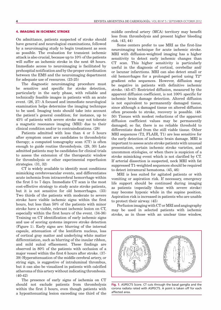
CT is widely available to identify most conditions mimicking cerebrovascular events, and differentiates acute ischemia from intracerebral hemorrhage within the first 5 to 7 days. Immediate CT scan is the most cost-effective strategy to study acute stroke patients, but it is not sensitive for old hemorrhages. (33) Two thirds of the patients with moderate to severe stroke have visible ischemic signs within the first hours, but less than 50% of the patients with minor stroke have a visible, relevant ischemic lesion on CT, especially within the first hours of the event. (34-36) Training on CT identification of early ischemic signs and use of scoring systems improve stroke detection (Figure 1). Early signs are: blurring of the internal capsule, attenuation of the lentiform nucleus, loss of cortical gray matter and underlying white matter differentiation, such as blurring of the insular ribbon, and mild sulcal effacement. These findings are observed in 80% of the patients with occlusion of a major vessel within the first 6 hours after stroke. (37-39) Hyperattenuation of the middle cerebral artery, or string sign, is suggestive of intraluminal thrombus, but it can also be visualized in patients with calcified atheroma of this artery without indicating thrombosis. (40-42)
The presence of early signs of ischemia on CT should not exclude patients from thrombolysis within the first 3 hours, even though patients with a hypoattenuating lesion exceeding one third of the
middle cerebral artery (MCA) territory may benefit less from thrombolysis and present higher bleeding risk. (43, 44)
Some centers prefer to use MRI as the first-line neuroimaging technique for acute ischemic stroke. MRI with diffusion-weighted imaging has a higher sensitivity to detect early ischemic changes than CT scan. This higher sensitivity is particularly useful in the diagnosis of cortical, cerebellar and/or lacunar infarctions. MRI can also detect small or old hemorrhages for a prolonged period using T2* gradient echo sequences. However, diffusion may be negative in patients with definitive ischemic stroke. (45-47) Restricted diffusion, measured by the apparent diffusion coefficient, is not 100% specific for ischemic brain damage detection. Altered diffusion is not equivalent to permanently damaged tissue, since although a damaged tissue on altered diffusion often proceeds to stroke, recovery is possible. (48-50) Tissues with modest reductions of the apparent diffusion coefficient values may be permanently damaged; so far, there is no reliable threshold to differentiate dead from the still viable tissue. Other MRI sequences (T2, FLAIR, T1) are less sensitive for the early detection of ischemic brain damage. MRI is important to assess acute stroke patients with unusual presentation, certain ischemic stroke varieties, and uncommon etiologies, or when there is suspicion of a stroke mimicking event which is not clarified by CT. If arterial dissection is suspected, neck MRI with fat suppressed T1-weighted sequences should be required to detect intramural hematoma. (45, 46)
MRI is less suited for agitated patients or with vomiting or aspiration risk. If necessary, emergency life support should be continued during imaging as patients (especially those with severe stroke) may become hypoxic while in the supine position. Aspiration risk is increased in patients who are unable to protect their airway. (45)
Perfusion imaging with CT or MRI and angiography may be used in selected patients with ischemic stroke, as in those with an unclear time window,
Fig. 1. ASPECTS Score. CT cuts through the basal ganglia and the corona radiata rated with ASPECTS. A point is taken off for each affected area.
CONSENSUS STATEMENT ON DIAGNOSIS AND TREATMENT OF ACUTE ISCHEMIC STROKE 5
or late admission, to aid on thrombolysis therapy decision making, although there is no clear evidence that patients with particular perfusion patterns are more or less likely to benefit from thrombolysis. Selected patients with intracranial arterial occlusion may be candidates for intra-arterial thrombolysis, although scientific evidence is limited. Patients with combined obstructions of the internal carotid artery (ICA) and MCA have less possibility of recovery with intravenous thrombolysis than patients with isolated MCA obstructions. (49, 50) In patients with main MCA occlusion, the frequency of extracranial carotid occlusive disease is significantly high.
Mismatch between the volume of critically hypoperfused brain tissue (which can recover after reperfusion) and the volume of infarcted tissue (which does not recover even with reperfusion) may be detected with MR diffusion/perfusion imaging with moderate reliability, but this is not yet a proven strategy to improve thrombolysis response up to 9 hours. (51) There is disagreement on how to best identify irreversible ischemic brain injury and define critically impaired blood flow. Quantification of MRI perfusion is problematic, and there are diverse associations between perfusion parameters and clinical and radiological results. (52) Decreases in cerebral blood flow in CT are associated with subsequent tissue damage, but the therapeutic value of CT perfusion imaging has not yet been established. Although infarct expansion may occur in a high proportion of patients with mismatch, up to 50% of the patients without unbalanced perfusion-diffusion may also have infarct growth and so might benefit from tissue salvage. Hence, neither CT or MRI perfusion imaging nor the mismatch concept can be recommended for routine therapy decision making. (53-60)
Microhemorrhages are present on T2* weighted gradient MRI in up to 60% of the patients with hemorrhagic stroke, and are associated with older age, hypertension, diabetes, leukoaraiosis, lacunar stroke, and amyloid angiopathy. In acute ischemic stroke patients, the incidence of symptomatic intracranial hemorrhage following thrombolysis was not increased when cerebral microbleeds on pre-treatment T2*-weighted MRI were present. (50, 51)
Vascular imaging should be promptly performed to identify patients with significant symptomatic arterial stenosis who could benefit from endarterectomy or angioplasty. Non-invasive imaging with color-coded duplex imaging of the extracranial and intracranial arteries, CT angiography, or contrast-enhanced MR angiography (CE-MRA) is widely available. These approaches are relatively risk-free, whereas intra-arterial angiography currently has a 1% lower risk of causing embolism in patients with symptomatic carotid lesions. (61, 62) Invasive angiography may be necessary in certain circumstances, for example, when other explorations have not been conclusive.
Recommendations- In patients with suspected TIA or stroke, urgent CT scan should be performed (Class I, Level of Evidence A), or alternatively, MRI (Class II, Level of Evidence A). - If MRI is performed, the inclusion of diffusion- weighted imaging, apparent diffusion coefficient (ADC), gradient echo sequences, and FLAIR (Class II, Level of Evidence A) are required. - In patients with TIA and ischemic stroke, immediate diagnostic work-up is necessary (ultrasound, CT-angiography, or RM-angiography) (Class I, Level of Evidence A).
5. GENERAL MEDICAL TREATMENT OF ISCHEMIC STROKE
The head of the bed should be tilted up at 30-45°, and a nasogastric tube to prevent aspiration of gastric contents should be inserted in case of consciousness deterioration. Up to 63% of the patients with ischemic stroke develop hypoxemia, which has been related to stroke severity, dysphagia, and age. Monitoring of arterial oxygen saturation and initial oxygen therapy are recommended if pulse oxygen saturation falls below 92%. Patients with airway obstruction or poor management of respiratory secretions may need orotracheal intubation. (63, 64) Both high blood pressure (BP) levels and low systolic blood pressure (SBP) have been related to a worse prognosis. Early mortality rate increases by 17.9% per each 10 mm Hg below 150 mm Hg in SBP. Different studies with hypotensive drugs have been conducted during the acute phase of ischemic stroke. Today, drugs such as ultra short-acting beta-blockers or angiotensin-converting enzyme inhibitors, which have shown BP reduction without changing cerebral blood flow, are recommended. In brief, treating hypertension during the acute phase should be conducted only in those patients with BP > 200/120 mm Hg in two consecutive measurements separated by a15 minute interval, provided other factors that may cause hypertension have been ruled out, or in patients with heart failure, aortic dissection, acute myocardial infarction, acute renal failure, pregnancy, or in candidates for thrombolytic therapy. In case of BP modification, a decrease below 15% of the initial value during the first 24 hours is suggested. (63, 64) Over the last years, several clinical studies have pointed out that hyperglycemia in the acute phase of ischemic stroke would reflect an often unknown pre-existing diabetes mellitus. In these patients, hyperglycemia > 120 mg/dl is a negative prognostic marker regardless of age, severity, and subtype of the event. Moreover, this deleterious effect is greater in non-diabetic patients. At present, strict monitoring of blood glucose levels during the acute phase is recommended, to achieve euglycemia avoiding the administration of dextrose solution, and initiate insulin therapy if blood glucose level is > 180 mg/dl. (63, 64)
REVISTA ARGENTINA DE CARDIOLOGÍA / VOL 80 Nº 5 / SEPTEMBER-OCTOBER 20126
Hydration should be performed with normal saline solution, keeping a neutral balance. The use of hypotonic solutions (such as dextrose or Ringer’s lactate) may cause cerebral edema and hyponatremia. (63, 64)
Body temperature should be regularly checked, and in case of armpit temperature > 37.5 ºC, antipyretic medication should be administered. In these cases, it is necessary to rule out infection, the most common being aspiration pneumonia or urinary infections.
Oral feeding should be started only after clinical examination to rule out an evident deglutition disorder. In patients with evidence of aspiration pneumonia feeding by tube is necessary. (63, 64)
Voiding dysfunction is a common complication in stroke patients, so it is important to rule out the presence of a distended bladder indicating acute urinary retention.
Patients should be mobilized prematurely to prevent bed sores and abnormal positions that may hinder their rehabilitation. Patients with severe motor deficit who cannot walk during the acute phase should receive doses of heparin to prevent deep vein thrombosis and pulmonary thromboembolism. (63, 64)
Levetiracetam therapy to prevent seizures is not a routine indication for patients with ischemic stroke; only those who have had seizures should receive this therapy. (63, 64)
Recommendations - Neurological status, heart rate, blood pressure, and oxygen saturation should be monitored in patients with ischemic stroke, an indication based on Good Clinical Practice Guidelines (GCP).- Oxygen should be administered if the oxygen saturation falls below 92% (GCP).- Maintain a neutral fluid balance and correct hydro- electrolyte disturbances in patients with ischemic stroke (GCP). - Use normal saline (0.9%) for hydrotherapy during the first 24 hours after ischemic stroke (GCP). - As a general rule, blood pressure lowering is not recommended during ischemic stroke (GCP). - Elevated blood pressure should be lowered in patients with extremely high blood pressure levels (> 220/120 mm Hg) on repeated measurements and with clinical evidence of acute myocardial infarction, severe cardiac failure, aortic dissection, pregnancy, renal failure, or hypertensive encephalopathy (GCP). - Treat with volume expanders low blood pressure secondary to hypovolemia or associated with neurological deterioration during acute ischemic stroke (GCP). - Monitor serum glucose levels (GCP). - Treat serum glucose levels >180 mg/dl with insulin (Class II, Level of Evidence B). - Correct hypoglycemia (< 50 mg/dl) with
intravenous dextrose or 10-20% glucose infusion (Class II, Level of Evidence B). - Lower temperature > 37.5 °C with paracetamol or physical measures (GCP). - Search for concurrent infection in case of hyperthermia (GCP). - Prescribe low-dose heparin to prevent deep vein thrombosis and pulmonary thromboembolism (Class I, Level of Evidence A).- Routine prophylactic administration of anticonvulsants is not recommended and should only be indicated in patients with a history of seizures (Class II, Level of Evidence B).
6. SPECIFIC MEDICAL TREATMENT OF ISCHEMIC STROKE
Thrombolytic therapy
Intravenous tissue plasminogen activator Thrombolytic therapy with rtPA (0.9 mg/kg body weight, maximum dose 90 mg) given within 3 hours after onset of ischemic stroke, significantly improves outcome after 3 months, with an absolute increase of 11 to 13% patients who improve with no deficit or with an insignificant impairment according to the study scales. The number needed to treat (NNT) to achieve a favorable clinical outcome after 3 months is 7. A pooled analysis of individual data of rtPA trials showed that, even within a 3-hour window, earlier treatment results in better outcome (0-90 min: OR 2.11; 95% CI 1.33 to 3.55; 90-180 min: OR 1.69; 95% CI 1.09 to 2.62). (65-67)
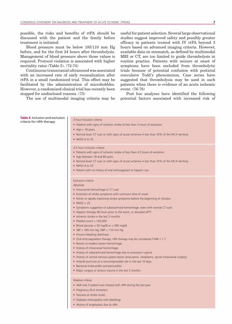
The meta-analysis of NINDS, ECASS I, ECASS II and Atlantis A and B studies suggests the benefit could extend up to 4.5 hours. (68, 69) This is further confirmed by the ECASS III study, which shows a benefit in patients < 80 years of age, with a NIHSS score < 25 points and with no history of oral anticoagulant agents, previous stroke or diabetes when they were treated within a 3 to 4.5-hour time window (Table 4). (70)
The NINDS (National Institute of Neurological Disorders and Stroke) study showed that the extent of early ischemic changes (using the ASPECT score) had no effect on treatment response within the 3-hour time window. However, European regulatory agencies do not advocate the treatment in patients with severe events (NIHSS ≥ 25), extended early signs of ischemia on CT scan, or age above 80 years (unlike the United States approval). Observational studies suggest that rtPA given within the first 3 hours of the event is safe and effective in patients over 80 years of age, but more randomized data are pending. The effect of gender on the response to rtPA is uncertain. (71)
Thrombolytic therapy appears to be safe and effective across various types of hospitals, if the diagnosis is established by an experienced physician on cerebrovascular disease and the brain CT is also assessed by an experienced physician. Whenever
CONSENSUS STATEMENT ON DIAGNOSIS AND TREATMENT OF ACUTE ISCHEMIC STROKE 7
possible, the risks and benefits of rtPA should be discussed with the patient and the family before treatment is initiated.
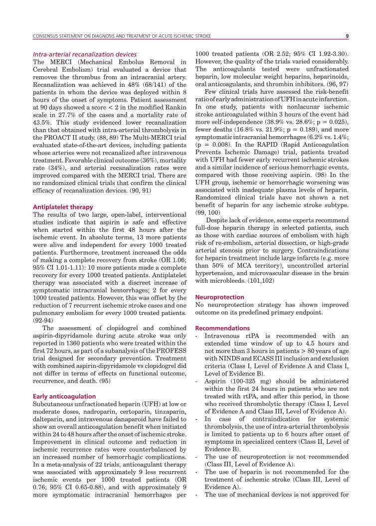
Blood pressure must be below 185/110 mm Hg before, and for the first 24 hours after thrombolysis. Management of blood pressure above those values is required. Protocol violation is associated with higher mortality rates (Table 5). (72-74)
Continuous transcranial ultrasound was associated with an increased rate of early recanalization after rtPA in a small randomized trial. This effect may be facilitated by the administration of microbubbles. However, a randomized clinical trial has recently been stopped for undisclosed reasons. (75)
The use of multimodal imaging criteria may be
useful for patient selection. Several large observational studies suggest improved safety and possibly greater efficacy in patients treated with IV rtPA beyond 3 hours based on advanced imaging criteria. However, available data on mismatch, as defined by multimodal MRI or CT, are too limited to guide thrombolysis in routine practice. Patients with seizure at onset of symptoms have been excluded from thrombolytic trials because of potential confusion with postictal convulsive Todd’s phenomenon. Case series have suggested that thrombolysis may be used in such patients when there is evidence of an acute ischemic event. (76-78) Post hoc analyses have identified the following potential factors associated with increased risk of
Table 4. Inclusion and exclusion criteria for rtPA therapy
3-hour Inclusion criteria
• Patients with signs of ischemic stroke of less than 3 hours of evolution.
• Age > 18 years.
• Normal brain CT scan or with signs of acute ischemia in less than 33% of the MCA territory.
• NIHSS 4 to 25.
4.5 hour inclusion criteria
• Patients with signs of ischemic stroke of less than 4.5 hours of evolution.
• Age between 18 and 80 years.
• Normal brain CT scan or with signs of acute ischemia in less than 33% of the MCA territory.
• NIHSS 4 to 25.
• Patient with no history of oral anticoagulant or heparin use.
Relative criteria
• AMI only if patient was treated with rtPA during the last year.
• Pregnancy (first trimester).
• Seizures at stroke onset.
• Diabetes (retinopathy with bleeding).
• History of anaphylaxis due to rtPA.
Exclusion criteria
Absolute
• Intracranial hemorrhage in CT scan
• Evolution of stroke symptoms with unknown time of onset.
• Minor or rapidly improving stroke symptoms before the beginning of infusion.
• NIHSS > 25.
• Symptoms suggestive of subarachnoid hemorrhage, even with normal CT scan.
• Heparin therapy 48 hours prior to the event, or elevated aPTT.
• Ischemic stroke in the last 3 months.
• Platelet count < 100,000.
• Blood glucose < 50 mg/dl or > 400 mg/dl.
• SBP > 185 mm Hg, DBP > 110 mm Hg.
• Known bleeding diasthesis.
• Oral anticoagulation therapy. rtPA therapy may be considered if INR < 1.7.
• Recent or evident severe hemorrhage.
• History of intracranial hemorrhage.
• History of subarachnoid hemorrhage due to aneurysm rupture.
• History of central nervous system lesion (aneurysms, neoplasms, spinal intracranial surgery).
• Arterial puncture at a noncompressible site in the last 10 days.
• Bacterial endocarditis and pericarditis.
• Major surgery or serious trauma in the last 3 months.
REVISTA ARGENTINA DE CARDIOLOGÍA / VOL 80 Nº 5 / SEPTEMBER-OCTOBER 20128
intracerebral hemorrhage complications following rtPA administration (79-80): - Hyperglycemia.- History of diabetes. - NIHSS > 22. - Age > 80 years.- Increased time to treatment. - Previous therapy with aspirin or oral anticoagulant agents.- History of congestive heart failure. - Low plasminogen activator inhibitor activity. - NINDS protocol violations.- Uncontrolled arterial hypertension
Intra-arterial and combined (IV+IA) thrombolysis Intra-arterial thrombolytic treatment of proximal MCA occlusion using pro-urokinase within 6 hours of the event was associated with a significantly better outcome in the PROACT II trial (Prolysis in Acute Cerebral Thromboembolism Trial). (81) Additional randomized clinical trials with pro-urokinase (PROACT I) or urokinase (MELT) and a meta-analysis of PROACT I, PROACT II and MELT studies indicate a benefit of intra-arterial thrombolytic therapy in patients with proximal MCA occlusions. Pro-urokinase is not available and intra-arterial thrombolytic therapy with rtPA is not substantiated by clinical trials, though observational data and non-randomized comparisons are available. (82, 83)
A randomized trial (IMS3) comparing standard intravenous rtPA therapy with a combined intravenous low dose (0.6 mg/kg) and intra-arterial strategy is in progress. (84, 85) In 1999, the Emergency Management of Stroke Study was completed. The bridging study consisting of low dose intravenous
rtPA (0.6 mg/kg) followed by an intra-arterial dose of 20 mg was compared with a group receiving only intra-arterial therapy. Patients under bridging therapy showed higher recanalization rates but failed to show associated functional improvement, evidencing a statistical trend towards higher hemorrhagic complications and death. (86)
Intra-arterial treatment of acute basilar artery occlusion with urokinase or rtPA has been available for more than 20 years, but has not been tested in a sufficiently ample randomized clinical trial, although encouraging results have been obtained in observational studies related to higher recanalization rates, but without significant clinical improvement. (87)
Table 4. (cont.) Inclusion and exclusion criteria for rtPA therapy
Table 5. BP management
*Note: in Asian patients, a 0.6 mg/kg rtPA dose is suggested due to higher risk of bleeding as a result of drug metabolism. (109, 110)
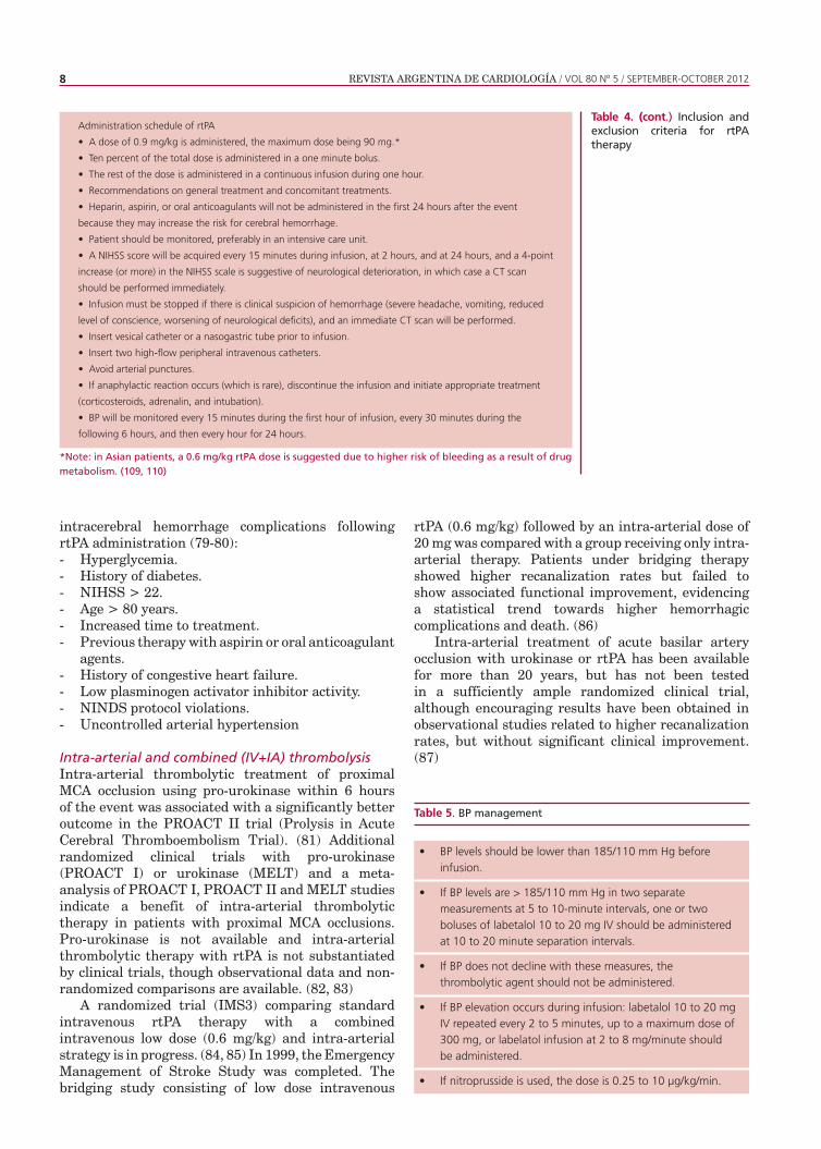
Administration schedule of rtPA
• A dose of 0.9 mg/kg is administered, the maximum dose being 90 mg.*
• Ten percent of the total dose is administered in a one minute bolus.
• The rest of the dose is administered in a continuous infusion during one hour.
• Recommendations on general treatment and concomitant treatments.
• Heparin, aspirin, or oral anticoagulants will not be administered in the first 24 hours after the event
because they may increase the risk for cerebral hemorrhage.
• Patient should be monitored, preferably in an intensive care unit.
• A NIHSS score will be acquired every 15 minutes during infusion, at 2 hours, and at 24 hours, and a 4-point
increase (or more) in the NIHSS scale is suggestive of neurological deterioration, in which case a CT scan
should be performed immediately.
• Infusion must be stopped if there is clinical suspicion of hemorrhage (severe headache, vomiting, reduced
level of conscience, worsening of neurological deficits), and an immediate CT scan will be performed.
• Insert vesical catheter or a nasogastric tube prior to infusion.
• Insert two high-flow peripheral intravenous catheters.
• Avoid arterial punctures.
• If anaphylactic reaction occurs (which is rare), discontinue the infusion and initiate appropriate treatment
(corticosteroids, adrenalin, and intubation).
• BP will be monitored every 15 minutes during the first hour of infusion, every 30 minutes during the
following 6 hours, and then every hour for 24 hours.
BP levels should be lower than 185/110 mm Hg before infusion.
If BP levels are > 185/110 mm Hg in two separate measurements at 5 to 10-minute intervals, one or two boluses of labetalol 10 to 20 mg IV should be administered at 10 to 20 minute separation intervals.
If BP does not decline with these measures, the thrombolytic agent should not be administered.
If BP elevation occurs during infusion: labetalol 10 to 20 mg IV repeated every 2 to 5 minutes, up to a maximum dose of 300 mg, or labelatol infusion at 2 to 8 mg/minute should be administered.
If nitroprusside is used, the dose is 0.25 to 10 μg/kg/min.
•
•
•
•
•
CONSENSUS STATEMENT ON DIAGNOSIS AND TREATMENT OF ACUTE ISCHEMIC STROKE 9
Intra-arterial recanalization devices The MERCI (Mechanical Embolus Removal in Cerebral Embolism) trial evaluated a device that removes the thrombus from an intracranial artery. Recanalization was achieved in 48% (68/141) of the patients in whom the device was deployed within 8 hours of the onset of symptoms. Patient assessment at 90 days showed a score < 2 in the modified Rankin scale in 27.7% of the cases and a mortality rate of 43.5%. This study evidenced lower recanalization than that obtained with intra-arterial thrombolysis in the PROACT II study. (88, 89) The Multi-MERCI trial evaluated state-of-the-art devices, including patients whose arteries were not recanalized after intravenous treatment. Favorable clinical outcome (36%), mortality rate (34%), and arterial recanalization rates were improved compared with the MERCI trial. There are no randomized clinical trials that confirm the clinical efficacy of recanalization devices. (90, 91)
Antiplatelet therapy The results of two large, open-label, interventional studies indicate that aspirin is safe and effective when started within the first 48 hours after the ischemic event. In absolute terms, 13 more patients were alive and independent for every 1000 treated patients. Furthermore, treatment increased the odds of making a complete recovery from stroke (OR 1.06; 95% CI 1.01-1.11): 10 more patients made a complete recovery for every 1000 treated patients. Antiplatelet therapy was associated with a discreet increase of symptomatic intracranial hemorrhages; 2 for every 1000 treated patients. However, this was offset by the reduction of 7 recurrent ischemic stroke cases and one pulmonary embolism for every 1000 treated patients. (92-94)
The assessment of clopidogrel and combined aspirin-dipyridamole during acute stroke was only reported in 1360 patients who were treated within the first 72 hours, as part of a subanalysis of the PROFESS trial designed for secondary prevention. Treatment with combined aspirin-dipyridamole vs clopidogrel did not differ in terms of effects on functional outcome, recurrence, and death. (95)
Early anticoagulation Subcutaneous unfractionated heparin (UFH) at low or moderate doses, nadroparin, certoparin, tinzaparin, dalteparin, and intravenous danaparoid have failed to show an overall anticoagulation benefit when initiated within 24 to 48 hours after the onset of ischemic stroke. Improvement in clinical outcome and reduction in ischemic recurrence rates were counterbalanced by an increased number of hemorrhagic complications. In a meta-analysis of 22 trials, anticoagulant therapy was associated with approximately 9 less recurrent ischemic events per 1000 treated patients (OR 0.76; 95% CI 0.65-0.88), and with approximately 9 more symptomatic intracranial hemorrhages per
1000 treated patients (OR 2.52; 95% CI 1.92-3.30). However, the quality of the trials varied considerably. The anticoagulants tested were unfractionated heparin, low molecular weight heparins, heparinoids, oral anticoagulants, and thrombin inhibitors. (96, 97)
Few clinical trials have assessed the risk-benefit ratio of early administration of UFH in acute infarction. In one study, patients with nonlacunar ischemic stroke anticoagulated within 3 hours of the event had more self-independence (38.9% vs. 28.6%; p = 0.025), fewer deaths (16.8% vs. 21.9%; p = 0.189), and more symptomatic intracranial hemorrhages (6.2% vs. 1.4%; (p = 0.008). In the RAPID (Rapid Anticoagulation Prevents Ischemic Damage) trial, patients treated with UFH had fewer early recurrent ischemic strokes and a similar incidence of serious hemorrhagic events, compared with those receiving aspirin. (98) In the UFH group, ischemic or hemorrhagic worsening was associated with inadequate plasma levels of heparin. Randomized clinical trials have not shown a net benefit of heparin for any ischemic stroke subtype. (99, 100)
Despite lack of evidence, some experts recommend full-dose heparin therapy in selected patients, such as those with cardiac sources of embolism with high risk of re-embolism, arterial dissection, or high-grade arterial stenosis prior to surgery. Contraindications for heparin treatment include large infarcts (e.g. more than 50% of MCA territory), uncontrolled arterial hypertension, and microvascular disease in the brain with microbleeds. (101,102)
Neuroprotection No neuroprotection strategy has shown improved outcome on its predefined primary endpoint.
Recommendations- Intravenous rtPA is recommended with an extended time window of up to 4.5 hours and not more than 3 hours in patients > 80 years of age with NINDS and ECASS III inclusion and exclusion criteria (Class I, Level of Evidence A and Class I, Level of Evidence B). - Aspirin (100-325 mg) should be administered within the first 24 hours in patients who are not treated with rtPA, and after this period, in those who received thrombolytic therapy (Class I, Level of Evidence A and Class III, Level of Evidence A).- In case of contraindication for systemic thrombolysis, the use of intra-arterial thrombolysis is limited to patients up to 6 hours after onset of symptoms in specialized centers (Class II, Level of Evidence B).- The use of neuroprotection is not recommended (Class III, Level of Evidence A).- The use of heparin is not recommended for the treatment of ischemic stroke (Class III, Level of Evidence A).- The use of mechanical devices is not approved for
REVISTA ARGENTINA DE CARDIOLOGÍA / VOL 80 Nº 5 / SEPTEMBER-OCTOBER 201210
the treatment of ischemic stroke (Class II, Level of Evidence B).
7. SURGICAL TREATMENT OF ISCHEMIC STROKE
Decompressive surgery Malignant MCA infarctionThe pooled analysis of 93 patients included in the DECIMAL (Decompressive Craniectomy in Malignant Middle Cerebral Artery Infarcts), DESTINY (Decompressive Surgery for the Treatment of Malignant Infarction of the Middle Cerebral Artery), and HAMLET (Hemicraniectomy after Middle Cerebral Artery Infarction with Life-Threatening Edema Trial) trials showed that, compared with the control group, the decompressive surgery group had more patients with a score ≤ 4 or ≤ 3 in the Rankin scale, and reduced mortality (NNTs 2, 4, and 2, respectively) one year after the event. Inclusion criteria for this combined analysis were: age 18-60 years, NIHSS > 15, decrease in the level of consciousness scoring 1 or more on the item 1a of the NIHSS, 50% or more infarct signs of the MCA territory in the CT scan or > 145 cm³ in diffusion sequencies and < 48 hours of infarction evolution. (103, 104)
Cerebellar infarctionVentriculostomy and decompressive surgery are considered treatments of choice for space-occupying cerebellar infarctions, although randomized clinical trials are lacking. As in space-occupying supratentorial infarction, the operation should be performed before signs of herniation are present. The prognosis among survivors can be very good, even in patients who are comatose before surgery. (105, 106)
Recommendations- Surgery in malignant infarctions is recommended, prior to signs of herniation, in young patients with infarctions in non-dominant hemisphere (Class II, Level of Evidence C).
BIBLIOGRAPHY
1. S1.http://www.msal.gov.ar/pngcam/pdf/GUIA_ADAPTACION.pdf.Último Acceso Septiembre 2011. 2. Mackay J, Mensah G, editores. Atlas of heart disease and. Geneva: WHO & CDC; 2004.3. Cushman M, Cantrell RA, McClure LA, Howard G, Prineas RJ, Moy CS, et al. Estimated 10-year stroke risk by region and race in the United States: geographic and racial differences in stroke risk. Ann Neurol. 2008;64:507-13. 4. Voeks JH, McClure LA, Go RC, Prineas RJ, Cushman M, Kissela BM, et al. Regional differences in diabetes as a possible contributor to the geographic disparity in stroke mortality: the REasons for Geographic And Racial Differences in Stroke Study. Stroke. 2008;39:1675-80.5. Melcon CM, Melcon MO. Prevalence of stroke in an Argentine community. Neuroepidemiology. 2006;27:81-8.6. Atallah A, Fustinoni O, Beigelman R: High prevalence of hemorrhagic stroke and untreated hypertension, frequent blood pressure lowering and inappropriate use of aspirin in the first Argentina National Stroke Registry (ARENAS). V World Stroke Congress, Vancouver, 2004 Abstract
7. Sposato LA, Esnaola MM, Zamora R, Zurrú MC, Fustinoni O, Saposnik G; ReNACer Investigators; Argentinian Neurological Society. Quality of IS care in emerging countries: the Argentinian National Stroke Registry (ReNACer). Stroke. 2008;39:3036-41.8. Albers GW, Hart RG, Lutsep HL, Newell DW, Sacco RL. AHA Scientific Statement. Supplement to the guidelines for the management of transient ischemic attacks: A statement from the Ad Hoc Committee on Guidelines for the Management of Transient Ischemic Attacks, Stroke Council, American Heart Association. Stroke. 1999;30:2502-11. 9. Alberts MJ, Hademenos G, Latchaw RE, Jagoda A, Marler JR, Mayberg MR, et al. Recommendations for the establishment of primary stroke centers. Brain Attack Coalition. JAMA. 2000;283:3102-9. 10. Hacke W, Kaste M, Skyhoj Olsen T, Orgogozo JM, Bogousslavsky J. European Stroke Initiative (EUSI) recommendations for stroke management. The European Stroke Initiative Writing Committee. Eur J Neurol 2000;7:607-23.11. Sacco RL, Adams R, Albers G, Alberts MJ, Benavente O, Furie K, et al. Guidelines for prevention of stroke in patients with IS or transient ischemic attack: a statement for healthcare professionals from the American Heart Association/American Stroke Association Council on Stroke: co-sponsored by the Council on Cardiovascular Radiology and Intervention: the American Academy of Neurology affirms the value of this guideline. Stroke 2006;37:577-617.12. Kwan J, Hand P, Sandercock P: A systematic review of barriers to delivery of thrombolysis for acute stroke. Age Ageing 2004;33:116-121.13. Gil Nunez AC, Vivancos Mora J. Organization of medical care in acute stroke: importance of a good network. Cerebrovasc Dis 2004;17 Suppl 1:113-23.14. Rothwell PM, Giles MF, Chandrateva A, Marquardt L, Geraghty O, Redgrave JN, et al. Effect of urgent treatment of transient ischaemic attack and minor stroke on early recurrent stroke (EXPRESS study): a prospective population-based sequential comparison. Lancet 2007;370:1432–42.15. Keskin O, Kalemoglu M, Ulusoy R. A clinic investigation into prehospital and emergency department delays in acute stroke care. Med Princ Pract 2005;14:408-12.16. Porteous GH, Corry MD, Smith WS. Emergency medical services dispatcher identification of stroke and transient ischemic attack. Prehosp Emerg Care 1999;3:211-6. 17. De Lemos CD, Atkinson RP, Croopnick SL, Wentworth DA, Akins PT. How effective are “community” stroke screening programs at improving stroke knowledge and prevention practices? Results of a 3-month follow-up study. Stroke 2003;34:e247-9.18. Harraf F, Sharma AK, Brown MM, Lees KR, Vass RI, Kalra L: A multicentre observational study of presentation and early assessment of acute stroke. BMJ 2002;325:17-21. 19. Kothari RU, Pancioli A, Liu T, Brott T, Broderick J. Cincinnati Prehospital Stroke Scale: reproducibility and validity. Ann Emerg Med. 1999;33:373-8.20. Kidwell CS, Starkman S, Eckstein M, Weems K, Saver JL. Identifying stroke in the field: prospective validation of the Los Angeles Prehospital Stroke Screen (LAPSS). Stroke. 2000;31:71-6.21. Lyden P, Brott T, Tilley B, Welch KM, Mascha EJ, Levine S, et al, and the NINDS PA Stroke Study Group. Improved reliability of the NIH Stroke Scale using video training. Stroke 1994;25:220-6. 22. Kelly-Hayes M, Robertson JT, Broderick JP, Duncan PW, Hershey LA, Roth LJ, et al. The American Heart Association Stroke Outcome Classification Stroke 1998;29:1274-80.23. Kasim KA, Brizzi M, Petersson J, Buchwald F, Sundgren PC. Combined clinical and radiological prognostic model in acute IS. Acta Neurol Belg 2010;110:239-45. 24. Saňák D, Herzig R, Zapletalová J, Horák D, Král M, Skoloudík D, et al. Predictors of good clinical outcome in acute stroke patients treated with intravenous thrombolysis. Acta Neurol Scand 2011;123:339-44. 25. Fryback DG, Thornbury JR. The efficacy of diagnostic imaging. Med Decis Making 1991;11:88-94. 26. Schramm P, Schellinger P, Klotz E, Kallenberg K, Fiebach J, Külkens S, et al. Comparison of perfusion CT and CTA source images with PWI and DWI in patients with acute stroke < 6 h. Stroke 2004;35:1562-68. 27. Barber P, Hill M, Eliasziw M, Demchuk A, Warwick Pexman J, Hudon M, et al. Neuroimaging of the brain in acute IS: A comparison of computed tomography and magnetic resonance diffusion weighted imaging. J Neurol Neurosurg Psychiatry 2005;76:1528-33. 28. Hand P, Wardlaw J, Rowat A, Haisma J, Lindley R, Dennis M. MR
CONSENSUS STATEMENT ON DIAGNOSIS AND TREATMENT OF ACUTE ISCHEMIC STROKE 11
brain imaging in patients with acute stroke - feasibility and patient-related difficulties. J Neurol Neurosurg Psychiatry 2005;76:1525-7. 29. The National Institute of Neurological Disorders and Stroke rt-PA Stroke Study Group: Tissue plasminogen activator for acute ischemic stroke. New Engl J Med 1995;333:1581-87. 30. Wardlaw J, Keir S, Dennis M. The impact of delays in computed tomography of the brain on the accuracy of diagnosis and subsequent management in patients with minor stroke. JNNP 2003;74:77-81. 31. Kidwell CS, Chalela JA, Saver JL, Starkman S, Hill MD, Demchuk AM, et al. Comparison of MRI and CT for detection of acute intracerebral hemorrhage. JAMA. 2004;292:1823-30. 32. Schellinger PD, Fiebach JB. Intracranial hemorrhage: the role of magnetic resonance imaging. Neurocrit Care. 2004;1:31-45.33. Wardlaw JM, Seymour J, Cairns J, Keir S, Lewis S, Sandercock P. Immediate computed tomography scanning of acute stroke is cost-effective and improves quality of life. Stroke. 2004;35:2477-83.34. von Kummer R, Bourquain H, Bastianello S, Bozzao L, Manelfe C, et al. Early prediction of irreversible brain damage after ischemic stroke at CT. Radiology. 2001;219:95-100.35. von Kummer R, Allen KL, Holle R, Bozzao L, Bastianello S, Manelfe C, et al. Acute stroke: usefulness of early CT findings before thrombolytic therapy. Radiology. 1997;205:327-33.36. Barber P, Demchuk A, Zhang J, Buchan A. Validity and reliability of a quantitative computed tomography score in predicting outcome of hyperacute stroke before thrombolytic therapy. Lancet 2000;355:1670-74. 37. Wardlaw J, Mielke O. Early signs of brain infarction at CT: Observer reliability and Outcome after thrombolytic treatment – systematic review. Radiology 2005;235:444-53. 38. Wardlaw J, West T, Sandercock P, Lewis S, Mielke O. The International Stroke Trials Collaborative Group: Visible infarction on computed tomography is an independent predictor of poor functional outcome after stroke, and not of haemorrhagic transformation. JNNP 2003;74:452-58. 39. von Kummer R. Effect of training in reading CT scans on patient selection for ECASS II. Neurology. 1998;51:S50-2. 40. Wardlaw J, Farrall A, Perry D, von Kummer R, Mielke O, Moulin T, et al. Factors influencing detection of early CT signs of cerebral ischaemia – an internet-based, international, multi-observer study. Stroke 2007;38:1250-6. 41. von Kummer R, Meyding-Lamadé U, Forsting M, Rosin L, Rieke K, Hacke W, et al. Sensitivity and prognostic value of early computed tomography in middle cerebral artery trunk occlusion. AJNR Am J Neuroradiol 1994;15:9-15. 42. Dzialowski I, Weber J, Doerfler A, Forsting M, von Kummer R. Brain tissue water uptake after middle cerebral artery occlusion assessed with CT. J Neuroimaging 2004;14:42-8. 43. Hill M, Rowley H, Adler F, Eliasziew M, Furlan A, Higashida R, et al. Selection of acute ischemic stroke patients for intraarterial thrombolysis with pro-urokinase by using ASPECTS. Stroke 2003;34:1925-31. 44. Patel S, Levine S, Tilley B, Grotta J, Lu M, Frankel M, et al. Lack of clinical significance of early ischemic changes on computed tomography in acute stroke. JAMA 2001;286:2830-8. 45. Chalela J, Kidwell C, Nentwich L, Luby M, Butmann J, Demchuk A, et al. Magnetic resonance imaging and computed tomography in emergency assessment of patients with suspected acute stroke: a prospective comparison. Lancet 2007;369:293-8. 46. Dimigen M, Keir S, Dennis M, Wardlaw J. Long-term visibility of primary intracerebral hemorrhage on magnetic resonance imaging. J Stroke Cerebrovasc Dis 2004;13:104-8. 47. Ay H, Oliveira-Filho J, Buonanno F, Schaefer P, Furie K, Chang Y, et al. “Footprints” of transient ischemic attacks: a diffusion-weighted MRI study. Cerebrovasc Dis 2002;14:177-86. 48. Fiehler J, Knudsen K, Kucinski T, Kidwell C, Alger J, Thomalla G, et al. Predictors of apparent diffusion coefficient normalization in stroke patients. Stroke 2004;35:514-9. 49. Oppenheim C, Lamy C, Touze E, Calvet D, Hamon M, Mas JL, et al. Do transient ischemic attacks with diffusion-weighted imaging abnormalities correspond to brain infarctions? AJNR Am J Neuroradiol 2006;27:1782-7. 50. Wardlaw J, Keir S, Bastin M, Armitage P, Rana A. Is diffusion imaging appearance an independent predictor of outcome after ischemic stroke? Neurology 2002;59:1381-7. 51. Hand P, Wardlaw J, Rivers C, Armitage P, Bastin M, Lindley R, et al. MR diffusion-weighted imaging and outcome prediction after
ischemic stroke. Neurology 2006;66:1159-63. 52. Kane I, Carpenter T, Chappell F, Rivers C, Armitage P, Sandercock P, et al. Comparison of 10 different magnetic resonance perfusion imaging processing methods in acute ischemic stroke: effect on lesion size, proportion of patients with diffusion/perfusion desacoplamiento, clinical scores, and radiologic outcomes. Stroke 2007;38:3158-64. 53. Kane I, Sandercock P, Wardlaw J. Magnetic resonance perfusion diffusion desacoplamiento and thrombolysis in acute ischaemic stroke: A systematic review of the evidence to date. JNNP 2007;78:485-90. 54. Coutts S, Simon J, Tomanek A, Barber P, Chan J, Hudon M, et al. Reliability of assessing percentage of diffusion-perfusion desacoplamiento. Stroke 2003;34:1681-83. 55. Albers GW, Thijs VN, Wechsler L, Kemp S, Schlaug G, Skalabrin E, et al. Magnetic resonance imaging profiles predict clinical response to early reperfusion: the diffusion and perfusion imaging evaluation for understanding stroke evolution (DEFUSE) study. Ann Neurol 2006;60:508-17. 56. Bandera E, Botteri M, Minelli C, Sutton A, Abrams K, Latronico N. Cerebral blood flow threshold of ischemic penumbra and infarct core in acute ischemic stroke: a systematic review. Stroke 2006;37:1334-9. 57. Carpenter T, Armitage P, Bastin M, Wardlaw J. DSC Perfusion MRI – Quantification and reduction of systematic errors arising in areas of reduced cerebral blood flow. Magn Reson Med 2006;56:1342-9. 58. Rivers C, Wardlaw J, Armitage P, Bastin M, Carpenter T, Cvoro V, et al. Do acute diffusion- and perfusion-weighted MRI lesions identify final infarct volume in ischaemic stroke? Stroke. 2006;37:98-104. 59. Dávalos A, Blanco M, Pedraza S, Leira R, Castellanos M, Pumar J, et al. The clinical-DWI desacoplamiento: a new diagnostic approach to the brain tissue at risk of infarction. Neurology 2004;62:2187-92. 60. Kent D, Hill M, Ruthazer R, Coutts S, Demchuk A, Dzialowski I, Wunderlich O, von Kummer R: „Clinical-CT desacoplamiento“ and the response to systemic thrombolytic therapy in acute ischemic stroke. Stroke 2005;36:1695-99. 61. Fischer U, Arnold M, Nedeltchev K, Brekenfeld C, Ballinari P, Remonda L, et al. NIHSS score and arteriographic findings in acute ischemic stroke. Stroke 2005;36:2121-5. 62. Allendoerfer J, Goertler M, von Reutern G. Prognostic relevance of ultra-early doppler sonography in acute ischaemic stroke: a prospective multicentre study. Lancet Neurology 2005;5:835-40. 63. The European Stroke Organisation (ESO) Executive Committee; ESO Writing Committee. Guidelines for management of ischaemic stroke and transient ischaemic attack 2008. Cerebrovasc Dis. 2008;25:457-507. 64. Adams HP Jr, del Zoppo G, Alberts MJ, Bhatt DL, Brass L, Furlan A. Guidelines for Early Management of Adults with Ischemic Stroke: A Guideline From American Heart Association / American Stroke Association Stroke Council, Clinical Cardiology Council, Cardiovascular Radiology and Intervention Council, and the Atherosclerotic Peripheral Vascular Disease and Quality of Care Outcome in Research Interdisciplinary Working Groups: The American Academy of Neurology affirm the value of this guideline as an educational tool for neurologist. Stroke 2007;38:1655-711. 65. Adams HP Jr, del Zoppo G, Alberts MJ, Bhatt DL, Brass L, Furlan A, Grubb RL, Higashida RT, Jauch EC, Kidwell C, Lyden PD, Morgenstern LB, Qureshi AI, Rosenwasser RH, Scott PA, Wijdicks EF; American Heart Association/American Stroke Association Stroke Council; American Heart Association/American Stroke Association Clinical Cardiology Council; American Heart Association/American Stroke Association Cardiovascular Radiology and Intervention Council; Atherosclerotic Peripheral Vascular Disease Working Group; Quality of Care Outcomes in Research Interdisciplinary Working Group. Guidelines for the early management of adults with ischemic stroke: a guideline from the American Heart Association/American Stroke Association Stroke Council, Clinical Cardiology Council, Cardiovascular Radiology and Intervention Council, and the Atherosclerotic Peripheral Vascular Disease and Quality of Care Outcomes in Research Interdisciplinary Working Groups: The American Academy of Neurology affirms the value of this guideline as an educational tool for neurologists. Circulation 2007 22;115:e478-534.66. Johnston SC, Nguyen-Huynh MN, Schwarz ME, Fuller K, Williams CE, Josephson SA, et al. National Stroke Association guidelines for the management of transient ischemic attacks. Ann Neurol 2006;60(3):301-13.67. Adams HP Jr, Leira EC. Treatment of acute ischemic stroke. Drugs Today (Barc) 1998;34:655-60.
REVISTA ARGENTINA DE CARDIOLOGÍA / VOL 80 Nº 5 / SEPTEMBER-OCTOBER 201212
68. Hacke W, Kaste M, Bluhmki E, Brozman M, Dávalos A, Guidetti D, Larrue V, Lees KR, Medeghri Z, Machnig T, Schneider D, von Kummer R, Wahlgren N, Toni D; ECASS Investigators. Thrombolysis with alteplase 3 to 4.5 hours after acute ischemic stroke. N Engl J Med 2008;359:1317-29.69. Tissue plasminogen activator for acute ischemic stroke. The National Institute of Neurological Disorders and Stroke rt-PA Stroke Study Group. N Engl J Med 1995;333:1581-7.70. Hacke W, Donnan G, Fieschi C, Kaste M, von Kummer R, Broderick JP, et al; ATLANTIS Trials Investigators; ECASS Trials Investigators; NINDS rt-PA Study Group Investigators. Association of outcome with early stroke treatment: pooled analysis of ATLANTIS, ECASS, and NINDS rt-PA stroke trials. Lancet. 2004;363:768-74.71. Thomalla G, Schwark C, Sobesky J, Bluhmki E, Fiebach JB, Fiehler J, et al; MRI in Acute Stroke Study Group of the German Competence NetworkStroke. Outcome and symptomatic bleeding complications of intravenousthrombolysis within 6 hours in MRI-selected stroke patients: comparison of a German multicenter study with the pooled data of ATLANTIS, ECASS, and NINDS tPA trials. Stroke. 2006;37:852-8. 72. Wahlgren N, Ahmed N, Eriksson N, Aichner F, Bluhmki E, Dávalos A, et al; Safe Implementation of Thrombolysis in Stroke-MOnitoring STudy Investigators. Multivariable analysis of outcome predictors and adjustment of main outcome results to baseline data profile in randomized controlled trials: Safe Implementation of Thrombolysis in Stroke-MOnitoring STudy (SITS-MOST). Stroke. 2008;39:3316-22.73. Bluhmki E, Chamorro A, Dávalos A, Machnig T, Sauce C, Wahlgren N, et al. Stroke treatment with alteplase given 3.0-4.5 h after onset of acute ischaemic stroke (ECASS III): additional outcomes and subgroup analysis of a randomised controlled trial. Lancet Neurol. 2009;8:1095-102.74. Wahlgren N, Ahmed N, Dávalos A, Hacke W, Millán M, Muir K, et al; SITS investigators. Thrombolysis with alteplase 3-4.5 h after acute ischaemic stroke (SITS-ISTR): an observational study. Lancet. 2008;372:1303-9. 75. Mikulik R, Dusek L, Hill MD, Fulep E, Grotta JC, Ribo M, et al; CLOTBUST Investigators. Pattern of response of National Institutes of Health Stroke Scale components to early recanalization in the CLOTBUST trial. Stroke 2010;41:466-70.76. De Silva DA, Brekenfeld C, Ebinger M, Christensen S, Barber PA, Butcher KS, et al; Echoplanar Imaging Thrombolytic Evaluation Trial (EPITHET) Investigators. The benefits of intravenous thrombolysis relate to the site of baseline arterial occlusion in the Echoplanar Imaging Thrombolytic Evaluation Trial (EPITHET). Stroke. 2010;41:295-9. 77. Mishra NK, Albers GW, Davis SM, Donnan GA, Furlan AJ, Hacke W, et al. Mismatch-based delayed thrombolysis: a meta-analysis. Stroke 2010;41:e25-33.78. Furlan AJ, Eyding D, Albers GW, Al-Rawi Y, Lees KR, Rowley HA, Sachara C, Soehngen M, Warach S, Hacke W; DEDAS Investigators. Dose Escalation of Desmoteplase for Acute ischemic Stroke (DEDAS): evidence of safety and efficacy 3 to 9 hours after stroke onset. Stroke 2006;37:1227-31.79. Wardlaw JM, Murray V, Berge E, del Zoppo GJ. Thrombolysis for acute ischaemic stroke. Cochrane Database Syst Rev 2009;(4):CD000213.80. Saver JL, Gornbein J, Grotta J, Liebeskind D, Lutsep H, Schwamm L, et al. Number needed to treat to benefit and to harm for intravenous tissue plasminogen activator therapy in the 3- to 4.5-hour window: joint outcome table analysis of the ECASS 3 trial. Stroke 2009;40:2433-7.81. Furlan A, Higashida R, Wechsler L, Gent M, Rowley H, Kase C, et al. Intraarterial prourokinase for acute ischemic stroke. The PROACT II study: a randomized controlled trial. Prolyse in Acute Cerebral Thromboembolism. JAMA 1999;282:2003-11.82. Mattle HP, Arnold M, Georgiadis D, Baumann C, Nedeltchev K, Benninger D, et al. Comparison of intraarterial and intravenous thrombolysis for ischemic stroke with hyperdense middle cerebral artery sign. Stroke 2008;39:379-83.83. Bourekas EC, Slivka A, Shah R, Mohammad Y, Slone HW, Kehagias DT, et al. Intra-arterial thrombolysis within three hours of stroke onset in middle cerebral artery strokes. Neurocrit Care 2009;11:217-22.84. Roberts HC, Dillon WP, Furlan AJ, Wechsler LR, Rowley HA, Fischbein NJ, et al. Computed tomographic findings in patients undergoing intra-arterial thrombolysis for acute ischemic stroke due
to middle cerebral artery occlusion: results from the PROACT II trial. Stroke 2002;33:1557-65.85. Lewandowski CA, Frankel M, Tomsick TA, Broderick J, Frey J, Clark W, et al. Combined intravenous and intra-arterial r-TPA versus intra-arterial therapy of acute ischemic stroke: Emergency Management of Stroke (EMS) Bridging Trial. Stroke 1999;30:2598-605.86. Shaltoni HM, Albright KC, Gonzales NR, Weir RU, Khaja AM, Sugg RM, et al. Is intra-arterial thrombolysis safe after full-dose intravenous recombinant tissue plasminogen activator for acute ischemic stroke? Stroke 2007;38:80-4.87. Schonewille WJ, Wijman CA, Michel P, Rueckert CM, Weimar C, Mattle HP, et al; BASICS study group. Treatment and outcomes of acute basilar artery occlusion in the Basilar Artery International Cooperation Study (BASICS): a prospective registry study. Lancet Neurol 2009;8:724-30.88. Shi ZS, Loh Y, Walker G, Duckwiler GR; MERCI and Multi MERCI Investigators. Endovascular thrombectomy for acute ischemic stroke in failed intravenous tissue plasminogen activator versus non-intravenous tissue plasminogen activator patients: revascularization and outcomes stratified by the site of arterial occlusions. Stroke 2010;41:1185-92.89. Shi ZS, Loh Y, Walker G, Duckwiler GR; MERCI and Multi-MERCI Investigators. Clinical outcomes in middle cerebral artery trunk occlusions versus secondary division occlusions after mechanical thrombectomy: pooled analysis of the Mechanical Embolus Removal in Cerebral Ischemia (MERCI) and Multi MERCI trials. Stroke 2010;41:953-60.90. Nogueira RG, Liebeskind DS, Sung G, Duckwiler G, Smith WS; MERCI; Multi MERCI Writing Committee. Predictors of good clinical outcomes, mortality, and successful revascularization in patients with acute ischemic stroke undergoing thrombectomy: pooled analysis of the Mechanical Embolus Removal in Cerebral Ischemia (MERCI) and Multi MERCI Trials. Stroke 2009;40:3777-83.91. Taschner CA, Treier M, Schumacher M, Berlis A, Weber J, Niesen W. Mechanical thrombectomy with the Penumbra recanalization device in acute ischemic stroke. J Neuroradiol 2011;38:47-52.92. Bradford AP, Khan SA, Lees KR, McInnes GT, Semple PF. Interpretation of IST and CAST stroke trials. International Stroke Trial. Chinese Acute Stroke Trial. Lancet 1997;350:440-51.93. CAST: randomised placebo-controlled trial of early aspirin use in 20,000 patients with acute ischaemic stroke. CAST (Chinese Acute Stroke Trial) Collaborative Group. Lancet 1997;349:1641-9.94. The International Stroke Trial (IST): a randomised trial of aspirin, subcutaneous heparin, both, or neither among 19435 patients with acute ischaemic stroke. International Stroke Trial Collaborative Group. Lancet 1997;349:1569-81.95. Bath PM, Cotton D, Martin RH, Palesch Y, Yusuf S, Sacco R, Diener HC, Estol C, Roberts R; PRoFESS Study Group. Effect of combined aspirin and extended-release dipyridamole versus clopidogrel on functional outcome and recurrence in acute, mild ischemic stroke: PRoFESS subgroup analysis. Stroke 2010;41:732-8.96. Sandercock PA, Counsell C, Kamal AK. Anticoagulants for acute ischaemic stroke. Cochrane Database Syst Rev 2008;(4):CD000024. Review.97. Sandercock PA, Counsell C, Tseng MC. Low-molecular-weight heparins or heparinoids versus standard unfractionated heparin for acute ischaemic stroke. Cochrane Database Syst Rev 2008;(3):CD000119. Review.98. Sherman DG. Prevention of venous thromboembolism, recurrent stroke, and other vascular events after acute ischemic stroke: the role of low-molecular-weight heparin and antiplatelet therapy. J Stroke Cerebrovasc Dis 2006;15:250-9.99. Sherman DG, Soltes S, Samuel R, Chibedi-Deroche D. Enoxaparin versus unfractionated heparin in the prevention of venous thromboembolism after acute ischemic stroke: rationale, design, and methods of an open-label, randomized, parallel-group multicenter trial. J Stroke Cerebrovasc Dis 2005;14:95-100.100. Bath P, Leonardi-Bee J, Bath F. Low molecular weight heparin versus aspirin for acute ischemic stroke: a systematic review. J Stroke Cerebrovasc Dis 2002;11:55-62.101. Paciaroni M, Agnelli G, Micheli S, Caso V. Efficacy and safety of anticoagulant treatment in acute cardioembolic stroke: a meta-analysis of randomized controlled trials. Stroke 2007;38:423-30.102. Padma V, Fisher M, Moonis M. Role of heparin and low-molecular-weight heparins in the management of acute ischemic stroke. Expert
CONSENSUS STATEMENT ON DIAGNOSIS AND TREATMENT OF ACUTE ISCHEMIC STROKE 13
Rev Cardiovasc Ther 2006;4:405-15.103. Jüttler E, Bösel J, Amiri H, Schiller P, Limprecht R, Hacke W, et al; DESTINY II Study Group. DESTINY II: DEcompressive Surgery for the Treatment of malignant INfarction of the middle cerebral arterY II. Int J Stroke 2011;6:79-86. doi: 10.1111/j.1747-4949.2010.00544.x.104. Hofmeijer J, Kappelle LJ, Algra A, Amelink GJ, van Gijn J, van der Worp HB; HAMLET investigators. Surgical decompression for space-occupying cerebral infarction (the Hemicraniectomy After Middle Cerebral Artery infarction with Life-threatening Edema Trial [HAMLET]): a multicentre, open, randomised trial. Lancet Neurol 2009;8:326-33.105. Vahedi K, Vicaut E, Mateo J, Kurtz A, Orabi M, Guichard JP, et al; DECIMAL Investigators. Sequential-design, multicenter, randomized, controlled trial of early decompressive craniectomy in malignant middle cerebral artery infarction (DECIMAL Trial). Stroke 2007;38:2506-17.106. Carter BS, Ogilvy CS, Candia GJ, Rosas HD, Buonanno F. One-year outcome after decompressive surgery for massive nondominant hemispheric infarction. Neurosurgery 1997;40:1168-75.
9. APPENDIX 1
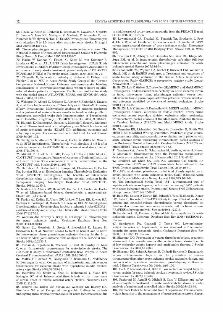
Definition of Class and Level of Evidence used by the American Heart Association
Class I: condition for which there is evidence and/
or general agreement that a given procedure or treatment is beneficial, useful, and effective.Class II: condition for which there is conflicting evidence and/or a divergence of opinion about the usefulness/efficacy of a procedure or treatment.Class IIa: the weight of evidence or opinion is in favor of a procedure or treatment.Class IIb: usefulness/efficacy is less well established by evidence or opinion.Class III: condition for which there is evidence and/or general agreement that a given procedure or treatment is not useful/effective and in some cases may be harmful.
Level of Evidence A: data derived from multiple randomized clinical trials.Level of Evidence B: data derived from a single randomized study or from non-randomized studies.Level of Evidence C: Expert opinions or case studies.
10. APPENDIX 2
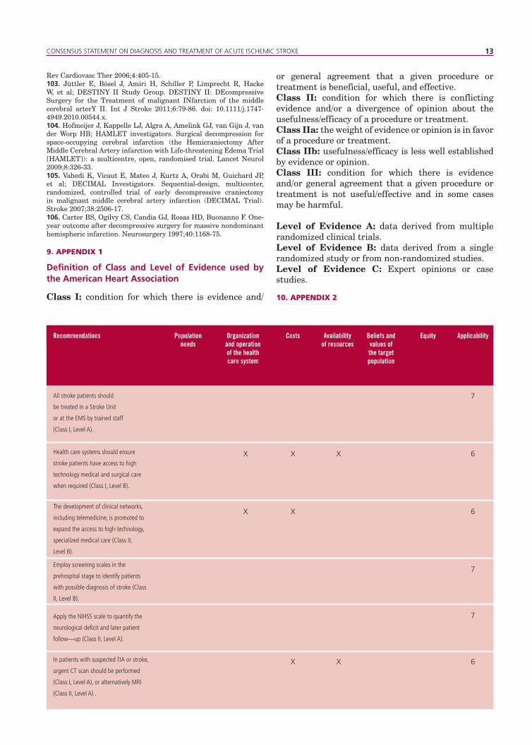
Recommendations
All stroke patients should
be treated in a Stroke Unit
or at the EMS by trained staff
(Class I, Level A).
Health care systems should ensure
stroke patients have access to high
technology medical and surgical care
when required (Class I, Level B).
The development of clinical networks,
including telemedicine, is promoted to
expand the access to high-technology,
specialized medical care (Class II,
Level B).
Employ screening scales in the
prehospital stage to identify patients
with possible diagnosis of stroke (Class
II, Level B).
Apply the NIHSS scale to quantify the
neurological deficit and later patient
follow—up (Class II, Level A).
In patients with suspected TIA or stroke,
urgent CT scan should be performed
(Class I, Level A), or alternatively MRI
(Class II, Level A) .
Populationneeds
Costs Equity ApplicabilityOrganization and operation of the health care system
Availability of resources
Beliefs and values of the target population
7
6
6
7
7
6
X
X
X
X
X
X
X
REVISTA ARGENTINA DE CARDIOLOGÍA / VOL 80 Nº 5 / SEPTEMBER-OCTOBER 201214
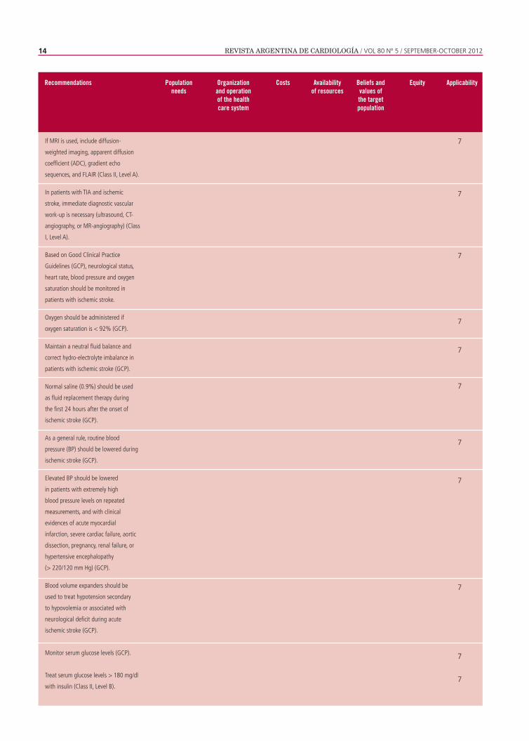
If MRI is used, include diffusion-
weighted imaging, apparent diffusion
coefficient (ADC), gradient echo
sequences, and FLAIR (Class II, Level A).
In patients with TIA and ischemic
stroke, immediate diagnostic vascular
work-up is necessary (ultrasound, CT-
angiography, or MR-angiography) (Class
I, Level A).
Based on Good Clinical Practice
Guidelines (GCP), neurological status,
heart rate, blood pressure and oxygen
saturation should be monitored in
patients with ischemic stroke.
Oxygen should be administered if
oxygen saturation is < 92% (GCP).
Maintain a neutral fluid balance and
correct hydro-electrolyte imbalance in
patients with ischemic stroke (GCP).
Normal saline (0.9%) should be used
as fluid replacement therapy during
the first 24 hours after the onset of
ischemic stroke (GCP).
As a general rule, routine blood
pressure (BP) should be lowered during
ischemic stroke (GCP).
Elevated BP should be lowered
in patients with extremely high
blood pressure levels on repeated
measurements, and with clinical
evidences of acute myocardial
infarction, severe cardiac failure, aortic
dissection, pregnancy, renal failure, or
hypertensive encephalopathy
(> 220/120 mm Hg) (GCP).
Blood volume expanders should be
used to treat hypotension secondary
to hypovolemia or associated with
neurological deficit during acute
ischemic stroke (GCP).
Monitor serum glucose levels (GCP).
Treat serum glucose levels > 180 mg/dl
with insulin (Class II, Level B).
Recommendations Populationneeds
Costs Equity ApplicabilityOrganization and operation of the health care system
Availability of resources
Beliefs and values of the target population
7
7
7
7
7
7
7
7
7
7
7
CONSENSUS STATEMENT ON DIAGNOSIS AND TREATMENT OF ACUTE ISCHEMIC STROKE 15
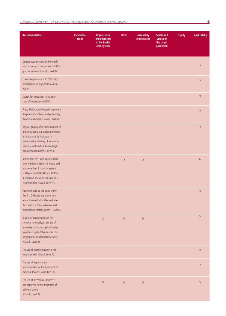
Correct hypoglycemia (< 50 mg/dl)
with intravenous dextrose or 10-20%
glucose infusion (Class II, Level B).
Lower temperature > 37.5 °C with
paracetamol or physical measures
(GCP).
Search for concurrent infection in
case of hyperthermia (GCP).
Prescribe low-dose heparin to prevent
deep vein thrombosis and pulmonary
thromboembolism (Class II, Level A).
Regular prophylactic administration of
anticonvulsants is not recommended.
It should only be indicated in
patients with a history of seizures or
ischemia with cortical hemorrhagic
transformation (Class II, Level B)
Intravenous rtPA with an extended
time window of up to 4.5 hours, and
not more than 3 hours in patients
> 80 years with NINDS and ECASS
III inclusion and exclusion criteria is
recommended (Class I, Level A).
Aspirin should be indicated within
the first 24 hours in patients who
are not treated with rtPA, and after
this period, in those who received
thrombolytic therapy (Class I, Level A)
In case of contraindication for
systemic thrombolysis, the use of
intra-arterial thrombolysis is limited
to patients up to 6 hours after onset
of symptoms in specialized centers
(Class II, Level B).
The use of neuroprotection is not
recommended (Class I, Level A).
The use of heparin is not
recommended for the treatment of
ischemic stroke (Class I, Level A).
The use of mechanical devices is
not approved for the treatment of
ischemic stroke
(Class II, Level B).
Recommendations Populationneeds
Costs Equity ApplicabilityOrganization and operation of the health care system
Availability of resources
Beliefs and values of the target population
7
7
7
7
7
6
7
5
7
7
5
X
X
X
X
X
X
X
X
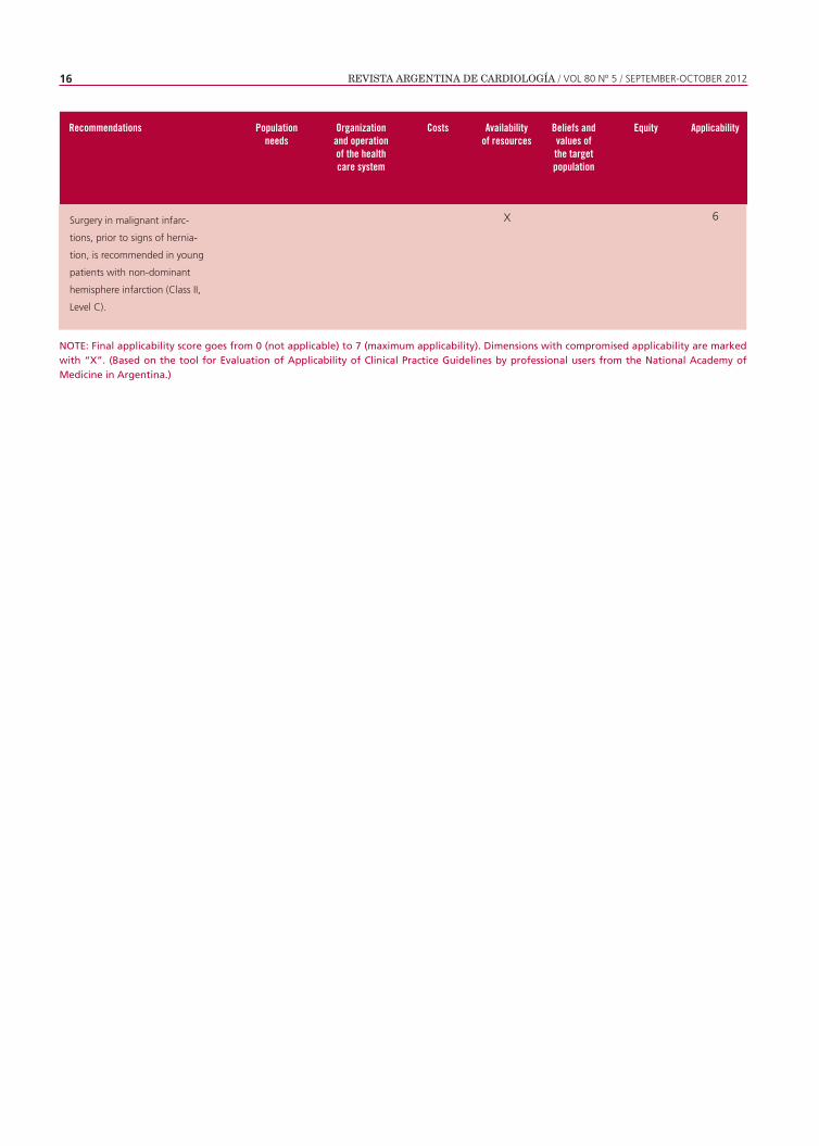
REVISTA ARGENTINA DE CARDIOLOGÍA / VOL 80 Nº 5 / SEPTEMBER-OCTOBER 201216
Recommendations Populationneeds
Costs Equity ApplicabilityOrganization and operation of the health care system
Availability of resources
Beliefs and values of the target population
Surgery in malignant infarc-
tions, prior to signs of hernia-
tion, is recommended in young
patients with non-dominant
hemisphere infarction (Class II,
Level C).
6X
NOTE: Final applicability score goes from 0 (not applicable) to 7 (maximum applicability). Dimensions with compromised applicability are marked with “X”. (Based on the tool for Evaluation of Applicability of Clinical Practice Guidelines by professional users from the National Academy of Medicine in Argentina.)