Consensus Recommendations On Advancing The … · 2 Robert J. Snyder, DPM, CWS* . Dr. Snyder is the...
24
Supported by a grant from Robert J. Snyder, DPM, CWS Robert S. Kirsner, MD, PhD Robert A. Warriner III, MD, FACA, FCCP, FCCWS, ABPM/UHM Lawrence A. Lavery, DPM, MPH Jason R. Hanft, DPM, FACFAS Peter Sheehan, MD Consensus Recommendations On Advancing The Standard Of Care For Treating Neuropathic Foot Ulcers In Patients With Diabetes Supplement to April 2010 This supplement is provided as a courtesy to the readers of WOUNDS. WOUNDS
Transcript of Consensus Recommendations On Advancing The … · 2 Robert J. Snyder, DPM, CWS* . Dr. Snyder is the...

Supported by a grant from
Robert J. Snyder, DPM, CWSRobert S. Kirsner, MD, PhDRobert A. Warriner III, MD, FACA, FCCP, FCCWS, ABPM/UHMLawrence A. Lavery, DPM, MPHJason R. Hanft, DPM, FACFASPeter Sheehan, MD
Consensus Recommendations OnAdvancing The Standard Of CareFor Treating Neuropathic Foot Ulcers In Patients With Diabetes
Supplement to April 2010
This supplement is provided as a courtesy to the readers of WOUNDS.
WOUNDS

2
Robert J. Snyder, DPM, CWS*. Dr. Snyder is the Medical
Director of the Wound Healing Center at University Hospital in
Tamarac, Fla. He is an Adjunct Professor at the Temple University
School of Podiatric Medicine in Philadelphia. Dr. Snyder is in pri-
vate practice in Tamarac, Fla.
Robert S. Kirsner, MD, PhD. Dr. Kirsner is the Director of
the University of Miami Hospital Wound CURE Center in
Miami. He is a Professor and the Vice Chairman of the Depart-
ment of Dermatology at the University of Miami Miller School
of Medicine.
Robert A. Warriner III, MD, FACA, FCCP, FCCWS,
ABPM/UHM. Dr. Warriner is Chief Medical Officer of Diversi-
fied Clinical Services in Jacksonville, Fla. He is the Emeritus Med-
ical Director and Founder of the Southeast Texas Center for
Wound Care and Hyperbaric Medicine at the HCA Conroe Re-
gional Medical Center in Conroe, Texas.
Lawrence A. Lavery, DPM, MPH. Dr. Lavery is a Professor in
the Department of Surgery at the Texas A&M Health Science
Center College of Medicine.
Jason R. Hanft, DPM, FACFAS. Dr. Hanft is the Director of
Research for the Podiatric Residency Program and is the Director
of Podiatric Education at the South Miami Hospital in Miami. He
is in private practice at The Foot and Ankle Institute of South
Florida in Miami.
Peter Sheehan, MD. Dr. Sheehan is the Chair of the Car-
diometabolic Risk Committee of the American Diabetes Asso-
ciation. He is a consultant at Sanford Health USD Medical
Center in Sioux Falls, S.D. Dr. Sheehan is in private practice in
New York City.
*Corresponding author: Please address correspondence to: Robert J. Sny-
der, DPM, CWS, 7301 N. University Drive, Suite 305, Tamarac, FL
33321; e-mail: [email protected]
Consensus Panel Members
Potential Conflicts Of InterestDr. Snyder has disclosed that he has received speaker honoraria
and served as a consultant or paid advisory board member for Ad-
vanced BioHealing. Dr. Snyder is the Medical Director of Systa-
genix Wound Management.
Dr. Hanft has disclosed that he has received speaker honoraria
and served as a consultant or paid advisory board member for Ad-
vanced BioHealing.
Dr. Kirsner has disclosed that he has received speaker honoraria
from Advanced BioHealing and Organogenesis.
Dr. Lavery has disclosed that he has received speaker honoraria
from KCI and Advanced BioHealing. Dr. Lavery serves as a con-
sultant or paid advisory board member for Chronic Disease Spe-
cialists, and has stock ownership in Chronic Disease Specialists.
Dr. Sheehan has disclosed that he has received research grant
funding from Tissue Repair Company, PamLab and Sanofi-Aven-
tis. Dr. Sheehan is the Director of Greystone Pharmaceuticals and
is a consultant or paid advisory board member for Advanced Bio-
Healing, Heal Or and ev3. Dr. Sheehan is also a consultant for
Sanford USD Medical Center, US Biotest, Izun, Hypermed and
Calretex. Dr. Sheehan is also a member of the Speakers’ Bureau
for ev3, Bristol-Myers Squibb/Sanofi-Aventis, Merck and
Organogenesis.
Dr. Warriner has disclosed that he has received speaker hono-
raria from Systagenix Wound Management.
Acknowledgments: This activity is supported by a grant from Advanced BioHealing. The authors thank Amy M. Horton,
PharmD, for editorial and manuscript assistance. The authors also thank David J. Margolis, MD, PhD, for editorial assistance.This supplement is provided as a courtesy to the readers of WOUNDS. This supplement was not subject to the peer review
process of WOUNDS.

3
Abstract: Neuropathic foot ulcers are a common and serious complication of diabetes mellitus. The presence of an unhealed
diabetic foot ulcer (DFU) increases the risk of infection, amputation and death. Low rates of DFU healing remain a challenge.
Recognizing these issues, a consensus panel recently was convened to review the evidence and practicalities for the evaluation
and treatment of patients with neuropathic DFUs. This consensus panel seeks to provide clinicians with the clinical markers, ev-
idence and recommendations that, used in conjunction with orderly decision-making and good clinical judgment, will advance
the standard of care for the treatment of neuropathic DFUs.
Key Words: advanced therapy, amputation, diabetic foot ulcer, debridement, hyperbaric oxygen therapy, infection control, livingskin equivalents, offloading, strain rate, wound bed preparation, wound classification systems
Index: Ostomy Wound Management. 2010;56(suppl 4):S1–S24.
Consensus Recommendations On Advancing TheStandard Of Care For Treating Neuropathic Foot Ulcers In Patients With DiabetesRobert J. Snyder, DPM, CWS; Robert S. Kirsner, MD, PhD; Robert A. Warriner III, MD, FACA, FCCP,FCCWS, ABPM/UHM; Lawrence A. Lavery, DPM, MPH; Jason R. Hanft, DPM, FACFAS; and PeterSheehan, MD

4
INTRODUCTIONDiabetes mellitus represents a group of chronic diseases charac-
terized by high levels of glucose in the blood resulting from de-
fects in insulin production, insulin action or both. Worldwide,
the number of cases of diabetes has been estimated to be 171
million and this number is projected to reach 366 million by
2025.1 Patients with diabetes are at risk for developing serious
health problems that may affect areas of the body including the
eyes, feet, skin, heart and kidneys. Among these health concerns,
foot ulceration is one of the most common complications. The
annual incidence rate for foot ulcers is 1% to 6.84% in individ-
uals with diabetes with a lifetime risk of 15% to 25%.2-6
The development of diabetic foot ulcers (DFUs) is thought to
result primarily from either peripheral arterial disease (PAD)
and/or peripheral neuropathy in addition to factors like defor-
mity, callus and trauma.4,5,7-9 However, the severity of PAD, in-
fection and deficiencies in the effective treatment of DFUs are
linked with secondary medical complications such as os-
teomyelitis and amputation. Approximately 15% of DFUs result
in lower extremity amputation.5,9 Diabetic foot ulcers are a con-
tributing factor in more than 85% of all diabetes-related lower
extremity amputations.8,10
The significant morbidity and mortality associated with dia-
betes is well known. A recent 10-year, prospective, population-
based study found a history of DFU to be a significant
independent predictor of mortality in patients with diabetes.11
This study found patients with diabetes with a history of DFU
had a 47% increased risk of mortality in comparison to patients
with diabetes who did not have a history of DFU. The five-year
mortality rates for patients with neuropathic and ischemic DFUs
were 45% and 55% respectively.12
The economic burden of DFUs and the complications aris-
ing from them are enormous. The estimated annual cost of care
for a patient with a DFU is $15,309 (1995 dollars) and direct
costs for a lower extremity amputation range from $22,700 to
$51,300 (2001 dollars).13,14 One of the most important cost-
saving considerations in caring for the patient with a DFU is
expeditious and complete wound healing to avoid complica-
tions such as amputation. Standard management strategies for
healing this malady typically have included preparation of the
wound bed, debridement, infection control, revascularization
and offloading. Despite the use of standard management strate-
gies, healing rates of DFUs remain low.
Clinical Decisions And Evidence-Based Medicine
Evidence-based medicine is defined as “the conscientious, explicit,
and judicious use of current best evidence in making decisions
about the care of patients” that integrates “individual clinical ex-
pertise with the best available external clinical evidence from sys-
tematic research.”15
Evidence-based medicine often involves searching published sources
for evidence to help guide an answer to a given clinical question. The
evidence often is obtained from sources such as PubMed
(http://pubmed.gov) or peer-reviewed sources like the Cochrane Col-
laboration (http://www.cochrane.org/). The practitioner then needs to
critically evaluate the evidence for its validity, importance and usefulness
in clinical practice.
Levels of evidence are used to rank the strength and validity of
the evidence from basic research to systematic reviews and meta-
analyses (see Figure 1). The treating clinician can subsequently in-
tegrate the critical evaluation of evidence with clinical expertise
and the patient’s individual problems and needs. The evidence ob-
tained from clinical trials is just one factor and must be interpreted
in light of individual patient factors, including the impact of co-
morbidities not addressed in clinical trials, economic issues and
psychosocial issues, in order to come to a clinical decision. Quite
often, the best level of evidence (ie, randomized controlled trial)
is not available on which to base a decision so all levels of evidence
must be considered.
Clinical guidelines and algorithms are readily available for as-
sisting in care decisions. Such guidelines, usually the result of
multidisciplinary teamwork, provide fast, accessible resources for
clinicians to make patient care decisions. Although they may vary
in regard to strength of scientific evidence, these guidelines and
Randomized Controlled Double Blind Studies
Systematic Reviewsand Meta-analyses
Cohort Studies
Case Control Studies
Case Series
Ideas, Editorials, Opinions
Animal research
In vitro (’test tube’) research
Case Reports
Figure 1. The Level Of Evidence Pyramid
Adapted from Sackett DL, Rosenberg WM, Gray JA, Haynes RB,Richardson WS.Evidence based medicine: what it is and what it isn't.BMJ. 1996;312(7023):71-2.

5
algorithms carry the weight of the governmental agencies, pro-
fessional organizations, universities, and authors who release
them.16 Table 1 lists several resources for guidelines and algo-
rithms specific to DFUs.
However, a systematic literature review of quality of care in the
United States revealed that 30% to 40% of patients are not being
treated according to evidence-based guidelines, and 20% to 30%
of care is either inappropriate, unnecessary or potentially danger-
ous.17 These data illustrate that there are apparent barriers to the
adoption of treatment guidelines and gaps exist between research
and clinical practice.
Recognizing these issues, a multidisciplinary panel of experts in
the field of wound care in patients with diabetes was recently con-
vened to review and discuss the evidence and practicalities for a
variety of modalities for DFU evaluation and treatment. The goal
of this panel was to provide straightforward and practical ap-
proaches for clinicians to adopt when treating patients with DFUs,
thus working to close the loop between research and practice.
In formulating these recommendations, the panel debated the
merits and disadvantages of these approaches, and held each to the
highest level of evidence available. They recognized that while ev-
idence-based guidelines may provide an ideal approach, clinicians
must consider and address the unique set of challenges inherent
to each individual patient.
ASSESSMENT OF THE DFUThe panel recognizes that a multidisciplinary team approach is
most advantageous for the treatment of the neuropathic ulcer in
patients with diabetes, and this is viewed as the standard of care.18
Furthermore, this approach should be considered as part of a con-
tinuum of care including acute care, home health care, subacute
intervention and the clinician’s office or wound care center.
However, the panel recognizes that multidisciplinary teams
are not always available. In these settings, referral to other spe-
cialists should be based on clinical judgment. The guidelines
listed in this section comprise the most important components
of the initial wound evaluation. Clinicians should be open to
additional testing beyond what is delineated below if clinical
impression warrants.
A comprehensive foot and ulcer evaluation for patients with dia-
betes encompasses several criteria. These key components include a
patient history and physical examination, laboratory screening, nu-
tritional evaluation, and a neurologic, musculoskeletal and vascular
assessment.19Wound history, a description and measurements of the
wound should be included in the evaluation. The clinician should
be looking for factors that may have led to wound formation and
may impair healing. Foot ulcers in patients with diabetes that exhibit
“stalled” healing after two to four weeks should be re-evaluated.
History And Physical Examination
A complete history and physical must be performed as part of an ap-
propriate evaluation. Information pertinent to the patient with a
DFU includes: the duration of diabetes, degree of glycemic control,
the presence of other diabetes-associated comorbidities and other
illnesses that may affect wound repair such as end-stage renal disease,
pulmonary disease, or cardiovascular disease (hypertension, dyslipi-
demia, myocardial infarction, transient ischemic attacks, angina, or
valvular heart disease). A review of systems and family history should
also be considered.
Clinicians also should assess factors such as the initial wounding
event, history of recurrent wounding, previous wound healing
problems, prior diagnostic testing, prior therapies and response,
functional impact of the wound on the patient, and a sufficient
social history to define potential adverse impact on an optimal
plan of care.
Table 1. Some Resources For Guidelines And Algorithms ForThe Care Of Patients With Diabetic Foot Ulcers
Association for the Advancement of Wound Care
www.aawconline.com
American College of Cardiology
www.acc.org/
American College of Footand Ankle Surgeons
www.acfas.org
American Diabetes Association
www.diabetes.org
American Orthopaedic Foot and Ankle Society
www.aofas.org
American Pharmaceutical Association
www.pharmacist.com
Cochrane Collaboration www.cochrane.org
Infectious Diseases Society of America
www.idsociety.org
International WorkingGroup on the Diabetic Foot
www.iwgdf.org
Wound Healing Society www.woundheal.org
Wound, Ostomy, Continence Nurses Society
www.wocn.org

6
Laboratory Screening
Since wound healing can be delayed by complications like anemia
and renal insufficiency, complete blood count and
creatinine/blood urea nitrogen tests may be included as part of
the baseline evaluation for patients with chronic wounds.20 If deep
tissue infection or osteomyelitis is suspected, erythrocyte sedimen-
tation rate (ESR) and C-reactive protein (CRP) testing may be
considered as secondary markers.21,22
Hemoglobin A1C. Although there is no concrete evidence
linking hemoglobin A1C to impaired wound healing, this test
should be used to assess the degree of glycemic control as an
overview of the patient’s disease state.23 Although optimal cut-
offs are still in debate, the American Diabetes Association (ADA)
generally recommends an A1C goal of <7% for adults with di-
abetes.24 Hemoglobin A1C should be ordered by the wound
care specialist if one has not been recently performed by the
patient’s internist or endocrinologist.
Lipid profile. A multitude of patients with diabetes who have
concomitant neuropathic ulcers are at high risk for developing or
have been diagnosed with cardiovascular disease.25 Therefore, it is
important to reduce dyslipidemia, hypertension and other cardio-
vascular risk factors. In this regard, evaluation of the patient’s lipid
profile (cholesterol, HDL and LDL) and homocysteine levels re-
mains critical, and wound specialists should work closely with the
patient’s primary care physician or cardiologist for any significant
change in this panel.
Nutritional Assessment
Although albumin is considered a gold standard for assessing
long-term nutritional status, there is little evidence to suggest
that it either predicts DFU healing or that correction of a low
albumin level improves healing.26 In patients with diabetes, low
albumin may be a marker for poor renal function rather than
poor nutrition.
Prealbumin has a shorter half-life when compared to albumin
and therefore may be a more appropriate marker in evaluating
protein deficiency. This test can be performed frequently to ascer-
tain whether a particular nutritional intervention is effective.26
If malnutrition is suspected, a dietary questionnaire should be
completed at screening by the patient and evaluated by the clini-
cian.27 Although no specific evaluation was recommended by the
panel, clinicians should choose the tool that works best for their
daily clinical practice.
Considerations include:
• Height
• Weight
• Unintentional change in weight > 10 lbs over the past six months
• Persistent or recurrent diarrhea
• Alcohol intake greater than three drinks per day
• Current dietary supplements including over-the-counter
vitamins
• Mouth, tooth or swallowing problems
• Tube feeding or TPN
• Limited access to food, missing two meals/day for more than two
days out of the week
• Morning fasting blood sugar
Responses may prompt further laboratory studies and nutritional
referral.27
Lifestyle/Psychosocial
Quality of life. General quality-of-life scoring systems exist but
may not be practical in day-to-day clinical practice because of their
length and complexity. Although the panel cannot recommend a
particular quality-of-life screener, a basic line of questioning should
be used at initial evaluation and subsequent visits to help guide
treatment decisions. For example: Do you have pain? How do you
feel? Do you feel worse or better? Are you working? What are
your daily activities? Are you able to carry out daily activities?
Smoking.Although there is little direct research on the effect
of smoking on healing of DFUs, there is some evidence that
chronic smoking has a negative impact on endothelial and smooth
muscle microcirculation in the skin, which could impair healing.28
This evidence, in addition to the known macrovascular and end-
organ complications associated with smoking, makes smoking ces-
sation a goal in the treatment of any DFU.
Additional factors include alcohol consumption and the pres-
ence of depression or other mental illness because these may affect
adherence with treatment recommendations.29,30
Neurologic Screening
There are several techniques that can be used to assess sensory func-
tion during neuropathic screening. The current recommendation
supported by the ADA advocates the use of the 10-g monofilament
in addition to one of the following techniques: pinprick sensation,
vibration perception with a 128-Hz tuning fork, ankle deep tendon
reflexes or vibration perception threshold testing.24The use of a 128-
Hz tuning fork and 10-g monofilament tests on the foot is recom-
mended by the consensus panel to assess neuropathy.
Vascular Evaluation
There is no universal noninvasive test that can completely evaluate
vascular health.31 However, a combination of testing, used where

7
indicated and appropriate, can support the assessment of vascular
supply in patients with diabetes. The panel recommends a tiered
testing approach to assist clinicians in evaluating DFUs. At screen-
ing, one or more measurements may be appropriate given the clin-
ical impression, equipment requirements and operator expertise.
These measurements include palpation of pulses, ankle brachial
index (ABI) and/or toe brachial index (TBI).
Palpation of pulses. Palpation of peripheral pulses, including
the femoral, popliteal and pedal vessels (dorsalis pedis and posterior
tibial), should be part of the routine physical examination. Al-
though the panel recognizes that palpation of pulses is subject to
a degree of variability among clinicians, it can provide evidence
for the presence of vascular disease but not necessarily its absence.32
In this regard, palpation of pulses is an inadequate screening tool for
PAD in patients with diabetes in the setting in which pulses are
present or absent.33
Ankle brachial index (ABI). The ADA recommends the
ABI as a reproducible and quantitative test for vascular evalu-
ation.32 Simple to perform, the ABI measures the patency of
the lower extremity arterial system using a hand-held Doppler
probe and a blood pressure cuff. The ABI is calculated as a ratio
of systolic blood pressure measured in the dorsalis pedis and
posterior tibial arteries of the ankle, taking the highest of the
two, divided by the systolic blood pressure in the brachial ar-
tery measured at the arm of a patient in a supine position for
five minutes.
Diagnostic interpretation indicates that low ABI ratios are as-
sociated with a high vascular risk (see Table 2).32 Normal values
range from 0.91 to 1.30 and ratios < 0.91 or > 1.30 could be
indicative of PAD. These individuals require further evaluation.
However, the ABI should be performed with an understanding
of the limitations of this test in patients with diabetes. For ex-
ample, results may be normal because the patient may be in a
transitional stage of diabetes. Conversely, an ABI value of >1.30
may be spurious secondary to medial arterial calcification in
this patient population.31
Toe brachial index.As the applicability of ABI may be po-
tentially limiting given that some patients with diabetes may de-
velop calcification in lower limb arteries that may result in a
falsely high ankle pressure, the TBI can be substituted. Clinicians
should remember that the TBI measurement requires specialized
equipment not commonly found in clinical settings and addi-
tional technical expertise.34 The TBI has shown to be superior
to the ABI in patients with medial arterial calcification and a
normal TBI (greater than 0.7 is normal) can exclude the pres-
ence of arterial disease.35 This outcome may well reduce con-
cerns of underdiagnosis in patients with diabetes and early stages
of incompressible vessels because a normal ABI does not neces-
sarily exclude systemic vascular disease.
If there is a high clinical index of suspicion that the wound is
ischemic or for individuals at high risk for PAD, a referral for sec-
ondary tier evaluations may include segmental pressure volume
and skin perfusion pressure (SPP) as well as transcutaneous oxy-
gen measurement (TCPO2), evaluation of lower limb indices and
waveforms (ie, segmental pressure volume recording). Tertiary
approaches for more aggressive care may include referral to a
vascular specialist for angiography and other interventions.
Segmental pressure volume. Segmental pressure pulse vol-
ume recording is considered a secondary tier approach for as-
sessing vascular health and is primarily used for patients with
poorly compressible vessels, or those with a normal ABI with
suspicion of peripheral vascular disease.32 Segmental pressure
volume is based on the principle that obstruction is proximal
to the level at which the pressure drops. To localize arterial le-
sions, systolic blood pressure cuffs are placed at several intervals
on the legs (thigh, calf and ankle) and pressures are recorded.
The shape of the observed pulse waveform is used to determine
the presence, severity and general location of vascular disease.36
Skin perfusion pressure (SPP). A laser Doppler measurement
that uses a blood pressure cuff at the ankle, SPP indicates the presence
(or lack thereof) of perfusion in the lower limbs. In essence, SPP is a
measure of cutaneous capillary circulation. Although SPP requires
specialized equipment, it has been shown be more sensitive than other
techniques for detecting lower extremity peripheral arterial disease.37
Transcutaneous oxygen measurement (TCPO2).TCPO2
measures oxygen tension in areas adjacent to a wound and has
been suggested as a diagnostic tool for assessing the probability of
wound healing. Two evidenced-based reviews support TCPO2 as
a screening tool for a wound population at high risk for vascular
Table 2. Diagnostic Interpretation Of Ankle-Brachial Index
Resting ABI Severity
0.91-1.30 Normal
0.70-0.90 Mild obstruction
0.40-0.69 Moderate obstruction
< 0.40 Severe obstruction
> 1.3 Poorly compressible
Adapted from American Diabetes Association. Peripheral arterial diseasein people with diabetes. Diabetes Care. 2003;26:3333-3341.

8
disease.38,39 When clinically indicated, TCPO2 should be used to
validate referral for vascular status and be used in conjunction with
hyperbaric oxygen (HBOT) intervention. Drawbacks of this test
may include variability secondary to technician experience and
technique although this can be true with other tests of perfusion
as well.39
Imaging.Although angiograms, duplex ultrasound, magnetic
resonance angiography, carbon dioxide angiography and com-
puted tomography angiography are not recommended as initial
screening tools, such methods may be necessary in further eval-
uation of these patients once clinical suspicion is ascertained. In
an observational study of 104 patients with a normal pulse, nor-
mal ABI or normal TCPO2, who were consecutively evaluated
with arteriography, 99% had hemodynamically significant le-
sions in the presence of an ulcer.33
Therefore, the panel agrees that if there is a high degree of
clinical suspicion of vascular disease, vascular consultation and
angiography should be considered to evaluate arterial disease
when an intervention is being considered in patients with dia-
betes who have non-healing wounds.
Additionally, the panel strongly suggests that if there is a high
clinical suspicion of critical limb ischemia, the patient should
be referred for a consult with a vascular specialist with whom
the clinician can have a dialogue concerning the potential for
wound healing, and help foster decisions regarding which pro-
cedures (ie, open procedures versus endovascular interventions)
would be most appropriate.
Foot And Ulcer Evaluation
The complete wound history is critical. Clinicians should re-
view and consider factors including: the initial wounding event;
history of recurrent wounding; previous wound healing prob-
lems; prior diagnostic testing, therapies and response; and the
functional impact of the wound on the patient. A sufficient so-
cial history is also helpful in defining potential adverse impacts
on an optimal plan of care.
The foot and ulcer examination should include:
• assessment of dermatologic changes in the surrounding skin in-
cluding callus, musculoskeletal deformity and muscle wasting;
• documentation of ulcer characteristics including location,
shape, and size of the wound (measurement of length, width,
and depth);
• clinical utilization of a probe to check for sinus tracts and a
positive probe-to-bone test;
• determination of the condition of the wound edges, wound
bed, wound base, periwound skin, and exudates; and
• determination of the presence of necrosis and wound
associated pain.40-42
Screening for complications, such as cellulitis, gangrene, os-
teomyelitis or Charcot deformity (neuropathic osteoarthropathy),
also should be performed. Wound depth, as measured by those
wounded anatomic structures (ie, dermis, fascia, muscle or bone), ap-
pears to be the most important clinical measurement of delayed heal-
ing.43Ankle mobility is also a key factor to assess with plantar ulcers.44
The panel recommends that both be evaluated by the clinician.
The Wound, Ostomy and Continence Nurses Society offers
further guidance for wound assessment that may prove helpful to
the clinician.42
• Determine localized inflammation by palpation and
dermal thermometry.
• Determine if edema is dependent or pitting, localized or
generalized, or bilateral or unilateral.
• Assess perfusion status by assessing skin temperature,
capillary refill, venous refill and color changes.
• Assess for ischemic skin changes including purpura, atro-
phy of subcutaneous tissue, shiny, taut skin, hair loss and
dystrophic nails.
• Assess musculoskeletal/biomechanical status for foot de-
formities, muscle weakness or gait abnormalities.
Wound Classification Systems
While several wound classification systems are available, there are
two well-established systems: the Wagner and the University of Texas
classifications. Although both systems provide descriptions of ulcers,
each has its own set of advantages and drawbacks.
The Wagner system uses six wound grades (scored 0 to 5) to assess
ulcer depth (see Table 3).45 However, the system is limited in its abil-
ity to identify and describe vascular disease and infection as inde-
pendent risk factors.46 For instance, superficial wounds that are
infected or dysvascular cannot be classified by this system.
The University of Texas system uses a matrix of grades (scored 0
to 3) and scales (scored A to D) to assess ulcer depth along with the
presence of wound infection and lower extremity ischemia.47 The
system allows identification of vascular disease and infection as in-
dependent factors regardless of ulcer anatomic depth (see Table 4).
Although the Wagner system is more widely implemented by health
care providers, is used in evaluation for hyperbaric oxygen therapy,
and may be required for reimbursement, the consensus panel favors
the University of Texas system because it has been shown to be a
better predictor of clinical outcome.48
The implementation of any wound classification system, when
used appropriately (ie, with a presenting grade in place – no

9
retroactive assessment), is a stride forward for patient data collec-
tion and quality reporting provided the ulcer measures are con-
sistent. However, a complete wound description should be
included so a clear picture of what the clinician is observing is
available to other specialists who may read the chart.
Infection Evaluation
Clinical assessment of wound infection in DFUs is imperative to
prevent complications such as amputation. Heat, pain, redness and
swelling are classic symptoms of wound infection. However, pa-
tients with diabetes are typically immunocompromised with an
impaired neuroinflammatory response and often fail to mount a
physiological response to infection. Therefore, clinicians should
look for secondary signs of infection including exudates, delayed
healing, friable granulation tissue, discolored granulation tissue,
foul odor, pocketing at the wound base and wound breakdown.49
Although clinical examination remains the standard for diag-
nosing skin, wound and soft tissue infection, recent evidence sup-
ports the use of erythrocyte sedimentation rate and C-reactive
protein as potential markers for infection in bone.50,51These factors
may help assist in diagnosing osteomyelitis along with evidence of
a positive probe-to-bone test and chronicity.52 Bone histology and
culture are the gold standard in diagnosing osteomyelitis.53 How-
ever, the consensus panel does not recommend routine culture as
a method to diagnose skin or soft tissue infections unless there are
clinical signs of infection, protracted healing and sensitivities are
required for appropriate antibiotic selection.
Radiography
Plain film radiography represents an important initial assessment
tool for evaluating infection, foreign bodies and deformity. Radi-
ographs of the affected foot represent the gold standard.53 How-
ever, if clinically indicated, bilateral radiographs should be
considered as a method for comparison.
Table 3. Wagner Classification System
Grade Lesion
0 No open lesions: may have deformity or cellulitis
1 Superficial ulcer
2 Deep ulcer to tendon or joint capsule
3 Deep ulcer with abscess, osteomyelitis or joint sepis
4 Local gangrene — forefoot or heel
5 Gangrene of entire foot
Adapted from Wagner FW. The dysvascular foot: a system for diagno-sis and treatment. Foot Ankle. 1981;2:64-122.
Table 4. University Of Texas At San Antonio Diabetic Wound Classification System
Wound depth
0 1 2 3
APre- or postulcerative lesionwith complete epithelialization
Superficial wound. No involvementof tendon, bone or capsule.
Wound penetrates to tendon or capsule.
Wound penetrates to boneor joint.
BPre- or postulcerative lesionwith complete epithelializationand infection
Superficial wound. No involvementof tendon, bone or capsule. Presence of infection.
Wound penetrates to tendon or capsule with infection.
Wound penetrates to bone or joint with infection.
CPre- or postulcerative lesionwith complete epithelializationand ischemia
Superficial wound. No involvementof tendon, bone or capsule. Presence of ischemia.
Wound penetrates to tendon or capsule withischemia.
Wound penetrates to boneor joint with ischemia.
DPre- or postulcerative lesionwith complete epithelialization,infection and ischemia
Superficial wound. No involvement of tendon, bone or capsule. Presence of infectionand ischemia.
Wound penetrates to tendon or capsule withinfection and ischemia.
Wound penetrates to bone or joint with infection and ischemia.
Adapted from Lavery LA, Armstrong DG, Harkless LB. Classification of diabetic foot wounds. J Foot Ankle Surg. 1996;35:528-531.
Presence of infection/ischemia
10
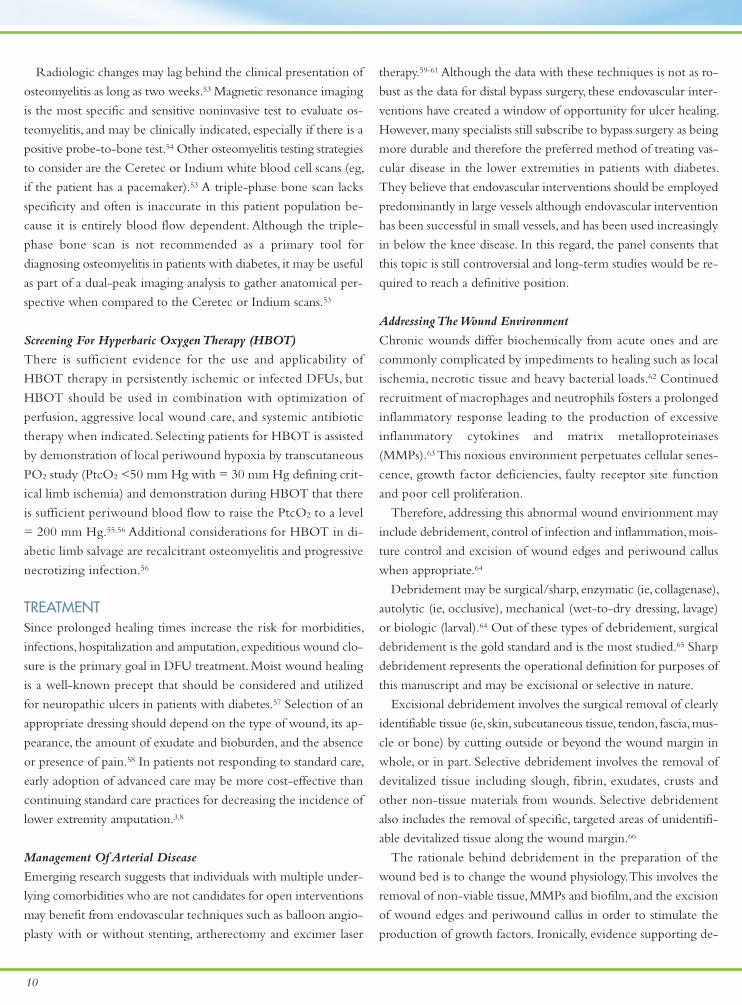
Radiologic changes may lag behind the clinical presentation of
osteomyelitis as long as two weeks.53 Magnetic resonance imaging
is the most specific and sensitive noninvasive test to evaluate os-
teomyelitis, and may be clinically indicated, especially if there is a
positive probe-to-bone test.54 Other osteomyelitis testing strategies
to consider are the Ceretec or Indium white blood cell scans (eg,
if the patient has a pacemaker).53 A triple-phase bone scan lacks
specificity and often is inaccurate in this patient population be-
cause it is entirely blood flow dependent. Although the triple-
phase bone scan is not recommended as a primary tool for
diagnosing osteomyelitis in patients with diabetes, it may be useful
as part of a dual-peak imaging analysis to gather anatomical per-
spective when compared to the Ceretec or Indium scans.53
Screening For Hyperbaric Oxygen Therapy (HBOT)
There is sufficient evidence for the use and applicability of
HBOT therapy in persistently ischemic or infected DFUs, but
HBOT should be used in combination with optimization of
perfusion, aggressive local wound care, and systemic antibiotic
therapy when indicated. Selecting patients for HBOT is assisted
by demonstration of local periwound hypoxia by transcutaneous
PO2 study (PtcO2 <50 mm Hg with = 30 mm Hg defining crit-
ical limb ischemia) and demonstration during HBOT that there
is sufficient periwound blood flow to raise the PtcO2 to a level
= 200 mm Hg.55,56 Additional considerations for HBOT in di-
abetic limb salvage are recalcitrant osteomyelitis and progressive
necrotizing infection.56
TREATMENTSince prolonged healing times increase the risk for morbidities,
infections, hospitalization and amputation, expeditious wound clo-
sure is the primary goal in DFU treatment. Moist wound healing
is a well-known precept that should be considered and utilized
for neuropathic ulcers in patients with diabetes.57 Selection of an
appropriate dressing should depend on the type of wound, its ap-
pearance, the amount of exudate and bioburden, and the absence
or presence of pain.58 In patients not responding to standard care,
early adoption of advanced care may be more cost-effective than
continuing standard care practices for decreasing the incidence of
lower extremity amputation.3,8
Management Of Arterial Disease
Emerging research suggests that individuals with multiple under-
lying comorbidities who are not candidates for open interventions
may benefit from endovascular techniques such as balloon angio-
plasty with or without stenting, artherectomy and excimer laser
therapy.59-61 Although the data with these techniques is not as ro-
bust as the data for distal bypass surgery, these endovascular inter-
ventions have created a window of opportunity for ulcer healing.
However, many specialists still subscribe to bypass surgery as being
more durable and therefore the preferred method of treating vas-
cular disease in the lower extremities in patients with diabetes.
They believe that endovascular interventions should be employed
predominantly in large vessels although endovascular intervention
has been successful in small vessels, and has been used increasingly
in below the knee disease. In this regard, the panel consents that
this topic is still controversial and long-term studies would be re-
quired to reach a definitive position.
Addressing The Wound Environment
Chronic wounds differ biochemically from acute ones and are
commonly complicated by impediments to healing such as local
ischemia, necrotic tissue and heavy bacterial loads.62 Continued
recruitment of macrophages and neutrophils fosters a prolonged
inflammatory response leading to the production of excessive
inflammatory cytokines and matrix metalloproteinases
(MMPs).63 This noxious environment perpetuates cellular senes-
cence, growth factor deficiencies, faulty receptor site function
and poor cell proliferation.
Therefore, addressing this abnormal wound envirionment may
include debridement, control of infection and inflammation, mois-
ture control and excision of wound edges and periwound callus
when appropriate.64
Debridement may be surgical/sharp, enzymatic (ie, collagenase),
autolytic (ie, occlusive), mechanical (wet-to-dry dressing, lavage)
or biologic (larval).64 Out of these types of debridement, surgical
debridement is the gold standard and is the most studied.65 Sharp
debridement represents the operational definition for purposes of
this manuscript and may be excisional or selective in nature.
Excisional debridement involves the surgical removal of clearly
identifiable tissue (ie, skin, subcutaneous tissue, tendon, fascia, mus-
cle or bone) by cutting outside or beyond the wound margin in
whole, or in part. Selective debridement involves the removal of
devitalized tissue including slough, fibrin, exudates, crusts and
other non-tissue materials from wounds. Selective debridement
also includes the removal of specific, targeted areas of unidentifi-
able devitalized tissue along the wound margin.66
The rationale behind debridement in the preparation of the
wound bed is to change the wound physiology. This involves the
removal of non-viable tissue, MMPs and biofilm, and the excision
of wound edges and periwound callus in order to stimulate the
production of growth factors. Ironically, evidence supporting de-
11
bridement as a primary treatment regimen to improve healing
rates is sparse. The evidence primarily consists of self-reporting
from treating physicians and post-hoc analysis of RCTs.65,67,68
Steed and colleagues evaluated debridement frequency as a
secondary endpoint to a double-blind randomized control trial
in patients with chronic neuropathic DFUs (N = 118) treated
with platelet-derived growth factor.65 All patients had aggressive
sharp debridement of DFUs before randomization and repeat
debridement of callus and necrotic tissue as needed. Across the
six treatment centers, 83% of patients who received frequent
debridement (81% of visits) healed compared with 20% who
received less frequent debridement (15% of visits). A potential
flaw of the study may have been its focus on center outcomes
as opposed to individual ulcer outcomes, and could reflect other
factors such as the general consistency of the center in following
the clinical trial protocol rather than the separate effect of de-
bridement alone.
In a randomized, controlled trial of a bilayered human skin equiv-
alent, Saap and Falanga rated the adequacy and performance of sur-
gical debridement.67The researchers found that patients with higher
scores (3–6) on the debridement performance index were 2.4 times
more likely to heal than those who had lower scores (0–2).
A post hoc analysis of two controlled, prospective, randomized piv-
otal trials of topical wound treatments evaluated surface area changes
and wound closure probabilities for 366 venous leg ulcers (VLUs)
and 310 DFUs over 12 weeks.68 Both VLUs and DFUs had a higher
median wound area reduction with debridement (34% and 24%,
P=0.019 and P=0.317, respectively), but an effect on healing was
not demonstrated.
Wound debridement has been proposed to be performed at
weekly intervals or as often as needed based upon the formation
of necrotic or fibrinous tissue.69 Anecdotally, if the ulcer bed is
clean, shows beefy red granulation tissue, is free of infection and
has no abnormality of the wound edge, debridement may not be
required. Conversely, Falanga and colleagues suggest that mainte-
nance debridement should be considered even in the face of a
seemingly healthy wound bed if the wound is not showing evi-
dence of closure.69
Despite the fact that debridement is universally accepted in the
care and treatment of neuropathic ulcers and chronic wounds, the
panel agrees there is very little high-quality evidence validating this
practice. This procedure, albeit an important part of any treatment
algorithm, is still considered intuitive and should be performed after
clinical evaluation. When debridement is considered, surgical de-
bridement is recognized as the gold standard and is recommended
by the panel.
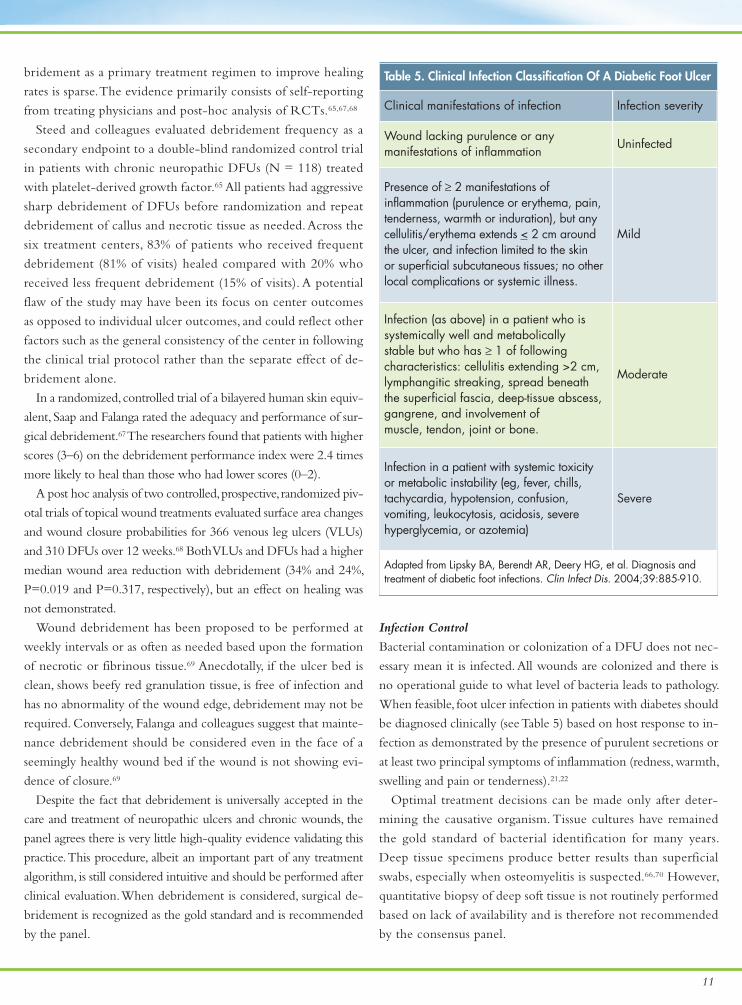
Infection Control
Bacterial contamination or colonization of a DFU does not nec-
essary mean it is infected. All wounds are colonized and there is
no operational guide to what level of bacteria leads to pathology.
When feasible, foot ulcer infection in patients with diabetes should
be diagnosed clinically (see Table 5) based on host response to in-
fection as demonstrated by the presence of purulent secretions or
at least two principal symptoms of inflammation (redness, warmth,
swelling and pain or tenderness).21,22
Optimal treatment decisions can be made only after deter-
mining the causative organism. Tissue cultures have remained
the gold standard of bacterial identification for many years.
Deep tissue specimens produce better results than superficial
swabs, especially when osteomyelitis is suspected.66,70 However,
quantitative biopsy of deep soft tissue is not routinely performed
based on lack of availability and is therefore not recommended
by the consensus panel.
Table 5. Clinical Infection Classification Of A Diabetic Foot Ulcer
Clinical manifestations of infection Infection severity
Wound lacking purulence or any manifestations of inflammation
Uninfected
Presence of ≥ 2 manifestations of inflammation (purulence or erythema, pain,tenderness, warmth or induration), but anycellulitis/erythema extends < 2 cm aroundthe ulcer, and infection limited to the skin or superficial subcutaneous tissues; no otherlocal complications or systemic illness.
Mild
Infection (as above) in a patient who is systemically well and metabolically stable but who has ≥ 1 of following characteristics: cellulitis extending >2 cm,lymphangitic streaking, spread beneath the superficial fascia, deep-tissue abscess, gangrene, and involvement of muscle, tendon, joint or bone.
Moderate
Infection in a patient with systemic toxicity or metabolic instability (eg, fever, chills, tachycardia, hypotension, confusion, vomiting, leukocytosis, acidosis, severe hyperglycemia, or azotemia)
Severe
Adapted from Lipsky BA, Berendt AR, Deery HG, et al. Diagnosis andtreatment of diabetic foot infections. Clin Infect Dis. 2004;39:885-910.
12
Although the panel does not specifically recommend one cul-
turing method, sharp debridement followed by culture using the
Levine technique has been shown to be accurate and consistent
with quantitative biopsy.71The Levine technique involves rotating
a culture swab in one area of the ulcer bed with gentle pressure,
thereby extracting fluid from the underlying tissue.72
Newer but not universally available diagnostic tests with greater
sensitivity have been developed that can better identify infections
or pathogens within hours instead of days.70 For instance, a poly-
merase chain reaction assay can detect gram-positive, gram-negative
and anaerobic organisms. An oligonucleotide array can detect genes
involved with resistance and toxins, and can also identify some spe-
cific species by their genotype. The clinical utility of these is not yet
clear. Finally, magnetic resonance imaging (MRI) is an emerging
technique for detecting infections in soft tissue and bone.
Infections in DFUs are usually polymicrobial, predominantly
comprising aerobic, gram-positive, cocci organisms.70 Staphylococcus
aureus is the most common pathogen found in chronic, non-healing
DFUs.70,72 Osteomyelitis often underlies an infected DFU. The bone
can be cultured and biopsied. However, less invasive diagnostic tech-
niques such as X-ray, MRI or computed axial tomography scans
also may be of benefit.66 Osteomyelitis can be difficult to cure.
Whenever feasible, the bone should be debrided. Intravenous an-
tibiotic therapy should be utilized for a maximum of two weeks if
all infected bone has been resected, four to six weeks after debride-
ment, and for several months if oral antibiotic therapy is used.66 In-
fectious disease consult should be considered in these cases.
The panel recommends diagnosing infection based on clinical
presentation. Quantitative biopsy of deep tissue specimens for culture
is not primarily recommended but the Levine swab technique may
be helpful for quantitative analyses.71,72
Offloading
From a practical standpoint, more widespread adoption of effec-
tive offloading modalities would make the most positive im-
provement in DFU treatment. Clinical studies have shown that
fewer wounds heal with offloading methods such as therapeutic
shoes, pads or custom insoles as compared to more aggressive
techniques like cast walkers and total contact casts (TCCs).74,75
There is little evidence that wheelchairs, bed rest or crutches fa-
cilitate wound healing.76
True offloading is crucial to decreasing pressure and strain
rate.77 Pressure is the force applied over a surface and is measured
as force per unit of area.78 Strain rate is force divided by time. The
duration of stress is very important in determining strain. The re-
lation between the shear stress and the rate of strain is linear.77
Many patients with DFUs are obese and suffering from atten-
dant comorbidities. Obese patients suffering from DFUs put 2 to
2.5 times their body weight on the wound with each step, and the
elastic modulus of skin is about 10 cm per kilogram squared.79
That means a 400-lb patient exceeds the maximum skin elasticity
by about threefold with each step.
Decreasing the strain rate, not just pressure, may improve wound
healing. The easiest way to decrease force over time is to decelerate
the foot into the ground and shorten the time the foot is on the
ground. However, most patients with DFUs are so neuropathic
that they do not decelerate the foot into the ground and strike
more rapidly than those without neuropathy.77
The literature supports the following devices as having repro-
ducible ability to heal wounds: TCC or instant total contact casting
(iTCC); cast walkers (eg, DH, Bledsoe, 3-D, CAM); Charcot Re-
straint Orthotic Walker (CROW)/total contact brace; patellar ten-
don-bearing (PTB) braces; and ankle-foot orthoses (AFOs) in
shoes.80-82 These methods work because they decelerate the foot
into the ground and decrease the weightbearing if they are used
for walking.
The key to effective offloading is to have an ankle brace or a
facsimile fixed to the foot bed. The TCC is the ideal method
for most patients and is supported by the highest level of evi-
dence.83-86 Molding the bottom of the cast to the bottom of the
foot causes the entire sole to participate in the force distribu-
tion, resulting in lower pressures. One non-randomized, single
center comparison study assessed the treatment of 1,350 diabetic
foot ulcers with either TCC, 3-D walkers with custom insoles
or custom healing sandals.87Within five weeks, the percentages
of closures were as follows:
TCC — 88%
3-D walkers with custom insoles — 63%
Custom sandals with three layers of foam — 55%
The results show superior healing with the TCC. However,
the other offloading modalities in this study provided rates of
healing better than those seen without offloading.87 Clinician
comfort with TCC is a barrier to its acceptance but training in-
services can smooth the transition to everyday practice.88
The key in casting is to increase the padding in high-risk
areas. Clinicians should ensure a well-molded cast, which allows
for no ankle motion (to transfer all forces to the tibia) and is
less likely to create secondary ulcerations.
Reimbursement issues are potential barriers to adoption.
However, there is time and effort to be saved by ensuring pa-
tients have better healing rates more quickly. If TCC is not avail-
able for any reason, there are ways to improvise and make

13
removable casts non-removable in order to ensure adherence.
Sometimes referred to as an instant TCC, plaster application
over a properly fitted CAM walker can be very effective.89 An-
other option is to staple together the ends of hook and loop
closures to assess adherence. If the staples are removed, the pa-
tient has removed the device.
Not all patients are appropriate candidates for a TCC or a
CROW walker. These devices are cumbersome relative to size
and weight. They also may not be appropriate for frail individ-
uals, patients with motor difficulties or morbidly obese pa-
tients.90,91 An option for these individuals is an AFO with felted
foam. Clinicians who are not entirely comfortable with TCC
or iTCC can opt for a CAM walker that can be obtained by
prescription from any durable medical equipment supplier.
The primary disadvantage of these removable devices is that
they are just that — removable. There is a probability that these
removable devices will be worn while the patient works (likely
seated) during the day and subsequently removed when the patient
is at home at night.82,89 However, this is when most of the patient’s
weightbearing will occur and may, therefore, render the removable
device useless. In addition, if the patient improperly dons a remov-
able device, the wearing of it may not be effective.
Pressure and strain rate reduction are imperative in healing
DFUs in people with neuropathy. The panel members agree that
total contact casting is the gold standard for offloading DFUs but
recognize this application may not be practical for some clinicians.
Fortunately, clinicians have many other effective offloading devices
available to them and their patients (see Figure 2).90 Given frequent
non-adherence with removable devices, the panel recommends
careful patient counseling.
Hyperbaric Oxygen Therapy (HBOT)
HBOT has been shown to reduce amputation rates in a prospec-
tive, randomized controlled clinical trial when compared to stan-
dard therapy that included aggressive revascularization,
debridement, treatment of infection and glycemic control.92 These
patients were severely ischemic, had underlying osteomyelitis (or
both) with threatened amputation below or above the knee at
presentation. Similar results have been reported in a small RCT
as well as a large (1,144 patients) cohort.93,94 The Wound Healing
Society clinical practice guidelines for diabetic foot ulcer care has
given HBOT in this setting a IA level of evidence.66 A Cochrane
review of HBOT trials found that HBOT may reduce the number
of major amputations in patients with diabetes and chronic foot
ulcers.95 Kalani and colleagues showed long-term durability with
limb salvage for patients receiving HBOT in this setting.96
Medicare has established the following guidelines for coverage
of HBOT for patients with diabetic foot ulcers.97
• Type I or Type II DM, lower extremity wound due to DM
• Wagner grade III or higher
• Failed standard wound care (no measurable signs of healing
for 30 days)
• Wound must be re-evaluated every 30 days during HBOT
therapy course
• Continued HBOT therapy will not be covered if there are
no measurable signs of healing during the 30-day period
Advanced Therapies
Good wound care practices are necessary to promote timely
and complete DFU healing. Despite management with good
wound care, many DFUs do not heal completely, become
chronic or infected.3,98 Major costs associated with managing
DFUs include hospitalizations due to osteomyelitis and ampu-
tation. Therefore, the economics of treatment point to healing
the ulcer and preventing these complications.
Margolis and colleagues evaluated the rate of neuropathic
ulcer healing in 10 control groups from prospective clinical tri-
als via meta-analysis.98 Control groups used good wound care,
which included debridement, offloading and either saline-
moistened gauze or placebo gel and gauze. Six-hundred and
twenty-two (n=622) patients were assessed; 172 in the 20-week
end point group and 450 in the 12-week end point group.
Weighted mean healing rates were 24.2% (95% CI 19.5–28.8%)
for the 12-week end point and 30.9% (95% CI 26.6–35.1%)
for the 20-week end point.
These surprisingly poor healing rates elucidate the challenges
of healing chronic wounds despite appropriate conservative
wound management and fosters the notion that advanced
wound therapies may be required to treat ulcers that fail to
progress with good wound care alone.
The importance of utilizing advanced therapies and products
such as human skin equivalents (HSEs), wound modulators and
growth factors is well documented.3,66,99,100 Clinicians, however,
continue to use these therapies as “last resorts” and may not
be sure when it is appropriate to use them earlier in the wound
healing process.
It has been increasingly suggested that after four weeks of stan-
dard DFU care, the wound should be reassessed for progress and
reduction in ulcer size should be used as a predictive marker.3,100
Sheehan and colleagues assessed the ability of the four-week
healing rate to predict complete healing over a 12-week pe-
riod. Sheehan and co-workers did a post hoc analysis of data
Continued on page 16

14
Figure 2. Offloading options.
Total Contact Cast CROW BootPrefabricatedWalker
DH OffloadingWalker
IPOS Shoe Ortho Wedge
Dorsal Digit
• • • • • •
Plantar Digit
• • • • • •
Plantar Metatarsal
• • • • • •
Medial Metatarsal
• • • • • •
Lateral Metatarsal
• • • •
Heel

15
PostOp Shoe Healing Sandal Reverse IPOS L’nard Splint PTB Brace MABAL Shoe
•
• •
• •
• •
• •
• • • •
Adapted from Snyder RJ, Lanier KK. Offloading difficult wounds and conditions in the diabetic patient. Ostomy Wound Management. 2002;48(1):22-35.

16
from a large prospective multicenter trial of 203 patients with
diabetes and foot ulcerations.100 (This analysis included patients
receiving an active treatment and control.) The midpoint be-
tween the percentage area reduction (PAR) from baseline at
four weeks in patients healed versus those not healed at 12
weeks was found to be 53%. Subjects with a PAR in ulcer size
greater than 53% in four weeks had a 12-week healing rate of
58% whereas those with a reduction in ulcer area less than 53%
in four weeks had a healing rate of only 9% (P <0.01).100
It was concluded that the PAR in foot ulcer area after four
weeks is a robust predictor of healing at 12 weeks and could serve
as a pivotal clinical decision point in the treatment algorithm of
diabetic foot ulcers for early identification of patients who may
not respond to standard care and may require advanced therapies.
Foremost, this study created a negative predictive value for those
ulcers that would not heal in 12 weeks.100
Snyder and co-workers found similar results by conducting post
hoc analyses of control participant data extracted from two previ-
ously published randomized, controlled trials of a human fibrob-
last-derived dermal substitute for treating DFUs.101 The total study
population size was 250 patients. Analyses showed that a 50% PAR
at four weeks was significantly associated with healing at 12 weeks
independent of baseline ulcer area (P < 0.001).
The lead author notes that additional analyses indicated that
approximately one-tenth or fewer of DFUs with a PAR < 50%
at weeks 2, 3 or 4 were healed by week 12 (12%, 9% and 4%
respectively) whereas more than half of the DFUs with at least
50% PAR measurements at weeks 2, 3 and 4 were healed by
week 12 (58%, 55% and 54% respectively).
Therefore, previous recommendations to re-evaluate wound
care at four weeks continue to hold true. In addition, it has
been recommended that the failure to reduce the size of an
ulcer after four weeks, despite standard wound care, should
prompt consideration of an advanced therapy.3
In addition to percent area reduction, several other wound-
specific characteristics (duration of wound, baseline wound
size, and location of the wound) have been predictive of DFU
healing.102,104The value of understanding the possible outcomes
associated with prognostic factors should not be underesti-
mated. A recent study demonstrated improved DFU healing
rates by merely providing clinicians with computer generated
prognostic data based on the ulcer baseline measurements and
changes in wound size at four weeks.104 No guidance for ad-
justing treatment was given with the prognostic data.
In a recent review of optimal treatment strategies for dia-
betic foot ulcers, Armstrong and colleagues advocated the use
of an active therapy such as a bioengineered skin substitute to
stimulate healing in non-responding wounds after four weeks
of treatment.12
Only a small handful of wound care technologies have proven
their value in accelerating DFU healing in prospective, randomized
trials. These include: becaplermin (Regranex®, Systagenix Wound
Management), a topical gel containing recombinant human
PDGF; and two living skin equivalents in the form of a bilayered
skin substitute (Apligraf®, Organogenesis) and a human fibroblast-
derived dermal substitute (Dermagraft®, Advanced BioHeal-
ing).62,99,105,106 These clinical trials involved well-perfused and
non-infected, partial to full thickness DFUs. Before these therapies
are applied, the wound should be clean and free of infection. Other
advanced modalities (with less rigorous trial data) that may be ef-
fective include vacuum-assisted wound closure and electrical stim-
ulation among others.107,108
Any incremental improvement in healing the wound and
avoiding serious complications — especially amputation — can
have a dramatic impact on overall health-care utilization costs.
The panel recognizes the prognostic value of 50% percent
area reduction of the wound at four weeks and recommends
utilization of this parameter as a clinical decision point for the
use of advanced therapies in healing DFUs. Use of advanced
modalities, when indicated, should be viewed as the new stan-
dard of care and these advanced modalities should not be a ‘last
resort’ in the treatment of DFUs. There are, however, certain
patients, who may benefit from even earlier intervention with
advanced therapies while others may require other interven-
tions first (ie, revascularization) to optimize the success of
wound healing.
Amputation
The decision to amputate when a wound has penetrated
through the dermis and affects tendon or bone is a difficult
one. Five-year mortality rates after lower extremity amputation
(LEA) range from 50% to 76%.109,110 Approximately 85% of
lower limb amputations in patients with diabetes are preceded
by ulceration.
Whether an amputation is minor (ie, occurring distal or
through the tarsometatarsal joint) or major (ie, proximal to the
tarsometatarsal joint), it is sometimes the best therapeutic op-
tion for the patient. Some studies using quality-of-life (QOL)
indicators have shown slightly better mean QOL scores with a
healed amputation than with a chronically open wound.111
In summary, patients and their treating clinicians would pre-
fer not to seek amputation as a primary endpoint. However, if
Continued from page 13

17
other therapies are not appropriate or do not prove effective,
one may consider amputation. Other key considerations in-
clude the patient’s quality of life and medical status. The alter-
natives and rationale of amputation should be discussed in
depth with the patient.
CONCLUSIONNeuropathic diabetic foot ulcerations are one of the most com-
mon complications in patients with diabetes. The economic
burden of DFUs is high and secondary complications of infec-
tion and amputation contribute most to the costs. Expeditious
and complete wound healing is the definitive goal in treating
DFUs. Standard management strategies include preparation of
the wound bed, debridement, infection control and offloading.
Despite the use of these strategies, healing rates of DFUs re-
main low.
The approaches of this consensus panel are designed to pro-
vide straightforward and practical management paths for clini-
cians to adopt when treating patients with DFUs, but clinicians
must recognize the unique challenges presented by individual
patients and adjust treatment approaches accordingly.
Assessment should include a comprehensive foot and ulcer
evaluation with key components of a patient history and phys-
ical examination, laboratory screening, nutritional evaluation,
and a neuropathic and vascular assessment. Wound status history
and a complete and accurate description of the wound, includ-
ing measurements of length, width, and depth, need to be in-
cluded in the evaluation.
Treatment should include appropriate preparation and
maintenance of the wound bed with special attention paid to
debridement, offloading and infection control. Clinicians must
take a holistic approach to healing DFUs and decision-making
is a proactive process that requires ongoing reassessment. When
wound healing stalls, early adoption of advanced therapies is
advocated to accelerate wound healing and decrease compli-
cations, and should be considered the new standard of care. n

18
ASSESSMENTHistory and Physical Exam
General• Determine the duration of diabetes and quality of glycemic control• Determine the presence of comorbidites (eg, end-stage renal disease, pulmonary disease, hypertension, hy-percholesterolemia, hyperlipidemia, myocardial infarction, transient ischemic attacks, angina or valvularheart disease)• Laboratory tests
• complete blood count and creatinine/blood urea nitrogen • HbA1c• lipid profile • prealbumin
• Dietary questionnaire• Quality of life questions• Smoking cessation
Neurologic exam: Vibration perception with a 128-Hz tuning fork and 10-g monofilament tests
Vascular exam: use a tiered approach as indicated or appropriate• 1º palpation of pulses, ankle brachial index (ABI), and/or toe brachial index (TBI)• 2º (if suspicion of vascular insufficiency) refer for segmental pressure volume and skin perfusion pressure(SPP) and transcutaneous oxygen measurement (TCPO2)• 3º refer for vascular consultation and angiography in patients being considered for intervention
Foot and ulcer evaluation• Determine:
• initial wounding event, history of recurrent wounding, previous wound healing problems • prior diagnostic testing, prior therapies and response• functional impact of the wound on the patient • sufficient social history to define potential adverse impact on treatment plan
• Assess dermatologic changes including callus, musculoskeletal deformity and muscle wasting• Document ulcer characteristics including location, shape and size of the wound (measurement of length,width and depth)• Clinical utilization of a probe to check for sinus tracts and a positive probe to bone test • Determine condition of the wound edges, wound bed, wound base, periwound skin and exudates• Determine presence of necrosis and wound associated pain
Wound classification: University of Texas system is recommended, Wagner system may be required for re-imbursement. Prognosis may be related to anatomically deeper wounds.
Consensus Panel Summary Of RecommendationsUse a multidisciplinary team approach to assessment and treatment.

19
Infection• Classic signs: heat, pain, redness and swelling • Secondary signs: exudates, delayed healing, friable granulation tissue, discolored granulation tissue, foulodor, pocketing at the wound base, and wound breakdown • Probe-to-bone test • If osteomyelitis is suspected, 2º markers are erythrocyte sedimentation rate and C-reactive protein • Not recommended: routine culture as an evaluation method unless an infection is apparent or sensitivities arerequired for appropriate antibiotic selection
Radiography • Plain film radiography• If the clinician suspects osteomyelitis (especially if there is a positive probe-to-bone test)
• MRI to evaluate for osteomyelitis• Other testing may include Ceretec or Indium white blood cell scans. While a triple phase bone scanlacks specificity, it may be used in conjunction with these tests for dual peak imaging.
TREATMENT
Debridement: Cold steel surgical debridement initially and then as needed based on condition of the wound (maintenance)
Infection control• Bacterial colonization ≠ infection• Infection is diagnosed from clinical findings whenever possible. Purulent secretions are present or > two orat least two principal symptoms of inflammation (eg, redness, warmth, swelling and pain or tenderness). • Given that patients with diabetes are typically immunocompromised, clinicians should also look for secondarysigns of infection including exudates, delayed healing, friable granulation tissue, discolored granulationtissue, foul odor, pocketing at the wound base and wound breakdown. • Culture:
• Levine swab technique• Quantitative biopsy (for bone only) • Testing (not universally available) via polymerase chain reaction assay, oligonucleotide array
Offloading• Total contact cast (TCC) • Instant TCC • See Figure 2.
Advanced therapies· Use 4-week treatment end point to assess need for advanced therapies· If wound is not progressing toward healing (percent wound area reduction < 50%), then advanced therapiesshould be considered· All previous assessment and treatment standards should continue to be utilized

20
References
1. Wild S, Roglic G, Green A, et al. Global prevalence of diabetes. Estimates for theyear 2000 and projections for 2030. Diabetes Care. 2004;27(5):1047-1053.
2. Reiber GE, Ledoux WR. Epidemiology of diabetic foot ulcers and amputations: ev-idence for prevention. In: Williams R, Herman W, Kinmonth AL, Wareham NJ (eds).The Evidence Base for Diabetes Care.Hoboken, NJ: John Wiley & Sons; 2002:641–665.
3. Boulton AJ, Kirsner RS, Vileikyte L. Clinical practice. Neuropathic diabetic foot ul-cers. N Engl J Med. 2004;351(1):48–55.
4. Boulton AJ, Vileikyte L, Ragnarson-Tennvall G, Apelqvist J. The global burden of di-abetic foot disease. Lancet. 2005;366(9498):1719–1724.
5. Sanders LJ. Diabetes mellitus: prevention of amputation. J Am Podiatric Med Assoc.1994;84(7):322–328.
6. Lavery LA, Armstrong DG, Wunderlich RP, et al. Diabetic foot syndrome: evaluatingthe prevalence and incidence of foot pathology in Mexican Americans and non-Hispanic Whites from a diabetes disease management cohort. Diabetes Care.2003;26(5):1435-8.
7. King H, Aubert RE, Herman WH. Global burden of diabetes, 1995-2025: prevalence,numerical estimates, and projections. Diabetes Care. 1998:21(9):1414-1431.
8. Apelqvist J, Larsson J. What is the most effective way to reduce incidence of ampu-tation in the diabetic foot? Diabetes Metab Res Rev. 2000:16:S75–S83.
9. Ramsey SD, Newton K, Blough D, et al. Incidence, outcomes, and cost of foot ulcersin patients with diabetes. Diabetes Care. 1999;22(3):382–387.
10. Pecoraro RE, Reiber GE, Burgess EM. Pathways to diabetic limb amputation. Basisfor prevention. Diabetes Care. 1990;13(5):513–521.
11. Iversen MM, Tell GS, Riise T, et al. History of foot ulcer increases mortality amongindividuals with diabetes: ten-year follow-up of the Nord-Trøndelag Health Study,Norway. Diabetes Care. 2009;32(12):2193-2199.
12. Armstrong DG, Granick MS, Marston WA, et al. A regimen to optimize the resultsof a living dermal substitute (Dermagraft) for healing diabetic foot ulcers. US En-docrine Review. 2007;(Spring b):60–66.
13. Harrington C, Zagari MJ, Corea J, et al. A cost analysis of diabetic lower-extremityulcers. Diabetes Care. 2000;23(9):1333-8.
14. Gordois A, Scuffham P, Shearer A, et al. The health care costs of diabetic peripheralneuropathy in the US. Diabetes Care. 2003;26(6):1790-1795.
15. Sackett DL, Rosenberg WM, Gray JA, et al. Evidence based medicine: what it isand what it isn't. BMJ. 1996;312(7023):71-2.
16. Eddy DM. Evidence-based medicine: a unified approach. Health Aff (Millwood).2005;24(1):9-17.
17. Schuster MA, McGlynn EA, Brook RH. How good is the quality of care inthre United States? Milbank Q. 1998;76;517-563. Stable URL:http://www.jstor.org/pss/3350511
18. Frykberg RG, Zgonis T, Armstrong DG, et al. Diabetic foot disorders. A clinicalpractice guifdeline (2006 revision). J Foot Ankle Surg. 2006;45(5 Suppl):S1-66.
19. Boulton AJ, Armstrong DG, Albert SF, et al. Comprehensive foot examination andrisk assessment: a report of the task force of the foot care interest group of the Amer-ican Diabetes Association, with endorsement by the American Association of Clin-ical Endocrinologists. Diabetes Care. 2008;39(5 Suppl):S1-60.
20. Seaman S. Considerations for the global assessment and treatment of patients with re-calcitrant wounds. Ostomy Wound Manage. 2000;46(1A Suppl):10S-29S; quiz 30S-31S.
21. Lipsky BA, Berendt AR, Deery HG, et al. Diagnosis and treatment of diabetic footinfections. Clin Infect Dis. 2004;39(7):885-910.
22. Lipsky BA, Berendt AR, Embil J, et al. Diagnosing and treating diabetic foot infec-tions. Diabetes Metab Res Rev. 2004;20(suppl 1);S56-S64.
23. Selvin E, Steffes MW, Zhu H, et al. Glycated hemoglobin, diabetes and cardiovas-cular risk in nondiabetic adults. N Engl J Med. 2010;362(9):800-811.
24. American Diabetes Association. Standards of medical care in diabetes – 2010.Diabetes Care. 2010;33(suppl 1):S11-S61.
25. Young MJ, McCardle JE, Randall LE, et al. Improved survival of diabetic footulcer patients 1995-2008: possible impact of aggressive cardiovascular risk man-agement. Diabetes Care. 2008;31(11):2143-7.
26. Pompeo M. Misconceptions about protein requirements for wound healing:results from a prospective study. Ostomy Wound Manage. 2007;53(8):30-2,34,36-38 passim.
27. Langemo D, Anderson J, Hanson D, et al. Nutritional considerations in woundcare. Adv Skin Wound Care. 2006;19(6):297-8,300,303.
28. Rossi M, Carpi A, Di Maria C, et al. Absent post-ischemic increase of bloodflowmotion in the cutaneous microcirculation of healthy chronic cigarettesmokers. Clin Hemorheol Microcirc. 2007;36(2):163-71.
29. Ahmed AT, Karter AJ, Liu J. Alcohol consumption is inversely associatedwith adherence to diabetes self-care behaviours. Diabet Med. 2006;23(7):795-802.
30. Katon W, Russo J, Lin EH, et al. Depression and diabetes: factors associatedwith major depression at five-year follow-up. Psychomatics 2009;50(6):570-9.
31. Snyder RJ. Controversies regarding vascular disease in the patient with diabetes:a review of the literature. Ostomy Wound Manage. 2007:53(11):40-48.
32. American Diabetes Association. Peripheral arterial disease in people with dia-betes. Diabetes Care. 2003;26(12):3333-3341.
33. Faglia E, Favales F, Quarantiello A, et al. Angiographic evaluation of peripheralarterial occlusive disease and its role as a prognostic determinant for major am-putation in diabetic subjects with foot ulcers. Diabetes Care. 1998;21(4):625-630.
34. Brooks B, Dean R, Patel S, et al. TBI or not TBI: that is the question. Is it betterto measure toe pressure than ankle pressure in diabetic patients? Diabet Med.2001;18(7):528-532.
35. Williams DT, Harding KG, Price P. An evaluation of the efficacy of methodsused in screening for lower-limb arterial disease in diabetes. Diabetes Care.2005;28(9):2206-2210.
36. Halperin JL. Evaluation of patients with peripheral vascular disease. ThrombRes. 2002;106(6):V303-V311.
37. Okamoto K, Oka M, Maesato K, et al. Peripheral arterial occlusive disease is moreprevalent in patients with hemodialysis: comparison with the findings of multide-tector-row computed tomography. Am J Kidney Dis. 2006;48(2):269-276.
38. Padberg FT, Back TL, Thompson PN, et al. Transcutaneous oxygen (TcPO2)estimates probability of healing in the ischemic extremity. J Surg Res.1996;60(2):365-369.
39. Fife CE, Smart DR, Sheffield PJ, et al. Transcutaneous oximetry in clinical prac-tice: consensus statements from an expert panel based on evidence. UnderseaHyperb Med. 2009;36(1):43-53.
40. Boulton AJM, Armstrong DG, Albert SF, et al. Comprehensive foot examinationand risk assessment: a report of the task force of the foot care interest group ofthe American Diabetes Association, with endorsement by the American Asso-ciation of Clinical Endocrinologists. Diabetes Care. 2008;31(8):1679-1685.
41. Keast DH, Bowering K, Evans AW, et al. MEASURE: a proposed assessmentframework for developing best practice recommendations for wound assess-ment. Wound Repair Regen. 2004;12(suppl):S1-S17.
42. Ratliff CR. WOCN’s evidence-based pressure ulcer guideline. Adv Skin WoundCare. 2005;18(4):204-208.
43. Margolis DJ, Kantor J, Santanna J, et al. Risk factors for delayed healing of neu-ropathic diabetic foot ulcers. Arch Dermatol. 2000;136(12):1531-1535.
44. Caselli A, Pham H, Giurini JM, et al. The forefoot-to-rearfoot plantar pressureratio is increased in severe diabetic neuropathy and can predict foot ulceration.Diabetes Care. 2002;25(6):1066-71.

21
45. Wagner FW. The dysvascular foot: a system for diagnosis and treatment. FootAnkle. 1981;2(2):64-122.
46. Oyibo SO, Jude EB, Tarawneh I, et al. A comparison of two diabetic foot ulcer clas-sification systems. The Wagner and the University of Texas wound classification sys-tems. Diabetes Care. 2001;24(1):84-88.
47. Lavery LA, Armstrong DG, Harkless LB. Classification of diabetic foot wounds. JFoot Ankle Surg. 1996;35(6):528-531.
48. Armstrong DG, Lavery LA, Harkless LB. Validation of a diabetic wound classificationsystem. The contribution of depth, infection, and ischemia to risk of amputation.Diabetes Care. 1998;21(5):855-859.
49. Gardner SE, Frantz RA, Doebbeling BN. The validity of the clinical signs and symp-toms used to identify localized chronic wound infection. Wound Repair Regen.2001;9(3):178-186.
50. Rabjohn L, Roberts K, Troiano M, et al. Diagnostic and prognostic value of ery-throcyte sedimentation rate in contiguous osteomyelitis of the foot and ankle. J FootAnkle Surg. 2007;46(4):230-237.
51. Fleischer AE, Didyk AA, Woods JB, et al. Combined clinical and laboratory testingimproves diagnostic accuracy for osteomyelitis in the diabetic foot. J Foot Ankle Surg.2009;48(1):39-46.
52. Lavery LA, Armstrong DG, Peters EJG, et al. Probe-to-bone test for diagnosing di-abetic foot osteomyelitis: reliable or relic? Diabetes Care. 2007;30(2):270-274.
53. Snyder RJ, Cohen MM, Sun C, et al. Osteomyelitis in the diabetic patient: diagnosisand treatment. Part 1: overview, diagnosis, microbiology. Ostomy Wound Manage.2001:47(1):18-30.
54. Schwegler B, Stumpe KDM, Weishaupt D, et al. Unsuspected osteomyelitis is fre-quent in persistent diabetic foot ulcer and better diagnosed by MRI than by 18F-FDG PET or 99mTc-MOAB. J Intern Med. 2008;263(1):99-106.
55. Fife CE, Buyukcakir C, Otto G, et al. The predictive value of transcutaneous oxygentension measurement in diabetic lower extremity ulcers treated with hyperbaricoxygen therapy: a retrospective analysis of 1144 patients. Wound Rep Reg 2002;10(4):198-207.
56. Warriner R, Hopf H. Enhancement of healing in selected problem wounds. In:Gesell LB, ed. Hyperbaric Oxygen Therapy Indications, 12th edition.Durham, NC: Un-dersea and Hyperbaric Medical Society; 2008: 67-84.
57. Winter GD. Oxygen and epidermal wound healing. Adv Exp Med Biol.1977;94:673-8.
58. Ovington LG. Hanging wet-to-dry dressings out to dry. Home Healthc Nurse.2001;19(8):477-83.
59. Adam DJ, Beard JD, Cleveland T, et al.; BASIL trial participants. Bypass versus an-gioplasty in severe ischaemia of the leg (BASIL):multicentre, randomised controlledtrial. Lancet. 2005;366(9501):1925-34.
60. Muhs BE, Gagne PJ, Maldonado T, et al. Minimally invasive revascularization strategies for chronic lower limb ischemia. Int J Low Extrem Wounds. 2006;5(1):35-9.
61. Laird JR, Zeller T, Gray BH, et al.; LACI Investigators. Limb salvage following laseras-sisted angioplasty for critical limb ischemia: results of the LACI multicenter trial. JEndovasc Ther. 2006;13(1):1-11.
62. Basu S, Ramchuran Panray T, Bali Singh T, et al. A prospective, descriptive study toidentify the microbiological profile of chronic wounds in outpatients. Ostomy WoundManage. 2009;55(1):14-20.
63. Doughty DB, Sparks-DeFriese B. Wound-healing physiology. In: Bryant RA, NixDP, eds. Acute and chronic wounds. Current management concepts. 3rd ed. St. Louis: Mosby;2007:56-81.
64. Brem H, Sheehan P, Rosenberg HJ, et al. Evidence-based protocol for diabetic footulcers. Plast Reconstr Surg. 2006;117(7 Suppl):193S-209S; discussion 210S-211S.
65. Steed DL, Donohoe D, Webster MW, et al. Effect of extensive debridement andtreatment on the healing of diabetic foot ulcers. Diabetic Ulcer Study Group. J AmColl Surg. 1996;183(1):61–64.
66. Steed DL, Attinger C, Colaizzi T, et al. Guidelines for the treatment of diabetic ul-cers. Wound Repair Regen. 2006;14(6):680-92.
67. Saap LJ, Falanga V. Debridement performance index and its correlation withcomplete closure of diabetic foot ulcers. Wound Repair Regen. 2002;10(6):354-9.
68. Cardinal M, Eisenbud DE, Armstrong DG, et al. Serial surgical debridement:a retrospective study on clinical outcomes in chronic lower extremitywounds. Wound Repair Regen. 2009;17(3):306-11.
69. Falanga V, Brem H, Ennis WJ, et al. Maintenance debridement in the treatment ofdifficult-to-heal chronic wounds. Recommendations of an expert panel. OstomyWound Manage. 2008;54:(suppl 6):2-13.
70. Lipsky BA. New developments in diagnosing and treating diabetic foot infections.Diabetes Metab Res Rev. 2008;24(suppl 1):S66–S71.
71. Gardner SE, Frantz FA, Saltzman CL, et al. Diagnostic validity of three swab techniquesfor identifying chronic wound infection. Wound Rep Reg. 2006;14(5):548–557.
72. Levine NS, Lindberg RB, Mason AD Jr, et al. The quantitative swab culture andsmear: a quick, simple method for determining the number of viable aerobic bac-teria on open wounds. J Trauma. 1976:16(2):89-94.
73. Gardner SE, Frantz RA. Wound bioburden and infection-related complications indiabetic foot ulcers. Biol Res Nurs. 2008;10(1):44–53.
74. Bus SA, Valk GD, van Duersen RW, et al. The effectiveness of footwear and offload-ing interventions to prevent and heal foot ulcers and reduce plantar pressure in di-abetes: a systematic review. Diabetes Metab Res Rev. 2008;24 Suppl 1:S162-80.
75. Mueller MJ, Diamond JE, Sinacore DR, et al. Total contact casting in treat-ment of diabetic plantar ulcers. Controlled clinical trial. Diabetes Care.1989;12(6):384-8.
76. Hanft J et al. “Off-Loading of Ulcers with Aperture Pads: Does This Really LimitCutaneous Blood Flow?”, American College of Foot and Ankle Orthopedics andMedicine, National Meeting, Los Angeles, CA, February 18, 1999.
77. Landsman AS, Meaney DF, Cargill RS 2nd, et al. 1995 William J. Stickel GoldAward. High strain rate tissue deformation. A theory on the mechanical etiology ofdiabetic foot ulcerations. J Am Podiatr Med Assoc. 1995;85(10):519-27.
78. Zou D, Mueller MJ, Lott DJ. Effect of peak pressure and pressure gradient on sub-surface shear stresses in the neuropathic foot. J Biomech. 2007;40(4):883-90.
79. Pinzur M, Freeland R, Juknelis D. The association between body mass index andfoot disorders in diabetic patients. Foot Ankle Int. 2005;26(5):375-7.
80. Lavery LA, Vela SA, Lavery DC, et al. Reducing dynamic foot pressures in high-risk diabetic subjects with foot ulcerations. A comparison of treatments. DiabetesCare. 1996;19(8):818-821.
81. Armstrong DG, Nguyen HC, Lavery LA, et al. Off-loading the diabetic foot wound:a randomized clinical trial. Diabetes Care. 2001;24(6):1019-1022.
82. Armstrong DG, Lavery LA, Kimbriel HR, et al. Activity patterns of patients withdiabetic foot ulceration: patients with active ulceration may not adhere to a standardpressure off-loading regimen. Diabetes Care. 2003:26(9):2595-2597.
83. Ali R, Qureshi A, Yaqoob MY, et al. Total contact cast for neuropathic diabetic footulcers. J Coll Physicians Surg Pak. 2008;18(11):695-8.
84. Tamir E, Heim M, Siev-Ner I. Total contact cast for management of neuropathiculceration of the foot. (Article in Hebrew). Harefuah. 2006;145(2):152-5, 163.
85. Birke JA, Sims DS Jr, Buford WL. Walking casts: effect on plantar foot pressures. JRehabil Res Dev. 1985;22(3):18-22.
86. Sinacore DR, Mueller MJ, Diamond E, et al. Diabetic plantar ulcers treated by totalcontact casting. A clinical report. Phys Ther. 1987;67(10):1543-9.
87. Hanft JR, Surprenant MS. Is total contact casting the gold standard for the treatmentof diabetic foot ulcerations? Abstract presented at: The American College of Footand Ankle Surgeons Joint Annual Meeting and Scientific Seminar; February 9, 2000;Miami, Fla.

22
88. Wu SC, Jensen DL, Weber AK, et al. Use of pressure offloading devices in diabetic footulcers: do we practice what we preach? Diabetes Care. 2008 Nov;31(11):2118-9.
89. Armstrong DG, Lavery LA, Wu S, Boulton AJ. Evaluation of removable and irre-movable cast walkers in the healing of diabetic foot wounds: a randomized controlledtrial. Diabetes Care. 2005;28(3):551–554.
90. Snyder RJ, Lanier KK. Offloading difficult wounds and conditions in the diabeticpatient. Ostomy Wound Management. 2002;48(1):22-35.
91. Van Deursen R. Footwear for the neuropathic patient: offloading and stability. Dia-betes Metab Res Rev. 2008;24 Suppl 1:S96-100.
92. Faglia E, Favales F, Aldeghi A, et al. Adjunctive systemic hyperbaric oxygen therapyin treatment of severe prevalently ischemic diabetic foot ulcer: A randomized study.Diabetes Care 1996; 19(2):1338-1343.
93. Abidia A, et al. The role of hyperbaric oxygen therapy in ischaemic diabetic lowerextremity ulcers: a double-blind, randomized-controlled trial. Eur J Vasc Endovasc Surg2003; 25(6):513-518.
94. Fife C, et al. Factors influencing the outcome of lower-extremity diabetic ulcerstreated with hyperbaric oxygen therapy. Wound Rep Regen 2007; 15(3):322-331.
95. Kranke P, et al. Hyperbaric oxygen therapy for chronic wounds. The Cochrane Data-base of Systematic Reviews 2004, Issue 1. Art. No.: CD004123.pub2. DOI:10.1002/14651858.CD004123.puib2.
96. Kalani M, Jorneskog G, Naderi N, et al. Hyperbaric oxygen (HBO) therapy in treat-ment of diabetic foot ulcers. Long-term follow-up. J Diabetes Complications 2002;16(2):152-258.
97. CMS Coverage Memorandum CAG 00060N 08.30.02, Transmittal AB-02-18312.22.02 Effective 04.01.03
98. Margolis D, Kantor J, Berlin J. Healing of neuropathic ulcers receiving standard treat-ment: a meta-analysis. Diabetes Care 1999;22(5):692-695.
99. Marston WA, Hanft J, Norwood P, et al. The efficacy and safety of Dermagraft inimproving the healing of chronic diabetic foot ulcers. Diabetes Care.2003;26(6):1701–1705.
100. Sheehan P, Jones P, Caselli A, et al. Percent change in wound area of diabetic footulcers over a 4-week period is a robust predictor of complete healing in a 12-weekprospective trial. Diabetes Care. 2003;26(6):1879–1882.
101. Snyder R, Cardinal M, Dauphinee D, et al. A post-hoc analysis of reductionin diabetic foot ulcer size at 4 weeks as a predictor of healing by 12 weeks.Ostomy Wound Manage. 2010:56(3):44-50.
102. Margolis DJ, Allen-Taylor L, Hoffstad O, et al. Diabetic neuropathic foot ulcers:predicting which ones will not heal. Am J Med. 2003;115(8):627-31.
103. Margolis DJ, Allen-Taylor L, Hoffstad O, et al. Diabetic neuropathic footulcers: the association of wound size, wound duration, and wound gradeon healing. Diabetes Care. 2002;25(10):1835-9.
104. Kurd SK, Hoffstad OJ, Bilker WB, et al. Evaluation of the use of prognostic in-formation for the care of individuals with venous leg ulcers or diabetic neuro-pathic foot ulcers. Wound Repair Regen. 2009;17(3):318-325
105. Veves A, Falanga V, Armstrong DG, et al. Graftskin, a human skin equivalent,is effective in the management of noninfected neuropathic diabetic footulcers: a prospective randomized multicenter clinical trial. Diabetes Care.2001;24(2):290-5.
106. Cavorsi J, Vicari F, Wirthlin DJ, et al. Best-practice algorithms for the useof a bilayered living cell therapy (Apligraf) in the treatment of lower-ex-tremity ulcers. Wound Repair Regen. 2006;14(2):102-9.
107. Blume PA, Walters J, Payne W, et al. Comparison of negative pressurewound therapy using vacuum-assisted closure with advanced moist woundtherapy in the treatment of diabetic foot ulcers: a multicenter randomizedcontrolled trial. Diabetes Care. 2008;31(4):631-6.
108. Peters EJ, Lavery LA, Armstrong DG, et al. Electric stimulation as an ad-junct to heal diabetic foot ulcers: a randomized clinical trial. Arch Phys MedRehabil. 2001;82(6):721-5.
109. Ghanassia E, Villon L, Thuan dit Dieudonne JF, et al. Long-term outcomeand disability of diabetic patients hospitalized for diabetic foot ulcers: a 6.5year follow-up study. Diabetes Care 2008;31(7):1288-1292
110. Resnick HE, Carter EA, Lindsay R, et al. Relation of lower extremity am-putation to all-cause and cardiovascular disease mortality in American In-dians: the strong heart study. Diabetes Care 2004;27(6):1286-1293.
111. Armstrong DG, Lavery LA, Wrobel JS, et al. Quality of life in healing dia-betic wounds: does the end justify the means? J Foot Ankle Surg.2008;47(4):278-82.

23
There exists a number of challenges in diagnosing and treating
diabetic foot ulcers (DFUs). Between the myriad comorbidities
that can contribute to stalled healing and striving to ensure the
best multidisciplinary care, it can be difficult for physicians to stay
abreast of the latest research let alone incorporate these findings
into the management of these patients in the clinic.
This consensus panel seems to have found a middle ground be-
tween evidence-based medicine and the practical realities of the
day-to-day pragmatic practice. For example, the panelists discuss
the supporting research behind total contact casting as well as this
technique’s practical limitations. They also explore the pros and
cons of alternative offloading techniques and modalities.
The consensus panel also reviews new tools for diagnosing in-
fection and pathology in DFUs, and considers the increasingly
emerging evidence about the earlier use of advanced therapies.
This practical yet evidence-based guide should go a long way
toward facilitating improved outcomes in healing chronic ulcers
in patients with diabetes.
— David G. Armstrong, DPM, MD, PhD,
Professor of Surgery
Director, Southern Arizona Limb Salvage Alliance (SALSA)
University of Arizona College of Medicine
Tucson, Ariz.
Expert Commentary The chronic complications of diabetes in the lower limbs now
represent the most prevalent cause of lower extremity amputation
(LEA). It has been estimated worldwide that a lower limb is lost
because of diabetes every 30 seconds and the prevalence is bound
to rise in relation to the projected increased incidence of type 2
diabetes in the near future.1
Far from being a local problem, the diabetic foot is the conse-
quence of the systemic involvement of both the vascular and pe-
ripheral nervous system in the lower extremity. It has been
demonstrated to be a marker of mortality for patients with diabetes
with a frequency, which has been compared to some of the most
severe types of cancer.2
Such a complex and evolutive pathology deserves a thorough
assessment, which encompasses both systemic and local character-
istics, and an integrated therapeutic approach, which addresses both
the vascular, neurologic and infective components. Revasculariza-
tion, surgical treatment and offloading (and systemic antibiotic
therapy when appropriate) are the cornerstones of therapy and
ensure healing in more than 90 percent of the cases when they
are applied in a timely manner.3
Increasing the knowledge and promoting an advancement of
the standards of care for the diabetic foot (through consensus rec-
ommendations such as these) is crucial to developing a stronger
medical understanding of this still neglected aspect of the compli-
cations of diabetes, which are often managed with empiricism and
still end frequently in dramatic consequences.
— Alberto Piaggesi MD
Head, Diabetic Foot Section
Depatment of Endocrinology and Metabolism
University of Pisa
Italy
References
1. Boulton AJ, Vileikyte L, Ragnarson-Tennvall G, et al. The global burden of dia-
betic foot disease. Lancet. 2005 Nov 12;366(9498):1719-24.
2. Armstrong DG, Wrobel J, Robbins JM. Guest Editorial: are diabetes-related wounds
and amputations worse than cancer? Int Wound J. 2007 Dec;4(4):286-7.
3. Schaper NC, Apelqvist J, Bakker K. The international consensus and practical
guidelines on the management and prevention of the diabetic foot. Curr Diab
Rep. 2003 Dec;3(6):475-9

Supported by a grant from
LLC,
www.hmpcommunications.com
2010-120-082
©2010 HMP Communications, LLC (HMP). All rights reserved. Reproduction in whole or in part prohibited. Opinions expressed by authors, contributors, and advertisers are their own and not necessarily those of HMPCommunications, the editorial staff, or any member of the editorial advisory board. HMP Communications is not responsible for accuracy of dosages given in articles printed herein. The appearance of advertisements inthis journal is not a warranty, endorsement, or approval of the products or services advertised or of their effectiveness, quality, or safety. HMP Communications disclaims responsibility for any injury to persons or propertyresulting from any ideas or products referred to in the articles or advertisements. Content may not be reproduced in any form without written permission. Reprints of articles are available. Rights, permission, reprint, andtranslation information is available at: www.hmpcommunications.com.
HMP Communications, LLC (HMP) is the authoritative source for comprehensive information and education servicing healthcare professionals. HMP’s products include peer-reviewed and non–peerreviewed medicaljournals, national tradeshows and conferences, online programs, and customized clinical programs. HMP is a wholly owned subsidiary of HMP Communications Holdings LLC. Discover more about HMP’s products andservices at www.hmpcommunications.com.